Animal Kingdom Basis Of Classification
Both plants and animals show a high degree of diversity. In the previous chapter, we have studied the classification of plants. Plants constitute the plant kingdom. Similarly, all animals belong to the kingdom Animalia or animal kingdom.
Even under the same kingdom, each species may exist in different forms. Classification helps us to deal with this enormous diversity. The study of the classification of the animal world is known as systematic zoology.
The animals are classified into different categories according to their general characteristics. These characteristics include body organization, body symmetry, presence of germ layer, etc.
Read and Learn More: WBCHSE Notes for Class 11 Biology
General Characteristics Of Animals
The general characteristics of animals, on the basis of which they are classified, are discussed below.
Habitat
The geographical region and environment, where an animal lives, is called its habitat.
According to the habitat, the animals are classified into three types—
- Aquatic,
- Terrestrial, and
- Aerial.
Aquatic animals: Animals that live in water are called aquatic animals.
Animal kingdom notes for NEET PDF
These animals are further classified into the following types—
Zooplankton: Microscopic organisms that float on water are called zooplankton. Example Microscopic animals and larvae of some higher animals.
Nektons: Organisms that can swim freely in water are called nektons (derived from the Greek word, nekton, meaning swimming). For example, Fish, prawns, etc.
Benthon: Organisms that are present in the lower region of the sea are called benthons (derived from the Greek word —benthos, meaning depths of the sea). Example Corals, sponges, etc.
Littoral: Organisms that live near the shores are called littoral. Example Small fish.
Neritic: Organisms that live in shallow regions of the water bodies are called neritic. Example Snail.
Neuston: Organisms that are found just below the water surface or on the water surface are called neuston (derived from the Greek word, neustos, meaning floating). Example Mosquito larvae.
Lcntic and lotic: Organisms that are found in still water (such as ponds or lakes) are called lentic. Example Small fish. Organisms that are found in flowing water (such as rivers) are called lotic. Example Big fish.
Terrestrial animals: Animals that live on land are called terrestrial animals. They are further divided into several types. Some of them are discussed below.
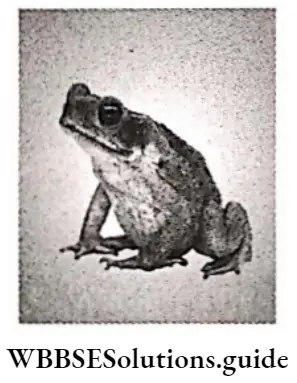
Amphibians: Animals that live on both land and water are called amphibians. Example Common Asian toad.
Arboreal: Animals that Live on trees are called arboreal animals. ExampleMonkey.
Cursorial: Animals that can run very fast are called cursorial animals. Example Horse.
Subterranean: Animals that live in underground holes are called subterranean. Example Earthworms.
Scansorial: Animals that can climb trees are called scansorial animals. Example Squirrel.
Fossorial: Animals that dig holes or burrows and live in them, are called fossorial animals. Example Rabbits, armadillos, etc.
Aerial animals: Animals that can fly in the sky, are called aerial animals. According to their adaptation, they are of two types—primary and secondary aerial animals.
Primary aerial animals: Animals that fly in the sky throughout most part of their life, are called primary aerial animals. The ability to fly in such animals is due to their ancestors who were adapted to flight. Example Birds.
Secondary aerial animals: Animals that can fly for certain intervals of time, but are not aerial in nature are called secondary aerial animals. Example Bats.
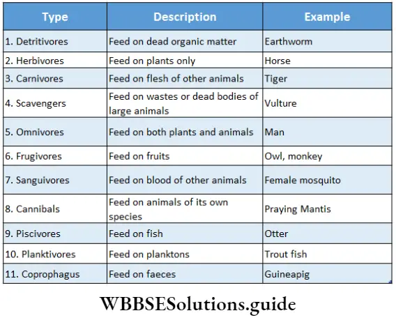
Food habit
Different types of food habits are found in the animal kingdom.
According to food habits, the classification of animals is as follows—
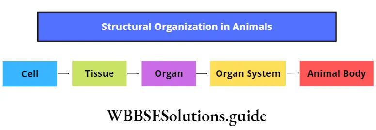
Structural organization
Different kinds of structural organizations are found in multicellular organisms. These may be at the cellular, histological, or organ system level. These have been discussed below.
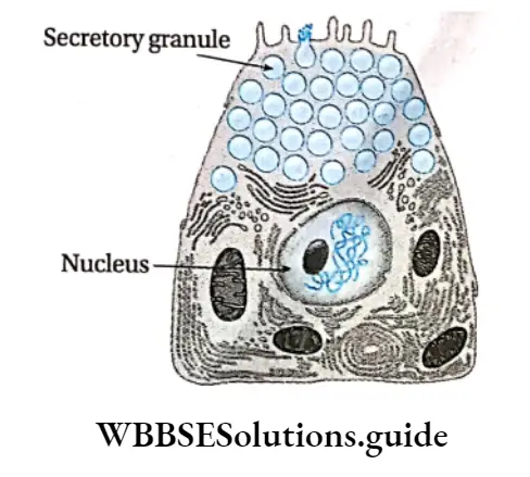
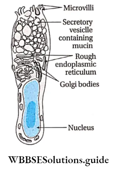
Cellular organization: In this case, several cells together form a body, but do not form any tissue. Members of the phylum Porifera have this kind of body organization.
Histological organization: In this case, several ceÿs together form tissues, and several tissues together form the body. Members of phyla Cnidaria and Platyhelminthes have this kind of body organization.
Morphological organization: In this case, several cells form tissue, several tissues form an organ, and several organs form an organ system. Several such organ systems form the body. Members of phylum Nemathelminthes and other higher phyla show this kind of body organization.
Body plan
Different multicellular animals show different types of body plans. The types of body plan are cell aggregate, blind sac, and tube within tube plan.
Cell aggregate plan: In the case of animals without tissues (cellular organization), several cells aggregate to form the body. This type of body plan is called the cell aggregate plan. Animals belonging to the Phylum Porifera show this type of body plan.
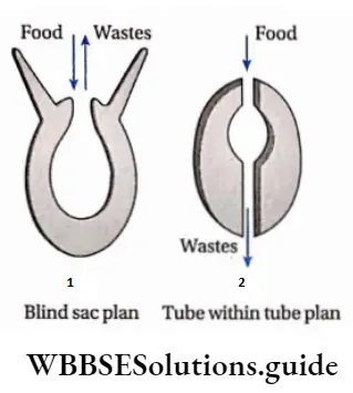
Blind sac plan: In the case of animals with tissues, a common opening (acting as both mouth and anus) is present, along with a blind sac-like structure or cavity. This type of body plan is called a blind sac plan. Animals belonging to the Phyla Cnidaria and Platyhelminthes, show this type of body plan.
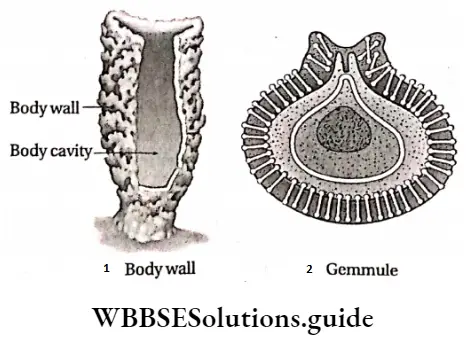
Tube within tube plan: In the case of higher animals that have organ systems, the body plan is that of a tube within another tube. The outer tube is the body wall, while the inner tube is the alimentary canal.
Classification Of Animal Kingdom
This type of body plan is called a tube-within-tube body plan. All the animals from Phylum Nemathelminthes to Phylum Chordata, show this type of body plan. This body plan is further divided into two types—protostome and deuterostome.
Protostome: When the blastopore in the embryo develops into the mouth, the body plan is called the protostome type. This implies that the mouth develops before the anus. This type of body plan is found in the members of the phyla Platyhelminthes, Nemathelminthes, Annelida, etc. These animals are known as protostomia.
Deuterostome: When the blastopore in the embryo develops into the anus, the body plan is called deuterostome type. This implies that the anus develops before the mouth. This type of body plan is shown by members of the Phyla Echinodermata and Chordata. These animals are known as deuterostomia.
Symmetry Of The Body
Multicellular organisms show both symmetry and asymmetry of the body.
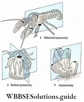
Symmetry: The similarity in the arrangement of organs along an axis that, divides the body into two equal halves, is called symmetry. Such organisms that show symmetry are called symmetrical. Different types of symmetry are discussed below.
Bilateral symmetry: In this type, the body of an organism can be divided into two equal halves along a longitudinal plane of division. Such organisms are called bilaterally symmetrical. In these organisms, both halves of the body appear as mirror images of each other (with respect to shape, structure, and color). Example Some invertebrates (Phyla Annelida, Arthropoda, etc.) and phylum Chordata (Fish, amphibians, reptiles, birds, and mammals).
Radial symmetry: The type of symmetry in which the body of an organism can be divided into equal halves along any vertical plane passing through the central axis of the body, is called radial symmetry. Such organisms are called radially symmetrical. Examples are Hydra, starfish, sea cucumber, etc.
Radial symmetry is further divided into two types—
- Biradial and
- Pentaradial symmetry.
Biradial symmetry: In this case, a body can be divided across the central axis, along two vertical planes. For example, Animals that belong to phylum Ctenophora.
Pentaradial symmetry: In this case, a body can be divided across the central axis, along five vertical planes, into five equal parts. Example Certain animals such as starfish, under Phylum Echinodermata. The larvae of echinoderms are bilaterally symmetrical, but the adult forms are pentaradially symmetrical.
Spherical symmetry: In this type, a spherical body, can be divided into two equal halves, along any plane. Such organisms are called spherically symmetrically. Example Noctiluca.
Asymmetry: This occurs when the body of the animal can never be divided into two or more equal parts. Such animals are called asymmetrical in nature. Example Amoeba.
Body axis and body surface
Different characteristics are observed in different animals, according to body axis and body surface. These include the ends of the body and different types of P|anes that Pass through it. These have been discussed below.
Anterior end: The part of the body, carrying the mouth, that is the first to move forward, during locomotion, is called the anterior end.
Posterior end: The part of the body, (opposite to the anterior end), that is the last to move forward, during locomotion, is called the posterior end.
Aboral end: The end of the body that lies opposite to the mouth, is called the aboral end.
Oral end: The end of the body, which bears the mouth, is called the oral end.
Proximal end: The part of any organ, that remains closer to the main body, is called its proximal end.
Distal end: The part of any organ, that remains at a distance from the main body is called its distal end.
Ventral plane: It is the plane of the body which remains closer to the ground. In the case of vertebrates, the plane that contains the reproductive organs and lies opposite to the spinal cord is called the ventral plane. This plane lies in front of the alimentary canal.
Dorsal plane: It is the plane of the body which remains away from the ground. In the case of vertebrates, it is the plane that contains the spinal cord. This plane lies behind the alimentary canal.
Lateral planes: The two planes on both sides of the body are called lateral planes.
Planes of Division
Different planes across which an animal body may be divided, are called planes of division. These planes of division have been discussed below.
Frontal or coronal plane: The plane extending along the longitudinal axis, parallel to the dorsal and ventral plane, is called the frontal or coronal plane.
Transverse plane: The plane extending perpendicular to the longitudinal axis, is called the transverse plane. The section of the body along the transverse plane is called the transverse section. The section of the body along the longitudinal or transverse plane, that passes through the vertical regions of the body, is called the vertical section.
Sagittal plane: The plane that divides the animal body into left and right parts, is called the sagittal plane. It extends over a longitudinal and dorsal-ventral axis. The section of the body along the longitudinal axis, is called longitudinal or sagittal section.
Metamerism or Segmentation
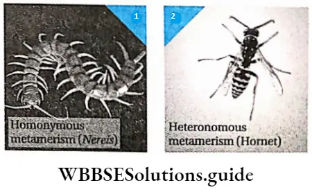
The process by which an animal body can be divided, both externally and internally, into linearly arranged, repeating, identical segments, is called metamerism or metamerisation. Each segment is known as a metamere or somite. Metamerisation is observed in some animals under the phylum Annelida, phylum Arthropoda, and phylum Chordata. Metamerism is of two types—homonymous and heteronomous.
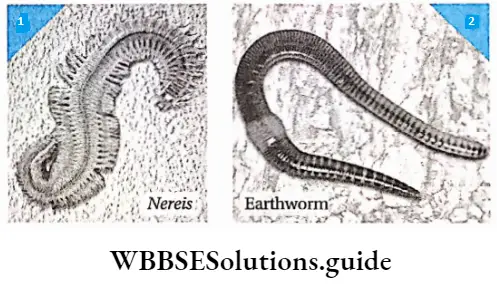
Homonymous metamerism: This type of metamerism is characterized by similar types of segments or metameres. Such type of metamerism is found in animals under Phylum Annelida. Example Nereis sp.
Heteronomous metamerism: This type of metamerism is characterized by different types of metameres. Out of these different metameres, the similar ones together perform similar functions. Such type of metamerism is found in animals under the phylum Arthropoda, Class Insecta. Example Hornet.
Tagmatisation
The process by which several adjacent segments of an animal body are grouped into larger specialized functional units or stigmas is called tagmatisation. These are observed in animals under the phylum Arthropoda.
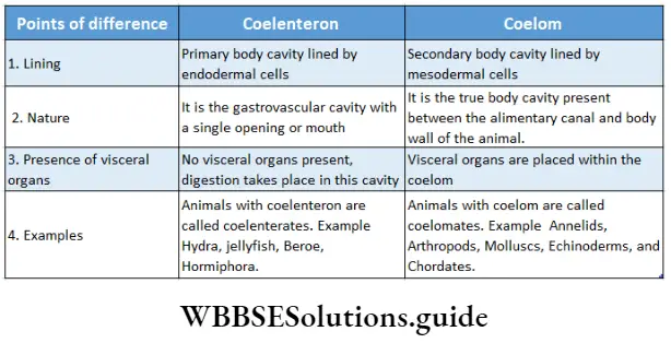
Coelom
The cavity that is covered both externally and internally by the peritoneum, extending up to the alimentary canal and body wall, is called coelom. Coelom is formed from mesoderm. Hence, it can be considered as an intermediate structure lying between somatic mesoderm (parietal layer of mesoderm) and splanchnic mesoderm (visceral layer of mesoderm). It is found in triploblastic organisms.
Types of coelom
The two types of coelom based on its origin, are described below.
Schizocoelic coelom: The coelom that has developed from a mesodermal chord is called schizocoelic coelom. This type of coelom is found in members of Phyla Annelida, Arthropoda, and Mollusca.
Enterocoelic coelom: The coelom that has developed from a sac-like structure (mesodermal pouch), produced from a primitive alimentary canal, is called enterocoelic coelom. It differentiates mesoderm and endoderm. This type of coelom is found in members of the phylum Echinodermata and Chordata. According to the presence of coelom, animals are of three types— acoelomate, pseudocoelomate, and coelomate.


Functions of coelom:
- It separates the muscles in the alimentary canal from those in the body wall. This makes the circulation easier within the muscles. It also allows the internal organs to maintain their position within the body.
- In some animals, excretory products and matured germ cells are collected within the coelom.
Coelomic fluid: The fluid present within the coelom, is called the coelomic fluid. This fluid functions as a hydrostatic endoskeleton (found in phylum Annelida) or circulatory medium for the transport of gases, nutrients, and wastes.
Coelomoduct: The duct produced from the mesoderm membrane, that connects the coelom to the external environment in some invertebrates, is called a coelomoduct. This duct is responsible for carrying the germ cells or excretory wastes out of the body.
Haemocoel: The cavities that carry blood or other fluids, within the body of invertebrates with the open circulatory systems, are called hemocoel. The fluid that fills the hemocoel is called haemocoelic fluid. Such animals are called haemo coelomates. This type of hemocoel is found in members of Phyla Arthropoda and Mollusca. They are also found in some members of phylum Annelida such as leeches.
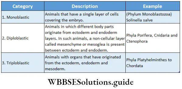
Germ Layers or Germinal Layers
The cellular layers of the embryo, from which different organs are formed, in the gastrula stage of the embryo, are called germ layers.
Types: Germ layers are of three types—
- ectoderm,
- endoderm, and
- mesoderm.
Ectoderm: The outermost layer of the gastrula, that forms the epidermis and epidermal outgrowths (such as ectodermal scales, feathers, nails, horns, enamel of teeth, etc.) is called ectoderm. Most of the nervous system and a part of the alimentary system also develops from the ectoderm.
Mesoderm: The intermediate layer, within the external and the internal layers of the gastrula, is called the mesoderm. Later, this layer forms the muscles, blood vessels, internal parts of the skin, etc.
Endoderm: The innermost layer of the gastrula, which later on forms the alimentary canal and its corresponding glands, is called the endoderm. A part of both the excretory and respiratory systems also develop from this layer.
Classification of animals according to germ layers: According to the types of germ layers, animals are divided into three groups—monoblastic, diploblastic, and triploblastic.
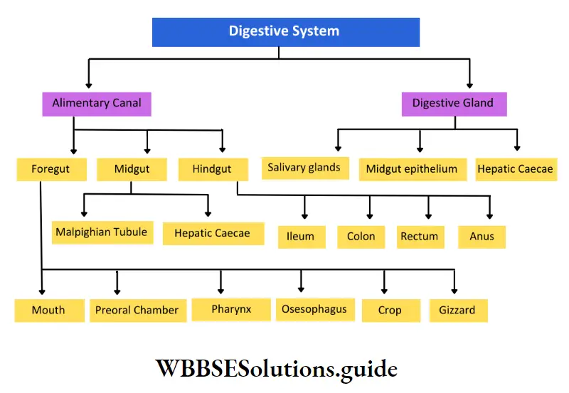
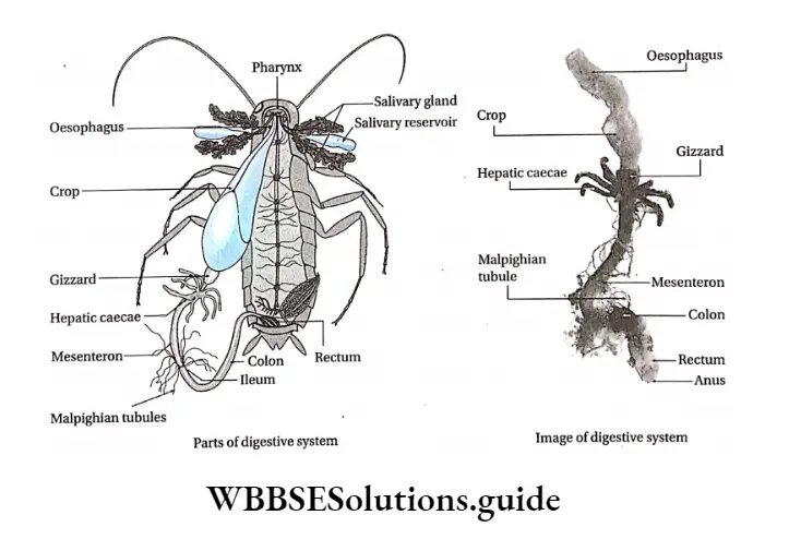
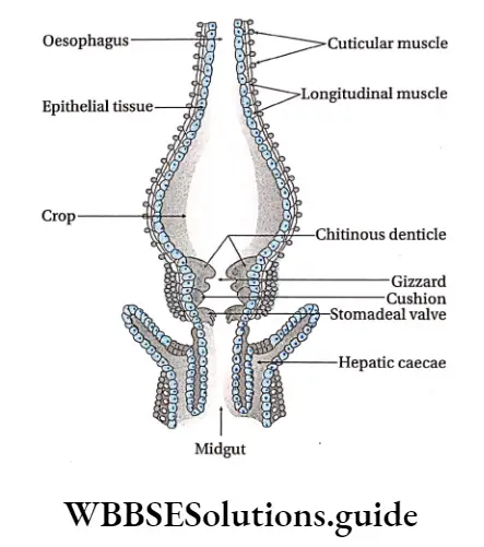
Digestive system
The organ system that helps to take in food, digest it, and absorb some of it, while ejecting the rest of it is called the digestive system. The central tube-like structure that is present in the animal body, to which other digestive organs remain associated, is called digestive organs remain associated, is called the digestive tract or alimentary canal.
Generally, there are two types of alimentary canals complete and incomplete.
Complete digestive tract: In this type of digestive tract, separate openings as the mouth and anus are present. Animals under Phyla Aschelminthes, Chordata, etc., have this type of digestive tract.
Incomplete digestive tract: In this type of digestive tract, only one opening is present, that serves as both mouth and anus, i.e, they are not separate Animals belonging to Phyla Cnidaria, Ctenophora, Platyhelminthes, etc., have this type of digestive tract.
Class 11 biology animal kingdom notes with diagrams
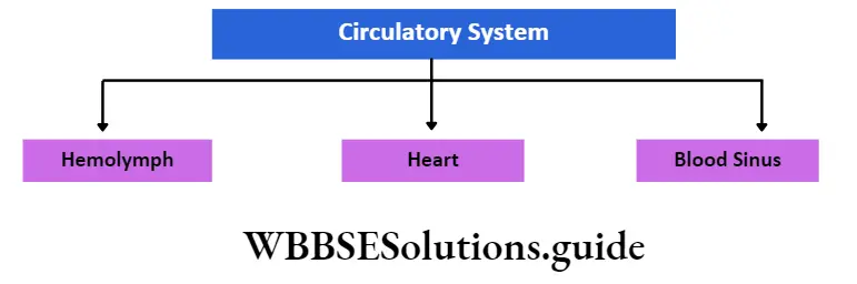
Circulatory System
The system that transports important constituents to the specific cells and their released products to the specific organs, both through a liquid medium, is called the circulatory system.
Circulatory systems are of two types—
- open and
- closed.
Open circulatory system: In this type of circulatory system, the circulating fluid does not flow through the blood vessels. Instead, it is released into the lumen. Animals under Phyla Arthropoda, Mollusca, etc., have this type of circulatory system.
Closed circulatory system: In this type of circulatory system, the circulating fluid flows through the blood vessels. It is not released into the lumen. Animals under Phyla Annelida, Chordata, etc., have this type of circulatory system.
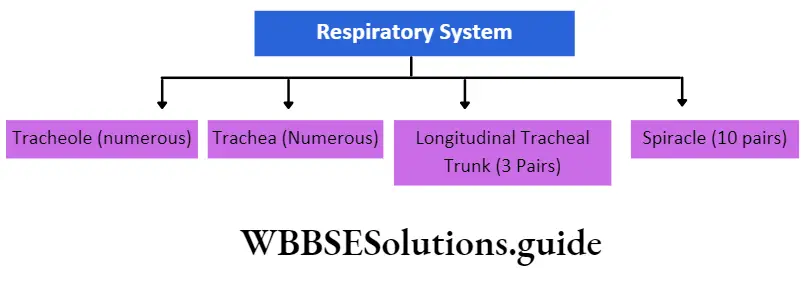
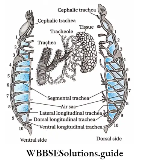
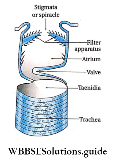
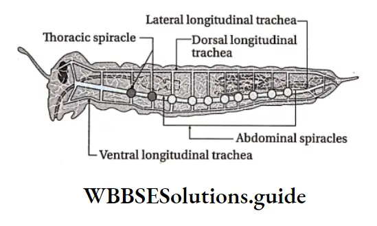
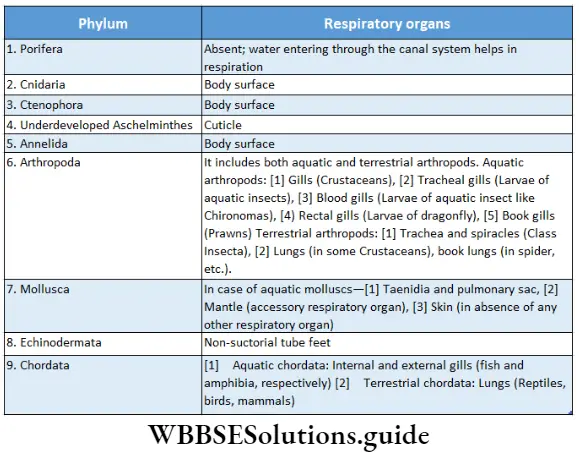
Respiratory system
The organ system that helps in the exchange of gases and in respiration, is called the respiratory system. With the rise in the complexity of the structural organization of the animals, the respiratory system is also modified. Some of them are discussed below.
Excretory system
The organs that help to carry out excretion are called excretory organs. Together they constitute the excretory system. Different animals have different types of excretory organs. Some of such organs have been mentioned in the table below.
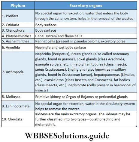
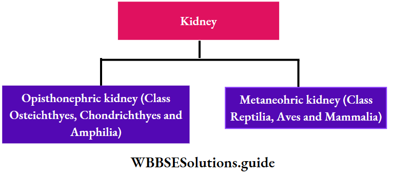
The most primitive type of vertebrate kidney is pronephros. It is functional in early larvae of fishes and amphibians.
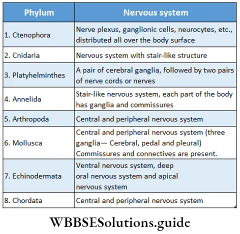
Nervous system
The system by which animals regulate and coordinate their various physiological processes, due to the effect of external and internal stimulants, is called the nervous system. Types of nervous systems found in animals of different phyla have been mentioned in the table below.
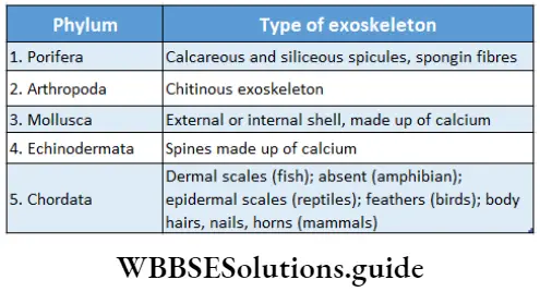
Skeletal system
The system which forms the skeleton of the animal body is called the skeletal system. There are two types of skeletal systems.
They are as follows—
- endoskeleton and
- exoskeleton.
Endoskeleton: The skeleton formed inside the body, in the case of vertebrates is called the endoskeleton.
Exoskeleton: The skeleton formed outside the body, in the case of invertebrates and some vertebrates, is called an exoskeleton. Some of these have been listed in the given table.
Fertilisation
Fertilization is a stage in sexual reproduction, where the sperm and ovum fuse to form the zygote which forms the embryo.
The different types of fertilization are—
Types according to the site of occurrence There are two types of fertilization according to the site of occurrence.
- External fertilization: This type of fertilization takes place outside the body. This is seen in the members of Class Pisces and Amphibia.
- Internal fertilization: This type of fertilization takes place inside the body. This is seen in the members of Class Reptilia, Aves, and Mammalia.
Types according to the nature of fertilization There are two types of fertilization according to its nature.
- Self-fertilization: This process takes place as the egg and sperm, produced in the same animal, fuse to form the zygote. It occurs mostly in bisexual animals. Example Tapeworm.
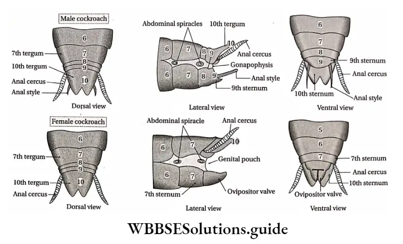
- Cross-fertilization: This process takes place as the egg and sperm, produced by two different animals, fuse to form the zygote. It occurs mostly in unisexual animals Example cockroaches, frogs, human beings, etc. Even though roundworm is bisexual, it shows cross-fertilization.
Reproduction
The process by which an organism produces its offspring is called reproduction. Different types of reproduction are discussed below.
Vegetative reproduction: This process by which an animal produces its offspring from a part of its own body, by cell division. Found in Protozoans.
Parthenogenesis: By this process, an animal produces its offspring from an unfertilized egg. Found in male wasps, male bees, etc.
Asexual reproduction: By this process, an animal produces its offspring by division, regeneration, or spore formation. Found in Planaria.
Sexual reproduction: By this process, an animal produces its offspring by fusion of its gametes. Found in all vertebrates and higher invertebrates.
Body temperature
According to body temperature, animals are of two types—poikilotherms and homeotherms.
Poikilotherms: The animals in which the body temperature varies with the external environment, are called poikilotherms or ectotherms. Example Amphibians, reptiles, etc.
Homeotherms: The animals that have their body temperatures constant irrespective of changes in the temperature of the external environment, are called homeotherms. The homeotherms that have an internal regulation system for body temperature are called endotherms. Examples, Birds and Mammals.
Sex organs
The animals that can reproduce sexually have reproductive or sex organs. These organs also help in determining the sex of the animals. They are of two types
Unisexual: The animals that have only one type of reproductive system (either male or female), are called unisexual. Examples Human beings, cockroaches, etc.
Bisexual: The animals that have both types of reproductive systems (male and female), are called bisexual. Example Roundworms, tapeworms, etc.
Classification Of Animals
Scientists have classified animals according to different criteria. R.H. Whittaker has classified the animal kingdom into 26 animal phyla, consisting of more than 1 million known animal species. Each phylum includes animals with similar characteristics.
Out of these, there are 11 major phyla (phylum Hemichordata, though classified as a major phylum, is considered to be a part of phylum Chordata. Hence, it has not been shown in the chart). The phylogenetic tree of the animal kingdom showing the major phyla only is given below.

Other than these major phyla, there are certain minor phyla within the classification. These minor phyla have not been explained to avoid the complexity of the chapter.
Subkingdom Classification
The Kingdom Animalia is further divided into subkingdoms. They are— Protozoa and Metazoa
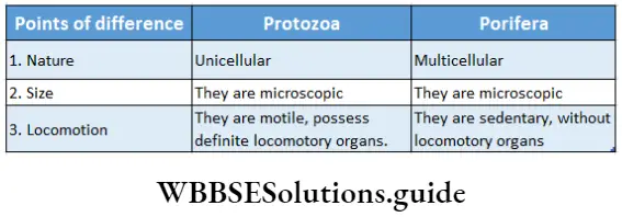
Subkingdom Protozoa
[Latin proto: first; zoom animal]
Protozoa is a group of simple, unicellular, microscopic organisms with different types of locomotory organelles. The term ‘Protozoa’ was coined by Goldfuss (1820). The group comprises about 80,000 species.
General features:
- Simple, unicellular, microscopic animal, either free-living or symbiotic or parasitic.
- The nucleus is single, vesicular (nucleus contains more nucleoplasm but less chromatin), or compact (nucleus contains less nucleoplasm but more chromatin) in majority. Few protozoa are multinucleated.
Phylum Porifera
(Latin porus: pore;/erre: to bear)
Animal Kingdom Biology
Porifera is a group of multicellular animals, sedentary in habit. They have numerous pores on their body surfaces. They have distinct canal systems lined internally by specialized cells called choanocytes. They are multicellular without the development of organ systems, division of labor, and specialization at the cellular level —cells function more or less independently. This phylum comprises about 10,000 species.
General features: The general features of members of the phylum Porifera are discussed below.
Nature:
- Most of them are aquatic, sedentary, and sessile inhabit.
- They may remain solitary or in colonies.
- The body is multicellular, elongated, cylindrical, sometimes branched, and irregular in shape.
- They are diploblastic with an outer ectoderm (pinacoderm) and an inner endoderm (choanoderm) separated by a non-cellular, jelly-like, loose mesenchyme layer. True body cavity or gut is absent.
- The body is asymmetrical or radially symmetrical.
Pores: The body is perforated with numerous minute pores, called ostia. Water enters through the ostia into a cavity called spongocoel or paragastric cavity. This water ‘released body, through a single, large aperture, at one end of the body, called an osculum. Separate mouth and anus are absent in the body.
Canal system: They possess a branched network of canals known as the canal system. Water containing food particles and oxygen enter through the Ostia. The canal system helps to circulate the water throughout the body.
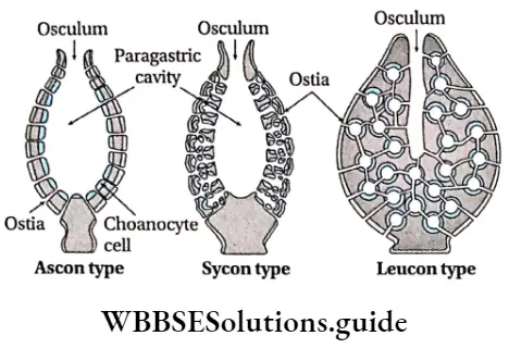
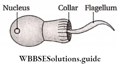
Choanocytes and other cells; The canal system is lined by a type of flagellated cells, called choanocytes. The movement of their flagella creates a water current which helps in the movement of the cells. These cells also help in the ingestion of food and the movement of sex cells, etc.
There are other cells like amoebocytes, pinacocytes, etc. Amoebocytes possess pseudopodia which helps in the movement of the cells, while pinacocytes provide contractibility.
Organ system: Though they are multicellular, they lack an organ system.
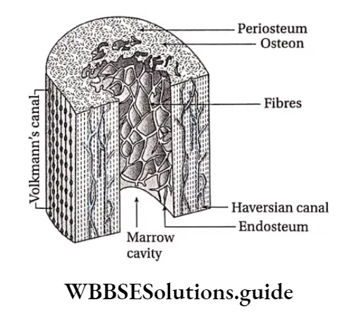
Endoskeleton: The body is provided with a large number of minute, pointed calcareous or siliceous spicules and spongin fibers, that form the internal skeletal framework.
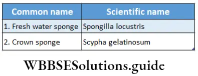
Reproduction: Reproduction occurs asexually by budding, fragmentation, and by formation of reproductive bodies, called gemmules. The sexual mode of reproduction occurs through male and female gamete formation. They possess the ability to regenerate lost body parts.
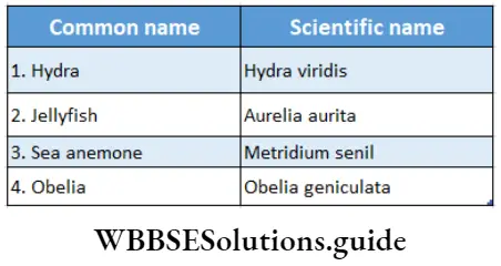
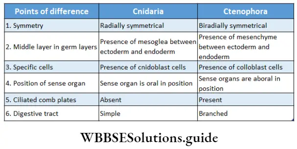
Phylum Cnidaria
(Greek Cnidos: thread)
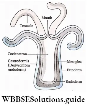
Cnidarians are a group of diploblastic, radially symmetrical animals. They possess special sensory cells (cnidoblast cells) and a gastrovascular cavity (coelenteron) with a single opening, called a mouth. In this phylum, the tissue grade of organization is observed for the first time. Cnidarians are considered true multicellular animals.
General features: The general features of the members of the phylum Cnidaria are discussed below.
Nature:
- Cnidarians are mostly marine while few are freshwater.
- They are multicellular and acoelomate in nature.
- The body is radially symmetrical, diploblastic with jelly-like, non-cellular mesoglea in between the outer ectoderm and an inner endoderm.
- They may remain solitary or form colonies.
- They may be unisexual or bisexual.
Cnidoblasts: They possess special sensory cells called cnidoblasts or nematoblasts, within the tentacles at the oral end. The cnidoblasts contain a sac called a nematocyst, which contains a tube. This tube may contain structures such as barbs and barbules, that help in offense, defense, and food capture. The cnidoblasts burst and release the barbs when touched or sensitized.
Coelenteron: The body has a central cavity, called coelenteron or gastrovascular cavity, lined with endodermal cells. It has a single opening, the mouth at the oral end.
Mouth: The mouth is present on the hypostome, encircled with tentacles in one or more whorls.
Digestion and organ systems: Digestion is of two types—extracellular, within the gastrovascular cavity, and intracellular, within the endodermal cells. Respiratory, circulatory, and excretory systems are absent. A primitive type of nervous system is present with a diffused network of nerve cells.
Reproduction: Asexual reproduction takes place through fission and budding, while sexual reproduction takes place through gamete formation.
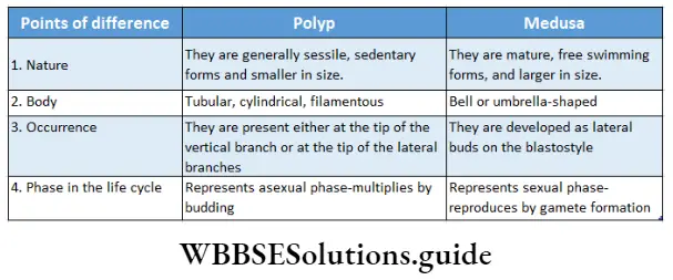
Life cycle: The life cycle has two phases, hence polymorphism is distinct. It has an asexual phase called polyp (cup-shaped) and a sexual phase called medusa (umbrella-shaped). They also show alternation of generations and metagenesis. Planula larva is found in the life cycle.
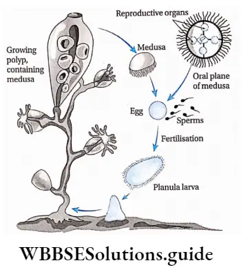
Polymorphism: The occurrence of structurally and functionally different forms within the same animal during its life history is known as polymorphism. In colonial Cnidarians, such as Obelia, polyp, and Medusa occur in different forms performing different functions.
Metagenesis: The interconversion of asexual and sexual phases is known as metagenesis.
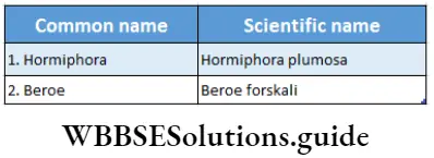
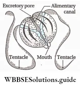
Phylum Ctenophora
(Greek Ktenos: comb; photos: bearing)
Ctenophorans are commonly known as sea walnuts or comb jellies. Ctenophorans represent a small group of animals with only 50 known species, distributed abundantly in the coastal waters. The first description of Ctenophora was given by Martens (1671). Hatschek (1839) placed Ctenophora under a distinct phylum.
General features: The general features of the phylum.
Ctenophora are discussed below.
Nature:
- Ctenophorans are exclusively marine animals.
- The members may exist as single units or in colonies, devoid of any skeleton-like structures.
- The body is transparent, flat, and biradially symmetrical.
Locomotion: They swim freely with the help of eight vertical ciliated comb plates. These plates are present throughout the body, at regular intervals.
Colloblasts: The tentacles, if present, occur in pairs and opposite to each other. Special adhesive cells called colloblast cells or lasso cells are present on the tentacles. Other than these, smooth muscle cells, nerve cells, amoebocytes, etc., are present. Nematocysts are absent.
Germ layers: The body is diploblastic with mesenchyme in between the outer ectoderm and inner endoderm, containing muscle cells and amoebocytes.
Short notes on animal kingdom for quick revision
Sensory organ: Statocyst is a sensory organ, present at the aboral end (the end of the body opposite to that of the mouth).
Digestion: The gastrovascular system or coelenteron is complete with a mouth and a highly branched digestive tract. The digestive system can perform both extracellular and intracellular digestion.
Organ systems: Respiration takes place by diffusion. The nervous system is underdeveloped but the sub-epidermal network of nerves is well developed beneath the comb plates.
Life cycle: The members of this phylum are hermaphrodites. Sexual reproduction is common and fertilization is external. No alternation of generation is seen in the life cycle. Cydippid larva is a ciliated spherical type of larva, found in their life cycle.
Similarities between Cnidaria and Ctenophora
There are some similarities between the phylum Cnidaria and Ctenophora.
They are as follows—
- Both are diploblastic in nature.
- Both have coelenteron.
- Both have tentacles.
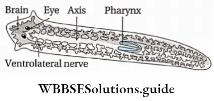
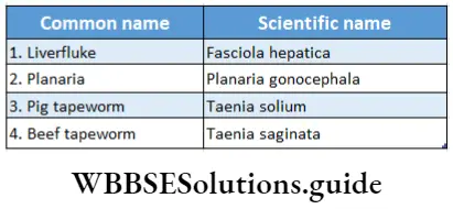
Phylum Platyhelminthes
(Greek Platy: flat; helminth: worm)
Phylum Platyhelminthes is a diverse group comprising 25,000 living species.
General features: The general features of the members of the phylum Platyhelminthes are discussed below.
Nature:
- The body is thin, dorsoventrally flattened, and bilaterally symmetrical.
- The body is ribbon-like or leaf-shaped, covered by a syncytial membrane.
- The true body cavity is absent, hence, acoelomate (a = without; coelom = body cavity).
- The members may be parasitic or free-living.
Germ layers: Triploblastic organization with ectoderm, endoderm, and mesoderm. Mesoderm is present between ectoderm and endoderm.
Segmentation: Metameric segmentation is absent.
Organ systems: Alimentary canal is incomplete. The mouth is present, but the anus is absent. Excretory organs are protonephridia or numerous flame cells or solenocytes. Circulatory and respiratory systems are absent. Gaseous exchange occurs by diffusion. The nervous system is simple, ladder-like with a brain and a double ventral nerve cord. The organ system is absent or reduced in parasitic forms.
Reproduction: They are hermaphrodite in nature. A complex reproductive system is generally present. Asexual reproduction occurs by transverse fission. Free-living Planaria has the power of regeneration.
Life cycle: The life cycle is completed through one or two hosts. Different larval forms (like cysticercus cellulose in pig tapeworm) are usually present.
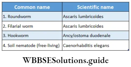
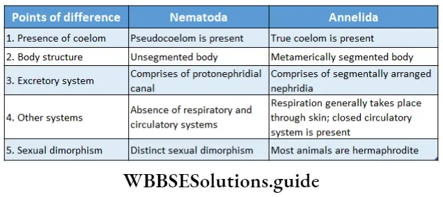
Phylum Nemathelminthes or Nematoda
(Greek Nematos: thread; eidos: form)
Nematodes are a diverse group of organisms comprising 15,000 living species. The members of this phylum were initially included in the phylum Aschelminthes(Grobben, 1910).
General features: The general features of the members of the phylum Nemathelminthes or Nematodes are discussed below.
Nature:
- The body is bilaterally symmetrical, thread-like, cylindrical, elongated, unsegmented, tapering at both ends, covered with cuticle.
- They are mostly parasitic, while some are free-living.
Germ layers: They are triploblastic and pseudocoelomate (body cavity is present, but not mesodermal in origin), with pseudocoelomic fluid.
Body wall: Longitudinal muscles are present in the body wall, but circular muscles are absent.
Digestive system: Alimentary canal is simple, straight with a distinct mouth and anus at opposite ends.
Excretory system: The Excretory system comprises a canal called protonephridial canal, without flame cells. In some cases, a type of cells called rennet cells, carry out excretion.
Nervous system: Nervous system comprises a brain in the form of a nerve ganglia surrounding the gut. This nerve ganglion is called the cricopharyngeal nerve ring.
Other organ systems: Respiratory and circulatory systems are absent.
Reproduction: Sexes are separate with distinct sexual dimorphism. Males are smaller, having a curved tail end with a pair of pineal setae, while females have a straight pointed tail end. Fertilisation is internal. Development is generally direct but in parasitic forms, the life cycle passes through infective larval stages like microfilariae, rhabditiform, etc.
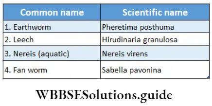
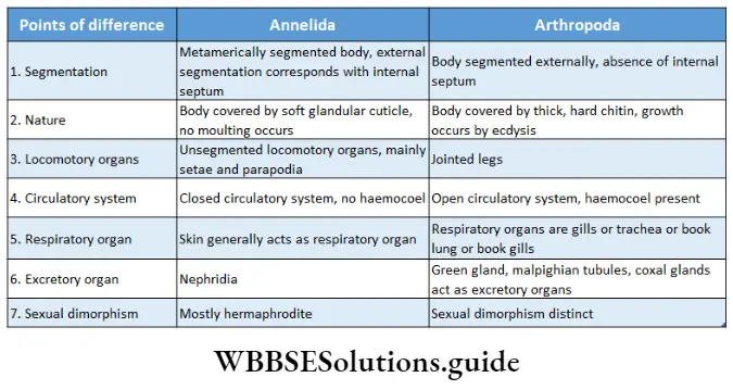
Phylum Annelida
(Latin annulus: ring; eidos: form)
Phylum Annelida exhibits great diversity of body form, comprising approximately 17,000 species. Lamarck (1802) coined the term ‘Annelida’ due to their ring-shaped body segments.
General features: The general features of members of phylum Annelida are discussed below.
Nature:
- The body is elongated, bilaterally symmetrical, and metamerically segmented, into several ring-like structures called somites or metameres.
- The body surface is covered with a thin cuticle.
Body cavity: The body is triploblastic and coelomate with a true body cavity lined by mesodermal cells.
Body wall: Both circular and longitudinal muscles are present in the body wall.
Germ layers: Triploblastic (ectoderm, mesoderm and endoderm are present).
Digestive system: Alimentary canal is complete. It is a muscular tube-like structure, with a separate mouth and anus at opposite ends.
Respiratory system: Respiration generally takes place through the skin; in some forms gills are present.
Circulatory system: Well-developed closed circulatory system with blood vessels. Hemoglobin or haemoerythrin remains dissolved in the plasma. Distinct heart-like structures and blood vessels appear for the first time in Annelida.
Excretory system: It comprises segmentally arranged nephridia.
Nervous system: The nervous system in annelids, comprises of brain or cerebral ganglion, circumoesophageal ring, and a double ventral nerve cord with a segmental ganglion (nerve ganglia arising from each segment). Statocysts, tentacles, etc., are present as sensory organs.
Locomotory organs: Locomotory organs are setae (stiff bristles on the body, that help in attachment, found in earthworms), parapodia (simple, unjointed, locomotory appendages, found in Nereis) or suckers (structures present at both anterior and posterior ends of the body, found in leech).
Reproduction: Most animals are hermaphrodite but some unisexual forms are also found. They show sexual reproduction.
Life cycle: Development may be direct or indirect. A free-swimming, ciliated, marine larva called trochophore larva is found in annelids, during indirect development.
Phylum Arthropoda
(Greek Arthron: jointed; photos: foot)
Kingdom Animalia Phylum Chart
Arthropoda is the biggest phylum with respect to the number of species, presently including approximately 9,00,000 species. They are one of the oldest and the most biologically adapted phylum. They can inhabit practically all spheres of earth—terrestrial, aquatic (sea and freshwater), and air.
General features: The general features of the members of the phylum Arthropoda are discussed below.
Nature:
- The body is triploblastic, bilaterally symmetrical, and coelomate.
- The body is segmented, but the segments are not separated internally by septa.
- Each segment contains a pair of jointed appendages.
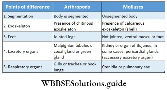
- These organisms have a chitinous exoskeleton and a haemocoelomic body cavity.
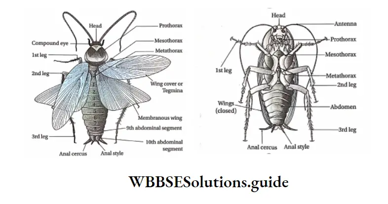
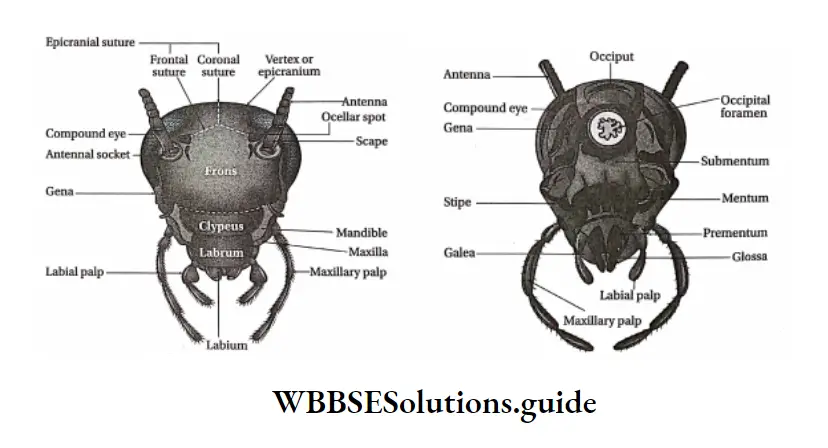
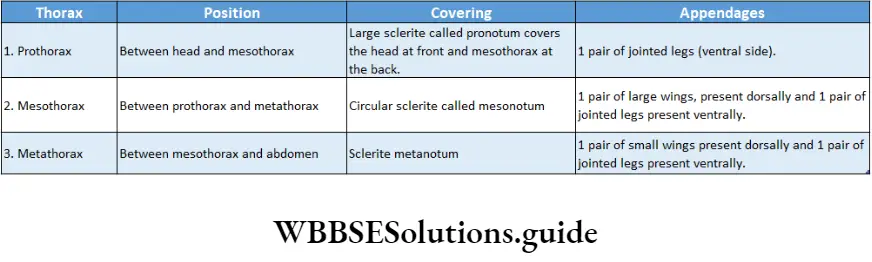
Body: The body is divided into three parts, thorax, and abdomen. In some cases, the head and thoracic regions are fused together, known as cephalothorax.
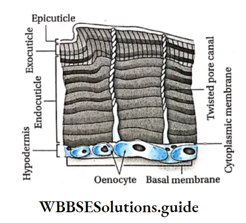
Exoskeleton: The body is enclosed in a thick and tough exoskeleton formed of a nonliving substance, called chitin. The exoskeleton is periodically shed off, being replaced by a new one. This process is called molting or ecdysis.
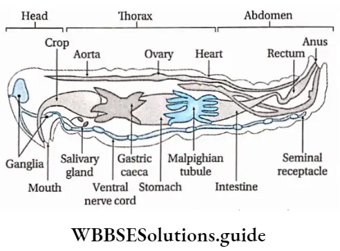
Digestive system: Alimentary system is complete and complex with the mouth and anus at opposite ends.
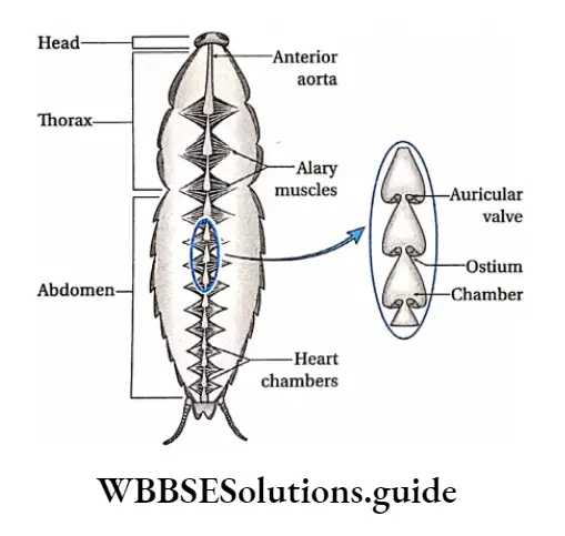
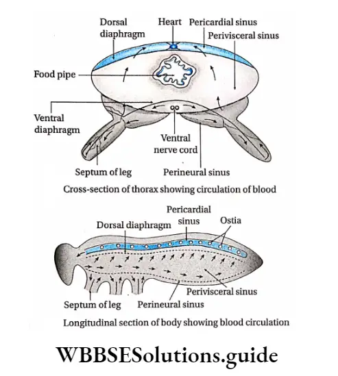
Circulatory system: The circulatory system’ is the open type with a dorsar heart and blood vessels opening into irregular blood-filled spaces, called the sinuses.
Respiratory system: The respiratory organs are gills (in aquatic forms like prawns), book gills (Limulus), trachea (in terrestrial insects), or book lungs (in scorpions) or the body surface which helps in respiration in certain forms.
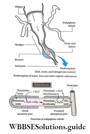
Excretory system: Excretion occurs through Malpighian tubules (coiled tubules, found in insects), coxal g|and (a type S that can co,, etc and excrete urine, found in scorpions) or green gland (a type of gland that acts like kidneys in terms of function, found in Prawn).
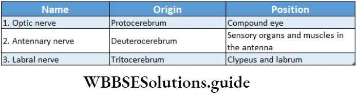
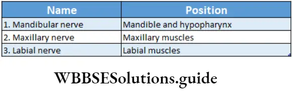
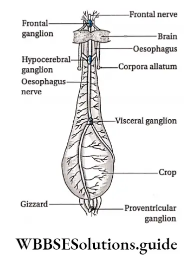
Nervous system: Nervous system is well developed. It is composed of a brain or cerebral ganglion and a pair of solid ventral nerve cords with thoracic and abdominal ganglia.
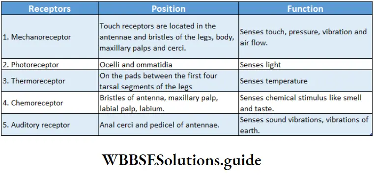
Sense organs are well-developed like antennae or feelers (for tactile sensations), simple or compound eyes (photoreceptors), statocysts (balancing organ), taste receptors on insect feet, and sound receptors in crickets.
Ommatidium: Compound eyes are made up of many similar units, called ommatidia. Each of these units has its own lens and help in the formation of several images (mosaic image).
Life cycle: Prominent sexual dimorphism is seen. Fertilisation is internal. Metamorphosis may be direct without a larval stage or indirect with one or more larval stages (like a caterpillar, grub, etc.).
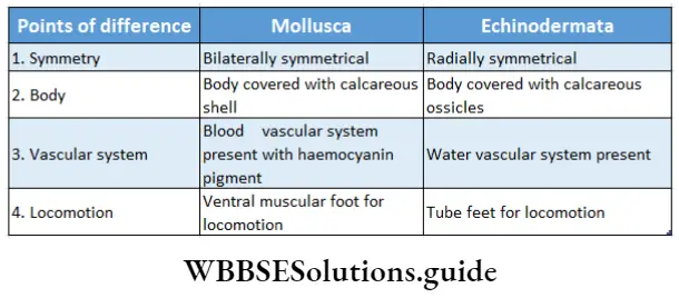
Phylum Mollusca
(Latin mollis: soft)
Mollusca is the second largest invertebrate phylum, after Arthropoda. It contains about 1,00,000 described living species and 35000 known fossil species.
General features: The general features of members of the phylum Mollusca are discussed below.
Nature: The body is soft, unsegmented, triploblastic, and asymmetrical or bilaterally symmetrical.
Body parts: A distinct head with a mouth, eyes, and tentacles are present. Buccal mass contains radula (except Bivalvia).
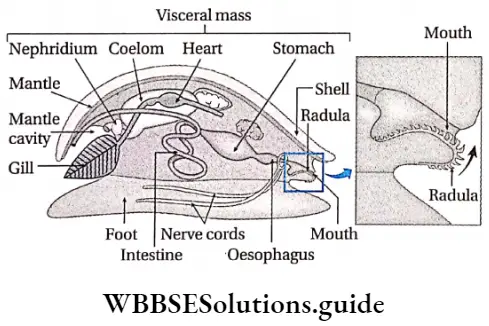
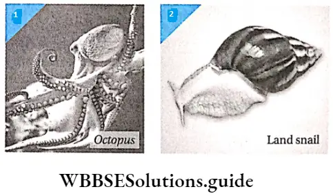
Shell: The body is generally enclosed by a calcareous shell. Mostly, the shell is external except for Loligo sp., Sepia sp., and Octopus sp., which have internal shells.
Mantle: The visceral mass is enclosed within a thick muscular fold of the body wall, called the mantle. The mantle is present below the shell and secretes substances that help in the formation of the shell.
Ventral muscular feet: Most of the species have a ventral, thick, muscular foot for locomotion, except cephalopods (Octopus, Sepia, Loligo). In cephalopods, the foot is modified into a circle of tentacles or arms.
Respiratory organs: Respiration occurs by ctenidia or gills in aquatic forms and lungs or pulmonary sacs in terrestrial forms.
Excretory organs: Excretory organs are kidneys or Organ of Bojanus. Other than these, in some cases, a gland named the pericardial gland or Keber’s gland acts as an accessory excretory organ.
Circulatory system: It is open lacunar type with a dorsal heart and a few blood vessels. Haemocyanin is a respiratory pigment.
Nervous system: The nervous system is well-developed with distinct brains, different ganglia, connectives, and commissures.
Sense organs: Sense organs are mainly eyes and tentacles. Other than this, sensory organ like osphradium (chemoreceptor) is also found. Osphradium can sense the change in the chemical nature of water.
Radula: They have a type of muscular, rasping organ, called radula, that helps in acquiring food.
Reproduction: Sexes are usually separate. Some are hermaphrodite. Fertilization is internal or external.
Life cycle: Development may be direct or through veliger or glochidium larva.
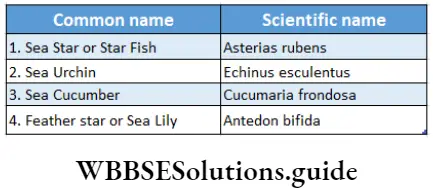
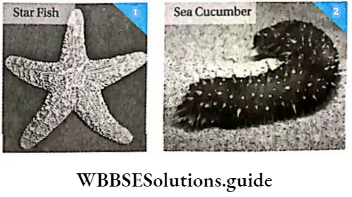
Phylum Echinodermata
(Latin chinos: spiny; derma: skin)
The phylum contains about 7000 living species and 20,000 fossil species. This group was established by Jacobklein (1734) who coined the term ‘echinodermata’for sea urchin.
General features: The general features of the members of the phylum Echinodermata are discussed below.
Nature: The members are exclusively marine, triploblastic, and coelomate.
Skin and endoskeleton: The body is covered with calcareous ossicles or spines. These ossicles together form an endoskeleton.
Symmetry: Adults are pentamerous radially symmetrical and larvae are bilaterally symmetrical.
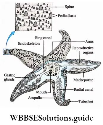
Body ends: Anterior and posterior ends are missing. Distinct oral and aboral surfaces are present.
Tube feet: Ambulacral groove (a tube-like structure with grooves) and tube feet (small flexible appendages, used for locomotion) remain on the oral surface. The tube feet are arranged in rows on both sides of the ambulacral groove. They are responsible for locomotion. A perforated plate called madreporite is present on the aboral surface that permits the entry of water into the water vascular system.
Water vascular system: A characteristic water vascular system consists of various canals such as radial canals, ring vessels, and tube feet. This type of vascular system helps in grasping objects, locomotion, food capture, and respiration.
Organ systems: The Alimentary canal is usually a coiled tube. Excretory and respiratory organs are absent in their body. Respiration occurs mainly by papulae, present on the skin. The nervous system is of reduced primitive type with circumoral ring and radial nerves. The brain is absent.
Reproduction: Sexes are separate. Fertilization is usually external. Development is indirect through different types of larvae (bipinnaria, brachiolaria, and doliolaria in Star Fish, echinopluteus in Sea Urchin, and auricularia in Sea Cucumber) in the life cycle.
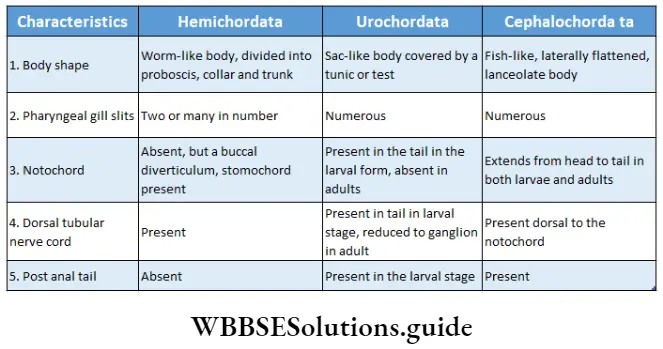
Phylum Hemichordata Or Stomochordata
(Greek hemi: half; Latin chorda: cord)
General features: The general features of the members of phylum Hemichordata or stomochordata are discussed below.
Nature: The members are mostly marine and exist either as single units or in clusters.
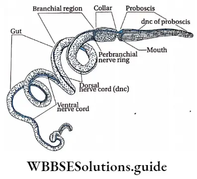
Body: The body possesses true coelom. Worm-like unsegmented body, divisible into proboscis, collar and trunk. Post-anal tail is absent.
Oral diverticulum: A hollow outgrowth called oral diverticulum arises from the roof of the buccal cavity and extends up to the proboscis. Previously, it was regarded to be equivalent to notochord. It is now called stomochord.
Pharyngeal gill-slits: The members of this group have pharyngeal gill-slits (except in Rhabdopleura).
Life cycle: It is completed through a free-swimming tornaria larval stage.
Phylum Chordata
(Greek Chorda: string/rod)
There are controversies and doubts among evolutionary biologists and taxonomists regarding the classification of chordates. The animals having the rod-like structure—’notochord’ are included in the phylum Chordata. It was established by Balfour in 1880. It includes about 48,000 living species.
General features: The general features of members of the phylum Chordata are discussed below.
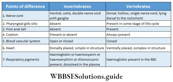
Nature: The body is coelomate, triploblastic, and bilaterally symmetrical.
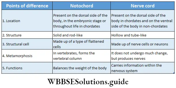
Notochord: There is a flexible, rod-like, slender notochord lying dorsal to the coelom and beneath the dorsal nerve cord. It is either persistent throughout life or present during early embryonic life.
Dorsal nerve cord: A dorsal, hollow, tubular nerve cord, lying above the gut and dorsal to the notochord, is present along the length of the body.
Pharyngeal gill-slits: Paired pharyngeal gill slits are found either during the early embryonic life or throughout the organism’s life span.
Tail: Post-anal tail is found in a majority of chordates.
Kingdom Animalia Phylum Table
Similarities between members of Echinodermata and Chordata: Both have triploblastic embryonic layers. Both have true coelom and body cavities. Both are unisexual and undergo sexual reproduction. Though members of the phylum Echinodermata are radially symmetrical, their larvae are bilaterally symmetrical like members of the phylum Chordata.
The classification of chordates has been done mainly on the basis of the scheme of classification by J.Z. Young.
According to this classification, phylum Chordata is divided into four subphyla—
Hemichordata or Adelochordata, Urochordata or Tunicata, Cephalochordata or Acraniata and Vertebrata or Craniata. The above-mentioned scheme of classification is discussed below.

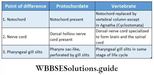
Hemichordata, Urochordata, and Cephalochordata are known as ‘Protochordates’ (means primitive chordates) as they possess a very simple type of body organization. They are also known as ‘invertebrate chordates’ as they lack vertebral columns.
Subphylum Hemichordata or Adelochordata
According to Barnes et al. (1993), the subphylum comprises nearly 100 known species. Hemichordates are considered as half-chordates because they possess at least half of the main features of the chordates, like pharyngeal gill slits and dorsal nerve cord.
As a result, a separate non-chordate phylum, Hemichordata, has recently been created by scientists, following a new system of classification. This is the reason why phylum Hemichordata has been placed earlier.
Earlier, scientists assumed a buccal diverticulum of Hemichordates as a notochord but recent research proves no similarity of notochordal cells with the cells of the oral diverticulum of Hemichordates.
Thus, they discarded the idea of the existence of notochord in Hemichordates. The scientists now call the diverticulum a stomochord.
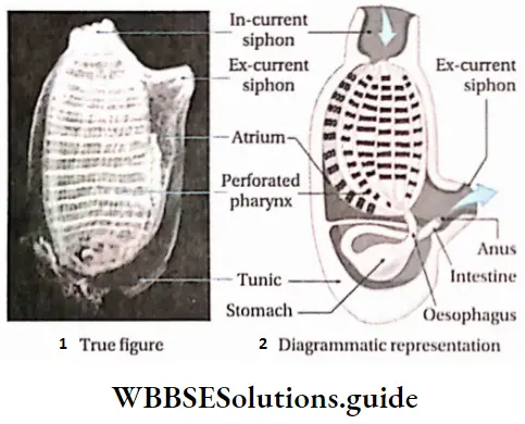
Subphylum Urochordata or Tunicata
(Greek Uro: tail; Latin chorda: rod/string)
Urochordata is a group of animals with a notochord and nerve cord confined to the tail region in the larval stage. The adult lacks notochord. These animals are also called sea squirts.
General features: The general features of the subphylum Urochordata are discussed below.
Nature: The members are sessile, mostly living at the bottom of the sea. The body is sac-like, covered by a; covering called a tunic or test. The tunic is made up of I protein tunicin and polysaccharide cellulose.
Oral and atriopore: The body has two openings— mouth or oral pore and atriopore.
Pharynx: Pharynx is sac-like and perforated with numerous gill slits or stigmata.
Notochord: Larvae are free-swimming tadpoles with a nerve cord and a notochord in the tail region. In adults, the nerve cord is reduced to a ganglion.
Life cycle: The larvae undergo retrogressive metamorphosis (a more developed larva changes into a degenerated adult) and lose the tail and notochord.
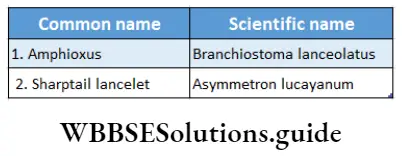
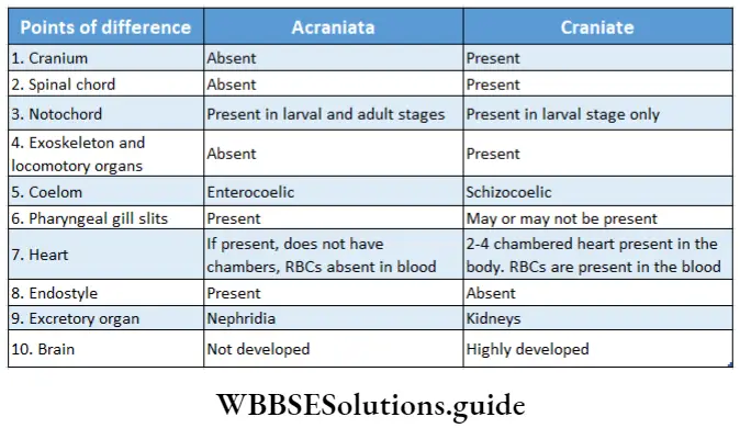
Subphylum Cephalochordata or Acraniata
[(Greek Cephalo: head; chords: rod) (o= without; cranial brain box)]
Cephalochordata is a group of marine animals, having fish-like elongated bodies. The body is provided with a notochord that extends throughout the entire length of the body up to the head region.
General features: The general features of the members of the subphylum Cephalochordata are discussed below.
Nature: The body is fish-like, laterally flattened. It is tapering at the anteroposterior ends, with unpaired dorsal, ventral, and caudal fins. This type of shape of the body is called lanceolate.
Myotome muscles: ‘V’ shaped segmental myotomes are present along the length of the body, on its lateral sides.
Notochord: It extends throughout the length of the body. Cranium is absent.
Nervous system: A dorsal hollow nerve cord is present. Brain and spinal cord, both are absent.
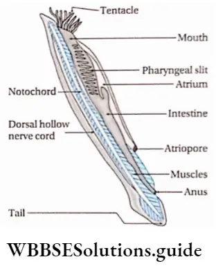
Oral hood: Presence of oral hood with thin tentacle-like strands called buccal cirri. It is surrounded by a transverse, muscular velum, in the posterior wall of the buccal cavity. The velum has a hole with an adjustable diameter. The walls of the buccal cavity in front of the velum, bear several ciliated grooves. These grooves make up the wheel organ that creates a vortex of water current that helps in filter-feeding.
Pharynx: Large, sac-like pharynx perforated with numerous gill slits.
Endostyle: A well-developed endostyle is present on the floor of the pharynx.
Excretory system: The excretory system comprises ciliated nephridia with solenocytes (long, narrow, flagellated cells that help to excrete nitrogenous wastes).
Circulatory system: The circulatory system is without a heart.
Reproduction: Gonads are without ducts. Members are unisexual.
Subphylum Vertebrata or Craniata
Vertebrata is a group of chordates with a well-developed vertebral column. In this group, the notochord is replaced by a bony vertebral column. Hence, they are called Vertebrata. The vertebrates are highly advanced due to their adaptability under various environmental conditions. Subphylum Vertebrata is the dominant group among the chordates. It comprises about 90% of all chordate species.
General features: The general features of the members of the subphylum Vertebrata are discussed below.
Vertebral column: The notochord is replaced by a bony vertebral column in adults. It is composed of several segmented vertebrae.
Cranium: The brain is enclosed within a well-developed, bony, or cartilaginous covering, called a brain box or cranium.
Animal kingdom classification chart with notes
Endoskeleton: The endoskeleton is made up of bones or cartilage.
Brain: The dorsal tubular nerve cord is specialized anteriorly into a complex brain. The rest of the nerve cord forms the spinal cord, extending from the brain to the posterior end of the body along the mid-dorsal line.
Respiratory organ: In aquatic vertebrates, gills act as respiratory organs, while in the case of terrestrial vertebrates, lungs serve the function.
Circulatory system: The circulatory system is of closed type with a ventral muscular heart. The heart shows increasing structural complexity from two-chambered (fish) to four-chambered (birds and mammals) in order to prevent the mixing of deoxygenated and oxygenated blood. Haemoglobin, the respiratory pigment is confined within the red blood corpuscles (RBC).
Excretory system: One pair of kidneys is the primary excretory organ. It is important for nitrogenous waste excretion and osmoregulation.
Nervous system: It is well developed with cranial and spinal nerves.
Locomotory organs: Paired lateral appendages in the form of fins or limbs are present.
Tail: Most vertebrates have post-anal tails. Though it is an extension of the body it is devoid of coelom.
Reproduction: Sexes are separate. They reproduce sexually.
Classification: The subphylum Vertebrata is classified into two superclasses—
- Agnatha and
- Gnathostomata

Both these superclasses are discussed under separate heads.
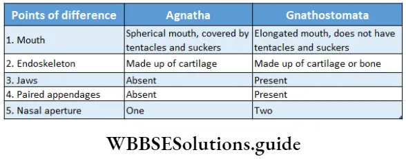
Superclass Agnatha
(Greek A: without; gnathos: jaw)
Agnatha is a group of fish-like aquatic animals, that do not have jaws surrounding their mouth.
General features: The general features of the members of superclass Agnatha are discussed below.
Jaws: The mouth is not bounded by jaws, hence called Agnatha.
Notochord: Notochord persists throughout life
Paired appendages: Paired appendages are absent.
Skin: Skin is soft and slimy without an exoskeleton.
Semicircular canal: One or two semicircular canals are present in the internal ear.
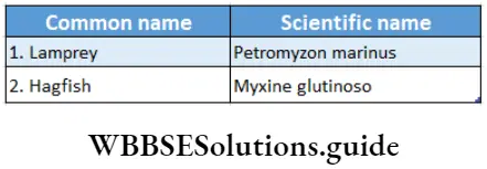
This superclass includes the class Cyclostomata with two orders—Petromyzontiformes and Myxinoidea.
Class Cyclostomata
(Greek Cyclos: circular; stoma: mouth)
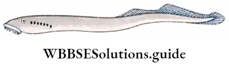
Cyclostomata is a group of jawless vertebrates.
General features: The general features of the members of class Cyclostomata are discussed below.
Body: The body is elongated and cylindrical in shape.
Mouth: The members have a circular and suctorial mouth without a jaw.
Endoskeleton: Endoskeleton is cartilaginous. The notochord is persistent.
Nasal aperture: Unpaired, median nasal aperture.
Skin: Skin is glandular, smooth, and without scales.
Locomotory organs: Median fin is present, paired fins are absent.
Respiratory organs: 5-16 pairs of sac-like gill pouches are present. Hence, Marsipobranch (Greek Marsipos: pouch; bronchia: gills).
Nervous system: Sub-epithelial nerve plexus (branched network of intersecting nerves), ganglionic cells, and neurites (projections from the cell body of the neuron) are present. Presence of lateral line sense organ (a system of sense organs in aquatic vertebrates, used to detect movement and vibration in the surrounding water).
Superclass Gnathostomata
(Greek Gnathos: jaw; stoma: mouth)
Gnathostomata is a group of vertebrates with a pair of jaws surrounding the mouth.
General features: The general features of members of the superclass Gnathostomata are described below.
Jaws: The mouth is bounded by jaws.
Vertebral column: Notochord is replaced by vertebral column.
Appendages: Paired appendages—fins or limbs are present.
Skin: Skin is soft and slimy or dry and covered with scales, feathers, or hairs.
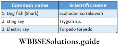
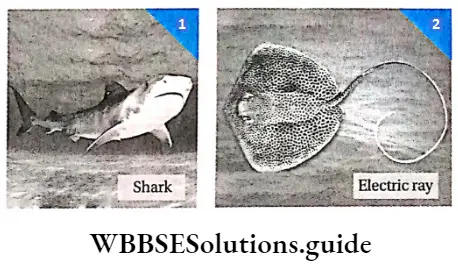
Class Chondrichthyes or Elasmobranchii
(Greek Chondros: cartilage, ichthyic: fish)
Animal Kingdom Types
Class Chondrichthyes is a group of vertebrates with jaws and comprises of approx 600 living species.
General features: The general features of the members of class Chondrichthyes are described below.
Nature: Marine, carnivorous and cold-blooded animals.
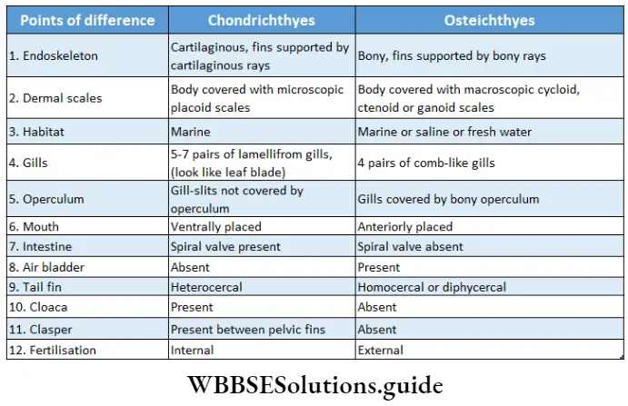
Endoskeleton: The Endoskeleton is cartilaginous.
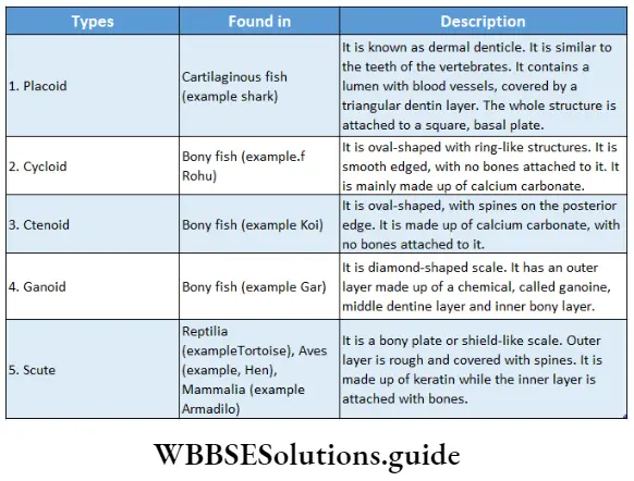
Scales: Body covered with microscopic placoid scales, that are dermal in origin.
Fins: Paired and unpaired fins supported by cartilaginous rays. The tail fin is heterocercal in nature, i.e., the segments of the tail are unequal. Pelvic fins are modified into clasper in males.
Mouth: The shape of the mouth is like a half-moon and it is ventrally placed.
Pharyngeal gill-slits: 5-7 pairs of lamelliform septal gill which open directly outside by 5-7 pairs of gill slits and not covered by operculum.
Swim bladder: Air bladder or swim bladder is absent.
Reproduction: Sexual reproduction is observed Fertilisation occurs internally. These animals may be oviparous or ovoviviparous.
Class Osteichthyes or Teleostomi
(Greek Osteon: bone; ichthyic: fish)
Animal Kingdom Phylum
Class Osteichthyes Is a group of vertebrates with jaws, comprised of about 25,000 living marine and freshwater species.
General features: The general features of the members of the class Osteichthyes are discussed below.
Nature: The members of this class are generally cold-blooded and live in fresh or saline water. They may be herbivorous or carnivorous.
Endoskeleton: The Endoskeleton is bony.
Scales: The body is covered with dermal scales that may be cycloid, ctenoid, or ganoid.
Fins: Paired and unpaired fins supported by bony rays. The tail fin is generally homocercal or diphycercal*. In some cases, the tail fin is heterocercal.
Operculum: Gills are present in 4 pairs, covered by a bony operculum.
Mouth: Mouth is placed anterior to the head.
Swim bladder: Air bladder or swim bladder is present.
Sense organs: Lateral line sense organ is present.
Heart: The heart is two-chambered with one auricle and one ventricle. Conus arteriosus and sinus venosus are present.
Excretory organs: The kidneys are the primary excretory organs.
Reproduction: Sexual reproduction is observed, Males do not have clasper (a pair of appendages in male sharks, insects, etc., for clasping the female during copulation). Fertilization occurs externally.
Lungfish
Lungfishes are freshwater cartilaginous fishes belonging to the subclass Dipnoi under class Osteichthyes. They have air bladder modified into lungs enabling them to breathe air. Today the three surviving genera of lungfishes live only in the southern hemisphere of the earth confined to Africa (Protopterns), South America (Lepidosiren), and Australia (Neoceratodus).
Coelacanth
The coelacanths belong to a rare order of fish that includes two extant species of the genus Latimeria: the West Indian Ocean coelacanth and the Indonesian coelacanth. They are the oldest living fish that have survived on the earth, from the Devonian period, and have retained their primitive features. The coelacanth is considered a Tiving fossil’ due to its apparent lack of significant evolution over the past millions of years.

Class Amphibia
(Greek Amphi: both sides; bios: life)
Amphibia is a class of vertebrates that spend some part of their life cycle on land and the remaining part in water. They comprise approximately 5000 living species.
General features: The general features of the members of class Amphibia are described below.
Nature: During the larval stage, they live mostly in water. During the adult stage, they change their habitat and start living on land. They are cold-blooded animals.
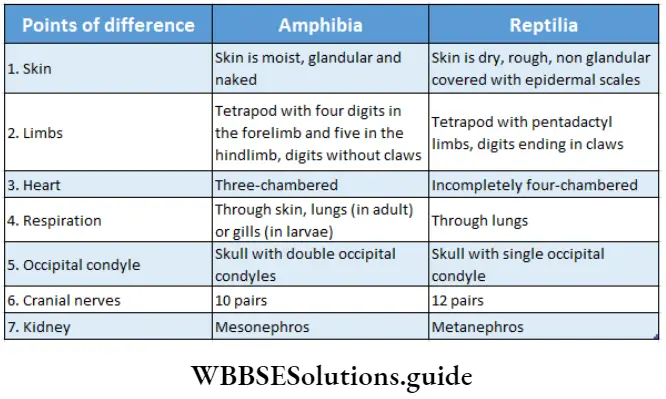
Skin: Body covered with moist, glandular, naked skin. Generally, scales are absent. If present, they remain deeply embedded within the skin.
Body: The body is divided into a head and a trunk. The neck is absent.
Appendages: Tetrapod (2 pairs of limbs) with four digits in the forelimb and five in the hindlimb, digits without claws. Webs are present between the toes.
Respiration: Respiration occurs through the lining of the buccopharyngeal cavity, lungs, or moist skin (in adults) or gills (in larvae).
Heart: The heart is three-chambered with two auricles and an undivided ventricle (mixed heart). In most cases, accessory chambers like sinus venosusm, and conus arteriosus are present in the heart.
Excretory organs: The primary excretory organ is the kidney. The kidney is mesonephric, and the animals are ureotelic.
Cranial nerves: 10 pairs of cranial nerves are present.
Tympanum: Distinct tympanum present, external pinna absent.
Occipital condyle: Skull with double occipital condyle (two rounded knobs present at the base of the skull, which articulates with the first vertebra).
Reproduction: Sexes are separate. They are oviparous and fertilisation takes place in water. A chamber receiving fecal matter and urinogenital product, called cloaca, is present. This chamber opens outside through an opening called the cloacal aperture. Development occurs through an aquatic larval stage, tadpole.
Class Reptilia
(Latin Reptilis: creeping)
Reptilia is a group of terrestrial vertebrates with dry skin covered with horny epidermal scales and clawed pentadactyl limbs. It comprises 6000 living species.
General features: The general features of members of Class Reptilia are described below.
Nature: The members of this group are terrestrial and cold-blooded animals.
Skin: Skin is dry, rough, and non-glandular covered with epidermal scales or spines or scutes.
Body: The body is divided into a head, neck, trunk, and tail. A developed post-anal tail is present.
Skull: Skull with single occipital condyle (monocondylic).
Teeth: Teeth are homodont, pleurodont and polyphyodont.
Respiratory organs: A pair of lungs acts as the respiratory organ.
Heart: The heart is incompletely four-chambered with two auricles and an incomplete ventricle. Sinus venous is present, but conus artists have split into 3 arches.
Locomotory organs: Tetrapod with pentadactyl limbs, digits ending in claws.
Cloaca aperture: Cloaca is present which opens to the outside through transverse cloacal aperture.
Excretory organs: Primary excretory organs are kidneys. These are of metanephros type. The members are uricotelic.
Cranial nerves: 12 pairs of cranial nerves are present.
Sense organs: Lateral line sense organs and external ears are absent. However, the middle and internal ears are present. Jacobson’s organ or Camaro-nasal organs are also present.
Reproduction: Sexes are separate. They are oviparous or ovoviviparous. Fertilization is internal and development is direct. Eggs are large, shelled, yolky, and macrolecithal. Foetal membranes—amnion, allantois and chorion are present.

Dentition of Reptiles
Polyphyodont dentition is the type of dentition in which there is replacement of teeth from time to time. This signifies that jaws are never left without teeth.
Characteristics Of Animal Kingdom
Pleurodont dentition is a type of dentition in which teeth are attached to the inner and upper sides of the jawbone. This brings a larger surface area of teeth in contact with the jawbone and hence attachment is stronger, as in lizards.
Homodont dentition is the type of dentition in which all teeth are functionally and anatomically of the same type, although size may vary. It is found in the majority of vertebrates such as fish, amphibia, and reptiles. Sometimes functionally some teeth may be specialized as fangs, found in snakes.
Class Aves
(Latin avis: bird)
Aves is a class of highly evolved, bipedal, feathered vertebrates, adapted for aerial mode of life.
Birds are regarded as ‘Glorified reptiles’, comprising about 9000 living species.
General features: The general features of members of class Aves, are discussed below.
Nature: They are warm-blooded animals.
Body: The body is streamlined and covered with an exoskeleton, made of feathers. Hindlimbs are covered with scales.
Locomotion: Forelimbs are modified into wings for flight. Hindlimbs are armed with claws for bipedal locomotion.
Beak: Jaws are replaced with beak or bill
Skin: Glands are absent in the skin except the oil gland. Oil glands are also known as the uropygial or preen glands. They are located at the base of the tail. This gland is a holocrine gland, that secretes oil.
Sense organs: Eyes are large, with specialized comb-like structures, called pecten, for acute vision.
Syrinx: A sound-producing organ, syrinx is present
Heart: The heart is four-chambered with two auricles and two ventricles. The right aortic arch is present.
Air sacs: Lungs are provided with nine air sacs for performing double respiration.
Skeleton: Skull with a single occipital condyle, sternum with keel, and pneumatic bones with air cavities (that help in flight) are present.
Excretory system: The kidney is metanephros in nature. These animals are uricotelic (uric acid is the excretory product). The urinary bladder is absent to reduce body weight.
Reproduction: Sexes are separate. Right ovary and oviduct absent. They are oviparous, eggs produced are large, yolky, and macrolecithal with a hard shell. Fertilisation is internal. Foetal membranes—amnion, allantois and chorion present. Development is direct.

Class Mammalia
(Latin mammals: breast)
Mammalia is a class of highly evolved vertebrates adapted to a variety of habitats. This class has several specialized characteristics like mammary glands, left aortic arch, corpus callosum in the brain, pinna, etc.
The term ‘Mammalia’ was given by Linnaeus (1758). The class comprises about 4500 living species including man.
General features: The general features of members of class Mammalia, are described below.
Nature: The members of this group are warm-blooded and their bodies are bilaterally symmetrical.
Body: The body is covered with epidermal hairs (Exception: Hair is absent in whales).
Mammary glands: Special glands that secrete milk, called mammary glands, are present. The young ones are nourished by milk secreted by these glands. Other than these, glands like sweat glands, and sebaceous glands are also present on the skin.
Ear pinna: Presence of external ear, pinna, and middle ear with three ear ossicles.
Diaphragm: The thoracic and abdominal cavities are separated by a muscular partition, called the diaphragm.
Heart: The heart is four-chambered; the left aortic arch is present. RBCs are non-nucleated, biconcave, and circular in nature.
Brain: A transverse band of nervous tissue, called corpus callosum, joins the two cerebral hemispheres. Optic lobes are divided into four parts, forming corpora quadrigemina.
Teeth: Teeth are thecodont (implanted in the sockets of the jaws), heterodont (different types), and diphyodont (two sets—milk set and permanent set).
Endoskeleton: The Endoskeleton is made of bones. Skull with double occipital condyle; vertebrae with aqueous centrum; cervical vertebrae seven in number; lower jaw composed of a single bone, dentary; double-headed ribs—capitulum and tuberculum, for articulation with the thoracic vertebrae.
Excretory system: The primary excretory organ is a pair of kidneys.
Reproduction: Testes are extra-abdominal, enclosed in scrotal sacs. Generally, the animals are viviparous, except for egg-laying mammals (Monotremes). The developing embryo is attached to the uterine wall by a membrane-like structure called the placenta, which provides nourishment and oxygen to the embryo.

Classification: Class Mammalia has been divided into the following two subclasses—
- Subclass Prototheria with one order—
- Monotremata (egg-laying mammal).
- Subclass Theria is further divided into two infraclasses—
-
- Infraclass Metatheria (Marsupial mammal with a skin pouch) and
- Infraclass Eutheria (Placental mammals)
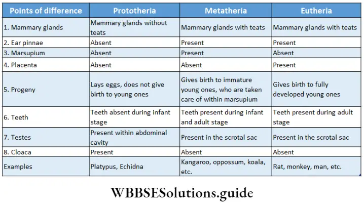
Subclass Prototheria
(Greek Protos: first; therion: wild animal)
General features: The general features of the members of subclass Prototheria are as follows—
- They are oviparous mammals—lay eggs from which young ones are hatched.
- Mammary glands are devoid of teats.
- Ear pinna is either absent or degenerating.
- Presence of cloaca, but absence of urinogenital apertures.
- Absence of teeth but beak is present.
- Testes are present within the abdominal cavity, the uterus is absent and the placenta is not formed.
- They have webbed toes.
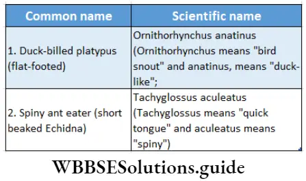
Monotremes constitute the only order under this subclass. They are considered as connecting links between mammals and reptiles because they exhibit features that are common to both reptiles and mammals.
Subclass Theria
(Greek Therion: wild animal)
General features: The general features of the members of this subclass are as follows—
- They are viviparous mammals—give birth to young ones.
- Mammary glands are with teats.
- Ear pinna is present.
- Teeth present.
- Cloaca absent.
- After birth, testes remain lodged within a sac-like structure called the scrotum.
Classification: Subclass Theria is further divided into two infraclasses—Metatheria and Eutheria.
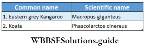
Infraclass Metatheria: (Greek Meta = nearby; therion = wild animal) This infraclass has one order— Marsupialia.
General features: The general features of the members of infraclass Metatheria are as follows—
- Females with a skin pouch or sac on the ventral abdominal surface, are called marsupium.
- The young ones are born in an immature condition and are kept in the marsupium (a pouch-like structure outside the body) for complete development.
- Mammary glands open inside the marsupium and young ones are nourished by the milk.
- The uterus and vagina, are both nourished by the milk.
- The uterus and vagina, both are two in number.
- The true placenta is absent.
Classification: Subclass Theria is further divided into two infraclasses—Metatheria and Eutheria.
Infraclass Metatheria: (Greek Meta = nearby; therion = wild animal) This infraclass has one order— Marsupialia.
General features: The general features of the members of infraclass Metatheria are as follows—
- Females with a skin pouch or sac on the ventral abdominal surface, are called marsupium.
- The young ones are born in an immature condition and are kept in the marsupium (a pouch-like structure outside the body) for complete development.
- Mammary glands open inside the marsupium and young ones are nourished by the milk.
- The uterus and vagina, both are two in number.
- The true placenta is absent.
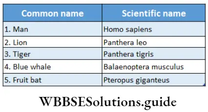
Infraclass Eutheria:
(Greek Eu: developed; therion: wild animal)
General features: The general features of members of the infraclass Eutheria are as follows—
- Complete development of the offspring occurs within the mother’s body. The gestation period is variable. Marsupium absent.
- Ear pinna is developed.
- Anus and the urinogenital apertures are separate.
- Young ones are nourished by the placenta.
- Single vagina and single uterus present. Subclass Theria
Animal Kingdom Notes
- Asexual Reproduction: the process by which an organism produces its own offspring by division, regeneration, or spore formation
- Blastopore: It is an opening through which the cavity of the gastrula (an embryonic stage in the developmental process of animals), communicates with the exterior
- Contractile: capable of contraction
- Cuticle: hard outer covering of the body in some animals
- Distal: away from the point of reference
- Exoskeleton: the external covering of the body
- Endoskeleton: the internal skeleton of the body
- Hypostome: any part of the mouth
- Hermaphrodite: The sex of the organism is not differentiated as male or female. So, the organism has both male and female reproductive organs and produces both male and female gametes in the same body
- Macrolecithal: egg with a large amount of yolk
- Obligatory Parasite: unable to complete its life cycle without a host
- Oviparous: animals that lay eggs that hatch out the produce young ones
- Ovoviviparous: animals producing young ones within eggs which are hatched inside their bodies
- Proximal: nearer to the point of reference
- Peritoneum: the lining of the abdominal cavity
- Pneumatic: containing air
- Pentadactyl: having five digits in limbs
- Sessile: fixed, immobile
- Sexual Dimorphism: having separate sexes of the same species
- Sexual Reproduction: the process by which an organism produces its offspring by fusion of its gametes
- Viviparous: animals that give birth to their young ones
Points To Remember
- Greek philosopher and naturalist Aristotle is known as the father of the animal kingdom.
- Organisms belonging to the animal kingdom are classified on the basis of their characteristic features such as habitat, embryonic germ layer, coelom, body organization, body symmetry, etc.
- The cellular layer of the gastrula from which organs of the body develop is called the germ layer. Higher organisms have three germ layers—endoderm, mesoderm, and ectoderm.
- The process of generation of the head in the anterior part of the body is called cephalisation.
- The cavity within the animal body which is present between the somatic layer and visceral layer of the mesoderm, and is surrounded by the peritoneum is called coelom.
- The organisms that have coelom derived from the splitting of the embryonic mesodermal cord are called schizocoelic. The organisms that have coelom derived from the gut of the embryo as a mesodermal pouch are called enterocoelic.
- The body cavity of arthropods and mollusks is filled with blood. This kind of body cavity which is filled with blood is called haemocoel.
- The organisms in which the mouth develops before the anus from the blastopore, at the time of embryonic development are called protostomes.
- The organisms in which anus develops before the mouth from the blastopre, at the time of embryonic development are called deuterostomes.
- The regular pattern following which organs of the body are arranged in it is called body symmetry.
- When a spherical body is divided into two equal halves in any plane, along the central axis, the symmetry is called spherical symmetry.
- When a body is divided longitudinally into two equal halves along the central axis, the symmetry is called bilateral symmetry.
- The process by which the body of an organism, is divided into several segments which can be identical or non-identical is called segmentation.
- The organisms whose bodies are made up of more than one type of cells are called metazoa. The multicellular organisms in which the tissue system is not formed are called parazoa. The multicellular organisms in which the tissue system is present are called enterozoa.
- In diploblastic (organisms with two germ layers, namely ectoderm, and endoderm) organisms, a gel-like acellular layer is present between ectoderm and endoderm. This gel-like layer is called mesoglea.
- In some organisms, a fluid-filled cavity consisting of mesodermal cells is present between the epidermis and visceral organs which is called a pseudocolor.
- Small pores are seen throughout the body of organisms belonging to the phylum Porifera. These pores are called ostia. A large exhalant aperture, osculum, is present.
- A network of canals connecting the Ostia with the spongocoel forms the canal system. The central cavity of the canal system is called paragastric or spongocoel.
- Different types of cells are seen in organisms belonging to porifera. these are choanocyte, amoebocyte, pinacocyte,sclerocyte, etc.
- The body of Poriferans bears spike-like structures called spicules. These spicules are calcareous, siliceous, or made of spongin fibers.
- The body cavity of organisms belonging to the phylum Cnidaria is called coelenteron or gastrovascular cavity.
- Cnidarians have a special type of cell called cnidoblast which consists of a toxic structure called nematocyst.
- Organisms belonging to the phylum Ctenophora have eight comb plates in their body.
- Colloblast or lasso cells are present in the body of ctenophores instead of cnidoblast.
- Platyhelminths bear a special type of cell called flame cell, as an excretory organ.
- Some organisms of phylum Nematoda (such as— Ascaris sp., Wuchereria sp., etc.) live as parasites in the human body and cause various diseases (such as ascariasis, filariasis, etc.).
- The locomotory organ of organisms of phylum Annelida is called seta.
- Each segment of the body of annelids bears a pair of excretory organs. These are called nephridia.
- The life cycle of annelids has a trochophore larval stage.
- The respiratory organs of some arthropods (like—prawns, crabs, etc.) are book gill. However, some other members of the phylum Arthropoda have book lungs as their respiratory organs (like—scorpions, spiders, etc.).
- A green gland is the excretory organ of some members of Arthropoda (like—prawns, crab, etc.). Again, some members of Arthropoda have the coxal gland as their excretory organ (like—scorpion, Limulus, etc.).
- The organ of Bojanus is the excretory organ of members of the phylum Mollusca.
- The life cycle of mollusks has free-swimming, ciliated larvae, called trochophore larvae, followed by another larval stage called the veliger stage.
- Some members of the phylum Echinodermata have grooves on their oral surfaces. These are called ambulacral grooves.
- A complex circulatory system is seen in the echinoderms.
- The longitudinal, elastic part present between the alimentary canal and nerve cord in the dorsal surface of organisms belonging to the phylum Chordata is called notochord or chorda dorsalis.
- The cavity present in the nerve cord of chordates is called neurocoel.
- The paired openings at both the lateral sides of the pharynx at any stage of the life cycle in chordates are called pharyngeal gill slits.
- The tornaria larva stage is seen in animals belonging to the subphylum Hemichordata.
- Embryos of some vertebrates are covered by a membrane called amnion. These organisms are called amniotes.
- The birds which cannot fly but can run are called ratitae or running birds.
- The birds which can fly in the sky are called carinatae or flying birds.
- A disc-like structure is present in the body of roundworms which is called trochal disc. With the help of this disc, these worms move in a circular pattern and so are called wheel animalcules.
- A thin, elastic cell membrane is called a pellicle. It is seen in Euglena sp.
- Living fossils of invertebrates are Peripatus and Limulus. Living fossils of vertebrates are Sphenodon (reptile), and Coelacanth (fish).