Cell Cycle And Cell Division Introduction
Cell Division Notes
All over the world, we can see various types of organisms.
All of them have originated from a single cell. Again, these single cells arise from pre-existing cells.
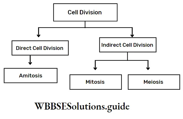
The process, by which new cells arise from pre-existing ones, sustain life and bring variation among organisms, is called cell division. It is of three types—amitosis, mitosis and meiosis.
A mature cell which undergoes cell division is called a mother cell or parent cell and the newly formed cells are known as daughter cells.
In the case of unicellular organisms, a single mother cell divides to form two daughter cells. Multicellular organisms, like human beings, are made up of billions of cells.
| Class 11 Biology | Class 11 Chemistry |
| Class 11 Chemistry | Class 11 Physics |
| Class 11 Biology MCQs | Class 11 Physics MCQs |
| Class 11 Biology | Class 11 Physics Notes |
Cell cycle and cell division notes for NEET PDF
Such a large number of cells originate from a single-celled zygote which undergoes repeated divisions to develop into an embryo. The embryo divides multiple times to develop into a complete organism.
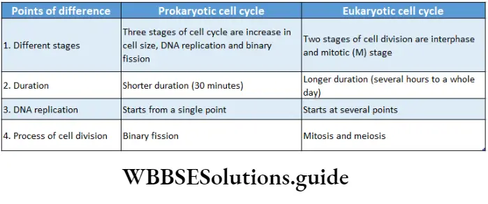
The cell division continues in a newly formed daughter cell till it reaches its mature form, in a cyclic and controlled manner. This is called the cell cycle. It has two phases—interphase and M phase.
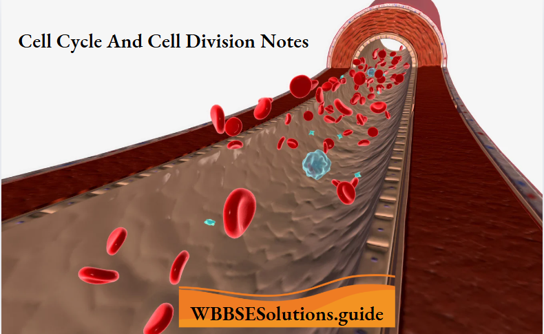
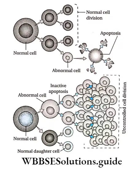
But, after a certain period of time, a ‘suicide’ programme is activated within the cells. It leads to cessation of cell division, fragmentation of the DNA, shrinkage of cytoplasm, membrane change and cell death without lysis or damage to neighbouring cells. This is known as apoptosis or programmed cell death.
Hence, the birth of young ones, their complete development and eventual death, all depend on the processes of cell cycle and cell division.
Cell Cycle
Cell Cycle Definition: The orderly sequence of events by which a cell duplicates its DNA, synthesises its other components and produces daughter cells is known as the cell cycle.
In an organism, cell division takes place all the time in different parts of the body.
Class 11 biology cell cycle and cell division notes with diagrams
A daughter cell, after division, grows, develops and divides again by passing through a series of stages collectively known as the cell cycle. The time period between two successive divisions is known as the generation time.
Phases Of Cell Cycle
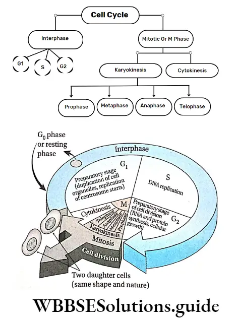
There are two phases of the cell cycle in a proliferating somatic cell—interphase and mitotic phase.
Interphase
Interphase Definition: Interphase is a metabolically active stage between two mitotic divisions during which the cell prepares itself for another division.
Previously, the interphase stage was considered as a resting phase of the cell cycle.
But now, it has been regarded as the metabolically most active phase of the cell cycle. It is involved in different types of synthetic activities like synthesis of DNA, RNA, protein, etc.
The word ‘interphase’ is derived from two different words— Inter is a Latin word, that means ‘between’ and phasis is a Greek word, that means ‘state’. Interphase is also known as ‘enter mitosis’.
Characteristics:
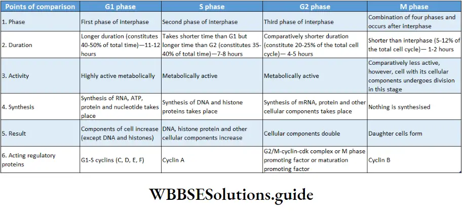
- It is the longest phase of the cell cycle. It lasts for approximately 22-23 hours (about 75-95% of the total span of a cell cycle).
- This stage exists between two successive cell divisions.
- In this phase, DNA replication takes place. RNA, histones and other nuclear proteins are also synthesised.
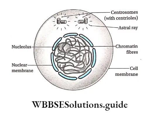
- Centrosome duplication to form two centrosomes in animal cells is seen. Each centrosome contains two centrioles. Hence, this phase is also known as the preparatory phase.
- The size and volume of the nucleus increase. Chromosomes undergo a condensation-decondensation cycle during this period. They cannot be seen under a light microscope at this stage.
- Cells spend most of their time in this intermediate non-mitotic state.
- During interphase (in the S phase), 4 copies of each gene form instead of the normal 2 copies in a diploid cell.
- Energy-rich ATP is synthesised and stored in cells. So, this phase is also known as the energy phase.
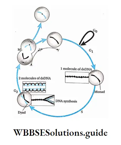
Phases of interphase: it is divided into three phases, G1-S-G2. The G1 and G2 phases stand for ‘gap phase 1’ and ‘gap phase 2’ respectively. The S phase stands for the ‘synthesis phase’ and in this phase, DNA replication occurs.
Short notes on cell cycle and cell division for quick revision
Within the G0 phase, sometimes, the cell stops dividing and enters into a sleeping stage or quiescent stage of the cycle called the G0 phase.
Different phases of the interphase have been discussed under separate heads.
G1 phase: The first gap phase of interphase, i.e., the phase between the M phase of the previous cell cycle and the S phase of the new cell cycle which is metabolically active, is called the G1 phase (growth phase I).
Characteristics:
- It is usually the longest period of the cell cycle. It lasts for about 11-12 hours (45-50%) of the total cycle.
- In some embryonic cells that are rapidly dividing, G1 may last only for a few minutes.
- It is a metabolically active phase.
- In this phase, carbohydrates, lipids, and all types of proteins and enzymes (DNA polymerase, RNA polymerase), that are required for DNA replication in the S phase, are synthesised.
- In this phase, three types of RNAs and structural proteins are also synthesised, but not DNA.
- Each chromosome is elongated, and thin and contains one DNA molecule. Therefore, the chromosome is monad.
- Different cellular organelles—mitochondria, Golgi bodies, plastids, ER, centrioles and ribosomes, increase in number. In this phase, ATP is stored in cells which is essential for the proper progression of cell division. So, the G1 phase is known as anaphase.
- Some cells, like nerve cells, never leave G2 and remain arrested in a stage called the G0 phase.
Monad and dyad
A chromosome with one chromatid is known as a monad. Chromosomes with two chromatids are known as dyads.
Cdk (Cyclin-dependent kinase)
The fate of a cell is determined by the regulatory enzyme; cyclin-dependent kinase, present at checkpoints of a cell cycle.
S phase: The phase of interphase that lies between two gap phases and is responsible for DNA replication and synthesis of histone proteins is called the S phase or synthesis phase.
S phase Characteristics:
It is the second phase of the interphase (next to the G1 phase).
Generally, the duration of the S phase is 7-8 hours. It occupies 35-40% of the total cell cycle. However, this duration varies in different stages stands for the ‘synthesis phase’ and in this phase, DNA replication occurs.
Within the G2 phase, sometimes, the cell stops dividing and enters into a sleeping stage or quiescent stage of the cycle called the G0 phase. Different phases of the interphase have been discussed under separate heads.
G2 phase: The first gap phase of interphase, i.e., the phase between the M phase of the previous cell cycle and the S phase of the new cell cycle which is metabolically active, is called the G2 phase (growth phase I).
G2 phase Characteristics:
It is usually the longest period of the cell cycle. It lasts for about 11-12 hours (45-50%) of the total cycle.
In some embryonic cells that are rapidly dividing, G1 may last only for a few minutes.
It is a metabolically active phase. In this phase, carbohydrates, lipids, all types of proteins and enzymes (DNA polymerase, RNA polymerase), that are required for DNA replication in the S phase, are synthesised.
In this phase, three types of RNAs and structural proteins are also synthesised, but not DNA.
Each chromosome is elongated, and thin and contains one DNA molecule. Therefore, the chromosome is monad.
Different cellular organelles—
Mitochondria, Golgi bodies, plastids, ER, centrioles and ribosomes, increase in number. In this phase, ATP is stored in cells which is essential for proper progression of cell division. So, the G1 phase is known as antiphase.
Some cells, like nerve cells, never leave G1 and remain arrested in a stage called the G1 phase.
M Phase Or Mitotic Phase
M Phase Or Mitotic Phase Definition: The shortest phase of a cell cycle which occurs after the completion of interphase and is involved in cell division is called the M phase.
M Phase Or Mitotic Phase Characteristics:
- It is the dividing phase of the cycle.
- Cell division occurs either by mitosis or meiosis.
- During this phase, the components of the cell that are synthesised in interphase, (chromosome, DNA, cellular organelles, cytoplasm, etc.), are distributed among the daughter cells.
- It occupies 5-12% of the total cell cycle, i.e., less than 1 hour or 1 hour.
- The phase includes the breakdown of the nuclear membrane, the condensation of chromosomes, their attachment to the mitotic spindle and the segregation of chromosomes to the two poles.
- It occurs in two phases—nuclear division or karyokinesis followed by cytoplasmic division or cytokinesis.
G0 phase (Quiescent phase)
- In some cells, at a certain point of the G2 phase, the cell cycle stops, i.e., the cell enters an inactive phase. This phase is known as the G0 phase.
- Mainly, a cell enters the G0 phase due to the absence of cell cycle-controlling factors (e.g. nitrogenous and energy-rich compounds).
- However, the cell is found to be metabolically active and viable but is not proliferative in this phase.
- Cells do not divide at this phase but act as reserve cells. Only under favourable conditions, do these cells re-enter the cell cycle and start division.
- Most cells do not re-enter the cell cycle. They grow and differentiate to play their roles in an organism’s body. For example, nerve cells remain in the G0 phase permanently.
- Mature brain cells become arrested in G0 and do not normally divide again during a person’s lifetime.
- Some cells de-differentiate to re-enter the cell cycle from the GQ phase and thus resume division. Examples are the parenchyma cells of plants and fibroblasts of animals.
Importance Of Cell Cycle
The importance of the cell cycle is as follows—
- Due to the cell cycle, amounts of DNA, RNA and protein increase in the cell.
- However, after the generation of daughter cells, these molecules are distributed equally.
- The number of cellular organelles increases as a result of the cell cycle.
- Daughter cells are produced due to the cell cycle and they are supplied with the required components.
Control Of Cell Cycle
The transformation of cells from one phase of the cell cycle to another is controlled by some external factors.
These factors control the cell cycle at certain points. In most cases, the first factor is present in the G1 phase.
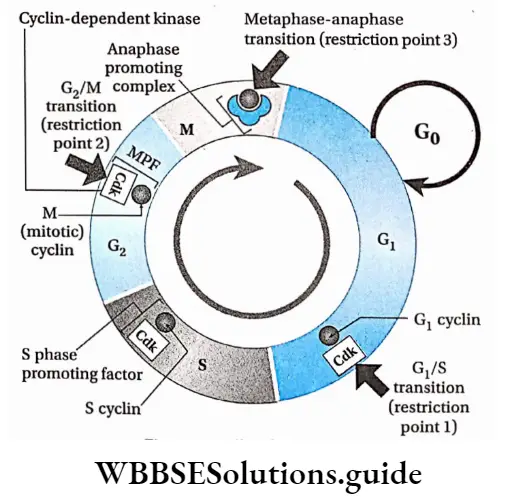
A number of regulatory systems of the cell which monitor the progress in the cell cycle and can inhibit subsequent stages in the event of failure to maintain normalcy are called cell cycle checkpoints.
Cell cycle checkpoints
There are three main checkpoints in a cell cycle. They are as follows—
G1-S checkpoint: This is the major checkpoint that determines the transition of the cell from the G1 to the S phase. If the DNA of a cell gets damaged or the cell has not attained proper volume, then the cell is arrested at the G0 phase.
The cell is not allowed to proceed to the S phase. Rather, the cell is directed to move to the G0 phase.
In yeasts, this checkpoint is known as START and in animal cells, it is known as restriction point or commitment point.
G2-M checkpoint: This checkpoint occurs at the G2 phase and it regulates the entry into the M-phase.
Unless all DNAs have been replicated in the S phase and the cell has attained proper volume, the cell is not allowed to enter the mitotic phase of the cell cycle from the G2 phase.
M checkpoint: This checkpoint occurs during the M-phase. Spindle fibres are formed in the mitotic phase during metaphase.
The chromosomes should align properly at the mitotic plate in order to trigger the separation of chromatids. This checkpoint stops the cell from undergoing mitosis unless these conditions are fulfilled.
Chemical regulation of checkpoints
The cell cycle is regulated chemically by a variety of regulatory proteins, namely cyclin and cyclin-dependent kinases (Cdks). They are the key components of the cell cycle and associate with one another.
Cyclins are proteins that get their name from their cyclically fluctuating concentration in the cell.
These undergo structural modification in different phases of the cell cycle.
Cell cycle and cell division chapter summary with important points
Animal cells have four types of cyclins—cyclin A, cyclin B, cyclin D and cyclin E. Cdk is present in all phases of the cell cycle but as an inactive protein kinase.
The cyclin-dependent kinases associate with cyclin to form active Cdk-cyclin complexes.
On the basis of the mode of activities in specific events in animal cells, cyclin–
CDK complexes are classified into three types-
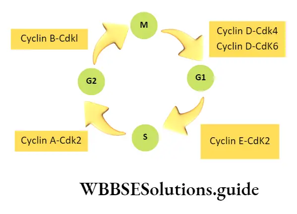
Gl/S-cyclin-Cdk complex: Gi/S-cyclin-Cdk complex has two structural components—cyclin D-Cdk 4/6 and cyclin E-Cdk 2. The transition from Gx to S is promoted by Cdk.
CdK becomes active in the presence of G2 cyclin and ATP which causes the transition of Gx to the S phase
Once the Gj cydins activate the Cdks, the levels of cyclins degrade due to proteolysis.
S-cyclin-Cdk complex: The component of this complex is cyclin A-Cdk 2. During the S phase, cyclin A is synthesised and associates with Cdk to form the S-cyclin-cdk complex. This complex controls DNA replication.
G2/M-cyclin-Cdk complex (MPF): The component of this complex is cyclin B-Cdk1 which binds with ATP to become active. The complex so formed is also known as the M phase promoting factor or the Maturation promoting factor.
This factor was first discovered in mature unfertilised eggs of frogs. MPF activation stimulates the G2-M transition. It controls the supercoiling of chromosomes, contraction of the nuclear membrane, arrangement of spindle fibres, etc., at the M phase.
At the end of the M phase, proteolytic degradation of cyclin B causes the termination of MPF activity. Cyclin B is marked for destruction by the Anaphase Promoting Complex (APC).
APC is the ubiquitin-protein ligase that targets cyclin B and destroys it to enable the transition of a cell from metaphase into anaphase.
Uncontrolled cell cycle and cell division in animal cells If, for any reason, cell cycle checkpoints do not function, cell division becomes uncontrolled.
The mass of a cell that develops due to unregulated cell division, is known as a tumour.
In the animal body, when the tumour cells spread to other parts of the body from their origin, via blood vessels and form new tumours, then it is known as malignant or cancerous.
Malignant cells have uncontrolled growth and show genetic and cellular changes. These changes enable the cancer cells to spread to distant locations from their original site.
This phenomenon or characteristic is called metastasis. The tumour which remains at the original site is known as benign. Most benign tumours do not cause serious problems.
Different types of uncontrolled cell proliferation
The proliferative growth of cells occurs due to various physiological factors and the presence or absence of effectors.
Some examples of proliferative cell growth are—
Hyperplasia: Overgrowth of a particular tissue or organ due to an increase in the rate of cell division of that region is known as hyperplasia. It happens due to increased hormone secretion that leads to an increase in metabolic activity of the cells of that tissue or organ.
Example development of breasts in females during pregnancy, and the thickening of endometrium in aged women.
Hypertrophy: It causes an increase in the size of an organ due to the enlargement of cells. This may occur due to any infection or increase in the metabolic activity of cells.
For example swelling of striated muscles during heavy exercise, and swelling of the endothelium of the alimentary canal due to infection by worms.
Metaplasia: It causes the transformation of mature cells into abnormal ones due to infection or injury.
For example during lung infection and in the case of smokers, the columnar epithelial cells of the bronchus transform into squamous epithelial cells.
In certain precancerous conditions, the normal existing epithelium is replaced by an epithelium from a nearby tissue by the process of metaplasia.
For example in Barrett’s oesophagus or Barrett’s oesophagitis, the normal squamous cells of the oesophageal lining are replaced by secretory cells that migrate from the stomach lining (metastatic Barrett’s epithelium).
Dysplasia: The production of abnormal tissue or cells, due to uncontrolled and abnormal cell division, that displays a transitional state between benign and pre-malignant growth is known as dysplasia.
Neoplasia: The uncontrolled, abnormal growth of cells in the body is known as neoplasia (neo meaning ‘new’). It occurs due to the presence of any effector.
Neoplastic growth may be of two types—benign or malignant. Benign tumours are confined or restricted to a particular site and are not harmful whereas malignant tumours are harmful and undergo metastasis.
Chromosomes
- The thread-like, self-replicating structures of the nucleus, which is made up of nucleoprotein and which transmit hereditary properties generation after generation through cell division, are known as chromosomes.
- During cell division, chromosomes are formed from chromatin fibres of the nucleus. Chromosomes were first observed by W. Hofmeister in 1848. However, it was named ‘chromatin1 by W.
- Flemming in 1879. Later, in 1888, W. Waldeyer coined the term ‘chromosome’, Generally chromosomes are thread or stalk-like structures. During the anaphase stage of cell division, they appear like V, ‘L’, ‘J’ or T.
- They are 0.5-30 /rm in length and 0.2-3.0 /im in diameter. Their structure becomes clearly visible at metaphase.
Chromosomes occur in homologous pairs in the body cells—
- One member of each pair is derived from the female parent (mother) and the other from the male parent (father).
- Gametes usually contain only one set of chromosomes. This number is called ‘haploid’ (n).
- The haploid set of chromosomes is known as the genome, while the number of chromosomes in somatic cells is ‘2n’. Hence, they are diploid.
Cell Division
Cell Division Definition: The fundamental and active biological process by which a parent cell, after replication of its components, divides to produce its daughter cells is called cell division.
Cell division is essential for growth, development and regeneration.
Types: There are three types of cell division—
- Amitosis,
- Mitosis and
- Meiosis.
Amitosis is the direct division which involves simple cleavage of the nucleus into two daughter nuclei.
The cytoplasm constricts and two daughter cells, each with a nucleus, are produced. Mitosis is essentially a duplication process.
It produces two genetically identical daughter (progeny) cells from a single parent (dividing) cell. Meiosis, on the other hand, is quite different.
It recombines the chromosomes to generate daughter cells that are distinct from one another and the original parent cell as well. Meiosis produces male and female gametes that undergo fertilisation to give rise to offspring.
So, basically, mitosis is for growth and maintenance, while meiosis is for sexual reproduction.
Conditions: Scientists say that normal cell division depends on certain conditions.
These are as follows—
Minimum growth: A newly formed cell does not undergo division immediately. It must attain a minimum level of maturity before it gains the ability to divide.
Karyoplasmic index: The ratio between the volume of the cell cytoplasm and the nucleus is known as the karyoplasmic index. Declination of this ratio leads to cell division.
Amount of nuclear material: When the DNA content of the nucleus doubles, then cell division takes place.
Cell volume and metabolism: With the increase of cell content due to a rise in metabolic activity, the volume of the cell increases without an increase in surface area. This causes cell division.
Mitogen: Some chemical factors which induce cell division are called mitogens. Examples are cytokines in plants, epidermal growth factor (EGF), platelet-derived growth factor (PDGF), etc.
Reproduction: In the case of unicellular organisms, cell division is an inevitable occurrence, as it is the means of reproduction. This is the only way by which they continue their existence.
Importance: The importance of cell division is—
Increase in number: Due to cell division, the number of cells increases which is essential for the development of the body.
Repair and healing: In multicellular organisms, cells continuously undergo wear and tear. These damaged cells are continuously replaced by new cells.
Reproduction: Unicellular and primitive multicellular organisms multiply and reproduce by cell division. Cell division produces gametes which help in reproduction in higher organisms.
Transmission of hereditary characters: Due to cell division, daughter cells acquire characteristics of the parent cell and also develop some new characteristics.
Variations in the offspring allow them to adapt to changing environments. Variation often leads to the evolution of new species.
Constant chromosome number of species: in sexually reproducing organisms, cell division occurs during gamete formation to maintain a constant chromosome number of a species.
Amitosis
Amitosis Definition: The simplest type of cell division in which two daughter cells are formed by simultaneous and direct division of both nucleus and cytoplasm by forming a constriction in the cell, without the formation of spindle fibre is known as amitosis (or direct cell division).
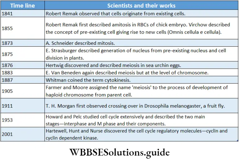
The amitotic type of cell division was first described by German biologist Robert Remak (1841). But, the term was proposed by histologist W. Flemming (1882).
The word is derived from two Greek words amitos and osis which mean ‘without thread’ and ‘state’ respectively.
Amitosis Characteristics:
- It is the simplest type of cell division in which the nucleus divides by constriction.
- Nuclear and cytoplasmic cleavage occurs without spindle formation.
- Maximal condensation of chromatin into chromosomes does not occur and thus chromosomes are not visible.
- The nuclear membrane does not disappear at all.
Site of occurrence: Amitosis is observed in bacterial cells, protozoans, mammalian cartilage cells, and foetal membrane cells.
Amitosis takes place in those cells which do not require equal distribution of cellular substances of parent cell to daughter cells, such as the formation of the large nucleus in Paramoecium, and the division of internodal cells in Chara.
Drawbacks Of Amitosis
Due to amitosis, chromatin material is unequally divided among daughter cells which causes structural and functional abnormalities in daughter cells.
In amitosis, there is no possibility of genetic recombination and there is a possibility of expression of unwanted recessive lethal genes.
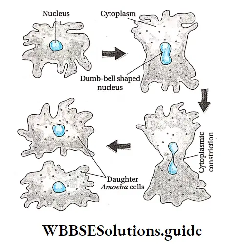
Amitosis Process:
- The nuclear membrane remains intact.
- The nucleus increases in length and two constrictions appear at the centre. As a result, the nucleus appears like a dumbbell.
- The constrictions of the nucleus gradually grow deeper and meet each other. Then the nucleus ultimately divides into two nuclei without the formation of spindle fibre.
- Cytoplasmic division occurs along with nuclear division.
- The cell membrane shrinks and invaginates towards the centre of the cell and forms a constriction between the two nuclei. Ultimately, the cell divides to form two daughter cells.
Amitosis Importance:
- It is a simple type of cell division without any complexity.
- It completes within a short duration and cell number increases rapidly in this process.
- Lower organisms like yeast, bacteria and protozoa reproduce mainly by this process.
Mitosis
Mitosis Definition: The indirect process of cell division by which the somatic parent cell divides once to produce two daughter cells, which are identical in shape and contain an equal number of chromosomes as the parent cell is called mitosis.
Walther Flemming (1879) first described and coined the term mitosis in 1882 in Salamander. Schneider (1879) also described the various stages of mitosis.
Mitosis Short Notes
The biochemical aspects of this process were explained by Cockraum and MacCanley (1960). The word ‘Mitosis’ is derived from two Greek words—mitos which means ‘thread’ and osis meaning ‘state’.
It is a type of equational division, that produces two identical daughter cells having the same genetic constituent (same chromosome number) as the parent cell.
Mitosis Characteristics:
The nucleus with chromosomes and cytoplasm of the cell divides only once.
The nucleus, cytoplasm, organelles and cell membrane of the parent cell are distributed among two daughter cells, containing roughly equal shares of these cellular components. Hence, it is called an equational division.
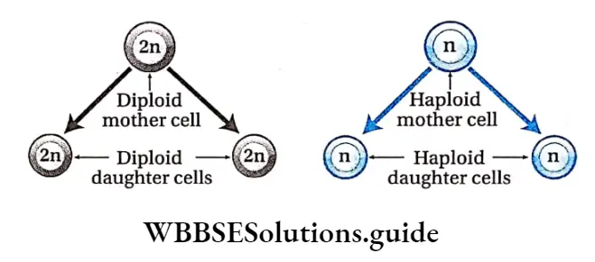
Site of occurrence:
- Mitosis occurs in almost all eukaryotic somatic cells.
- In some single-celled organisms, mitosis forms the basis of asexual reproduction.
- In advanced multicellular organisms, mitosis occurs during the formation of the embryo from the zygote, the development of a complete individual from an embryo and the subsequent growth and development of the organism.
- In some special parts of advanced organisms, ‘mitosis occurs throughout life. For example, mitosis occurs in meristematic tissue (plants) found in root tips, shoot tips, buds and leaf primordia. In the case of animals, this is found in cells of skin and bone marrow.
- Gamete mother cells also undergo mitotic cell division to increase their number in gonads.
- It occurs in the damaged organs of plant and animal bodies during wound healing.
Duration of mitosis: Duration of mitosis varies in different species.
In actively dividing animal cells, the whole process takes about one hour.
But generally, it takes 30 minutes to 3 hours. The time span of different phases of mitosis actually depends on different external and internal conditions of the dividing cell and it is very much tissue-specific.
Process Of Mitosis
Though mitosis occurs in several tissues of plants and animals, it is best to study the phases of mitosis from the stained squash of the root tip. Acetocarmine is commonly used for staining chromosomes of root tip cells.
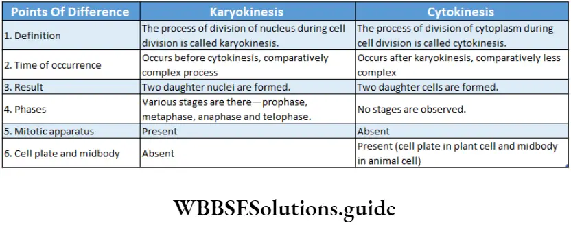
The process of mitosis occurs in two stages—karyokinesis and cytokinesis.
Karyokinesis
Karyokinesis Definition: The division of the nucleus at the time of cell division is called karyokinesis.
It is a process of indirect nuclear division. The nucleus passes through a sequence of events and forms two daughter nuclei from a single division.
Different phases: Karyokinesis is conventionally divided into four phases—prophase, metaphase, anaphase and telophase. These have been discussed below under separate heads.
Prophase
Prophase Definition: The first and the longest-running stage of karyokinesis where chromatin condenses to transform into chromosomes that eventually unwind to form two chromatids with the dissolution of the nucleolus and nuclear membrane, is called prophase.
Prophase Characteristics: Prophase is divided into three subphases.
The characteristics of each subphase are—
Early prophase:
The nucleus is diffused, and granular in appearance.
In the interphase, the refractive index of the chromatin fibre is almost the same as that of the nucleoplasm. Hence, the chromatin is not visible.
As the nucleus enters into prophase, a refractive index of chromatin changes from that of nucleoplasm due to dehydration of the nucleus. As a result, chromatin fibres become visible.
Refractivity and viscosity of cytoplasm also increase.
Chromatin begins to coil and condense to form thin, thread-like chromosomes. The sister chromatids which were intertwined during the G2 phase become untangled during chromatin condensation.
Condensation occurs due to the coming together of scaffolding proteins and folding of individual chromatin through spiralisation.
Due to spiralisation, chromosomes appear short, thick and stacked on each other like woollen balls. This stage is known as the supreme stage.
In animal cells and in the cells of some lower organisms, each of the centrosomes gives rise to two daughter centrioles by replication.
They associate with a newly formed centrosome followed by the shifting of a pair of centrioles to one pole and the other pair to the opposite pole.
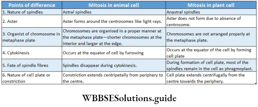
In animal cells, each centrosome produces fine, thin microtubular fibrils forming a star-shaped body called aster.
The microtubular fibrils radiating out of the surface of the aster are known as astral rays.
Middle prophase:
- The ends of the chromosomes become more visible. They undergo further thickening and shortening.
- Chromosomes are seen to be composed of two chromatids attached by centromere.
- The pair of centrioles move further to opposite ends of the cell.
- Astral rays increase in length.
- At this stage, the number of chromosomes (containing a pair of chromatids) is considered to be equal to the number of centromeres.
- The nucleolus remains associated with a specific secondary constriction of the chromosome.
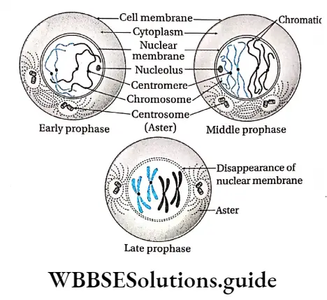
Late prophase:
- The nuclear envelope disintegrates into small vesicles as the centrosomes continue to move towards opposite poles. Nucleolus disappears.
- Chromosomes become thicker as a result of further condensation and coiling.
- Two centrosomes occupy two poles with radiating microtubules.
- Mitotic spindle begins to form from centrosomes by forming spindle microtubular fibrils.
Prometaphase
- The transitional stage between the end of prophase and the beginning of metaphase is called prometaphase.
- In this shortest phase, the nuclear membrane disappears completely, thus cytoplasm and nucleoplasm merge. In plant cells, centrosomes are absent.
- So mitotic spindle is developed from the cytoplasmic and nuclear sap and is known as the anastral spindle.
- In animal cells, the poles of the spindle are formed by two asters at two poles. So this type of spindle is called an amphiastral spindle.
Metaphase
Definition: The second, short durational stage of karyokinesis in which chromosomes with two clearly visible, chromatids are arranged along the equator of the metaphase plate is called metaphase.
Metaphase Characteristics:
- The two chromosomal fibres attached to each chromosome and connected to opposite poles start to contract.
- Thus, the chromosomes are arranged at the equator, on a specific plane of the spindle fibres. It is known as the metaphase plate.
- Shorter chromosomes are arranged at the interior while the longer chromosomes are arranged at the periphery of the metaphase plate. This arrangement of chromosomes at the metaphase plate is called congression.
- Spindle fibres bind themselves to the chromosomes through the kinetochore at the metaphase plate.
- The centromere remains connected with spindles while the arms of chromosomes remain suspended.
- The chromatids of the chromosomes are clearly visible.
- At the end of metaphase, the centromere of a chromosome splits and the two sister chromatids separate.
- Metaphase chromosomes can be stained and they show distinctive.
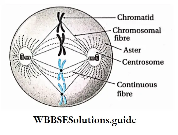
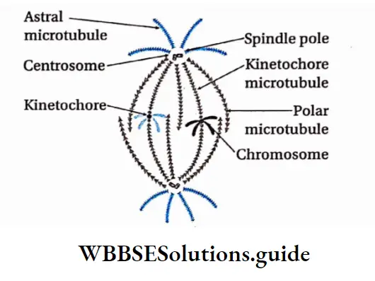
Anaphase
Anaphase Definition: The phase in karyokinesis in which chromosomes with single chromatids separate and move from the metaphase plate to the respective poles is called anaphase.
Characteristics:
- Centromeres divide separating the pair of chromatids.
- Each sister chromatid with one centromere is called a monad chromosome. At this stage, the number of chromosomes becomes double that of the parent cell.
- Each daughter chromosome binds with the spindle fibres through the kinetochore, located in the centromere.
- Before the onset of anaphasic movement of the monad chromosomes, a third type of fibre appears between the two separating centromeres known as interzonal fibres.
- Due to chromosomal repulsion, contraction of chromosomal fibres and expansion of interzonal fibres between daughter chromosomes, they move to opposite poles in equal halves. This movement of chromosomes is known as anaphasic movement. As the daughter chromosomes are now pulled towards the spindle poles, centromeres are found to lead the path while the arms of chromosomes trail behind.
- The daughter chromosomes become short and thick, hence, clearly visible.
- Poles of the spindle apparatus are pushed apart as the cell elongates. When an equal number of chromosomes reach their respective poles, the chromosomal fibres disappear. The number of chromosomes at the poles is equal to the number of chromosomes in the parent cell before division. So, equational division takes place. Anaphase results in the distribution of one complete diploid complement of genetic information to each daughter cell.
- In animal cells, the cell constricts from the middle and so the spindle fibres aggregate to form a structure, called a stem body. The stem bodies extend cylindrically, pushing the daughter chromosomes at the poles.
- At anaphase, the chromosomes with one chromatid bind with spindle fibres through the centromere and take various shapes— metacentric chromosome as V, submetacentric as T, acrocentric as T and telocentric as ‘I’.
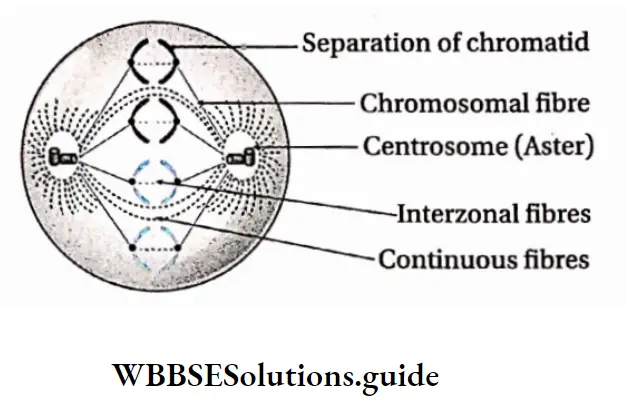
Some Facts About Anaphase
Reasons For Anaphasic Movement:
- The daughter chromatids separate due to the shortening or depolymerisation of the kinetochore microtubules from the poles of the chromosomal or centromeric fibres.
- It also involves simultaneous elongation of the continuous fibres due to polymerisation of the polar microtubules.
- Extension of interzonal fibres between two daughter chromosomes.
- Repulsion between daughter chromosomes that causes their sliding movement.
- Some facts about anaphase
Reasons for anaphasic movement:
- The daughter chromatids separate due to the shortening or depolymerisation of the kinetochore microtubules from the poles of the chromosomal or centromeric fibres.
- It also involves simultaneous elongation of the continuous fibres due to polymerisation of the polar microtubules.
- Extension of interzonal fibres between two daughter chromosomes.
- Repulsion between daughter chromosomes causes their sliding movement fragments of the nuclear membrane and ER.
- Nucleoli reappear in the nucleolus organiser regions (NORs) of SAT (satellite) chromosomes in both nuclei. Regeneration of nuclear membrane.
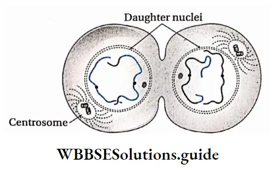
The density of cytoplasm and its refractive index decrease and chromosomes become invisible as chromatin fibre. Thus reconstruction of daughter nuclei is completed.
In animal cells, spindle fibres completely disappear. In plant cells, spindle fibres disappear from the poles but remain in the equatorial region. Golgi complex and endoplasmic reticulum are regenerated.
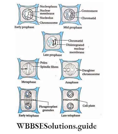
Types Of Mitosis
The different types of mitosis encountered in different organisms are discussed as follows—
Promitosis or intranuclear mitosis: It is a type of primitive intranuclear mitotic division which occurs without the formation of aster and disappearance of the nuclear membrane.
Thus, the nucleus divides within the nuclear membrane. For example, Amoeba, Yeast, etc., are divided by this process.
Eumitosis or extranuclear mitosis: This is the typical mitosis. This division involves the disappearance of the nuclear membrane at the end of the prophase.
Spindle forms and chromosomes are arranged at the equatorial plane by binding with spindle fibres. Example mitosis in plant and animal cells.
Endomitosis or Endoduplication or Endopolyploidy: The process in which repeated reduplication of chromosomes occurs during interphase without nuclear division (formation of daughter nuclei or daughter cells) is known as endomitosis or endoduplication or endopolyploidy.
Here, the segregation of chromosomes does not take place. Example polytene chromosomes in the salivary gland of Drosophila sp.
Paramitosis: The type of mitosis in which the spindle forms and the nuclear membrane disappears but the chromosome does not undergo coiling is known as paramitosis. Example division in Dinoflagellata.
Handwritten notes on cell cycle and cell division PDF download
Free nuclear division: The type of mitosis in which nuclear division is not followed by cytoplasmic division leading to a multinucleated condition is known as free nuclear division.
Plant cells containing more than one nucleus are called coenocytes and animal cells containing more than one nucleus are called syncytiums.
For example, the liquid endosperm of coconut, Mucor, Rhizopus, bone marrow cells and bone cells of animals contain more than one nucleus.
Cryptomitosis: The type of mitosis in which chromosomes coil, the nuclear membrane disappears, and typical spindles form but chromosomes are not arranged at the equatorial plate is called cryptomitosis.
Example division in parasitic protozoa like Plasmodium, and Heptazoon.
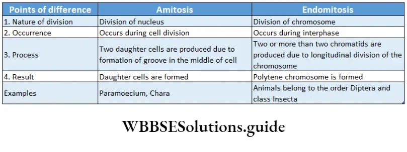
Significance of mitosis
The significance of mitosis is as follows—
Overall growth and development of the organism: The embryo forms from a single-celled zygote by mitosis and it divides multiple times to develop into a complete organism.
Thus, it is essential for increasing the number of cells in multicellular organisms.
Reproduction: It is a method of multiplication or reproduction in some unicellular organisms such as Amoeba, Paramoecium, etc.
Genetic stability: Due to mitosis, the number of chromosomes and genetic constitution like the total amount of DNA, RNA remain conserved in the daughter cell with respect to the parent cell.
Daughter cells are genetically almost identical to the parent cell and no variation in genetic material can therefore be introduced during mitosis.
So, mitosis results in genetic stability within the population of cells.
Repair and regeneration of worn-out parts:
Damaged cells are regularly replaced by new cells because of mitosis. It helps to heal wounds.
Replacement of older cells: By mitosis, older cells are replaced by newer cells.
Maintenance of surface-volume ratio:
The metabolic activity of a cell reduces when cell volume increases. Due to mitosis, the surface-volume ratio of a cell is maintained.
Maintenance of nucleoplasmic ratio: In mitotic cell division, the nucleoplasmic ratio of the cell is maintained.
Formation of gametes: Haploid or gametophytic organisms (n) produce gametes by mitosis.
Vegetative propagation: Mitosis helps in vegetative reproduction and micropropagation. New tissue or organs are formed by the process of mitotic cell division.
Meiosis
Meiosis Definition: The indirect process of cell division in which the chromosomes of parent cells divide once, but the nucleus divides twice to form four daughter cells, each containing half the number of chromosomes compared to the parent cell is called meiosis.
Van Beneden (1883) first reported that the reduction in chromosome number occurred during cell division. T. Bovery (1887) described the process in the gonads of Ascaris. J.B. Farmer and Moor coined the term ‘meiosis’.
chromosomes occur only once leading to a reduction in the chromosome number to half. The word ‘meiosis’ is also derived from two Greek words—meion means ‘to less’ and osis means ‘state’.
It is also known as reductional division because the chromosome number becomes haploid in the daughter cells produced. In all the sexually reproducing organisms meiosis occurs to produce haploid gametes from diploid cells.
Meiocytes And Meiospores
The diploid cells in which meiotic cell division occurs, are known as meiocytes. Example spermatocyte, and oocyte.
The haploid spores obtained by meiotic cell division, are known as meiospores.
Meiocytes And Meiospores Characteristics:
It is an indirect division of cells and occurs in several stages.
The process involves DNA replication during the ‘S’-phase of interphase followed by two successive nuclear and cytoplasmic divisions.
A diploid parent cell (2n), after division, produces four haploid daughter cells (n). The division takes place in two stages—meiosis 1 and meiosis 2.
During meiosis I, due to crossing over and chiasma formation between the non-sister chromatids, genetic recombination occurs.
The chromosomes divide once during meiosis 2.
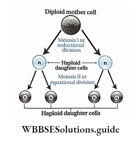
The two daughter cells produced after meiosis I, carry a haploid number of chromosomes as compared to the diploid number of chromosomes in the parent cell.
Site of occurrence: Meiosis occurs in the meiocytes. In plants like bryophytes, pteridophytes, gymnosperms and angiosperms, it occurs in the sporophytes, i.e., spore mother cells of anthers of stamen and ovules of the ovary.
ln animals, it occurs in the primary gametocytes, i.e., primary spermatocytes of the testis and primary oocytes of the ovary. ln lower plants like algae and fungi, it occurs in the diploid zygote.
Meiocytes And Meiospores Types: The process of meiosis is generally similar in both plant and animal cells but cytologists classified meiosis into three types—gametic or terminal meiosis, sporic or sporogenic or intermediate meiosis and zygotic or initial meiosis.
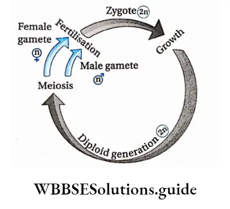
Gametic or terminal meiosis: Meiosis occurs during gamete formation in almost all animals.
The diploid primary gametocytes (spermatocytes and oocytes) undergo gametic meiosis to produce haploid gametes (sperm and ovum).
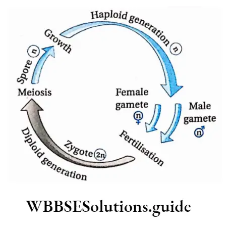
Sporic (sporogenic) or intermediate meiosis: In plants like bryophytes, pteridophytes, gymnosperms and angiosperms, the sporophytes reproduce by spore formation.
Haploid spores are produced from diploid spore mother cells by the process of sporadic or intermediate meiosis.
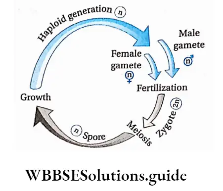
Zygotic or initial meiosis: When meiosis takes place immediately after the formation of the zygote, it is called zygotic or initial meiosis. It occurs in lower plants like algae (Spirogyra), certain fungi (Mucor) and in some sporozoan protozoa (Plasmodium).
Outline Of Different Stages Of Meiosis
Meiosis is a specialized and complex type of cell division. It occurs only in diploid reproductive cells during the formation of haploid gametes or sex cells.
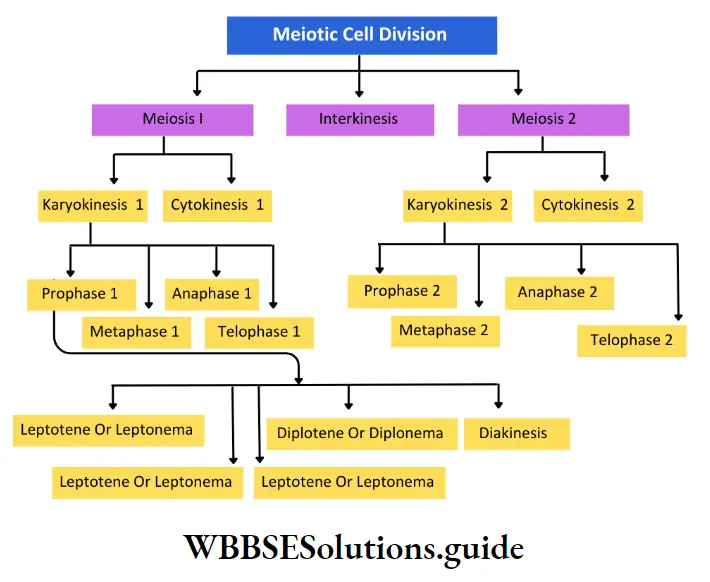
The process of meiosis consists of two complete divisions of a diploid cell—
1st meiotic division heterotypic cell division or reductional cell division.
2nd meiotic division is homotypic cell division or equational cell division. The process of meiosis starts in the interphase (like mitosis).
The DNA replication (=duplication) takes place at the ‘S’ phase of interphase.
Some biochemical mechanisms like protein synthesis take place at the G2 phase. The short period between meiosis I and meiosis II is called interkinesis.
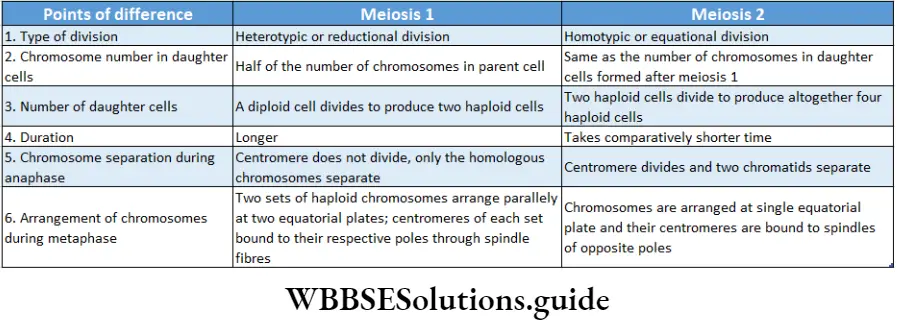
DNA replication does not occur at this stage. Meiosis I is called heterotypic or reductional division because the two daughter cells so formed, have half the number of chromosomes of that of the parent cell.
Meiosis II is called homotypic or equational division because the two daughter cells have equal chromosome numbers as that of their parent cell.
Both these meiotic divisions have several subphases. These phases and their subphases are discussed under separate heads
Meiosis Or Meiotic Division
Meiosis I starts after the end of the interphase. Before the first meiotic division, the meiocyte swells up. After meiosis I, two haploid cells form.
This phase is divided into two stages—karyokinesis I and cytokinesis I.
Karyokinesis
It is the process of the first nuclear division. It has been m divided into four phases—
- Prophase I,
- Metaphase I,
- Anaphase I and
- Telophase I.
It is the process of the first nuclear division. It has been divided into four phases—prophase I, metaphase I, anaphase I and telophase I.
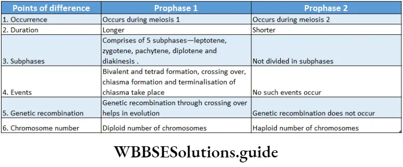
Prophase I: The first phase of the first meiotic division in which a progressive sequence of chromosomal changes occurs through spiralisation, formation of synapsis and tetrad, genetic recombination and terminalisation of chiasmata is known as prophase I.
Leptotene or Leptonema
Leptotene or Leptonema Definition: The subphase of prophase I where chromosomes appear as thin threads with bead-like chromomeres visible most of the time, along the length of the chromosome is called leptotene or leptonema.
The characteristics of leptotene are as follows—
- The size of the nucleus increases. Viscosity and refractivity of the nucleus increase due to dehydration of nucleoplasm.
- The reticular form of chromosomes opens up and the chromosomes become thread-like. Condensation and spiralisation of chromosomes begin. As a result, they become more clearly visible as long single threads.
- Homologous chromosomes exist in pairs.
- Although the chromosomes are dyads, but, due to their thin structure and more coiling, they appear as monads.
- Threads of DNA, wrapped with nuclear proteins and histones, gradually become visible.
- These threads often have chromomeres that appear as “bead-like” swellings along their length.
- In plant cells, chromosomes clump at one side of the nucleus like a bouquet, called synizesis.
- In some other groups of plants, chromosomes in this subphase come in contact at one point to diverge again.
- This point of contact is known as a synthetic knot. In animal cells, the terminal end of chromosomes remains bound to the nuclear membrane through an attachment plate.
- They appear like multiple loops. This arrangement is known as the bouquet stage.
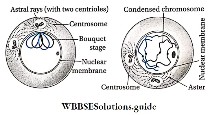
- These chromosomes are also known as polarised chromosomes.
- As leptotene progresses, chromosomes coil up more and become visible.
- In animal cells, the centrosome divides and astral rays start forming spindle fibres. At the end of this subphase, the two asters move apart from each other.
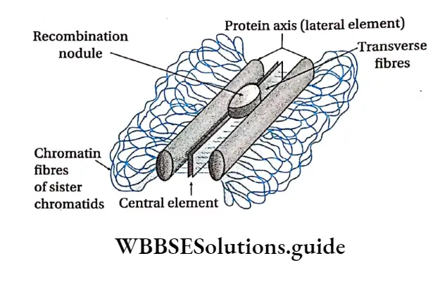
- At this time, components of the synaptonemal complex start gathering.
Homologous chromosomes
In a diploid cell (2n), chromosomes of identical shape, length and characteristics, exist in pairs. These chromosomes in a pair are known as homologous chromosomes.
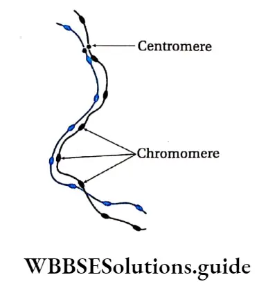
- For example, if the gene that determines the length of fingers, is present at a locus of one chromosome, then its homologous chromosome must carry the same gene at the same locus.
- Either both the genes would code for longer fingers or one would code for longer and the other for short fingers. But they must carry the same character-determining gene at a particular locus.
- Zygote (2n) is formed by the union of sperm (n) and ova (n). Zygote obtains two homologous chromosomes, one each from male and female gametes.
Zygotene Or Zygonema
Zygotene or zygonema Definition: The subphase of prophase I of meiosis I in which homologous chromosomes form synapsis is called zygotene or zygonema.
The characteristics of zygotene are as follows—
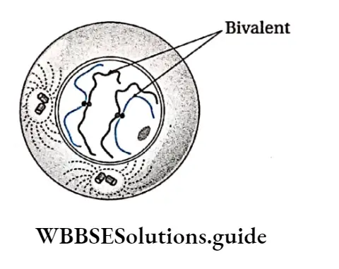
The homologous chromosomes get attracted towards each other and pair up lengthwise.
This temporary pairing of homologous chromosomes is known as synapsis and the paired chromosomes are known as bivalents.
Central transverse fibre is connected with a recombination nodule which contains enzymes helping in crossing over.
Pachytene
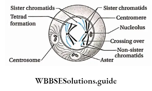
Pachytene Definition: The subphase of prophase in which the paired homologous chromosomes form a tetrad structure, followed by the exchange of genetic material among the non-sister chromatids through the process of crossing over, is called pachytene.
The characteristics of pachytene are as follows—
- This stage is of longer duration.
- Although the longitudinal replication of each chromosome has already taken place in the ‘S’ phase, the two chromatids remain invisible till zygotene.
- Due to continuous molecular packaging and condensation of each bivalent, two chromatids of each chromosome now become visible. The four chromatids of each bivalent are called tetrads.
- The two chromatids of the same chromosome are known as sister chromatids. The chromatids of two different chromosomes of the homologous pair are known as non-sister chromatids.
- Rounded or nodular structures begin to appear along the parts between the non-sister chromatids that occur at these nodules.
- In the recombination nodule, the recombinase complex mediates crossing over.
- The recombinase enzyme complex consists of two enzymes having opposite functions—endonuclease and ligase.
- The two non-sister chromatids of the same homologous pair undergo one or more transverse breaks.
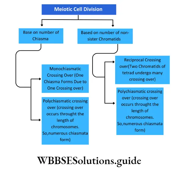
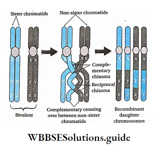
- The breaks are followed by the interchange of chromatid segments between the non-sister chromatids of the homologue. This process is known as crossing over.
- According to Stern and Hotta (1969), chromatid break occurs due to the action of the endonuclease enzyme.
- Ligase helps in joining the broken ends after the exchange. Due to crossing over, the exchange of genetic character between homologous chromosomes and gene recombination takes place.
- In animal cells, asters move away from each other.
- The synaptonemal complex between two homologous chromosomes disintegrates. The nucleolus remains associated with the NOR of a specific chromosome.
Diplotene Or Diplonema
Diplotene Definition: The subphase of prophase I in which paired chromosomes begin to repel each other along with the formation of chiasmata is known as diplotene.
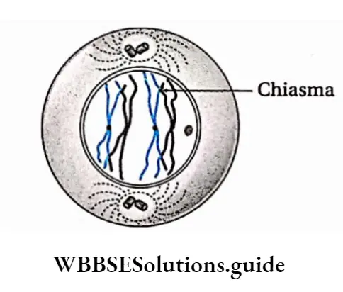
Such points become visible as ‘X’ like structures called chiasmata (singular-chiasma; Greek chiasma means ‘cross’). The position of chiasma may be interstitial, subterminal or terminal.
As the bivalents separate from each other, the chiasma begins to shift towards the terminal end of the chromosome. Thus, the terminalization of chiasmata begins.
Rotation of the arms of chromosomes, with single chiasma, is usually initiated in the late diplotene, forming cross-like (X Structures.
Nucleus Is Still Present. Asters Present In Animal Cells Separate Further Away From Each Other.

Diakinesis
Diakinesis Definition: The subphase of prophase I in which condensation of bivalents increases with fully terminalized chiasmata is known as diakinesis.
The characteristics of diakinesis are as follows—
- Bivalents are fully contracted, condensed, shortened and deeply stained.
- Rotation and terminalisation continue pushing the chiasmata at the extreme ends of the bivalents forming cross and ring-like structures.
- The shape of bivalents depends on the number of chiasmas—cross-like when the number of chiasmas is one, ring-like when the number of chiasmas is two and like a chain of loops when the number is more than two.
- Nucleolus disappears.
- The nuclear membrane starts disintegrating.
Metaphase 1: The phase of meiosis 1 when the maximally condensed bivalents are arranged at the equatorial plate is known as metaphase 1.
Phases of the cell cycle and types of cell division notes
Characteristics:
In this stage, the structure of the mitotic apparatus is completed. X-shaped, ring-shaped, diamond-shaped maximally contracted and condensed bivalents are arranged at the equator of the spindle.
Amphiastral spindles and anastral spindles form 1 in animal cells and in plant cells respectively.
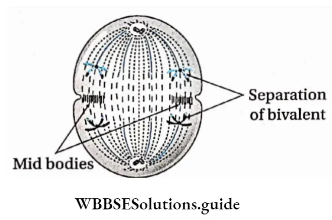
Homologous pairs of chromosomes (bivalents) are so: arranged at the equator of the spindle that their chromatids lie over the equator while their centromeres are bent in the direction of the poles. This special arrangement of the chromosomes makes a double metaphase plate at random.
The chromosomes of a bivalent remain bound to chromosomal fibres belonging to two poles through the centromeres. Kinetochores of the chromosomes bind to spindle fibres from opposite 1 poles.
The centromere of each chromosome is directed towards the opposite poles.
Prometaphase 1
It is a transitional phase between diakinesis and metaphase of meiosis I when the bivalents are maximally condensed in the form of Y or ring.
Characteristics of Prometaphase:
- X-shaped, ring-shaped, diamond-shaped bivalents reach their maximum contraction and condensation.
- The nuclear membrane completely disappears.
- Spindle formation begins.
- In animal cells, the microtubules are arranged in the form of a spindle apparatus in between the two centrosomes occupying the two opposite poles.
- In plant cells, a spindle arises from the cytoplasmic microtubules in the absence of centrosomes.
Anaphase I: The phase of meiosis I at which the homologous chromosomes separate and move to the opposite poles when pulled by the spindle microtubules is known as anaphase I.
Characteristics:
- The chiasmata joining the; homologous chromosomes dissolve allowing the separation of the maternal and paternal homologs,
- Spindle fibres pull the homologous pair towards opposite poles of the spindle.
- The smaller homologue separates more quickly than the larger one because the latter has more number of interstitial chiasmata that take time to dissolve.
- The separated homologs, each consisting of two chromatids united by a centromere, move towards the opposite poles.
- Thus, each pole receives half of the total number of chromosomes, reducing the chromosome number to haploid in the daughter cells.
- This movement of chromosomes towards opposite poles is known as anaphasic movement.
- Anaphasic movement takes place through the elongation of the continuous fibres and contraction of the equatorial zone. The spindle also elongates vertically.
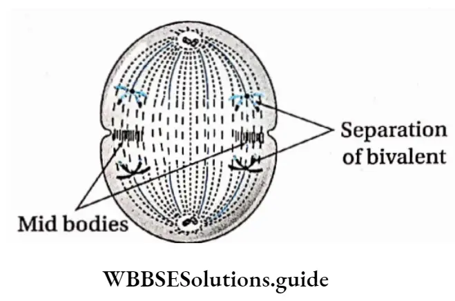
Telophase 1: The phase of meiosis I when the nuclear membrane is reorganised around each group of daughter chromosomes, that spiralised into thin elongated threads forming a reticulum, is known as telophase I.
Characteristics:
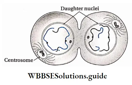
- Telophase I is marked by the presence of chromosomes which are half in number than that of the parent cell, at the poles.
- The nucleolus and the nuclear membrane reappear.
- Water diffuses in the nucleus, so chromosomes are not clearly visible.
- The chromosomes undergo deserialization and become elongated into thread-like structures forming a nuclear reticulum.
- Spindle fibres disappear.
- At the end of telophase, two daughter nuclei with a haploid number of chromosomes (n) are produced.
Cytokinesis
Cytokinesis Definition: The phase of meiosis during which the cell cytoplasm divides into two after the division of the nucleus, leading to the formation of two daughter cells is known as cytokinesis.
Cytokinesis Characteristics:
Cytokinesis can be of two types—simultaneous or successive. In the former separate cytokinesis, I cannot be distinguished.
- In the latter, distinct cytokinesis can be seen after both meiosis 1 and meiosis 2.
- In animal cells, a constriction appears in the equatorial region of the parent cell.
- The constriction gradually deepens to form a narrow furrow and divides the cell into two equal halves. The daughter cells thus produced, have a haploid number of elongated dyad chromosomes.
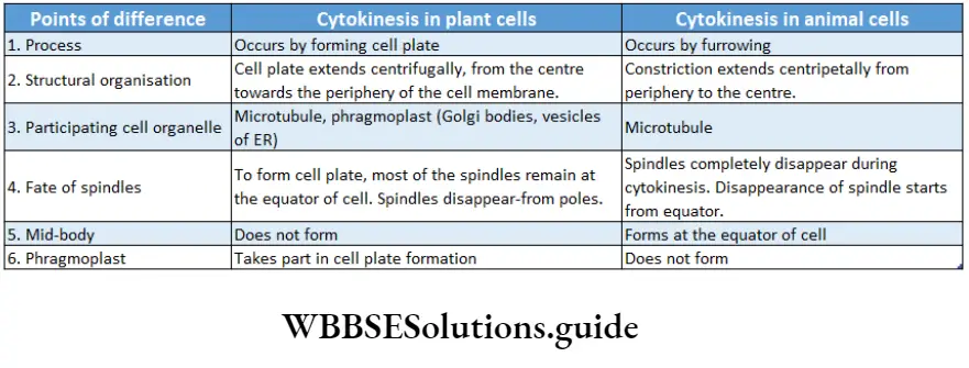
- In most plant cells, daughter cells are produced by the formation of phragmoplast or cell plate.
- Small vesicles or phragmosomes from the Golgi body appear in the equatorial region. They fuse with each other to form the cell plate or phragmoplast.
- The cell plate forms the middle lamella on which the primary cell wall and secondary cell wall are deposited.
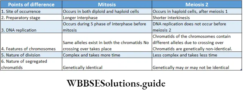
Interkinesis or intrameiotic interphase: The short period between the end of telophase of meiosis I and the beginning of prophase of meiosis 2, where replication of DNA does not take place, is known as interkinesis or intrameiotic interphase.
Cytokinesis Characteristics:
- RNA and proteins are synthesised, but DNA replication does not take place. Hence, the S phase is absent.
- Many plants skip telophase I and enter prophase 2.
- Very little ATP is synthesised.
Meiosis 2 or meiotic division 2
The events of meiosis 2 are analogous to those of a mitotic division, although half the number of chromosomes participate in this division.
It results in the formation of two daughter cells from each of the daughter cells formed after meiosis
They contain an equal number of chromosomes as that of their parent cells.
So, this division is also called homotypic or equational cell division. Meiosis 2 occurs in two stages—karyokinesis 2 and cytokinesis 2.
Karyokinesis 2
This is the nuclear division which occurs in four phases—prophase 2, metaphase 2, anaphase 2 and telophase 2.
Prophase 2: The phase of meiosis 2 during which elongated spiralised chromosomes with two chromatids joined at the centromere, become visible due to dehydration and condensation, is known as prophase 2.
Characteristics:
Due to dehydration, chromosomes become visible as they condense. Each chromosome consists of two chromatids. The chromatids are joined together in the region of centromere.
The chromatids coil thickens and becomes short and visible.
The nuclear envelope and nucleoli disappear and the spindles form. In animal cells, the centrosome divides into two, which move towards opposite poles. Asters form again and spindles form between asters.
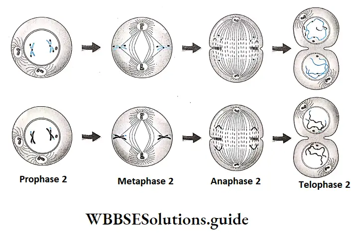
Metaphase 2: The phase of meiosis 2 during which the shortest and thickest chromosomes in the daughter cells arrange themselves at the equatorial plate, is known as metaphase 2.
Characteristics:
- Spindle formation between the two centrioles is completed.
- The shortened and condensed chromosomes become oriented at the
equatorial plate. - The kinetochore of the sister chromatids becomes attached to the spindle microtubules.
Anaphase 2: The phase of meiosis 2 during which the sister chromatids separate and move to the opposite poles when pulled by the spindle microtubules, is known as anaphase 2.
Characteristics:
- The centromere of the chromosome divides into two. As a result, the two sister chromatids (monad) separate.
- The chromatids are pulled to the opposite poles due to the shortening of the chromosomal spindle fibres.
- Interzonal fibres form between the daughter chromosomes. Expansion of interzonal fibres and contraction of chromosomal fibres result in anaphasic movement.
- Telophase 9: The phase of meiosis 2 during which the nuclear membrane is reorganised around each group of chromatids or daughter chromosomes at the poles, that spiralised into thin elongated threads forming a reticulum, is known as telophase 2.
Characteristics:
- Chromatids reach the opposite poles and clump together to form chromosomes.
- Chromosomes begin to uncoil and decondense. They become elongated into indistinct thread-like structures forming a nuclear reticulum.
- The nuclear membrane and nucleolus form again in each of the daughter nuclei.
- The spindle fibres and astral rays usually disappear.


Cytokinesis 2
Cytokinesis Definition: The phase of meiosis 2 during which the cell cytoplasm divides into two after the division of the nucleus, leading to the formation of four daughter cells is known as cytokinesis 2.
Characteristics:
Cytoplasm divides for the second time and two haploid (n) daughter cells of meiosis I, give rise to four haploid (n) daughter cells.
Cytokinesis occurs in animal cells by cleavage and in plant cells by the formation of cell plates.
Significance Of Meiosis
Sexual Reproduction: Meiosis Is responsible for the formation of haploid gametes in sexually reproducing organisms.
Maintains chromosome number: During fertilisation, the nuclei of the two gametes fuse and produce a zygote.
Thus, fertilisation doubles the chromosome number in the zygote. Meiosis halves the number of chromosomes in the gametes to maintain the chromosome number constant and stable for each species.
Source of variation: As a result of crossing over between two homologous chromosomes during prophase I, the exchange of genetic material occurs.
This leads to new combinations of alleles in the chromosomes of the gametes which result in genetic recombination.
Maintains chromosome number: During fertilisation, the nuclei of the two gametes fuse and produce a zygote. Thus, fertilisation doubles the chromosome number in the zygote.
Meiosis halves the number of chromosomes in the gametes to maintain the chromosome number constant and stable for each species.
Source of variation: As a result of crossing over between two homologous chromosomes during prophase I, the exchange of genetic material occurs.
This leads to new combinations of alleles in the chromosomes of the gametes which result in genetic recombination.
Meiosis in plant cell
In plant cells, meiosis occurs in two phases—meiosis I (reductional division) and meiosis II (equational division).
In both the phases karyokinesis is followed by cytokinesis.
Meiosis 1 has two stages—karyokinesis I and cytokinesis I.
Meiosis 2 also has two stages—karyokinesis 2 and cytokinesis 2. At interphase, the nuclear membrane, nucleoplasm and chromatin fibres of the nucleus of the plant cells become visible.
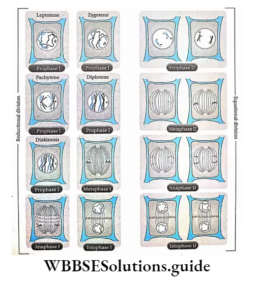
Meiosis 1 starts after interphase. It is followed by a short interkinesis phase after which meiosis 2 takes place. The different stages of meiosis.
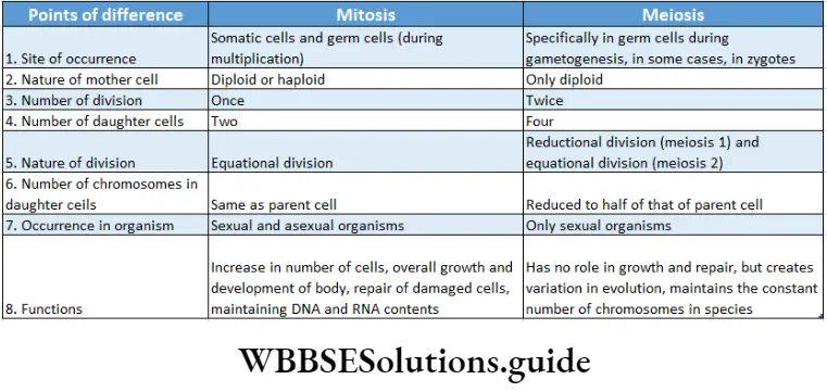


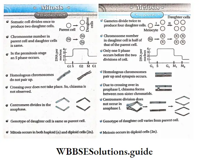
Cell Cycle And Cell Division Notes
- Cyclin: A protein involved in the cell cycle. It goes through cycles of synthesis and degradation during the cell cycle and activates cyclin-dependent protein kinases.
- Cyclin-dependent protein kinase (CDK): Protein found in eukaryotic cells which remains active when associated with cyclin. It regulates the cell cycle.
- Cyclin: A protein involved in the cell cycle. It goes through cycles of synthesis and degradation during cell cycle and activates cyclin-dependent protein kinases
- Cyclin-dependent protein kinase (CDK): Protein found in eukaryotic cells which remains active when associated with cyclin. It regulates the cell cycle.
NEET cell cycle and cell division revision notes with MCQs
Point Of Remember
- The cell generates from an existing cell by division.
- The phase between two successive cell divisions is known as interphase.
- The cell cycle occurs in two main phases, one is the M phase and the other is the I phase or interphase.
- During the Gx phase of interphase, RNA, protein, amino acid, ATP and nucleotides are synthesised.
- DNA and histone proteins are synthesised during the S phase of interphase.
- A mature cell that is no longer capable of undergoing mitosis enters the G0 phase and remains there permanently. This type of cells is known as post-mitotic cells. A neuron or nerve cell is an example.
- The cell cycle is controlled by cyclin and cyclin-dependent kinase.
- Cell division is of three types—amitosis, mitosis and meiosis.
- Amitosis is called direct nucleus division because in this process—
- Spindles do not form,
- Cells divide directly and
- No stages of cell division are found.
- Amoeba and bacteria divide by amitosis (binary fission) to achieve asexual reproduction.
- Alkaloid colchicine, obtained from the plant Colchicine autumnal (Liliaceae), inhibits spindle formation and is known as mitotic poison.
- Mitosis is known as equational division because it results in the formation of two identical daughter cells with an equal number of chromosomes as in the parent cells.
- In plant cells, spindle apparatus forms from microtubules of cytoplasm.
- In animal cells, the spindle apparatus forms by division of centriole and aster. So, spindles of animal cells are known as amphiaster.
- Contraction of the spindle, repulsion between two split centromeres and formation of interzonal fibres together help the daughter chromosomes to move towards the opposite poles (anaphasic movement).
- Presentation of all the chromosomes. pairs of a species
in a diagram or photograph is called a karyogram. - Cytokinesis occurs in plant cells by cell plate formation and in animal cells by furrowing or cleavage.
- Mitosis can be intranuclear or extranuclear. Again, mitosis can skip karyokinesis and can cause endoploidy or endomitosis by duplication of chromosomes.
- Three types of meiotic cell divisions occur. They are—
- Zygotic or initial meiosis,
- Gametic or terminal
- meiosis and
- Sporogenetic or intermediate meiosis.
- Segments of chromatids are exchanged in crossing over. This causes genetic variability.
- Crossing over starts at the pachytene subphase but becomes visible in the diplotene subphase.
- Subphase pachytene in prophase I of the first meiotic division is the longest phase.
- During anaphase I, homologous chromosomes separate and move away from each other. This process is known as disjunction.
- A bivalent contains four chromatids, which are together known as tetrad and this stage is called the tetrad stage.
- Excessive growth of a tissue or organ due to the increase in volume or shape of its constituent cells is known as hypertrophy.
- The process of programmed cell death is known as apoptosis.
- The crossing over between non-sister chromatids that results in the formation of chromosomes with new properties and new gene rearrangement is known as recombination.
- Malignant cells spread to different parts of the body from their primary origin through blood and lymph. This is known as metastasis.
- Two chromatids of a chromosome are known as a dyad.
- Two chromatids of one chromosome in a bivalent are known as sister chromatids. Two chromatids of two different chromosomes of a bivalent are known as non-sister chromatids.
