
Locomotion And Movement Introduction
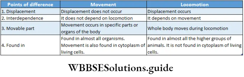
Definition Of Movement: Movement is the Process by Which An Organism Is Able To move its organs Or body parts in response to stimuli without changing the location of the whole body.
Definition of locomotion: Locomotion is the process by which an organism can move from one place to another in response to stimuli.
While watching the Olympic games, don’t you become amazed to see the performance of the athletes? Some of them can jump very high, some can run very fast, some can lift heavy weights and so on. All these abilities depend on the functions of various muscles and bones present in the body.
Movement is a change of position of any part or organ of the body of a living organism.
- Movement can be done by both plants and animals. An animal can also change its location by simply moving different parts of its body. It is termed locomotion.
- A large number of animals move around using their limbs. The limbs are moved by muscles and the muscles are attached either to the endoskeleton or the exoskeleton.
- In almost all vertebrates, movement and locomotion are controlled by muscles and bones. Some organisms such as worms, snails, protozoans, etc., do not have limbs.
- They move around either by changing their body shape or by using appendages or by floating in water.
- Among all the multicellular organisms, only animals explore their environment in an active way, through locomotion.
Significance Of Movement And Locomotion
The significance of movement and locomotion is discussed below.
Search for food: All living organisms have to move to places in search of food. Plants synthesise their own food through photosynthesis.
Thus, locomotion is not that necessary for them. Animals cannot produce their food through photosynthesis. Hence, they need to move from one place to another in search of food.
Reproduction: Locomotion is also important for reproduction in animals and in some lower groups of plants (algae and bryophytes).
Certain fishes (Example salmon) live in sea water but they require fresh water for reproduction. So, they move from sea to fresh water at the time of reproduction.
Shelter: All living beings require a safe and comfortable shelter for living and reproducing.
| Class 11 Biology | Class 11 Chemistry |
| Class 11 Chemistry | Class 11 Physics |
| Class 11 Biology MCQs | Class 11 Physics MCQs |
| Class 11 Biology | Class 11 Physics Notes |
“locomotion and movement neet notes “
Animals move from one place to another in search of a suitable shelter where they will have enough food and water as well as less threat from other predatory animals.

Search for a favourable environment: All plants and animals require various environmental components such as light, air, water, etc., for their survival. So, they move from one place to another in search of favourable environmental conditions required for their survival.
Different types of movement are observed in living organisms.
Ciliary Movements
Ciliary Movements Definition: The type of movement which includes rhythmic movements of cilia, present on the cell surface, is known as a ciliary movement.
Cilia are microscopic hair-like organelles found in eukaryotic cells. They project from the surface of the cells. They show movement which helps cells in locomotion. They also help the cells to remain anchored in the tissues.
Ciliary Movements Example:
- In multicellular organisms such as human beings, cilia are found in some internal organiser
- Cilia present in the respiratory tracts help to remove pathogens and dust particles. Cilia are also found in reproductive tracts. They help in the transportation of egg and sperm cells.
- In unicellular organisms such as Paramoecium, cilia are used for locomotion.
- Cilia is also found in the flame cells present in tapeworm. These cells are the excretory cells of tapeworms. The cilia of flame cells help to filter the body fluid by removing waste products from it.
Locomotion and movement notes for NEET PDF
Ciliary Movements Process:
- The organisms use their cilia similar to an oar used in a boat.
- Cilia can move both in anterior and posterior directions.
- Cilia are arranged in bands or clumps. The movement of each cilium must be closely coordinated with the movements of other cilia.
- Paramoecium uses its holotrichous cilia as a locomotory organiser During swimming, cilia move in a whip-like motion by producing two types of strokes water-power stroke and recovery stroke.
- During a power stroke, cilia push the water backwards and give a forward push.
- The power stroke is followed by the recovery stroke. In this movement, the cilia relax and return to their original position.
- In this way, the rows of cilia move one by one which helps to move the animal in water.

Flagellar Movement
Flagellar Movement Definition: Some organisms’ locomotion by movement of a type of cytoplasmic filament called flagella, is known as flagellary movement.
A flagellum (Latin: flagellum means ‘whip’) is a lash-like appendage that protrudes from the cell body of certain prokaryotic and eukaryotic cells.
The primary role of the flagellum is the locomotion of the organism in a liquid medium, but it also often functions as a sensory organ.
Flagellar Movement Example:
- Some unicellular organisms such as Euglena, Trypanosoma gambiense, etc., use their flagella for locomotion.
- In some multicellular organisms such as sponges, a water canal system is found. The large, central cavity included in this system is known as spongocoel.
- The inner wall of this spongocoel contains special flagellated cells known as choanocytes or collar cells.
- Each cell contains a central flagellum. This flagellum helps in the unidirectional flow of water into the spongocoel.
Flagellar Movement Process:
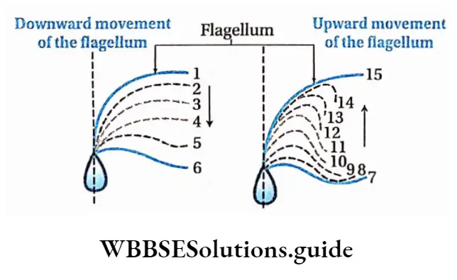
- Euglena and other flagellated protozoans move by the spiral undulation of their flagellum.
- Euglena swims by lashing the flagellae sideways and the body is directed obliquely backward. This movement helps Euglena to move forward by pushing the water backwards.
- The energy required for the movement is obtained by the breakdown of ATP into ADP and inorganic phosphate.
Amoeboid Movement
Amoeboid Movement Definition: The movement or locomotion that depends on the movement by extensions of the cell body called pseudopodia, is known as amoeboid movement.
Amoeboid movement is the most common mode of locomotion in eukaryotic cells. It is a crawling-like movement accomplished by cytoplasmic protrusion of the cells called pseudopodia.
Example:
- This kind of movement can be found in different cells such as leucocytes and macrophages, etc., present in our body.
- Unicellular organisms such as Amoeba, move from one place to another by this type of movement.
Amoeboid Movement Process:
- By changing the viscosity of its protoplasm and with the help of microfibrils inside the cells, Amoeba gives rise to pseudopodia.
- The pseudopodium attaches to the substratum (on which movement of Amoeba occurs) and microfibrils inside the cell contract to bring the rest of the cell towards the pseudopodium.
- By repeating this movement, Amoeba moves from one place to another.
Muscular Movement
Muscular Movement Definition: The movement of the body which occurs with the help of muscles is known as muscular movement.
Example: Muscular movement is found in vertebrates and some invertebrate animals (annelids, arthropods, molluscs, etc.).
Muscular Movement Process:
- Typical nature of muscles are excitability, contractility and elasticity.
- Due to nervous stimulation, the muscle fibres contract and then return to their original state.
- Some physiological processes occur due to the involuntary action of muscles. Some of such actions are peristalsis, movement of the heart muscles, etc.
- The voluntary action of muscles also helps the organisms in the movement of their body parts as well as locomotion.
chapter 20 biology class 11 notes
Skeletal Muscle Contractile Proteins And Muscle Contraction
Skeletal Muscle Contractile Proteins And Muscle Contraction Definition: The muscles which are attached to the bones, can contract voluntarily and are mainly involved in the movement of the bones, are known as skeletal muscles.
Skeletal Muscle Contractile Proteins And Muscle Contraction Location: In humans and other vertebrates, these muscles are attached to the endoskeleton by means of a thick band of collagenous connective tissues called tendons.
As these muscles are attached to the skeletal system, they are named as skeletal muscles.
Skeletal Muscle Contractile Proteins And Muscle Contraction Characteristics:
- Skeletal muscle is responsible for the movement of various parts of the body and locomotion. These muscles are attached to the bones with the help of tendons.
- These muscles are able to contract voluntarily.
- These are striated.
- Muscle fibres are the individual contractile units of muscle.
- A number of undifferentiated, mononucleated muscle cells (myoblasts), unite to form a single, cylindrical, multinucleated cell (syncytium). These syncytia play a fundamental role in muscle function as they form the muscle fibres.
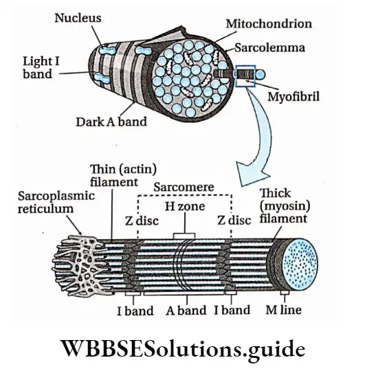
- Every muscle cell is covered with a thin transparent sheath, known as sarcolemma. The cytoplasm of muscle cells is called sarcoplasm. The sarcolemma contains some deep invagination called T-tubules (or transverse tubules).
- The bulk of fibre is arranged in the form of myofibrils that have characteristic striations due to the precise organisation of the contractile proteins actin and myosin.
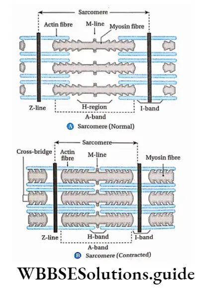
- The myofibrils are arranged in a section, called a sarcomere joined end to end all along the length of a muscle fibre.
- Sarcomeres are separated by thin, comparatively dense, zigzag lines or discs called Z-line or Z-disc.
- A myofibril has dark and light bands. The dark bands are called the A-band or anisotropic bands. The light bands are called l-band or isotropic bands.
- The two bands are separated by the H-zone (Hensen zone).
- The centre of the H-zone has a line called the M-line.
- The myofibrils have characteristic striations on them due to the precise organisation of two contractile protein filaments—
- Actin and
- Myosin.
- Adult skeletal muscle fibres have diameters between 10 and 100 /μm and lengths that may extend up to 20 cm.
Connective Tissue Covering Of Muscle Fibres
Skeletal muscle fibres are covered with layers of connective tissues.
Blood vessels, nerves, lymphatic vessels, etc., are present in this covering. This covering is divided into three layers—
- Endomysium: The thin layer of areolar connective tissue around each muscle fibre is known as endomysium.
- Perimysium: The thin sheath of connective tissue that covers a bundle of muscle fibres which are already surrounded with endomysium, is known as perimysium. Each bundle of muscle fibres covered with perimysium is known as fasciculus (plural: fasciculi).
- Epimysium: The layer of connective tissue that surrounds some bundles of muscle fibres together is known as epimysium.
Class 11 biology locomotion and movement notes with diagrams
Skeletal Muscle Contractile Proteins Physiological properties: The physiological properties of skeletal muscles are as follows—
- Excitability: If a suitable stimulus is applied to a voluntary muscle fibre, then it will get excited. This phenomenon is known as excitability. The stimulus can be chemical, electrical, mechanical, thermal, etc.
- Contractility: Muscle fibres contract due to the excitation caused by the appropriate stimulus. This is known as contractility. It is an important property of muscle fibres. Mainly contractile proteins are responsible for the contraction of the muscle fibres.
- Refractory period: A voluntary muscle fibre contracts when a threshold stimulus (an optimum stimulus that could generate a contraction) is applied to it. After this, for a small interval of time, the muscle fibre will not contract further, if another stimulus is applied at this stage.
- The small interval of time is known as the refractory period. In mammals, the absolute refractory period is 0.002-0.005 seconds.
This period can be categorised into two types—
- The absolute refractory period is the initial period of the refractory period during which the muscle fibres will not contract even if a very strong stimulus is applied.
- The relative refractory period is the end period of the refractory period when the muscle starts to contract by the effect of a strong stimulus.
All-or-none law:
- According to this law or principle, the response of a muscle fibre to a stimulus is independent of the stimulus.
- If the muscle fibre is given a stimulus that exceeds the threshold potential, the nerve of the muscle fibre will give a complete response; otherwise, there is no response.
- The contraction will not increase further even if the force of the stimulus is increased. This is the all-or-none law.
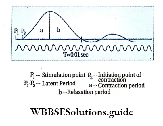
Latent period: It is the time gap between the application of a stimulus and the generation of the response.
Simple Muscle Curve
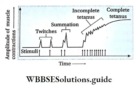
Contraction and relaxation of muscle fibres occur due to excitation. The recording of relaxation and contraction of muscle fibres by the kymograph is known as a simple muscle curve.
Summation of stimuli: In case of a stimulus lower than the threshold level, no contraction occurs within the voluntary muscles.
But if the same stimulus is applied repeatedly, between the latent period and refractory period of the first stimulus, then contraction of muscles can be observed.
Individually the stimulus could not evoke a contraction. However, the summation of those separate stimuli was able to generate a contraction as their cumulative value exceeded the threshold level.
Muscle twitch: A muscle fibre contracts only once if it is stimulated by a single stimulus of adequate strength. It relaxes immediately after the contraction. Together, this individual event of contraction and relaxation of muscle is known as muscle twitch.
Tetanus: If a muscle fibre is introduced to a rapid succession of stimuli for a certain period of time, then the rate of stimulation will be so high that the muscle fibres will be unable to relax between the two stimuli.
This will lead the muscle to a state when muscle twitches are converted to sustained contraction as long as the stimuli continue. This is known as tetanus.
Rigour mortis: After death, the muscle cells can no longer produce ATP and therefore the cross-bridges between muscle fibres cannot be broken. This causes muscle stiffness called rigour mortis.
Fatigue: Muscle fatigue refers to the use-dependent decrease in the ability of a muscle to generate force.
The causes of fatigue are not entirely understood. In most cases, however, muscle fatigue is correlated with the production of lactic acid due to excessive working of the muscles.
Lactic acid is produced by the anaerobic respiration of glucose, which is obtained from glycogen present in the muscles and blood. Lactate production and muscle fatigue are therefore also related to the depletion of muscle glycogen.
Muscle Fatigue
During excess muscular activity or if a muscle fibre is stimulated continuously, the contracting ability of the muscle gradually decreases and ultimately fails to contract for some time. This condition is known as muscle fatigue.
Muscle Fatigue Causes:
- The large amount of energy required for contraction and relaxation of muscles is produced by the aerobic oxidation of glycogen.
- However, due to excess oxidation, the muscle cells receive insufficient oxygen.
- As a result of which, partial oxidation of the glycogen takes place. This oxidation produces lactic acid.
- Thus, the concentration of lactic acid increases in the muscles. The stored lactic acid stops the muscle contraction for some time and causes muscle fatigue.
Relief from muscle fatigue: If the fatigued muscles remain in the resting stage for some time, then they will get a sufficient amount of oxygen. As a result, complete oxidation occurs and the muscles can overcome the fatigued condition slowly.
Structure of skeletal muscle: A detailed description of this topic has been provided.
Muscle Fatigue Types: Depending on structure and function, skeletal muscles are of different types.
On the basis of function: Details are discussed in the table given below.

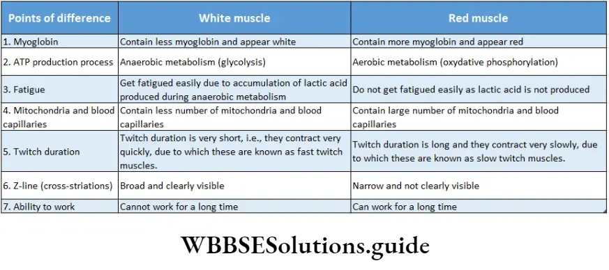
On the basis of structure: On the basis of structure, skeletal muscles are of two types—white and red muscles.
White muscles: Some of the muscles possess a very small quantity of myoglobin and therefore, appear pale or whitish. These are categorised as white muscles.
The number of mitochondria is also few in them, but the amount of sarcoplasmic reticulum is high. They depend on anaerobic processes for energy.
Example: White muscles are mostly found in amphibians and birds.
Red muscles: Some muscles contain a high concentration of myoglobin and hence appear red in colour.
These muscles are called the red muscles. They contain a large number of mitochondria which produce ATP by aerobic metabolism.
These muscles, therefore, can also be called aerobic muscles. Example: Red muscles are found in mammals.
Structure Of Contractile Proteins
- Every skeletal muscle is composed of myofibrils. These myofibrils are composed of myofilaments.
- The proteins present in the myofilaments are responsible for the contraction and relaxation of myofibrils.
- These are known as contractile proteins. Mainly, two types of contractile proteins are found in human beings.
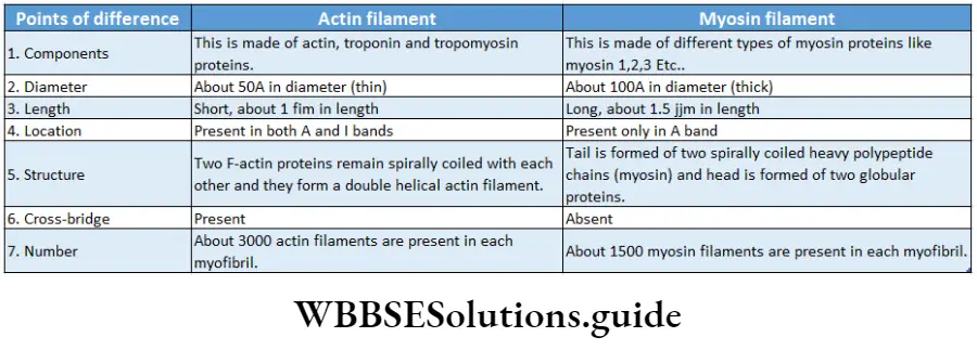
Actin Filament
The thin filamentous protein among the contractile proteins of myofibrils is known as actin filament. It is also known as secondary myofilament.
Actin Filament Number: Each myofibril is composed of about 3,000 actin filaments.
Mechanism of muscle contraction in locomotion and movement
Actin Filament Characteristics:
- Actin filaments are present in between two Z-lines (except the H-zone), generally all over the sarcomere.
- The length and diameter of each fibre are lum and SOA respectively.
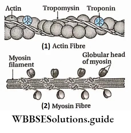
- Actin filament is composed of mainly three types of proteins—actin, tropomyosin and troponin.
- One end of these fibres remains attached to the Z-line (a zigzag line that separates two adjacent units of muscle or sarcomere) and another end is attached between the two myosin filaments.
Actin Filament Structure: The actin filament is composed of three types of proteins. They are discussed below.
- Actin: An actin filament consists of actin monomers polymerised into a large molecule that looks like two strands of pearls twisted together.
- Actin is found in two forms —G-actin and F-actin.
- When actin is present in the form of small monomers, then it is known as G-actin. Its molecular weight is about 42,000 Da.
- G-actin proteins form fibrous polymers in the presence of Mgz+. The filamentous structure formed by these fibrous polymers is known as F-actin.
- Two F-actin fibres form a double helix which forms a single main actin filament (lpm in length).
- During muscle contraction, myosin fibre attaches to the active site of the F-actin.
Tropomyosin:
- Tropomyosin is a type of long and thin protein molecule, present around the grooves of the actin chains.
- Its molecular weight is about 70,000 Da and its length is 40nm.
- During normal conditions, tropomyosin molecules remain attached to the active site of the F-actin.
Troponin: Troponins are small globular proteins present in tropomyosin at intervals. Each troponin molecule has three peptide subunits—
- Troponin-T: Connects the troponin with tropomyosin,
- Troponin-I: Prevents the attachment of myosin and actin.
- Troponin-C: Helps in muscle contraction in the presence of Ca2+ ions. The troponin complex (troponin-C, troponin-l and troponin-T) together with tropomyosin, form the calcium-sensitive switch that activates muscle contraction.
Myosin Filament
The thick filamentous protein among the contractile proteins of myofibrils is known as myosin filament. It is also known as primary myofilament.
Myosin Filament Number: Each myofibril is composed of 1,500 myosin fibres.
Myosin Filament Characteristics:
- These fibres are present in the A-band of the sarcomere.
- The length and diameter of each myosin fibre are about 1.6 fim and 100A respectively.
- Different types of myosin proteins are present in myosin fibres. Among them, myosin-II protein (also known as conventional myosin) helps in the contraction of skeletal muscles.
- Both ends of the myosin fibres are free.
muscular movement
Myosin Filament Structure:
- Each filament contains about 200 myosin molecules.
- Each myosin molecule is composed of six polypeptide chains. Among these, two are heavy chains and four are light chains.
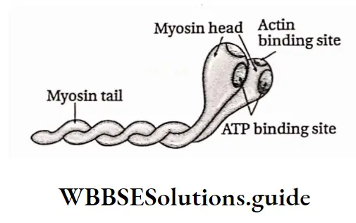
- Each myosin fibre has a head region and a long tail part.
- The long tails (heavy chains) of two myosin fibres wrap around each other to form a dimer, which in turn aggregates to form the myosin filament.
- Their head regions (light chains) are lined precisely opposite to the actin filaments.
- When viewed in transverse sections, the actin filaments form a hexagonal pattern around each myosin filament.
- The bundles of myosin filaments are held in a centred position within the sarcomere by a protein called titin.
- The myosin head contains a specific binding site for actin fibre.
- The head can act as an ATPase enzyme. Due to this property, it gathers energy for contraction by hydrolysis of the ATP molecules.
Contraction Of Skeletal Muscle
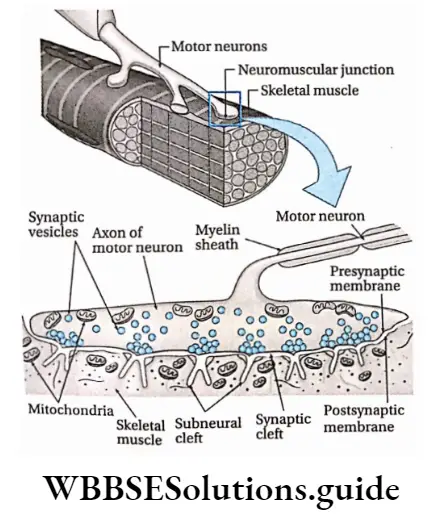
- The region of the sarcolemma that lies just below the terminal end of the axon, is known as the motor endplate.
- The synapse or junction formed between the axon terminal and the motor endplate is known as a neuromuscular junction.
- Each branch of the axon ends in an axon terminal that lies in close proximity to the sarcolemma of a muscle fibre.
- These neurons release acetylcholine with the help of Ca+2 ions in response to action potentials (i.e., change in j electrical potential in cells during a nerve impulse).
- Acetylcholine is a chemical substance that transmits the impulse between the nerve and muscle cell, hence, called a neurotransmitter. Acetylcholine is present at the neuromuscular junction.
- The contractile proteins (actin and myosin) cause contraction of muscle fibres when the impulse is transmitted.
- Muscle fibres are innervated and are stimulated to contract by motor neurons.
- These neurons have their cell body in the spinal cord and their axons are projected outside to enter the effector organiser
- The axon of one motor neuron has several branches and can stimulate a few to several muscle fibres of a particular muscle.
Neuromuscular Junction
- The junction between the axon terminal of a motor neuron and the skeletal muscle fibre is known as a neuromuscular junction or motor end plate.
- The nerve impulses from the motor neurons cross the neuromuscular junction and are transported to skeletal muscles by the neurotransmitter molecules.
- Here the end of the motor neurons become flat, broad and extended which is known as end feet.
- The membrane present at the nerve end is known as the presynaptic membrane. This portion is rich in mitochondria. The spaces between the motor neurons and the sarcolemma of the skeletal muscles are known as synaptic clefts.
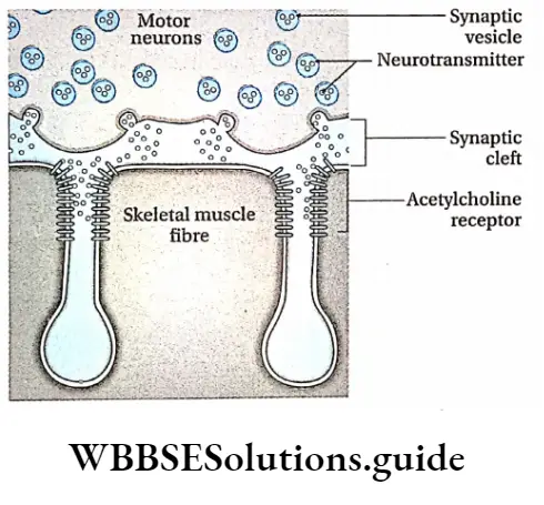
- Synaptic cleft contains numerous vesicles containing acetylcholine, a neurotransmitter. These vesicles are called synaptic vesicles. The sarcolemma is folded at the neuromuscular junctions of the skeletal muscles.
- These are known as postsynaptic membranes. The folds that occur in the membranes are known as subneural clefts.
- These clefts contain many receptor proteins. The acetylcholine molecules are released by the synaptic vesicles in the synaptic cleft.
- These neurotransmitters are transported to skeletal muscle fibres by binding with acceptor proteins present in the subneural clefts.
Mechanism of contraction of skeletal muscle was The mechanism of contraction of skeletal muscle best explained through—‘Sliding filament theory’. This was proposed by two groups of scientists—A. F.
Huxley and R. Nierdergerke, 1954; H. E. Huxley and J. Hansen, 1954.
According to this theory, events of muscle contraction are divided into two parts—
Physical events of muscle contraction, and biochemical and electrical events of muscle contraction.
Physical Events of Muscle Contraction:
- During muscle contraction, thick myosin fibres and thin actin fibres combine with each other by forming a cross-bridge. After this, myosin fibres pull the actin fibres towards the centre of the sarcomere.
- At this time Z-lines of the muscle fibres come closer to each other. As a result, the length of the sarcomere decreases.
- Contraction of l-band and H-zone (Heller zone from ‘heller’ in German, meaning brighter) takes place. The M-line crossing the H-zone centrally is also called the Micellar line or programme line.
- Only the length of the A-band remains unchanged.
- Due to contraction, the length of the muscle decreases but the volume remains the same.
Biochemical and electrical events during muscle contraction: The steps of biochemical and electrical events during muscle contraction are as follows—
Discharge of neurotransmitter by motor nerve: The motor neurons arising from the spinal cord or brain stem, release the neurotransmitter acetylcholine at the neuromuscular junction. In response to this, action potentials travel down the axon towards the skeletal muscle cell.
Types of movement in humans and animals with examples
Binding of neurotransmitter at skeletal muscle fibre: The amount of acetylcholine released is proportional to the frequency of action potentials travelling down the axon. Acetylcholine diffuses across the synaptic cleft and binds to a receptor protein, the nicotinic receptor, on the muscle cell membrane.
Generation of Action Potential at Skeletal Muscle Fibre:
- The nicotinic receptors form channels that allow sodium and potassium ion influx to the muscle cells.
- This causes partial depolarisation of the cell membrane in the synaptic cleft.
- This depolarization generates an endplate action potential or action potential. This action potential acts as an indicator of muscle contraction.
Release of Ca2+ ions:
- Muscle fibres store Ca2+ in a modified endoplasmic reticulum known as sarcoplasmic reticulum (SR).
- When a muscle fibre is stimulated to contract, an electrical impulse travels into T tubules. This triggers the release of Ca2+ from the sarcoplasmic reticulum.
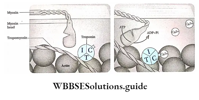
Release of the Active site of Actin Filament by Ca2+ ion:
- When a muscle is relaxed, myosin heads are unable to bind to actin.
- This is because the active sites (attachment sites) for the myosin heads on the actin are blocked by tropomyosin.
- Thus, myosins are unable to bind with actin. Tropomyosin is held in place by troponin.
- When the concentration of Ca2+ is raised in the sarcoplasm, Ca2+ binds to troponin. This causes the conformational change in troponin. This causes the troponin-tropomyosin complex to be shifted away from the active sites.
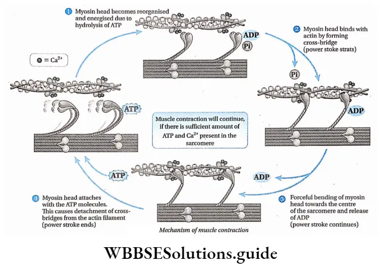
Formation of Energised Myosin Heads:
- An ATP molecule attaches to the active site of the myosin head. The head acts as an ATPase enzyme.
- This causes hydrolysis of the ATP in the presence of Ca2+ and Mg2+ to produce ADP and Pi. The myosin head takes up the energy released and becomes highly energised.
Formation of Cross-Bridges:
- The energised myosin heads are attached to actin by forming cross-bridges.
- After binding, the myosin heads release the inorganic phosphate and initiate an inward pull called power stroke.
- This causes inward bending of myosin filament, thereby shortening the sarcomere.
- The myosin heads then release the ADP molecule but still remain tightly bound to the active, until a new ATP molecule is bound to it. Then the heads separate from the actin filament.
- This process occurs in a cyclic manner as long as ATP and Ca2+ are present in the sarcoplasm. This is how actin slides and contraction of muscles takes place.
Types of Muscle Contraction are—
- Isometric muscle contraction, and
- Isotonic muscle contraction.
Isometric Muscle Contraction: The muscle contractions in which the length of the muscle remains the same but tension or force increases, are called isometric muscle contractions.
The term ‘isometric1 has been derived from the Greek words isos meaning equal and metric meaning measuring
The characteristics of Isometric Muscle Contractions are—
Isometric contractions occur as a phase of normal muscle contraction but also provide tautness and stability to the body.
- The weight of the muscles is more than the force of this contraction.
- No mechanical work is carried out by this type of contraction in muscles.
- Most of the free energy produced during this type of contraction of the muscles is released as heat energy.
Example: The contraction of muscles during chewing, writing, etc.
Isotonic Muscle Contraction: The muscle contractions in which the tension within the muscle during contraction remains the same but the length of the muscle changes are called isotonic muscle contractions.
The term ‘isotonic’ has been derived from two Greek words—isos meaning equal and tonic meaning strength.
Changes occurring during muscle contraction The changes occurring during muscle contraction can be divided into several types.
Physical Change: The length of the muscle fibres decreases and thickness increases during muscle contraction. The volume of the muscles remains unchanged.
Chemical Change: The energy required for muscle contraction is produced by hydrolysis of ATP.
Due to repeated hydrolysis of ATP, its amount gradually decreases in the cells. It is replenished by various chemical reactions. These chemical reactions are discussed under separate heads.
Glycolysis, Krebs cycle and terminal respiration: Synthesis of ATP, C02, water, lactic acid, etc., take place due to these chemical reactions.
Changes in creatine phosphate or phosphagen: Creatine phosphate is a highly energised compound. This compound is present in large amounts in muscle cells. It supplies inorganic phosphates during the conversion of ADP into ATP during muscle contraction.
Change in pH: During the initial stage of normal muscle contraction, the pH remains basic. But if the muscle contracts for a prolonged period of time, then due to the accumulation of lactic acid, the pH becomes acidic. ATP synthesis also occurs during this stage.
Thermal change: During muscle contraction, some of the energy is released in the form of heat energy. This energy is released in three forms—
The heat of activation: The heat energy released from the muscles just after receiving the stimulus, prior to contraction is called heat of activation. The amount of heat gradually decreases with muscle contraction.
Heat of shortening: The heat energy produced due to various structural changes during muscle contraction is called heat of shortening.
Recovery heat: The heat energy produced after muscle contraction, during re-synthesis of substances dissociated during muscle contraction, is called recovery heat.
Electric charge: The electric potential found in the muscles during their resting stage, is known as resting potential.
Its value is -70 mV. During muscle contraction, different ions are transported between the intracellular and extracellular fluids in the stimulated region of the sarcomere.
As a result, the resting potential is converted to the action potential. Its value is +35 mV.
Functions Of Skeletal Muscle
The functions of skeletal muscles are discussed below.
Movement of body parts: The skeleton muscles allow flexible body movement. These muscles remain attached to bones with the help of tendons. So, the muscle contraction leads to movement of different body parts.
Storage of glucose: Excess glucose present in the blood converts into glycogen by the process of glycogenesis. The glucose remains stored in the skeletal muscles as glycogen.
Maintainance of body structure: Skeletal muscles along with the bones maintain the structure of the body.
Maintainance of body balance: Skeletal muscles help to maintain the body balance.
Respiration: Skeletal muscles, especially the intercostal muscles, help in respiration.
Locomotion in animals is accomplished by the force of muscles acting on a rigid skeletal system.
The skeletal system consists of the bones and joints, along with cartilage and ligaments.
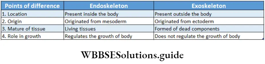
There are two types of skeletal systems found in the animal kingdom—exoskeleton, and endoskeleton. These are discussed under separate heads.
Exoskeleton: Exoskeletons can be of different forms, including horns, claws, nails, hooves, scales, etc.
- In general, they are hard outer coverings, that provide physical protection to soft internal body parts of animals. Exoskeletons also provide sites for muscle attachment. In most of the species, this structure surrounds the body as a rigid hard case.
- Arthropods, such as crustaceans and insects, have exoskeletons made of the polysaccharide, chitin. Having an exoskeleton also limits the size of the animal.
- Animals with exoskeletons cannot grow too large. This is because if the animal grows larger, its exoskeleton will become thicker and heavier and will cause hindrance in movement and locomotion.
Endoskeleton: An endoskeleton consists of hard, mineralised structures located within the soft tissues of organisms. The endoskeleton is present in vertebrates.
An example of a primitive endoskeletal structure is the spicule of sponges. The bones of vertebrates are composed of tissues, whereas sponges have no true tissues.
Endoskeletons provide support to the body, protect internal organs, and allow movement through the contraction of muscles, attached to the skeleton.
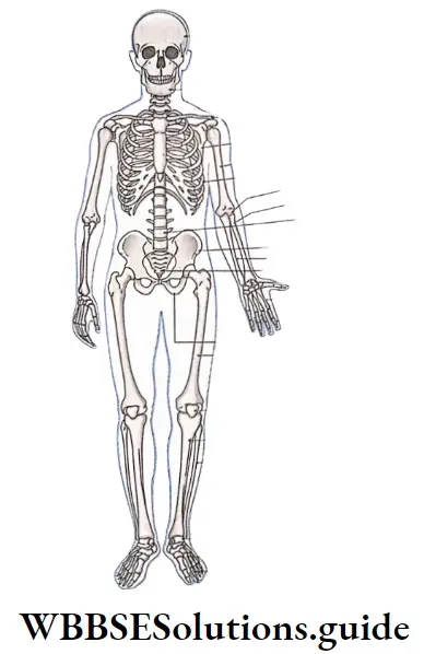
Description Of The Human Skeletal System
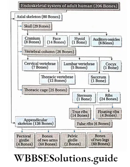
The skeletal system, in an adult body, is made up of 206 individual bones.
The endoskeleton is arranged into two major divisions—the axial skeleton and the appendicular skeleton.
The tissues of the axial and appendicular skeletons are bone (both compact and Fh spongy), cartilage (hyaline, fibrocartilage, and elastic cartilage), and dense connective tissue (a type of fibrous connective tissue).
Axial Skeleton
Axial Skeleton Definition: The bones of the skeleton, which form the main longitudinal axis of the body in humans and other vertebrates, are together known as the axial skeleton, It is formed of 80 bones.
The appendicular skeleton is attached to the axial skeleton. The main parts of the axial skeleton are—the skull, thoracic cage and vertebral column.
Skull
The skull is the bony, hollow, round-shaped structure of the axial skeletal system, that protects our brain.
Characteristics:
- The skull is composed of 29 bones, that are fused together, except the mandible.
- These skull bones are not fused in children, but rather loosely attached with connective tissues.
- This allows the growth of the skull. In adults, the skull bones fuse to give added strength and protection to the soft tissues and brain.
Parts: The four main parts of the skull are—the cranium, facial bones, hyoid bone and auditory ossicles.
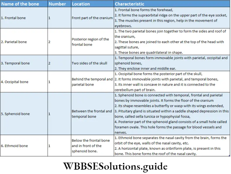
Cranium: The box-shaped structure made of 8 flat bones, present at the superior portion of the skull is known as the cranium.
The characteristics of the cranium are as follows—
- It is formed of 8 flat bones.
- These bones are separated from each other by immovable joints called sutures.
- The cranium is composed of one frontal bone, two parietal bones, one occipital bone, two temporal bones, one sphenoid bone, and one ethmoid bone.
- An orifice, known as the foramen magnum, is present behind the skull. The spinal cord remains attached to the brain through this orifice.
- The cranium is also known as the neurocranium and rainbow, as it contains the brain. It protects the brain from damage.
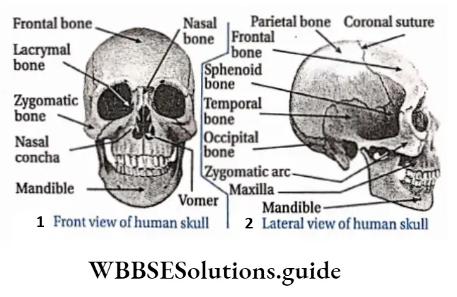
Facial bones: The bones of the inferior and anterior portions of the skull are known as facial bones.
The characteristics of facial bone are as follows—
- There are 14 facial bones.
- These bones generally form both the jaws, some sense organs like eyes, nose, ears, mouth and a part of the nasal passage.
- The face is composed of one mandible (the lower jaw), the only movable bone in the skull.
- Other bones of the face are two maxillae (upper jaw), two palatine, two zygomatic, two lacrimal, two nasal, one vomer and two inferior nasal conchae.
- These bones support the eyes nose and mouth.
Hyoid bone: The hyoid bone is a small, U-shaped bone found just inferior to the mandible.
It is the only bone in the body that does not have any joint and it is a floating bone.
The functions of the hyoid bone are as follows—
- The hyoid bone helps in various movements of the tongue, larynx and pharynx.
- Its function is to hold the trachea open and to form a bony connection for the tongue muscles.
Auditory ossicles:
- The three bones present in the ear malleus, incus, and stapes, are collectively known as the auditory ossicles.
- They are found in a small cavity inside the temporal bone. These bones are among the smallest bones in the human body.
- Malleus, incus and stapes are commonly known as hammer, anvil and stirrup, respectively.
The functions of auditory ossicles are as follows—
The ossicles help to transmit vibrations of sound waves, from the tympanum through the middle ear to the oval window of the inner ear.
They also protect the ear from damage caused by loud sounds.
Functions of the skull:
- The skull protects the brain.
- It also protects our main sensory organs—eyes, ears, nose and tongue.
- Jaws and teeth help in the mastication of food.
- The skull also protects the pituitary gland located at a depression (sella turcica) on the sphenoid bone of the skull.
- The opening present at the end of the skull called the foramen magnum, connects the spinal cord to the brain.
Thoracic Cage (Rid Cage)
A frame-like structure formed by flat bones, that is present within the thoracic region is known as a thoracic cage.
Thoracic Cage (Rid Cage) Characteristics:
- The rib cage, sometimes called the thoracic cage is the arrangement of bones in the thorax region.
- It is formed by thoracic vertebrae 12 of the vertebral column, ribs and associated cartilages, and sternum.
- The rib cage encloses the heart and lungs.
Parts: The parts of the rib cage have been described under separate heads below.
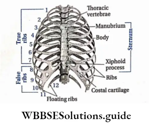
Ribs: The 12 pairs of narrow and flat bones, originating from both sides of the vertebral column, are known as ribs.
Characteristics:
- All twelve pairs of ribs are attached to the thoracic vertebrae of the vertebral column,
- The first seven pairs of ribs (upper ribs) connect the thoracic vertebrae directly to the sternum and are known as “true ribs”,
- The next three pairs of ribs (Ribs 8, 9, and 10) originated from the 8th, 9th and 10th thoracic vertebrae and are connected to the sternum indirectly via the costal cartilage of the 7th rib, and due to this they are considered as “false ribs.”
- The last two pairs of ribs (11 and 12), are not attached to the sternum. They remain free in front, and are termed as “floating ribs”.
Function: The functions of the ribs are, the formation of the thoracic cage and also protection of internal organs such as the lungs, heart, etc. The elasticity of the false ribs allows movement of the rib cage during respiratory activity.
Sternum; The sternum, or breastbone, is a thin, knife-shaped bone located at the anterior side of the thoracic region along the midline of the body.
Characteristics:
- The sternum is connected to the ribs by thin bands of cartilage, called the costal cartilage,
- The sternum is composed of three bones—manubrium, body, and xiphoid process, that fuse during foetal development.
Function:
- The sternum participates in the formation of the thoracic cavity,
- It helps to join different muscles.
- It also protects the heart, aorta, vena cava, etc.
Functions of the thoracic cage:
- The thoracic cage protects the heart, lungs and the main blood vessels.
- The functions of ribs are dependent on the function of intercostal muscles.
- Due to the action of these muscles, ribs help the lungs to expand and relax during breathing. Thus, the ribs play a key role in respiration.
- In humans, ribs contain red bone marrow throughout their life. So, they produce RBCs, some WBCs and platelets in them.
- The two pairs of floating ribs protect the liver and other organs in the abdominal cavity.
Vertebral column
A long pillar-like structure, composed of bones called vertebrae, present as the longitudinal axis, at the dorsal side of the body, is called the vertebral column.
Characteristics:
- The vertebral column, also known as the backbone or spine, extends from the skull to the pelvis.
- It consists of 33 bones, (In an adult person it becomes 26 in total due to the fusion of some lower vertebrae) the vertebrae.
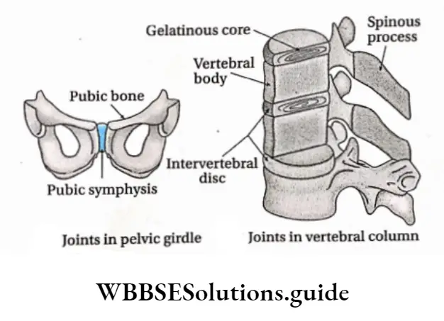
- They are separated by pads of fibrocartilage, known as the intervertebral discs.
- The vertebral column forms the vertical axis and is located in the middorsal region.
- The skull rests on the superior end of the vertebral column.
- The vertebral column supports the rib cage and serves as a point of attachment for the pelvic girdle.
- The vertebral column also protects the spinal cord (part of the central nervous system), which passes through a hollow space called the vertebral canal, created by vertebrae.
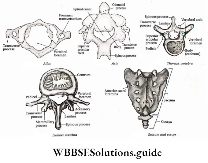
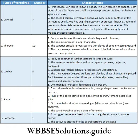
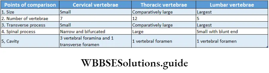
- They are named according to their location, such as—cervical vertebrae
- Present at the neck region, thoracic vertebrae
- (12) Present at the thorax region, lumbar vertebrae (5) present at the lower back region, sacrum or sacral vertebra (1) present at the end of the spine and coccyx or caudal vertebra (1) present as the tailbone.
- With the exception of the sacrum and coccyx, each vertebra is named as the first letter of its region and its position along the superior-inferior axis.
- For example, the most superior thoracic vertebra is called Tx and the most inferior is called T12.
- The cervical, thoracic and lumbar vertebrae remain separate and are known as movable vertebrae.
- In adults, 5 vertebrae fuse together to form the sacrum and 4 vertebrae fuse to form the coccyx.
- Every vertebra has a large, anterior middle portion called the centrum or body. Each vertebra also contains a central hole, called vertebral foramen (plural—foramina). The foramina of the vertebrae together form a hollow channel, called a neural canal. The spinal cord passes through this channel.
- The vertebral column is not a straight structure, rather it is curved.
- Curvatures of the vertebral column: There are four curves present in the vertebral column known as curvatures.
- Cervical curvature: The posterior convex curve, present in the region of neck vertebrae, is known as the cervical curve or curvature.
- Thoraciccmvatme:‘The anterior concave curve, present at the chest region, is known as thoracic curvature.
- Lumbar curvature: The posterior convex curve, present in the abdominal region, is known as lumbar curvature.
- Sacral curvature: The anterior concave curve, present Sacml curvature.
Functions of the vertebral column:
- It acts as the longitudinal axis of the axial skeleton, and the skull is situated on it.
- The vertebral column protects the spinal cord running through it.
- It also maintains body balance.
- The intervertebral discs provide the flexible movement of the vertebral column.
Appendicular Skeleton
Appendicular Skeleton Definition: The skeleton, attached to the axial skeleton that includes the bones of girdles and limbs is known as the appendicular skeleton.
The appendicular skeleton is made up of 126 bones. The appendicular skeleton has four parts. These pectoral girdle, arms, pelvic girdle, and legs.
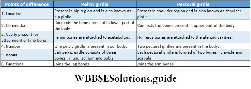
Pectoral Girdle
The part of the skeleton, which connects the arm bones to the axial skeleton, is known as the pectoral girdle.
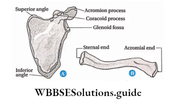
Pectoral girdle Characteristics:
- The pectoral girdle connects the upper limb (arm) bones to the axial skeleton.
- The pectoral girdle (shoulder girdle) contains four bones— two clavicles and two scapulae.
- The bones of this girdle are weakly attached and held in place by ligaments and muscles.
Pectoral Girdle Functions: It supports the arms and serves as a point of attachment for muscles, responsible for the movement of the arms.
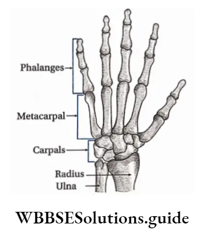
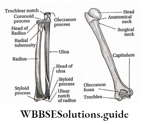
Arms
The arms are part of the skeleton, which includes the bones of the upper arm, forearm, wrist joint, palm and five fingers
Arms Characteristics: Arms are attached to the pectoral girdle. They are composed of 60 (30 in each upper limb) bones.
The characteristics of the bones of the arms are discussed in the table below.
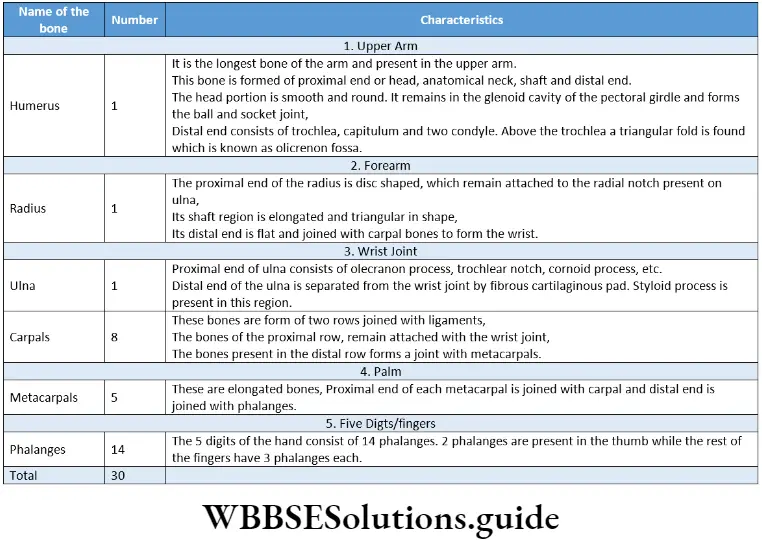
Arms Functions: Arms Help The Animal to perform different works such as grasping climbing etc.
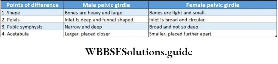
Pelvic Girdle
The part of the skeleton, connecting the bones of the legs to the axial skeleton, is known as the pelvic girdle.
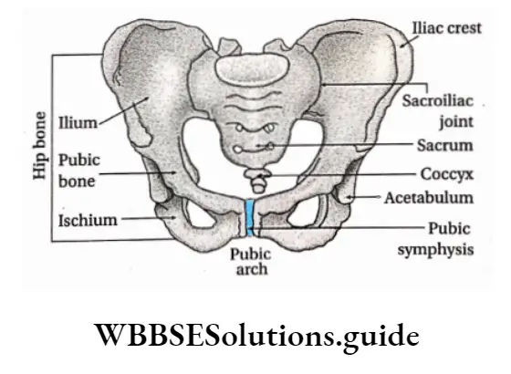
Pelvic Girdle Characteristics:
- The pelvic girdle is formed by the left and right of the coxae or hip bones.
- The pelvic girdle also contains the sacrum and coccyx, wedged in between two hip bones.
- The true pelvis is the portion of the trunk bounded by the sacrum, lower ilium, ischium, and pubic bones. It is inferior to the false pelvis.
- The upper part of the ilium is broad and flat, which is connected to the sacral segment of the vertebral column and forms the sacroiliac joint.
- Pubis and ischium remain attached at the lower part of the ilium.
- The obturator foramen (a hole) is present between the pubis and ischium.
- The cup-shaped cavity of the pelvic girdle is known as the acetabulum. The femur remains attached to the pelvic girdle by forming a ball and socket joint with this acetabulum.
- The ends of the pubis are connected with slightly movable joints and are known as symphysis pubis.
Pelvic Girdle Functions:
- The strong bones of the pelvic girdle bear the weight of the body.
- The pelvis also serves as the point of attachment for the lower limbs. It provides protection to the urinary bladder, reproductive organs, and a portion of the large intestine.
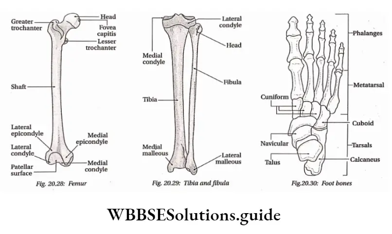
Legs
The legs are the lower parts of the skeleton which include the bones of the thigh (femur), kneecap (patella), leg (tibia and fibula) and foot (tarsals, metatarsals and phalanges)
Joints and their functions in locomotion notes
Legs Characteristics: Legs are attached to the pelvic girdle. They are composed of 60 (30 in each leg) bones. The characteristics of the bones of the legs are discussed in the table below
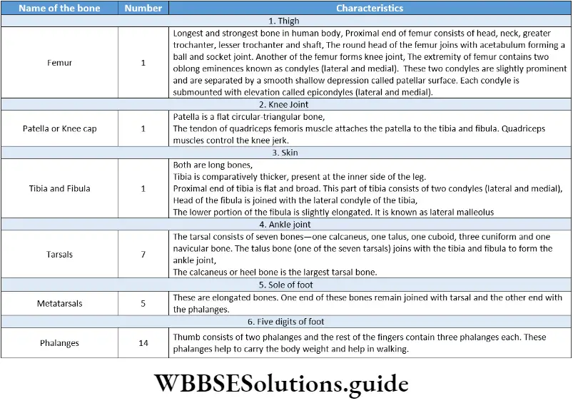
Legs Functions:
- In infants, the patella gives support to the knee during walking and crawling.
- The tibia bears almost the whole weight of the body.
- The fibula is mainly a point of muscle attachment and it maintains the body balance.
Functions Of The Human Skeletal System
The endoskeleton of a human being is composed of bones, cartilage, ligaments, etc. It plays various roles in our body.
Human Skeletal System Structure formation: It forms a rigid structure of the body and provides mechanical strength to it.
The endoskeleton is covered with muscles, skin, etc., which provide a definite shape to the body.
Human Skeletal System Role in increasing length: During adolescence, the cartilage present at the end of the long bones divides frequently causing the length of the bones to increase.
As a result, height increases during adolescence. Protects various internal organs: It protects various important parts of our body, such as the brain, neural canal, lungs, etc., from external damage.
Human Skeletal System Helps in movement and locomotion: Skeletal muscles move due to the activity of voluntary muscles. This causes movement of different parts of the body and bipedal locomotion in human beings.
Human Skeletal System Acts as storage: Stores minerals, such as calcium and phosphate. It also maintains the balance of minerals in the blood.
Human Skeletal System Production of blood cells:
- During birth red bone marrow is present in all the bones. These are the main sites for RBC formation.
- Besides RBCs, some WBCs, such as neutrophils, basophils, eosinophils, etc., are also formed from red bone marrow.
- In human beings, platelets are formed from human beings, platelets are formed from megakaryocyte cells.
Human Skeletal System Role in hearing: Helps in hearing by transmitting sound waves to the inner ear through the middle ear.
The auditory ossicles act as an amplifying device for sound waves and also protect the inner ear from loud sound.
Human Skeletal System Role in breathing: The sternum and ribs of the thoracic cage help in breathing. They enable the expansion and relaxation of the lungs.
Bone Joints Or Articulations
Bone Joints Or Articulations Definition: A joint or articulation is a point of contact between bones, between a bone and a cartilage, or between a bone and a tooth.
The study of joints is termed as arthrology.
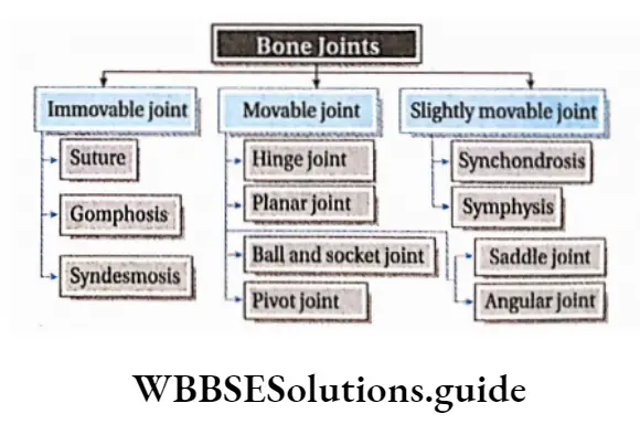
Bone Joints Or Articulations Types: Depending on structure and function, there are various types of joints in our body.
Immovable Bone Joints Or Synarthroses
Immovable Bone Joints Or Synarthroses Definition: The synarthroses (singular: synarthrosis) or immovable bone joints are the type of joints, where two or more bones are joined together by fibrous tissue and are unable to move.
These are also known as fibrous joints. Immovable joints are mainly of three types—
- Suture, bones with irregular surfaces, are tightly joined with each other by fibrous connective tissues. These types of fibrous joints are known as sutures. Example joints present between bones of the cranium.
- Gomphosis, which are immovable fibrous joints, where a pointed or peg-like portion of a bone is inserted into the socket or cavity of another bone, are known as gomphosis (singular: gomphosis). Example joints that bind the teeth in their sockets in the maxillary bone and mandible.
- Syndesmosis, are immovable joints, where the bones are connected by connective tissue or collagen fibres, are known as immovable fibrous joints or syndesmoses (singular: syndesmosis). For example the joint between the tibia and the fibula.
Slightly Movable Bone Joints Or Amphiarthroses
Slightly Movable Bone Joints Or Amphiarthroses Definition: The slightly movable bone joints or amphiarthroses (singular: amphiarthrosis) joints are the type of joints, where two or more bones are joined together by collagen fibres or cartilage and are able to move slightly.
These are also known as cartilaginous joints. Slightly movable joints are mainly of two types—
- Synchondrosis, these are slightly movable joints, where the bones are joined with each other by cartilage, known as synchondroses (singular: synchondrosis). For example the first pair of ribs are joined with the sternum by synchondrosis.
- Symphysis, these are slightly movable joints, where the bones are joined with one another by fibrous cartilage, are known as slightly movable cartilaginous joints or symphysis (singular: symphysis).
- For example joints between two adjacent vertebrae and pubic symphysis between two hip bones.
Freely Movable Bone Joints Bone Or Synovial Bone Joints Or Diarthroses
Diarthroses Definition: The freely movable joints or synovial bone joints or diarthroses (singular: diarthrosis) are the type of joints, where the bones are connected to each other through a joint cavity.
On the basis of structure and function, synovial joints are of six types.
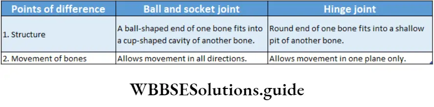
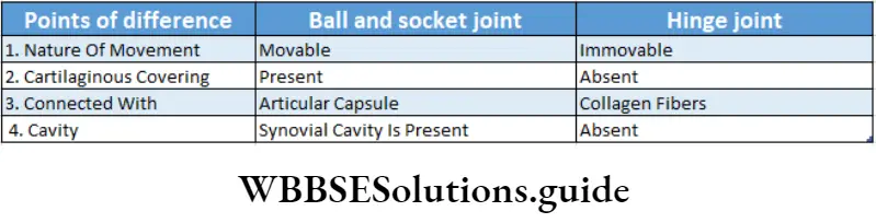
Ball and socket joint: In a ball-and-socket joint, a rounded, ball-like end of one bone fits into a cup-like socket of another bone. This organisation allows movements of the bones in all directions. Example the shoulder and hip joints.
Hinge joint: In the hinge joint, the slightly rounded end of one bone fits into the slightly hollow end of the other bone.
These bones are attached in such a manner so that movement will be possible only in one plane. The movement of these joints is similar to the hinge of a door. Example the elbow and knee joints.
Plane or planar joint or gliding joint: In the plane or planar joints, bodies with flat or slightly curved articulating surfaces are packed closely together or held in place by ligaments.
These joints allow gliding movements and hence, these joints are also referred to as gliding joints. Example the articulation between carpal bones in the hand and the tarsal bones of the foot.
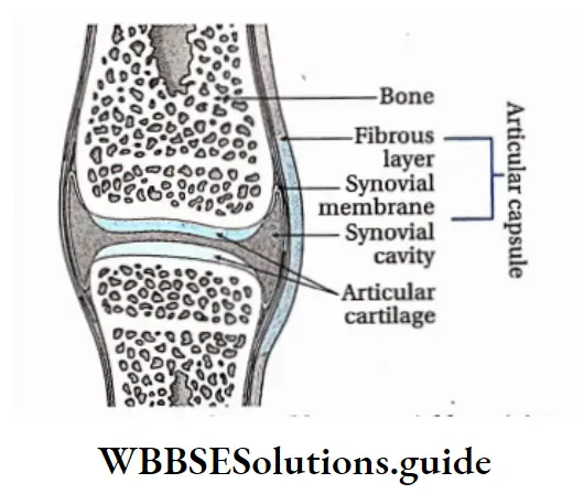
Structure Of Synovial Joint
Articular cartilage: The cartilage, that lines the epiphyses of the bones, present at the synovial joint, is known as articular cartilage.
The cartilaginous covering makes the end of the bones smoother reducing friction during movement of the joint. It contains a layer of connective tissue known as perichondrium.
Capsular ligament: These ligaments form capsules and are present at the outer surface of the synovial joints. These ligaments are known as extra-capsular ligaments.
Sometimes inner parts of the bones present at these joints also remain connected with some ligaments. These are known as intra-capsular ligaments. The capsular ligaments keep the bones in their proper position.
Synovial membrane and synovial fluid: A soft layer of glandular epithelial tissue is found between the articular capsule and the synovial cavity.
The space covered with the synovial membrane between the bones of the synovial joint is known as a synovial cavity.
The glands of the synovial membrane secrete a fluid known as synovial fluid. The synovial cavity is filled with this synovial fluid. Synovial fluid provides nutrients to the surrounding cells and reduces friction during movement.
NEET locomotion and movement revision notes with MCQs
Pivot joint or trochoid joint: In the pivot joint, the rounded end of one bone fits into a shallow pit formed by the other bone.
This structure allows rotational movement, as the rounded bone moves around its own axis. For, the joint of the first and second vertebrae of the neck that allows the head to move back and forth is of this type.
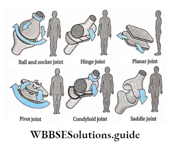
Angular or ellipsoid or condyloid joint: In a condyloid joint, an oval-shaped end of one bone fits into a similar oval-shaped hollow of another bone.
This is also sometimes called an ellipsoidal joint or angular joint. This type of joint allows movement in two directions, side to side and back to forth. For example, the joints of the wrist and fingers can move both sideways. and also up and down.
Saddle joint: In a saddle joint, concave and convex portions of the two bones fit together, just like a saddle and a rider.
Saddle joints allow movements similar to condyloid joints. For example, the thumb joint, can move back and forth and up and down. It can move more freely than the wrist or fingers.
Disorders Of Muscular And Skeletal System
Improper functioning of the muscular and skeletal system can cause several disorders. Some of them are discussed below.
Myasthenia Gravis
Myasthenia Gravis Definition: Myasthenia gravis is an autoimmune disorder that affects the neuromuscular junction at the post-synaptic level.
Myasthenia Gravis Symptoms:
- It is characterised by weakness, fatigue and paralysis of voluntary muscles.
- In some patients, myasthenia gravis may affect only the muscles of the eye, while others experience multi-system involvement.
- Muscle weakness in the eye may lead to blurred or double vision (diplopia). Difficulty in chewing, speaking, or swallowing may also be the initial symptoms.
Myasthenia Gravis Cause: It is an autoimmune disease. It is caused by the production of antibodies produced by one’s own body against nicotinic acetylcholine receptors (AchR) of the muscles.
In the case of myasthenia gravis, antibodies attack and destroy the acetylcholine receptors needed for muscle contraction. As a result, the muscles become weak.
Myasthenia Gravis Prevention: A high amount of cortisol hormones are used to treat this disease
Tetany
Tetany Definition: Tetany is a disorder marked by rapid muscular spasms, caused by malfunction of the parathyroid gland and consequent hypocalcemia.
Tetany Symptoms:
- It is neuromuscular activity and associated sensory disturbance.
- Tetany is characterised by muscle cramps, spasms or tremors. These occur within the muscles when muscle contracts uncontrollably.
- Tetany may occur in any muscle of the body, such as in the face, fingers or calves. The muscle cramping associated with tetany can be long-lasting and painful.
Tetany Cause: A common cause of tetany is very low levels of calcium in the body. Other common causes of tetany include—alcohol abuse, alkalosis (elevated pH of the blood), hypoparathyroidism, malnutrition, pancreatitis, vitamin D deficiency, etc.
Tetany Prevention: It can be treated by introducing a proper dose of calcium.
Human skeletal system and its role in locomotion notes
Muscular Dystrophy
Muscular Dystrophy Definition: Muscular dystrophy is a rare hereditary muscle disease, characterised by progressive damage of skeletal muscle.
Muscular Dystrophy Symptoms:
- Weakness and wasting (atrophy) of various voluntary muscles of the body are clinical features of this disease.
- Depending on the muscular dystrophy subtype, the disorders affect different muscles and have different effects with respect to age, severity and pattern of inheritance.
Muscular Dystrophy Cause: It is a genetic disorder. The absence or defective structure of dystrophin, a muscle cell structural protein, is the basic cause of this disorder.
Muscular Dystrophy Prevention: Presently, no cure for muscular dystrophy is available. Physiotherapy and physical exercise may be helpful to patients suffering from this disorder.
Arthritis
Arthritis Definition: Arthritis is a disease caused by inflammation of one or more joints, leading to pain and difficulty in movement.
Arthritis may be of different types.
Osteoarthritis form of arthritis or osteoarthrosis many bone joints of the body may undergo degenerative changes. This disease is very common in elderly persons.
Arthritis Symptoms:
Painful and swollen joints are the main symptoms of this disease. The pain may reduce temporarily after resting.
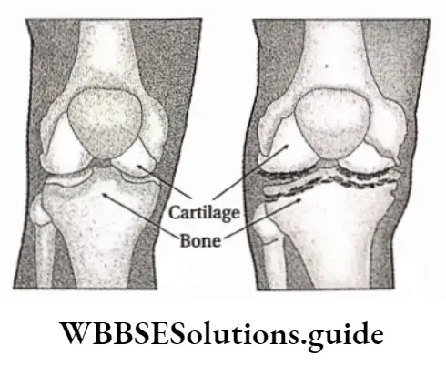
Generally, the cartilaginous covering of bones within the joint gets damaged, partially or completely.
This causes friction between the osteoarthritis of arthritis or osteoarthrosis many bone joints of the body may undergo degenerative changes. This disease is very common in elderly persons.
Arthritis Symptoms:
- Painful and swollen joints are the main symptoms of this disease. The pain may reduce temporarily after resting.
- Generally, the cartilaginous covering of bones within the joint gets damaged, partially or completely. This causes friction between the bones. This flattens the bones at the articulation.
- Due to the loss of the cartilage, the joint loses its flexibility and joint becomes stiff. Some patients experience a grating sensation when they move the joint or walk.
- The synovial membrane swells up due to continuous friction, causing excess synovial fluid to be collected within the joints. This may result in swelling of the joints.
Arthritis Cause:
- Osteoarthritis begins in the cartilage.
- In obese people of age 65 or above, the cartilage begins to get damaged slowly.
- This causes frictional erosion of two opposing bones which results in inflammation of the outer capsule of the joint.
Arthritis Prevention:
- Osteoarthritis cannot be cured.
- It can be controlled by regular exercise, reducing body weight and changing lifestyle.
- In severe cases, joint replacement through a surgical process may be done to get relief.
Rheumatoid Arthritis
It is the most common type of autoimmune disease which is triggered by faulty immune systems.
Rheumatoid Arthritis Symptoms:
- The patient often finds that the same joint on both sides of the body becomes painfully swollen and stiff.
- The smaller joints are usually noticeably affected. The joints of fingers, arms, legs and wrists are the most affected ones.
- The joint is tender to touch.
Rheumatoid Arthritis Cause:
- It is an autoimmune disease.
- It is caused when a special antibody or rheumatoid factor attacks the tissues.
- It damages the synovial membrane at the joints. It is more common in females than males.
Rheumatoid Arthritis Prevention: Presently several disease-modifying antirheumatic drugs (DMARDs) are used to stop the progression of the disease and to protect joints under the supervision of doctors
Osteoporosis
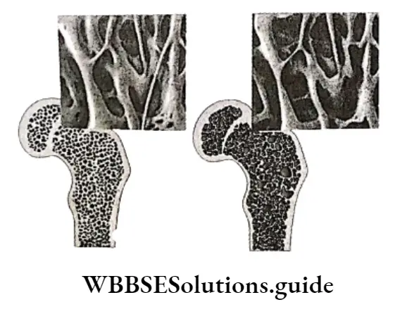
Osteoporosis Definition: A bone disorder, in which the density and strength of bones get reduced, is known as osteoporosis.
Osteoporosis, literally meaning porous bone, is a severe disorder of bones.
Osteoporosis Symptoms:
- Bones become so weak that they are no longer able to support the body weight.
- Bones can break, even under slight pressure.
- Chronic back pain is another symptom caused by osteoporosis. This pain can worsen even when a person makes very less movements, such as during regular activities, or while coughing, laughing and sneezing.
Osteoporosis Cause:
- Osteoporosis occurs when bone tissues and minerals are lost faster than they are replaced.
- In females, at the menopause stage, oestrogen secretion reduces gradually.
- This can cause osteoporosis.
- In males, with increasing age secretion of testosterone reduces gradually, which can cause osteoporosis.
Osteoporosis Prevention:
- A proper diet is essential to prevent osteoporosis.
- Vitamin D and a calcium-rich diet are essential to prevent as well as to treat this disease.
Fracture And Dislocation
Fracture: It is a medical condition where the continuity of the bone has been broken.
It can occur due to high force or stress. When a bone breaks into two pieces it is known as a simple fracture. When it breaks into more than two pieces then it is known as a compound fracture.
Dislocation: It is the separation of bones from a joint. In this condition, bones are no longer in their normal position.
Gout
Gout Definition: Gout is an ancient and common form of inflammatory arthritis caused by to accumulation of uric acid crystals within the bone joints.
Gout Symptoms:
- It is the most common inflammatory arthritis among men. Gout causes sudden painful joint inflammation, usually in one joint.
- Joint inflammation causes pain and redness of the joint.
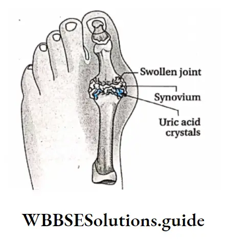
Gout Cause: Gout is a rheumatic disease, resulting from the accumulation of monosodium urate (sodium salt of uric acid) crystals at the joint. Uric acid is a byproduct formed due to the breakdown of purines.
Hyperuricemia includes heredity, obesity, certain medications such as diuretics and chronic kidney malfunction.
Gout Prevention: Chronic gout can be treated by using medications that lower the uric acid level in the body.
Locomotion And Movement Notes
- Autoimmune disorder: A disorder in which the immune system of our body recognizes our healthy cells as foreign and destroys them.
- Dystrophin: It is a rod-shaped cytoplasmic protein which connects the cytoskeleton of a muscle fibre to the surrounding extracellular matrix.
- Holotrichous cilia: Possesing cilia all over the cell surface.
- Hypocalcemia: Low calcium in blood.
- Hypoparathyroidism: A condition when the parathyroid gland is not able to produce enough parathyroid hormone.
- Hyperuricemia: Excess uric acid level in blood.
- Immune response: It is the reaction of the cells and the fluids due to the presence of unknown, foreign substances in the body.
- Iris: The thin circular structure of the eye which is responsible for controlling the diameter of the pupil and thus the amount of light reaching the eye.
- Pancreatitis: It is the inflammation of the pancreas.
- Sella turcica: It is the small cavity present in the sphenoid bone of the cranium. The pituitary gland lies in this cavity.
Points To Remember
- The study of locomotion and movement is known as kinesiology.
- The study of bones is known as osteology.
- The study of muscles is known as mycology.
- The minimum stimulus required for the contraction of a muscle is known as the threshold stimulus.
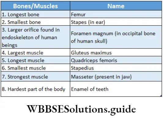
- Stapes are the smallest bone present in the human body.
- About 260 bones are present in newborns. Some of those fuse with age and the number of bones become 206 in adults.
- There are 8 cranial bones—1 frontal, 2 parietal, 1 occipital, 2 temporal, 1 sphenoid and 1 ethmoid bone.
- The bone, which forms the upper jaw, is known as the maxilla and the bone which forms the lower jaw, is known as the mandible.
- The density and strength of bones decrease due to a deficiency of calcium and phosphorus in bones. This disorder is known as osteoporosis.
- Osteopetrosis is an inherited disorder of bones. In this disorder, bones become harder and denser.
- An abnormal condition, where a person can feel his or her missing body part attached in its proper position in the body, is known as phantom limb In deficiency of ATP after death the cross-bridges formed between actin and myosin fibres do not break and the dead body becomes stiff. This condition is known as rigour mortis.
- The tension that occurs during the muscle contraction is known as muscle tension.
- The minimal stimulus of infinite duration which results in muscle contraction is known as rheobase.
- The minimum time, needed for a stimulus to double the strength of the rheobase to cause the muscle contraction, is known as chronaxie.
- The muscles, which are light in colour due to lack of myoglobin, are known as white muscle. The rate of contraction is high in these muscles, hence known as fast muscles.
- The muscles, which are dark in colour due to a huge amount of myoglobin, are known as red muscles. The rate of contraction is low in these muscles, hence they are known as slow muscles.
- When a stimulus is introduced to the muscles, the muscle first contracts and then relaxes. The contraction and relaxation together are known as muscle twitch.
- If a muscle fibre is introduced to a stimulus continuously for a certain period of time, then the rate of stimulation will be so high that the muscle fibre will be unable to relax between two stimuli. In this condition, the muscle twitches are converted to smooth, sustained contractions, known as tetanus.
- During excess muscular activity or when a muscle fibre is stimulated continuously without any break, the contracting ability of the muscle gradually decreases and ultimately the muscle fails to contract for some time. This condition is known as muscle fatigue.
- The cyclic pathway, in which the lactic acid produced by glycolysis in the muscles is transported to the liver, converted to glucose, again returned to muscles and metabolised into lactic acid, is known as the Cori cycle.
- The method of evaluating and recording the electrical activity of the skeletal muscles is known as electromyography.
- The Y-shaped bones found at the ventral side of the tail in reptiles, such as lizards, snakes, etc., are known as chevron bones. Its main function is to protect nerves and blood vessels in the tail.
- The tibia or shin bone is the strongest bone of the body.
- The two bones which help the birds to fly are pectoralis major and pectoralis minor.
- The disorder related to inflammation of joints is known as arthritis.
- In some people, an extra floating rib is found, this is known as gorilla rib.
- A short band of tough, flexible fibrous connective tissue which connects two bones together is called a ligament and a flexible but inelastic cord of strong fibrous collagen tissue attaching a muscle to a bone is called a tendon.