
Morphology Anatomy And Different Systems Of An Insect Cockroach
Cockroaches are one of those insects, which are found in almost every household. They are harmful as they can several diseases.
But cockroaches have a cause significant biological role, besides their harmful effect.
This chapter will give an insight into the structure and physiology of the cockroach. We will also discuss its biological as well as harmful role. Let’s explore!
Nomenclature: Cockroach derived its name from the word ‘Cucaracha’, which is a Spanish term for ‘cockroach’.
Common species: All over the world, there exist 3500-4500 species of cockroaches. Out of them, about 30 species live in human habitats. Among these 30 species, 4 species are well-known pests.
Read and Learn More: WBCHSE Notes for Class 11 Biology
Some common species of cockroaches are—
- German cockroach—Blattella germanica,
- American cockroach —Periplaneta americana,
- Asian cockroach —Blattella a shinai,
- Oriental cockroach—Blatta orientalis.
- we will discuss the species Periplaneta americana.
Systematic position:
- Kingdom—Animalia
- Phylum—Arthropoda
- Subphylum—Hexapoda
- Class—Insecta
- Subclass—Pterygota
- Order—Blattodea
- Family—Blattidae
- Genus—Periplaneta
- Species—Americana
Morphology and anatomy of cockroach notes
Characteristic features:
- Cockroaches are omnivorous, i.e., they feed on both animals and plants.
- They are nocturnal (remain active at night instead of day) and so they set out in search of food at night.
- They feed on dead organisms (both plants and animals), and other materials like paper, clothing, etc. Thus, they may be called ’scavengers’.
- They can run fast and are called cursorial. They have wings and hence fly as well.
- They cause damage to products used by humans and have been categorized as pests.
- They can survive without oxygen for about 45 minutes. They remain alive even after being drowned in water.
- In case of any danger, they release a foul smell from their body as a defense mechanism.
- They are unisexual and oviparous in nature.
- Young cockroaches are called nymphs which are wingless. Nymphs cannot reproduce. They transform into adults by molting or ecdysis.
| Class 11 Biology | Class 11 Chemistry |
| Class 11 Chemistry | Class 11 Physics |
| Class 11 Biology MCQs | Class 11 Physics MCQs |
| Class 11 Biology | Class 11 Physics Notes |
Habitat: Cockroaches generally live in warm and humid places all over the world. They are quite common in kitchens, bathrooms, gutters, godowns, storerooms, hospitals sewage channels, and all damp places throughout the world.
Origin: Cockroaches appeared on this planet more than 300 million years ago. They probably originated in Southern Asia or Africa but currently, they are found all over the world.
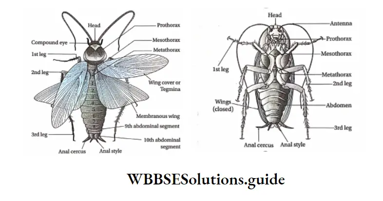
External Morphology
The external morphology of a cockroach (Periplaneta americana) is described below.
Size, shape, and color:
- The body of the cockroach is dorsoventrally flattened, longitudinal and, symmetrical.
- The adult male cockroach, Periplaneta americana is about 34-53 mm long and an adult female cockroach is about 29-37 mm long.
Exoskeleton: The entire body is covered with a hard, brownish, chitinous structure called an exoskeleton.
- The exoskeleton is made up of hard plate-like structures called sclerites. The sclerites present at the dorsal end are called tergites or terga while those at the ventral end are called sternites or sterna.
- The lateral sclerites (sclerites present on the sides) are called pleurites or pleura. The sclerites are joined to each other by a thin and flexible membrane called an articular membrane or arthrodial membrane.
- Sclerite provides rigidity to the body structure. These also serve as a surface for the attachment of body muscles. They also prevent water loss from the body.
Body segmentation: The whole body of the cockroach is segmented. However, it is divided into three distinct regions—head, thorax, and abdomen. Each of these parts is described as follows.
Head Position: The head is the triangular, most anterior part of the body. It lies at a right angle to the longitudinal body axis, bending downwards.
It is attached to the thorax by a narrow neck or cervical. The head is supported by a pair of dorsal and ventral sclerites called cervical sclerites.
External and internal anatomy of cockroach
The external structure of the head:
The head is surrounded by numerous sclerites, that form a structure known as vertex or head capsule, or epicranium.
The anterior part of the head capsule is made up of six segments. The segments have fused margins called sutures.
The dorsal part or vertex of the head is covered by a pair of epicranial plates, joined in front by an inverted Y-shaped epicranial or medial suture.
Lying between and below the arms of the epicranial suture is a broad, unpaired plate called frons.
Below the front, forming the lower part of the face is a broad rectangular plate called clypeus.
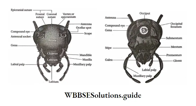
It is separated from the front by the front-clypeal suture. The two sides of the head are covered by two vertical plates called the cheek or genae, lying below the eyes. On each side, the gena is separated from the front by a front-genal suture.
The upper lip or labrum is movably attached to the lower border of the clypeus by labor-clypeal suture. The head can move in all directions as the neck is flexible.
The posterior end of the head capsule bears a cavity called the occipital foramen. It is surrounded by two plates or sclerites— occipital and post-occipital.
Different parts of the head
Mouth: It is located at the anterior end of the head.
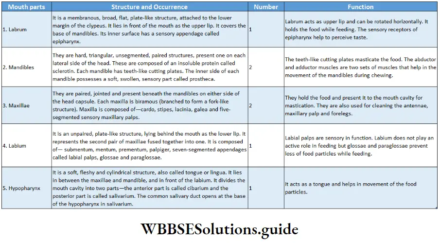
Mouth parts: The different structures associated with the mouth are known as mouthparts.
The mouth parts of cockroaches are mainly used for biting and chewing. They consist of a labrum, a pair of mandibles, a pair of maxillae, a labium, and a hypopharynx. These structures are described in the following table.
cockroach nervous system diagram
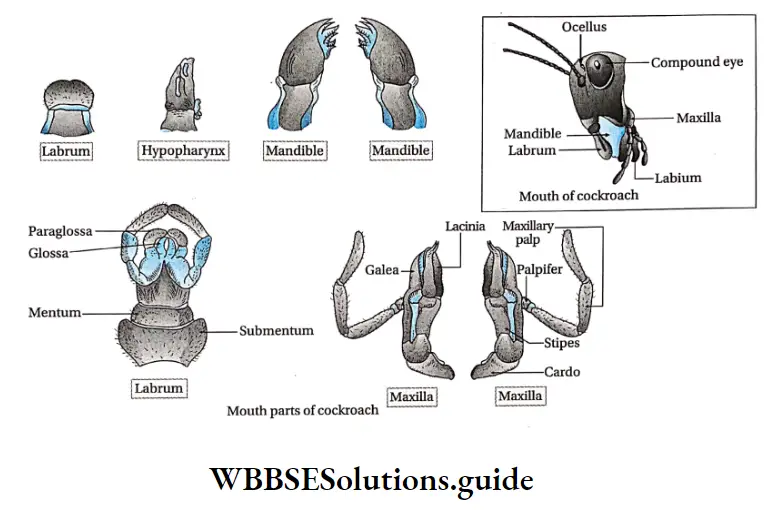
Sense organs: Different sense organs are present in the head. They are—
- Compound eyes are a pair of large, black, bean-shaped organs, situated dorsolaterally, one on each side of the head.
- Antennae are a pair of long, segmented, thread-like structures that remain articulated in membranous depressions called antennal sockets, in front of each eye.
- They can be moved freely in all directions. Each antenna has three parts—a large basal scape followed by a short pedicel and a long, numerously jointed flagellum. They possess small sensory bristles that can sense touch and smell.
- Hence, the antennae act as receptor organs of touch and smell
- Fenestra or Ocellus is a pair of small, circular, whitish parts, present on the inner side of the antennal sockets. They represent the simple eye. They are sensitive to the intensity of light.
cockroach characteristics
Thorax
- Thorax Position: Thorax lies between the head and abdomen of a cockroach.
- Thorax Structure: This part is made up of thoracic segments, jointed legs and wings
Thoracic segment: It consists of three parts—
- Prothorax,
- Mesothorax, and
- Metathorax.
Each part of the thorax is surrounded by four sclerites.
- There are altogether twelve sclerites, four of which surround each part of the thorax.
- A set of four sclerites includes a dorsal tergum, a ventral sternum, and two lateral pleura.
- The tergum of prothorax, known as pronotum, is the largest. It is a shield-shaped part that covers the neck and a part of the head.
- The tergum of meso- and metathorax are mesonotum and metanotum, respectively.
Parts of tergum, sternum, and pleura Tergum that form the dorsal meso-and metanotum are divisible into—
- Prescutum,
- Scutum,
- Scutellum.
The sternum constitutes the ventral side and is divisible into—
- Presternum,
- Basisternum,
- Sternellum.
Pleura on each lateral side is divisible into—
- Episternum and
- Epimeron
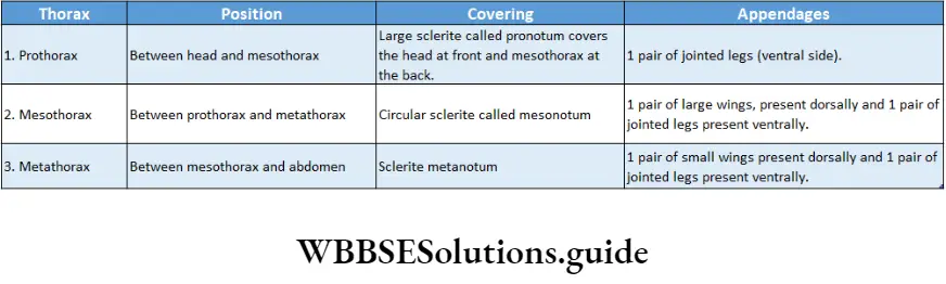
Jointed legs: A pair of jointed legs arise from the ventral side of each segment.
Hence, there are three pairs of jointed legs—prothoracic, mesothoracic, and metathoracic legs.
Detailed morphology of cockroach for biology students
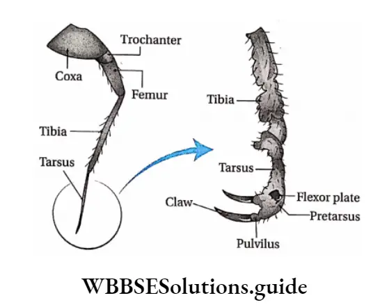
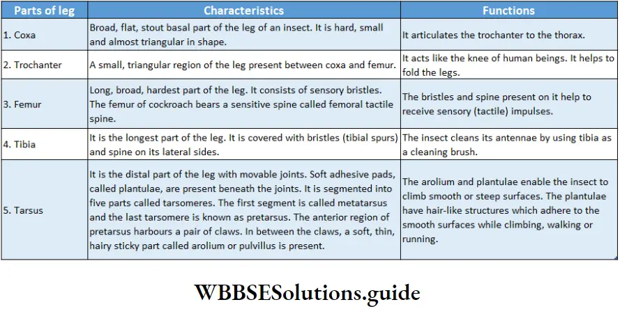
They are also known as prolegs mesolegs and metalegs respectively. Each jointed leg is made up of five segments called podomeres.
The first segment is the coxa, the second is the trochanter, the third is the femur, the fourth is the tibia and the fifth segment is the tarsus. These jointed legs are meant for walking, running, and climbing. Bristles on the legs are sensory in function.
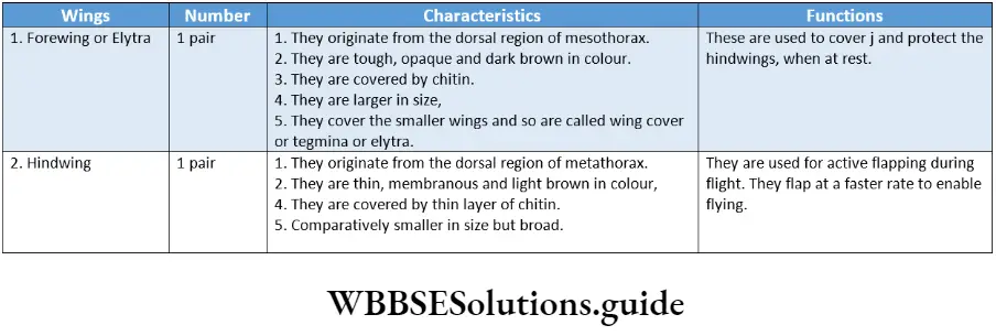
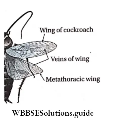
Wings: The cuticle (outermost, thick, hard, and non-living layer of the body) extends to form wings.
The wings are of two types—forewings or mesothoracic wings and hindwings or metathoracic wings.
Wings extend beyond the tip of the abdomen in males. In females, wings are shorter and extend only up to the abdomen.
Abdomen
Abdomen Position: The abdomen is the posterior part of the cockroach, placed next to the metathorax.
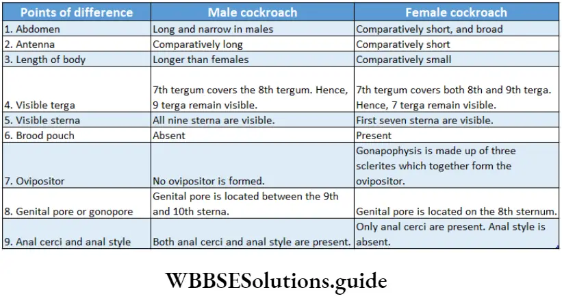
Abdomen Structure: The dorsoventrally flattened abdomen is long and narrow in males but short and broad in females. The abdomen consists of ten segments in adults. In the case of the embryo, there are eleven segments.
Each segment of the abdomen is covered by sclerites. The terminal segments carry appendages, apertures, and stink glands.
Abdomen Sclerites: The thick, hard, jointed, dark brown, chitinous plate-like structures that form the exoskeleton of cockroaches are known as sclerites. The different parts of the sclerites are as follows—
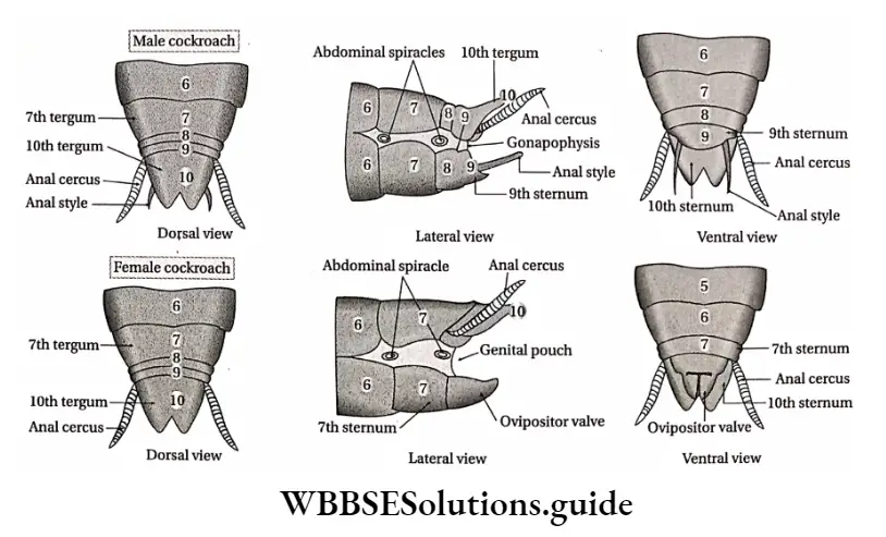
Abdomen Terga: The dorsal portion of each sclerite is known as tergum (plural: terga). There are ten tergas in the abdomen of a cockroach. Sometimes, a tergum is divided into smaller units known as tergites.
In males, the 8th tergum and in females, both 8th and 9th terga are not visible as they remain overlapped by the 7th tergum.
Sterna: The ventral portion of each sclerite is known as the sternum (plural: sterna). There are nine sterna in the abdomen of a cockroach.
In adult males, all nine sterna are visible but in adult females, only the first seven sterna are visible. In females, the 7th sternum is boat-shaped. This sternum is known as hypogenium. The 8th and 9th sterna remain fused to form a chamber called gynatrium.
Together, the three (7th, 8th, and 9th) sterna form a pouch-like structure called brood or genital pouch.
The anterior part of the genital pouch contains female gonophores, spermathecal pores, and collateral glands.
[These structures have been discussed in detail under the topic ‘Female Reproductive System’.]
Abdomen Pleura: The lateral, soft, membranous sclerites are called pleurites. They help in the articulation of the terga and sterna.
Abdominal appendages
Anal cerci: The 10th segment bears a pair of jointed filamentous structures with sensory bristles. These structures are called anal cerci (Sing, anal cercus) and are found in both sexes.
Each anal circus bears fifteen segments. The sensory bristles present are sensitive to touch and auditory impulses.
Anal style: The thin, needle-like appendages associated with the 9th sternum of male cockroaches are known as anal style. They help in copulation. These are unsegmented in nature.
Gonapophysis: Small, irregular, hard, chitinous structures are present surrounding the genital aperture in cockroaches. These structures are called gonapophyses or external genitalia.
In male cockroaches, the gonapophyses are formed of three appendages called phallomeres.
They arise from the 9th segment and act as the copulatory organ. In female cockroaches, gonapophysis is formed of three pairs of appendages.
They originate from the 8th and 9th segments to form a structure called an ovipositor. The ovipositor helps in transferring the fertilized egg towards another chamber called the oothecal chamber.
Aperture: The abdomen possesses three apertures— anus, genital aperture or gonopore, and abdominal spiracles.
Anus: Anus is a slit-like opening supported by two triangular podical plates (also known as parrots). It is located between the 10th tergum and the 9th sternum. It helps to release the undigested food materials from the body.
Genital aperture: The genital aperture, genital pore, or gonopore of a female cockroach is located on the 8th sternum. It leads to the brood pouch. In male cockroaches, the genital aperture is located between the 9th and 10th sternum.
Abdominal spiracles: The lateral surface of the body bears ten paired openings, called stigmata or spiracles, to facilitate respiration. Among them, two pairs are located in the thorax. The remaining eight pairs are located on each side of the eight segments of the abdomen. These eight pairs are called abdominal spiracles.
Stink gland: This gland is present in the arthrodial membrane, between the 5th and 6th abdominal terga. This gland is responsible for the stinky smell of cockroaches.
Internal Morphology
The internal morphology of cockroaches is described below in detail.
Integument
The outermost layer of the body of a cockroach is called integument. It is chitinous in nature.
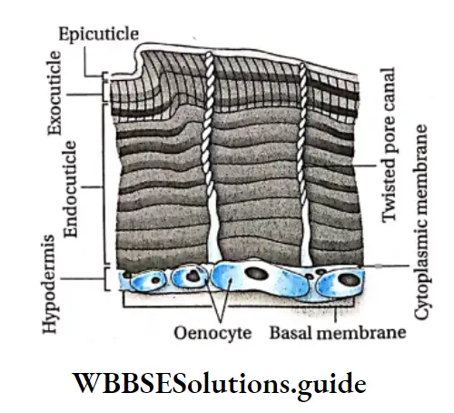
Parts of an integument: The structure of the integument of a cockroach is made up of three layers— the outermost cuticle, followed by the cellular epidermis or hypodermis, and the inner basement membrane.
Cuticle: The outermost layer of the body is called the cuticle. It forms the exoskeleton.
It is hard, thick, brownish, and resistant to water. It consists of dead cells, containing chitin. Chitin is chemically an acetate of glucosamine (acetylglucosamine).
Position: It is present in the outermost layer of the body, anterior and posterior parts of the esophagus, outer wall of the trachea, and ovarian duct.
Structure: The cuticle again has three layers, namely, epicuticle, exocuticle, and endocuticle. The exo- and endocuticle are together known as procuticle.
- The epicuticle is the outermost layer of the cuticle. It is further made up of a lipoprotein cement layer on the surface, a wax layer in the middle, and a layer of polyphenol on the inner side,
- The exocuticle is the middle layer of the cuticle. lt is chitinous in nature. It contains melanin pigment,
- The endocuticle is the innermost layer of the cuticle. it is made up of chitin and is the thickest layer.
Functions:
- Epicuticle protects the underneath layers of soft tissue from dehydration,
- The exocuticle gives rigidity and flexibility to the structure of the cuticle,
- The endocuticle is lysed just before each molting or ecdysis so that the new procuticle can be formed.
Hypodermis or epidermis: The layer made up of columnar epithelial cells, that lie below the cuticle is called hypodermis or epidermis.
Hypodermis or epidermis Position: Epidermis lies beneath the cuticle.
Hypodermis or epidermis Structure: It is made up of a single layer of columnar epithelial cells. Some of the epithelial cells are modified into other cell types like trichogen cells, tormogen cells, dermal gland cells, and oenocytes.
Hypodermis or epidermis Function: The epithelial cells of the epidermis secrete the cuticle.
The main functions of the modified cells are—
- Trichogen cells give rise to movable bristles under the body,
- Tormogen cells form sockets in the integument layer for the attachment of bristles,
- Dermal gland cells secrete a waxy substance over the cuticle,
- Oenocytes help in wax secretion and molting.
Basement membrane: The thin, innermost layer of the body wall, lying below the hypodermis is called the basement membrane. It is composed of flattened cells. It is composed of mucopolysaccharides.
Epidermal gland: The integument of cockroaches contains two types of glands. These are—the cervical and abdominal glands.
Cervical gland: It secretes a protein called peripatetic which induces molting
Abdominal gland: This gland secretes a substance with a pungent smell that helps to keep away predators.
Epidermal coloration: Every time after molting, the body of the cockroach becomes soft and pale.
The exocuticle has melanin pigment. Under the effect of the hormone bursicon, cuticle hardens and dark brown pigmentation takes place.
Functions Of Integument
Protection:
- The integument is a protective covering for the delicate internal organs.
- The cuticle is a waterproof layer that protects the body from harsh environments.
- A stinky, pungent substance is secreted from the abdominal gland which keeps away the predators and helps in protection.
Sense organ: Bristles on the body wall are formed due to the modification of the hypodermis. They are sensory in function.
Secretion: The hypodermis secretes the cuticle which hardens to form the exoskeleton. In some cases, hypodermis secretes pheromone.
Coelom
- The cavity enclosed by the peritoneum is called the coelom. Typical coelom of cockroaches can be seen at the embryonic stage.
- The coelom is greatly reduced in adults and is called hemocoel. It is filled with hemolymph.
- Coelom is divided into three cavities or sinuses by two membrane-like structures called diaphragms. These are—pericardial, perivisceral, and perineural sinus.
Muscle
- The muscles of cockroaches are of two types—skeletal and visceral muscles.
- Skeletal muscles are present in mouth parts, thoracic legs, wings, and genital appendages.
- They help to move the body parts such as the legs, wings, etc.
- Visceral muscles are present in the lining of the heart and gut. They help in the circulation of hemolymph Through the Heart And the Digestion Of Food In The gut.
Digestive system Definition: The physiological system that is associated with the intake of food, its breakdown into simpler forms, absorption and elimination of the wastes generated, is known as the digestive system.
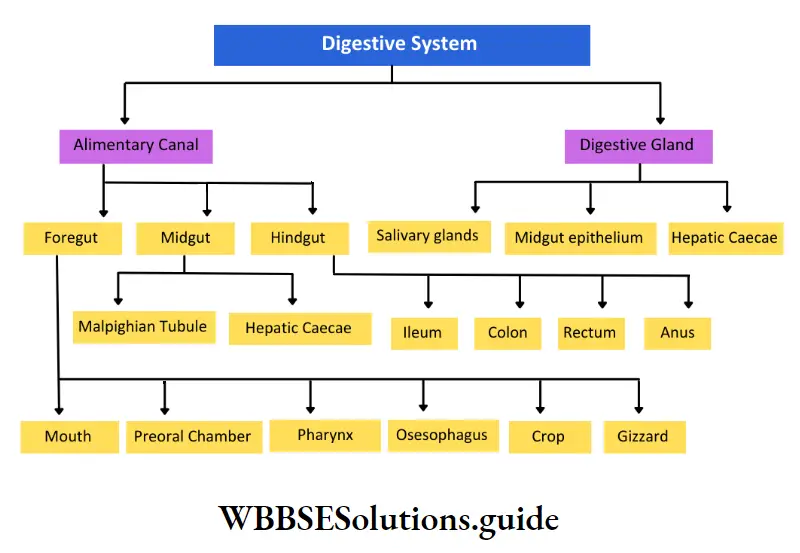
The digestive system of cockroaches comprises the alimentary canal and digestive glands.
Different parts of the digestive system re discussed below.
Alimentary Canal Definition: The canal extending from the mouth up to the anus, which is responsible for intake, digestion, absorption, assimilation of food, and elimination of the wastes generated, is known as the alimentary canal.
The alimentary canal of a cockroach is complete in nature i.e., contains openings (mouth and anus) at both ends.
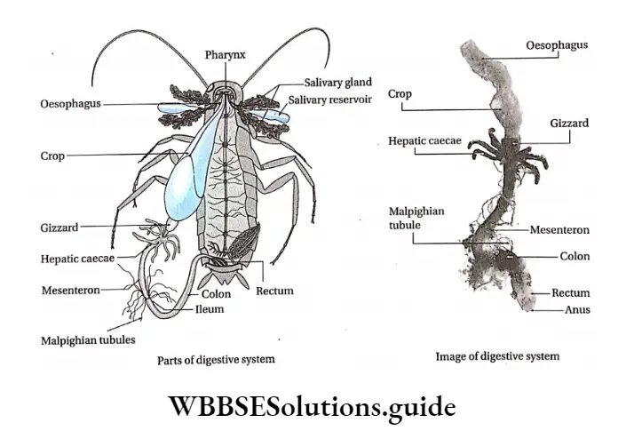
Its length is about 6.7 cm. The alimentary canal is divided into three regions—foregut, midgut, and hindgut.
Foregut: The foregut or stomodaeum is ectodermal in origin.
Different parts of the foregut are as follows—
Mouth: The mouth is the opening of the alimentary canal, present at the anterior region of the head. Its function is to receive food.
Mouth cavity: The cavity that lies next to the mouth is the mouth cavity. It is surrounded by mouthparts like the mandible and maxillae.
Cockroach uses these mouth parts, along with other such mouth parts to engulf the food and send it to mouth cavity J through the mouth.
The mouth cavity has two regions—
Anterior cibarium and posterior salivarium. From the posterior region of the mouth cavity, the salivary duct projects out.
Its function is to mix the food with saliva and pass it to the next region.
Digestive system of cockroach diagram and explanation
Pharynx: It is a small, tubular region present after the mouth cavity. Its function is to direct the food toward the esophagus.
Oesophagus: The narrow tubular passage, present after the pharynx is called the esophagus (food pipe), The opening between the pharynx and the trachea (windpipe) is guarded by sphincters. Its function is to direct the food to the crop.
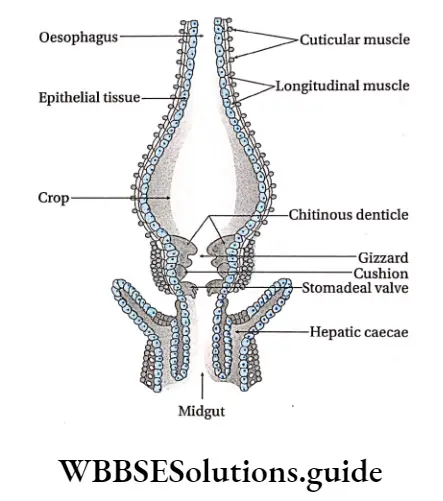
Crop: A crop is a sac-like structure present next to the esophagus. It extends up to the 3rd or 4th abdominal segments.
Its main characteristics are—
- It is pear-shaped,
- It is the largest part of the foregut.
- Its wall is thin,
- The cuticular layer is visible in the inner wall. The function of the crop is to store food.
Gizzard: The crop is followed by another structure called gizzard or proventriculus.
Its characteristics are—
- It is a thick muscular, triangular cavity,
- It has two regions— the anterior part is called armarium and the posterior part is called the stomodeal valve,
- The anterior (armarium) lining has—(a) 6 large criticized plates called teeth or denticles and
- 6 cushion-like pads covered with long bristles,
- The posterior part projects into the midgut as a funnel called the stomodeal valve.
The functions of the gizzard are—
- It acts as a grinding mill and filtering apparatus. Denticles help in grinding the food particles,
- The cushions act as filters. Also, they push the food particles to the next part of the alimentary canal, i.e., the midgut.
- The stomodeal valve prevents the food from returning to the gizzard from the midgut.
Histological structure of alimentary canal
Foregut originates from the ectoderm. It is internally lined by the cuticle. The gizzard of the foregut has an outer thick layer of circular muscles.
Midgut originates from the endoderm. It is made up of tall columnar glandular cells. The midgut lacks a cuticle. The internal lining has folds (villi) and is covered by a very thin transparent layer called peritrophic membrane.
Hindgut has originated from the ectoderm. It is internally lined by the cuticle. The inner lining of the colon is wrinkled and that of the rectum has six thick longitudinal folds, the rectal papillae.
Midgut: Next to the foregut, lies the midgut. It acts like the stomach.
This region is made up of the following parts—
Hepatic caecae: A ring of 6-8 hollow tubules called hepatic or gastric caecae is present at the junction of the foregut and midgut. One end of each of these tubules is closed and the other end is open.
Its function is to release the digestive juice that is secreted by the epithelium of the inner wall.
Midgut segment: It is a narrow tube of uniform diameter present next to the hepatic caeca. It secretes digestive juice and helps in digestion and absorption.
Within the midgut, a thin, semi-permeable membrane is secreted by certain cells. This membrane lines the midgut segment as well as forms a covering around the food particles.
This membrane is called the peritrophic membrane. It protects the inner lining of the gut from any injury by the food particles.
Malpighian tubule: At the junction of midgut and hindgut, there are 60-150 yellowish thread-like structures called Malpighian tubules. One end of these tubules is open and the other end remains closed.
The closed end remains suspended in the body fluid, while the open end connects with the gut. They do not have any significant role in digestion but are the primary excretory organs of the body.
Hindgut: The Midgut finally opens into the hindgut or proctodaeum. The hindgut is broader than the midgut and is differentiated into the ileum, colon, and rectum.
Ileum: The short, tubular structure next to the midgut is the ileum. There are six triangular lobes within the ileum. The function of the ileum is to digest and absorb food.
Colon: The next tubular region of the hindgut after the ileum is the colon. It is broader and more coiled than the ileum. Its function is to absorb the food.
Rectum: The cavity that lies next to the colon is the rectum. It consists of rectal papillae.
Its functions are to—
- Store faeces,
- Perform osmoregulation.
Anus: The rectum opens out into a structure called the anus. The anal aperture is located below the 10th tergum.
- There are certain muscles, called sphincter muscles, present near the anal aperture or opening.
- The contraction and relaxation of these muscles regulate the opening and closure of the anal aperture.
- The function of the anus is to eliminate the undigested substances out of the body.
Digestive Glands Definition: The exocrine glands that are associated with the digestive system and secrete enzymes that help in digestion are called digestive glands.
Digestive Glands Types: Cockroaches have three types of digestive glands—
- Salivary glands,
- Epithelium of midgut or mesenteron and
- Hepatic cancer.
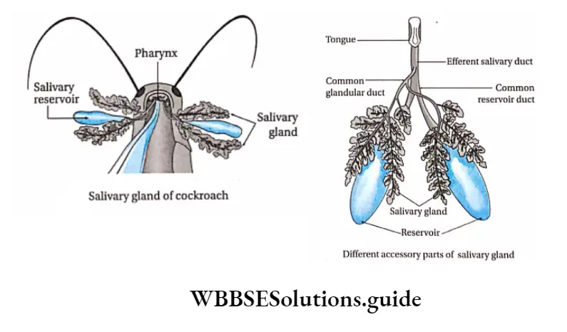
Salivary glands: The exocrine glands which secrete saliva are known as salivary glands. A pair of salivary glands is located one on each side of the crop in the thoracic cavity.
Characteristic features of salivary glands are—
They remain associated with the alimentary canal,
Each gland comprises two glandular portions and a bag-like salivary reservoir or
receptacle, which stores saliva,
The salivary glands are made up of many segments or lobules. These are made up of two types of cells—
- Secretory granular cells and
- Cells constituting intercellular ducts and numerous microvilli,
- Salivary ducts arising from the two glandular portions, unite to form a common duct. The common duct opens at the base of the hypopharynx in the preoral cavity,
- Saliva is secreted from each acinus and released into the common duct through salivary ducts.
Its functions are—
- Saliva moistens the food for easy passage through the alimentary canal.
- Salivary juice contains enzymes like amylase for the digestion of starch, cellulase for the digestion of cellulose, and chitinase for chitin digestion. The mucus of saliva makes the food slimy.
Midgut epithelium:
- It is the internal lining of the tubular midgut.
- The glandular epithelium is made up of tall columnar endodermal cells,
- Other parts of the midgut are also covered by a peritrophic membrane which disintegrates and then regenerates sometimes.
Its functions are—
- Secretion of amylolytic, proteolytic, and lipolytic enzymes for digestion of carbohydrates, proteins, and lipids respectively,
- Helping in the absorption of the digested food.
Hepatic cancer:
- It lies at the anterior end of the midgut.
- It is comprised of 8 glandular, finger-like, tubular, blind processes,
- They secrete digestive juice and help in digestion.
Process of nutrition: The nutrition of cockroaches is holozoic in nature. Nutrition occurs in five stages. These are—
- Ingestion,
- Digestion,
- Absorption,
- Assimilation, and
- Egestion.
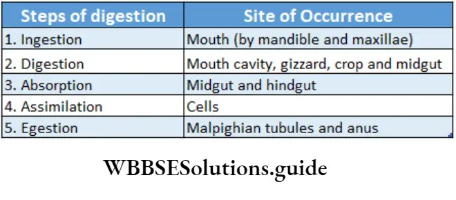
Ingestion: It is the process of intake of food and grinding it into smaller and simpler forms before digestion begins. The different mouth parts of cockroaches are modified to carry out different functions.
The food collected with the help of forelegs, labrum, and labium is passed to mandibles for biting and chewing.
The food materials are mixed with saliva during mastication. After chewing, the mouth swallows the food, with the help of the hypopharynx.
Digestion
Digestion in the mouth: Enzymes like amylase, present within the salivary juice, hydrolyze complex carbohydrates present in the food. It converts the starch into glucose that can be easily digested.
Digestion in crop: Food, that is mixed with mucus, passes through the pharynx, and esophagus and reaches the crop. This movement is carried out by peristalsis. The food is then digested in the crop by digestive juices secreted by the hepatic caecae.
Digestion in gizzard: The gizzard grinds and crushes the food particles into finer ones. Later, it mixes the food with the digestive juices regurgitated from the midgut. This, in turn, speeds up the process of digestion.
Digestion in midgut: The midgut secretes the following enzymes—
- Amylolytic enzymes like invertase, amylase convert carbohydrates into glucose,
- Proteolytic enzymes like peptidases and trypsin that digest protein into amino acids,
- Lipolytic enzymes that act upon lipids or fats and convert them into fatty acids and glycerol.
Digestion in hindgut: The cellulose of food is digested by the enzyme cellulase. It is secreted by the microorganisms present in the hindgut of cockroaches. Cellulose is converted to glucose by the enzymatic action of cellulase.
Absorption
Absorption in midgut: The cells lining the midgut and hepatic caecae absorb the end products such as glucose, amino acids, fatty acids glycerol, etc., generated during digestion.
Absorption in hindgut: Primarily water absorption takes place in the hindgut. Maximum water absorption takes place by the rectal papillae.
Assimilation: The absorbed food is carried to different cells by the hemolymph. It is transported to different parts of the body for utilization by the cells.
The absorbed food is assimilated within protoplasm so that it can be used to provide energy for metabolic purposes. The excess food material is stored in the fat body as glycogen and fat.
Egestion: The undigested residue, left after assimilation of the food, is stored in the rectum.
Nervous system of cockroach structure and function
The undigested food is almost solid due to maximum water absorption by the rectal papillae within the rectum. This is discharged out through the anus in the form of small dry pellets.
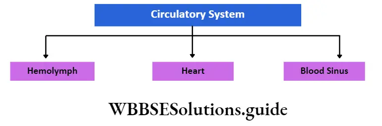
Circulatory system Definition: The organ system that is responsible for the transportation of substances throughout the body is known as the circulatory system.
A circulatory or vascular system of a cockroach is an open circulatory system. It means during circulation, blood directly flows into the body cavity instead of blood vessels.
The body cavity acts as a hemocoel and the organs and tissues are directly bathed by blood. The important parts of the circulatory system of cockroaches are— hemolymph, heart, and blood sinus.
Haemolymph: The blood of cockroaches is colorless (like the lymph) and is known as hemolymph. It transports all the necessary substances to the various organs and tissues.
Components: The hemolymph is mainly composed of colorless plasma, with certain cells known as hemocytes. The components of hemolymph are mentioned in the given chart.
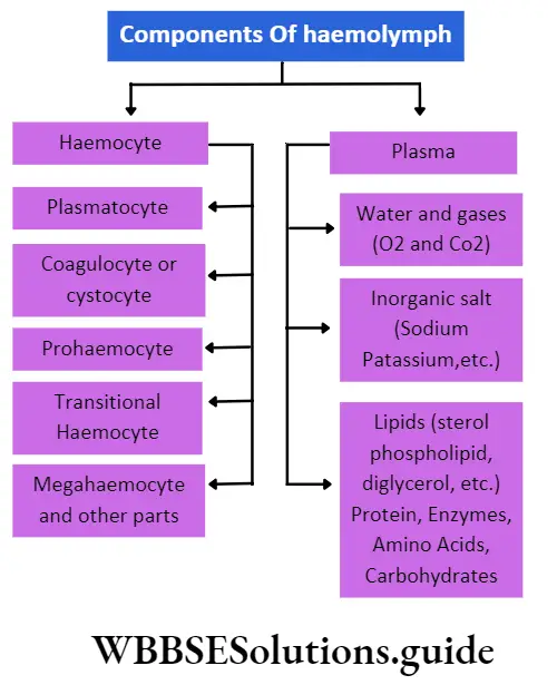
Plasma: The colorless fluid portion of hemolymph is called plasma. It contains about 70% water and a large number of organic molecules (such as carbohydrates, proteins, fats like sterol, phospholipids, triglycerides, etc.) arid various inorganic ions (salts of sodium and potassium, etc.),
Genital pouch brood pouch or gynatrium
Genital pouch brood pouch or gynatrium Definition: The genital or reproductive pouch is an accessory part in female cockroaches that comprises the female gonopore, openings of the collateral gland, spermatheca, and oothecal chamber.
Genital pouch brood pouch or gynatrium Position: This part is formed within the 7th sternum at the posterior end of the abdomen.
Genital pouch brood pouch or gynatrium Structure:
- The genital pouch is a large, boat-shaped sac-like structure,
- The sternum of the 7th abdominal segment forms the floor, while the sternum of 9th segment forms the roof. It is anteriorly surrounded by the sternum of the 8th segment,
- It is also known as the gym atrium.
- It is divided into the anterior genital chamber and a larger posterior oothecal chamber,
- The spermatheca, collateral gland, and genital pore open into a cavity called the genital atrium. Within this cavity, a fusion of sperm and ova takes place to produce a zygote,
- Within the oothecal chamber or vestibulum, the egg case or ootheca forms. The ootheca contains 16 fertilized ova.
Function: The genital pouch receives spermatophore and forms ootheca during copulation.
Gonapophyses Definition: The appendages at the terminal end of the body of a female cockroach that help in ootheca formation and copulation are known as gonapophyses or external genitalia.
Gonapophyses Position: They lie between the anal pore and the gonopore.
They remain concealed inside the genital pouch.
Gonapophyses Structure:
Gonapophyses are formed of three pairs of chitinous sclerites. Together these are called ovipositor valves,
The posterior gonapophysis is formed by the two pairs of sclerites, while the anterior gonapophysis is formed by the third pair of sclerites.
The mid-part of the 8th sternum forms a structure called sclerite spermathecal papilla that fuses at the spermathecal pore.
Gonapophyses Function: The ovipositor of gonapophyses conducts the fertilized eggs to the oothecal chamber and holds them till the ootheca is formed. It also helps in copulation.
Life cycle—copulation, fertilization, ootheca formation, and development
The copulation and fertilization process of cockroaches is described in this section.
Gonapophyses Copulation: During copulation, the following changes can be observed within cockroaches—
A female cockroach secretes a pheromone, that attracts the male cockroach.
The influence of pheromones on male cockroaches causes some behavioral changes, such as—
- It keeps its wings open and flaps them vigorously.
- It moves fast,
- It also moves its antenna fast,
The olfactory sensillae in the antenna of the male cockroach search for the female cockroach, till it finds one.
- Female cockroach performs copulation once in their lives but keeps giving birth to young ones for the rest of it.
- Once it finds a female cockroach, a male cockroach moves beneath the female cockroach.
- The left phallomere of the male cockroach is inserted into the genital pouch of the female allowing the pseudopenis into the female gonopore. The rest of the phallomeres hold the cockroaches in position.
- Now, the male and female cockroaches turn 180° and remain in this position for about 1 hour.
- Copulation generally occurs at night. During this time, secretion of neurohormone and pheromone increases.
- Copulation lasts for about an hour, after which the two cockroaches separate.
- During the next 20 hours, the sperm from the spermatophores pass into the spermatheca within the female cockroach.
Fertilization: Their fertilization is internal, i.e. takes, place within the body.
- During copulation, sperm from the male cockroach are transferred into the female cockroach.
- During this transfer, all the sperms coagulate into a pear-shaped capsule-like structure. This capsule or mass is formed by the secretion of the genital pouch. This is called a spermatophore. A tough covering is formed over the spermatophore.
- The spermatophore is released through the ejaculatory duct and deposited on the spermathecal papillae.
- The conglobate gland now releases its secretion on the spermatophore to form its outermost covering which takes about 2 hours to harden.
- The sperms remain alive for about 20 hours within the spermatheca of the female cockroach. After the entry of sperms in the spermatheca, the covering of the spermatophore is broken and the sperms are released.
- During this time, one ova from each ovariole i.e., a total of 16 ova, is released into the vagina.
- The eggs pass through the female gonopore into the genital chamber.
- The eggs are fertilized in the genital chamber by the sperm released from the spermatheca.
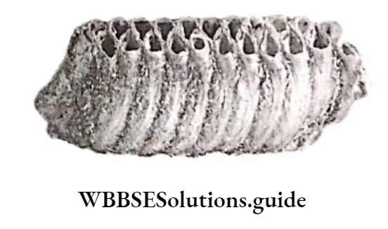
Ootheca formation: The special structure that contains several fertilized ova within it is called ootheca.
The features of ootheca are—
- Each ootheca contains 16 fertilized eggs (ova) arranged in two rows, standing vertically.
- Ootheca are dark brown in color and 8 mm in length.
- The covering of the ootheca contains scleroprotein and water. It takes 20 minutes to form an oothe1ca.
- The ovipositor valve helps in ootheca formation.
- Each ootheca is oblong in shape and its lateral surface shows black grooves.
- After formation, it protrudes from the oothecal chamber and is finally released.
- Female cockroach releases ootheca in a dark, swampy, hot environment.
Development: The stages of growth and Differentiation i.e., development Of An ootheca to an adult as follows—
- The embryonic development takes place in the ootheca and requires 5-13 weeks. This process is temperature-dependent.
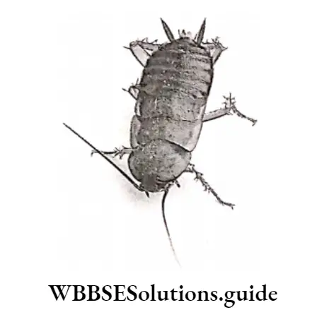
- The young ones that come out of the ootheca resemble the adult in structure and feeding habits but are soft, smaller in size, white, or pale in color. They are also devoid of wings; and have under-developed gonads. These young ones are called nymphs.
- During the nymphal period, the cockroach feeds and grows. It sheds its old exoskeleton and develops a new cuticle by the process of molting, triggered by molting hormone or ecdysone.
- Nymphs undergo several rounds of molting and metamorphose into adults within 6-8 months. (11 molts are required in males while 12 molts are in females)
Metamorphosis
The process by which changes occur within the body as an organism develops from a young stage into an adult stage is called metamorphosis. Types of Metamorphosis in cockroach—
Hemimetabolic or incomplete metamorphosis:
It is the mode of development that includes three distinct stages in the life cycle—egg, nymph and adult stages. The nymph resembles the adult but lacks wings and functional reproductive organs.
Paurometabolic metamorphosis: It is the gradual transformation of nymph to adult with hardly any significant change in the characteristics.
Interaction With Human
Cockroaches influence the daily lives of human beings in many ways. Most of them are harmful but some are useful. These effects are discussed as follows.
Harmful Effects
A pest: Cockroaches are noted as dangerous household pests. They feed on a variety of products, like meat, grease, starch-containing food, sweets, and other uncovered materials.
When cockroaches run over food, they leave filth and dirt over it. They secrete an oily liquid that has an offensive and sickening odour. This makes the food unhealthy.
Spreads diseases: Cockroaches are important potential vectors of several diseases.
They thrive in dirty places like drains, gutters, etc. So, they can easily transmit viruses, bacteria, fungi, and helminths.
They are responsible for diseases in humans like food poisoning, gastroenteritis, diarrhea, nausea, vomiting, etc.
Causes allergy: They also induce some allergic reactions in humans.
Useful effects
As medicine: The powdered cockroach brain was used to fight disorders like vomiting, diarrhea, respiratory problems, pneumonia, and skin infections.
In the laboratory: Cockroach serves as a good laboratory animal for studying insects.
As food: Cockroaches are consumed as food by the people of Myanmar and South American countries; as a delicacy.
Control of cockroach
Cockroaches can be controlled by mechanical, chemical, and biological control methods. These methods are discussed under separate heads.
Mechanical control: Different mechanical control methods by which cockroaches can be controlled are as follows—
Prevention of entry: Sealing entry points like gaps under doors or around pipes where they hide can help prevent cockroaches from entering the home.
Protection of foodstuffs: Food should be stored in containers or in sealed plastic bags.
Proper sanitation: All waste food and spillages should be cleaned immediately. Food debris can attract several insects, including cockroaches, which may be harmful. Organic wastes should be disposed of in sealed bins.
Use of baits (Vegas roach trap): Traps are used to catch cockroaches. Here, a deep, smooth-surfaced jar with food inside is placed, against a wall. The jar should be placed such that its open mouth remains touching the wall. Cockroaches can crawl up the wall.
If any of them slip and fall, they may fall into the jar, placed below. This way cockroaches can be trapped. Vaseline is smeared on the inner surface of the jar to make it slippery so that the trapped cockroach cannot climb back out.
This inexpensive method is effective with the American cockroach. It is called the “Vegas roach trap” as it was popularised by a TV channel in Las Vegas.
Chemical control: Different chemical control methods by which cockroaches can be controlled are as follows—
Diatomaceous powder: Diatomaceous earth (DE) is obtained from deposits of fossilized sedimentary layers of tiny phytoplanktons called diatoms.
The diatomaceous powder is a non-toxic insecticide, harmless to humans but can eliminate cockroaches.
Hydramethylnon: Hydramethylnon is an organic compound that acts as a metabolic inhibitor. It is used primarily as bait for cockroaches. It is used in the form of gel.
Deltamethrin and pyrethrin: They are synthetic pyrethroids, neuro-toxic in nature. Deltamethrin can kill insects by direct contact. It can even kill the insects if they eat it.
Pyrethrin, in greater concentration, induces cockroaches to abandon their hiding places. This, in turn, kills the cockroaches.
Pandan: Pandan leaves contain a number of essential oils and chemicals like 2-acetyl-l-pyrroline that cockroaches find unpleasant. Hence, these leaves can be used to drive out cockroaches.
Circulatory system of cockroach with labeled diagram
Biological control: Different biological control methods by which cockroaches can be controlled are as follows—
By wasps and bees: Wasps and bees are the predators of adult and nymphal cockroaches. The wasps paralyze the cockroach with their stings and drag it into the burrow.
The wasps now lay their eggs within the ootheca of the paralyzed cockroach.
The wasp larvae feed on the cockroach embryo within the ootheca. Ultimately, the cockroach dies.
By centipedes: The centipedes found in houses prey upon cockroaches. They can be used as the most effective control agents of cockroaches.