
Human Reproduction Notes
Reproduction is the production of young ones by an organism. Humans are sexually reproducing and viviparous.
1. Male Reproductive System: It consists of paired testes, Accessory ducts, Accessory glands, and external genitalia (penis).
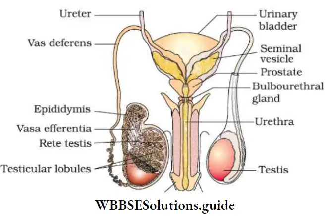
Male Reproductive System Paired Testes::Primary sex organs that produce sperm and testosterone.
“human reproduction class 12 “
Read And Learn More: NEET Biology Class 12 Notes
- Testes are formed within the abdomen. Soon after the birth or at the 8th month of pregnancy they descend into the scrotal sac (scrotum) through the inguinal canal.
- The low temperature (2-2.50 C less than the body temperature) of the scrotum helps for proper functioning of the testes and for spermatogenesis.
- Each testis is oval-shaped. Length 4-5 cm, width: 2-3 cm.
- Each testis has about 250 testicular lobules.
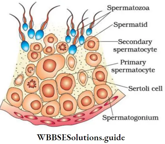
- Each lobule contains 1-3 coiled seminiferous tubules.
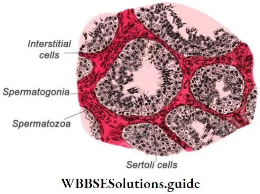
- The seminiferous tubule is lined internally with spermatogonia (male germ cells) and Sertoli cells (supporting cells). Sertoli cells give shape and nourishment to developing spermatogonia.
- The regions outside the seminiferous tubules (interstitial spaces) contain small blood vessels, interstitial cells (Leydig cells), and immunologically competent cells.
- Leydig cells secrete testicular hormones (androgens).
Class 12 Biology Notes For Neet
NEET Biology Class 12 Human Reproduction Notes
Male Reproductive System Accessory ducts (Duct system): Include rete testis, vasa efferentia, epididymis, and vas deferens. They conduct sperms from testis as follows:
- Seminiferous tubules → rete testis (irregular cavities) → vasa efferentia (series of fine tubules) → epididymis (stores sperms temporarily) → vas deferens → join with duct of seminal vesicle to form common ejaculatory duct → urethra → urethral meatus.
- Urethra receives ducts of prostate and Cowper’s glands.
“human reproductive system notes pdf “
Male Reproductive System Accessory glands: Include a prostate gland, a pair of seminal vesicles, and a pair of Cowper’s glands (bulbourethral glands).
- Their collective secretion (seminal plasma) is rich in fructose, Ca, and enzymes.
- Seminal plasma + sperms → semen.
Class 12 Biology Notes For Neet
Functions of seminal plasma:
- Helps transport sperms.
- Supplies nutrients to sperms.
- Provides alkalinity to counteract the acidity of uterus.
- Secretions of Cowper’s glands lubricate the penis.
- Secretions of the epididymis, vas deferens, seminal vesicle, and prostate help for maturation and motility of sperms.
Male Reproductive System Penis (external genitalia): It is a copulatory organ made of erectile spongy tissue.
When spongy tissue is filled with blood, the penis erects. It facilitates insemination. The cone-shaped tip of the penis is called a glans penis. It is covered by prepuce (foreskin).

“human reproduction project pdf class 12 “
2. Female Reproductive System: It includes Ovaries, Accessory ducts, and External genitalia.
Female Reproductive System Paired ovaries: Primary sex organs that produce ova (female gamete) and steroid ovarian hormones (estrogen and progesterone).
Human Reproduction NEET Notes
- Each ovary is 2-4 cm in length. They are located on both side of the lower abdomen and connected to the pelvic wall and uterus by ligaments.
- Each ovary is covered by a thin epithelium which encloses the ovarian stroma. The stroma has outer cortex and inner medulla.
- Ovary contains groups of cells (Ovarian follicles). Each follicle carries a centrally placed ovum.
Reproductive Health Class 12 Notes For Neet
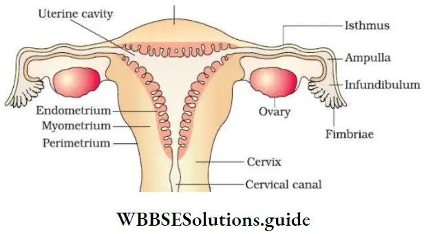
Female Reproductive System Accessory ducts (Duct system): Include 2 oviducts (Fallopian tubes), a uterus, and vagina.
- Oviducts: Each oviduct (10-12 cm long) has 3 parts:
- Infundibulum: Funnel-shaped opening provided with many finger-like fimbriae. It helps to collect the ovum.
- Ampulla: Wider part.
- Isthmus: Narrow part. It joins the uterus. The ciliated epithelium lines the lumen of the oviduct drives the ovum toward the uterus.
- Uterus (womb): It is inverted pear-shaped. It is supported by ligaments attached to the pelvic wall. The uterus has 3 parts- The upper fundus, the middle body, and the terminal cervix. Cervix opens to the vagina.
- The uterine wall has 3 layers:
- Perimetrium: External thin membrane.
- Myometrium: Middle thick layer of smooth muscle.
- Endometrium: Inner glandular and vascular layer.
- Vagina: It opens to the exterior between urethra and anus. The lumen of vagina is lined by a glycogen-rich mucous membrane consisting of sensitive papillae and Bartholin’s glands. Bartholin’s glands secrete mucus that lubricates the penis during sexual act.
Class 12 Biology Notes For Neet
Female Reproductive System External genitalia (vulva or pudendum): Consist of Mons pubis, vestibule, hymen and clitoris.
- Mons pubis: A cushion of fatty tissue covered by pubic hair.
- Vestibule: A median channel. It includes
- Labia majora: Large, fleshy, fatty, and hairy outer folds. Surrounds vaginal opening.
- Labia minora: Small, thin, and hairless inner folds.
- Hymen (Maidenhead): A membrane that partially cover the vaginal opening. It is often torn during the first coitus. It may also be broken by a sudden fall or jolt, insertion of a vaginal tampon; active participation in some sports items, etc. In some women, the hymen persists after coitus. So the hymen is not a reliable indicator of virginity.
- Clitoris: A highly sensitive organ lying just in front of the urethral opening.
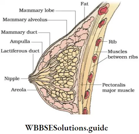
Mammary glands (breasts): A pair of mammary glands contains glandular tissue and fat.
- Glandular tissue of each breast has 15-20 mammary lobes containing clusters of cells (mammary alveoli). Cells of alveoli secrete milk. It is stored in lumen of alveoli.
- The alveoli open into mammary tubules. The tubules of each lobe join to form a mammary duct. Several mammary ducts join to form a wider mammary ampulla which is connected to lactiferous duct through which milk is sucked out.
“what is human reproduction class 12 “
Reproductive Health Class 12 Notes For Neet
NEET Biology Human Reproduction Important Notes
Gametogenesis
It is the formation of gametes in the gonads. It is 2 types: Spermatogenesis and Oogenesis.
Class 12 Biology Notes For Neet
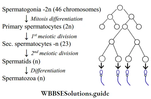
1. Spermatogenesis: It is the process of formation of sperms (spermatozoa) in seminiferous tubules of testis. It has 2 stages:
- Formation of spermatids: In this, spermatonia (Sperm mother cells or immature male germ cells) produce spermatids.
- Spermiogenesis: Spermatids transform into sperm.
Schematic representation of spermatogenesis
“reproduction in humans “
4 spermatids are formed from each primary spermatocyte. After spermiogenesis, sperm heads are embedded in Sertoli cells to get nourishment. Then they are released to the lumen of seminiferous tubules. It is called spermiation.
Reproductive Health Class 12 Notes For Neet
Role of Hormones in Spermatogenesis: The hypothalamus releases Gonadotropin-releasing hormone (GnRH).
- GnRH stimulates the anterior pituitary gland to secrete 2 gonadotropins such as Luteinizing hormone (LH) and follicle-stimulating hormone (FSH).
- LH acts on the Leydig cells and stimulates the secretion of androgens. Androgens stimulate the spermatogenesis. FSH acts on the Sertoli cells and stimulates secretion of some factors for spermiogenesis.
Best Short Notes for Class 12 Biology Human Reproduction
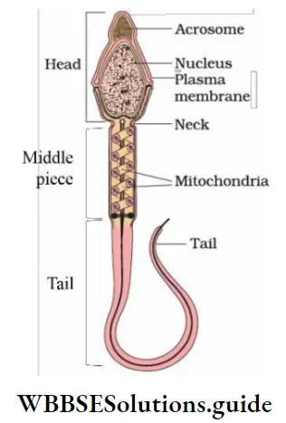
Structure of spermatozoa (Sperm): A mature sperm is about 60 μ (0.06 mm) long. A plasma membrane envelops the whole body of sperm. A sperm has 3 regions:
- Structure of spermatozoa Head: Oval-shaped. Formed of nucleus and acrosome. Acrosome is formed from Golgi complex. It contains lytic enzymes. Behind the head is a neck.
- Structure of spermatozoa Middle piece: Composed of axial filament surrounded by mitochondria and cytoplasm. Mitochondria produce energy for the sperm motility.
- Structure of spermatozoa Tail: Consists of a central axial filament. The sperm moves in fluid medium and female genital tract by the undulating movement of the tail.
Human Reproduction Class 12 NEET Key Concepts and Summary
Man ejaculates 200-300 million sperms during a coitus. For normal fertility, at least 60% sperms must have normal shape and size. 40% of them must show vigorous motility.
Reproductive Health Class 12 Notes For Neet
2. Oogenesis: It is the process of formation and maturation of ovum. It takes place in Graafian follicles. Oogenesis is initiated in embryonic stage when millions of egg mother cells (oogonia) are formed within each ovary.
NEET Biology Class 12 Chapter Human Reproduction Detailed Notes
No more oogonia are formed and added after birth. Oogonia multiply to form primary oocytes. They enter prophase-I of the meiosis and get temporarily arrested at that stage.
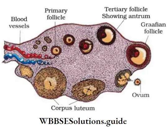
- Each primary oocyte gets surrounded by a layer of granulosa cells to form primary follicle. Many primary follicles degenerate during the phase from birth to puberty. Therefore, at puberty, only 60,000-80,000 primary follicles are left in each ovary.
- Primary follicles get surrounded by more layers of granulosa cells and a new theca to form secondary follicles. The secondary follicles transform into a tertiary follicle. It has a fluid-filled cavity (antrum). The theca layer forms an inner theca interna and an outer theca externa.
- The primary oocyte in tertiary follicle grows and undergoes the first unequal meiotic division to form a large secondary oocyte (n) and a tiny first polar body (n). So, the secondary oocyte retains nutrient-rich cytoplasm of the primary oocyte.
- It is unknown that whether the first polar body divides further or degenerates. The tertiary follicle further changes into the mature follicle (Graafian follicle).
- Secondary oocyte forms a new membrane (zona pellucida). Graafian follicle now ruptures to release the secondary oocyte (ovum) from the ovary. This is called ovulation.
“reproduction in humans “
Schematic representation of oogenesis:

NEET Study Material for Human Reproduction Chapter
Spherical and non-motile. About 0.2 mm in diameter. Ovum has 3 membranes:
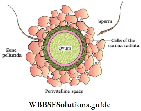
- Plasma membrane: Innermost layer.
- Zona pellucida: Outer to the plasma membrane.
- Corona radiata: Outer layer formed of follicle cells.
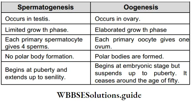
Spermatogenesis And Oogenesis A Comparison
Reproductive Health Class 12 Notes For Neet
Human Reproduction Class 12 NCERT Notes for NEET
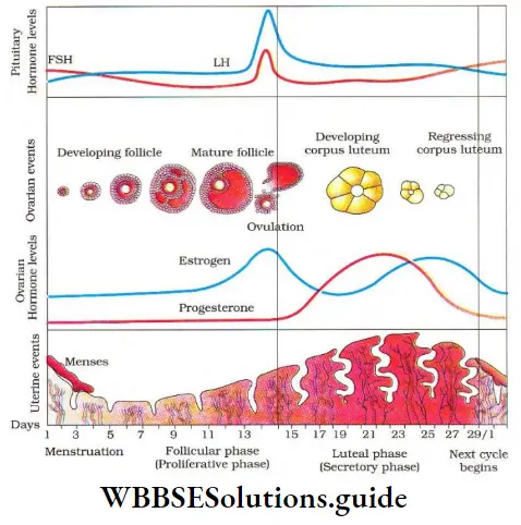
Menstrual Cycle – Reproductive Cycle
It is the cyclic events starting from one menstruation till the next during the reproductive period (from puberty to menopause) of a woman’s life. Its duration is 28 or 29 days. Menstrual cycle is also seen in other primates. Menstrual cycle includes the Ovarian cycle (changes in the ovary) and Uterine cycle (changes in the uterus, oviduct, and vagina).
Menstrual cycle has the following phases:
1. Menstrual phase: 1-5th day: The cycle starts with menstrual flow (bleeding). It lasts for 3-5 days.
- Menstruation occurs if the released ovum is not fertilized. It results in the breakdown of the endometrial lining and uterine blood vessels that come out through the vagina.
- Lack of menstruation indicates pregnancy. It may also be caused due to stress, poor health, etc.
- Menarche: The first menstruation during puberty.
2. Follicular (Proliferative) phase: 5-13th day
- It starts from 5th day after menstruation and is completed within 8-12 days.
- In this phase, the action of gonadotropins (FSH and LH) from the pituitary occurs. FSH stimulates
- Development of primary follicles into Graafian follicles.
- Secretion of estrogens by Graafian follicles.
“reproduction in humans “
- Oestrogens stimulate
- The proliferation of ruptures uterine endometrium and mucus lining of oviduct and vagina.
- Development of secondary sexual characters.
- Suppression of FSH secretion.
- Secretion of LH (Luteinizing hormone).
3. Ovulatory phase: 14th day
- LH and FSH attain a peak level in the middle of cycle.
- Rapid secretion of LH (LH surge) induces rupture of the Graafian follicle and thereby ovulation (on 14th day).
Class 12 Biology Notes For Neet
4. Secretory (Luteal) phase: 15-28th day
- After ovulation, the Graafian follicle is transformed into a yellow endocrine mass called Corpus luteum. It secretes progesterone.
- Functions of progesterone:
- Makes the endometrium maximum vascular, thick and soft. Thus, the uterus gets ready for implantation.
- Inhibits the FSH secretion to prevent the development of a second ovarian follicle.
If fertilization does not occur, the corpus luteum degenerates. It causes disintegration of the endometrium. It leads to next menstruation and new cycle. If a woman becomes pregnant, all events of the menstrual cycle stop and there is no menstruation.
Reproductive Health Class 12 Notes For Neet
Gametogenesis, Fertilization, and Embryonic Development NEET Notes
Menstrual hygiene:
- Take bath and clean yourself regularly.
- Use sanitary napkins or clean homemade pads.
- Change sanitary napkins or homemade pads after every 4¬5 hrs as per the requirement.
- Dispose the used sanitary napkins properly.
- Do not throw the used napkins in the drainpipe of toilets or in the open area.
- After handling the napkin, wash hands with soap.
Fertilization And Implantation
During copulation, semen is released by the penis into the vagina. It is called insemination. Fusion of a sperm with an ovum is called fertilization. It occurs in Ampullary region of fallopian tube.
Sperms → vagina → cervical canal → uterus → isthmus → Ampullary region → Fertilization → Ovum (from ovary) → fimbriae → infundibulum
- Fertilization happens only if ovum and sperm are transported simultaneously. So all copulations do not lead to fertilization and pregnancy. A sperm contacts with zona pellucida. It induces changes in the membrane that block entry of additional sperms.
- The secretions of the acrosome help sperm to enter the egg cytoplasm via zona pellucida and plasma membrane. This causes a second meiotic division of secondary oocyte to form an ovum (ootid) and a second polar body.
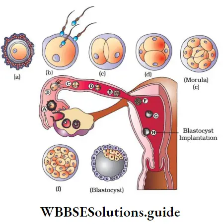
- The haploid nuclei of the sperm and ovum fuse together to form a diploid zygote. Zygote undergoes mitotic division (cleavage) as it moves through the isthmus towards the uterus and forms 2, 4, 8, and 16 daughter cells called blastomeres.
- The embryo with 8-16 blastomeres is called a morula. Morula continues to divide and transforms into a blastocyst.
- In blastocyst, blastomeres are arranged into trophoblast (outer layer) and an inner cell mass attached to trophoblast. The trophoblast layer gives nourishment to the inner cell mass. Also, it gets attached to endometrium.
- After attachment, uterine cells divide rapidly and cover the blastocyst. Thus, the blastocyst becomes embedded in the endometrium. This is called implantation.
- The inner cell mass gets differentiated to 3 germ layers (outer ectoderm, middle mesoderm, and inner endoderm). This 3-layered structure (gastrula) forms the embryo.
Reproductive Health Class 12 Notes For Neet
Pregnancy And Embryonic Development
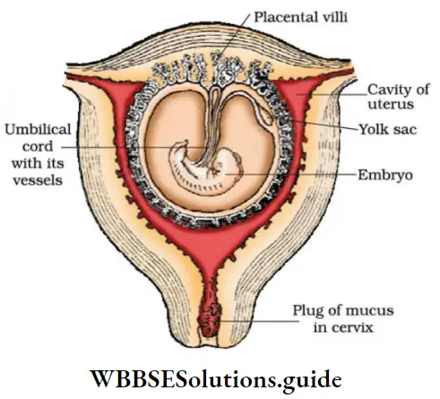
After implantation, finger-like projections (chorionic villi) appear on the trophoblast. They are surrounded by uterine tissue and maternal blood. The chorionic villi and uterine tissue are interdigitated to form the placenta. It is a structural and functional unit b/w embryo (fetus) and maternal body. Placenta is connected to the embryo by an umbilical cord. It transports substances to and from the embryo.
Functions of placenta
- Acts as a barrier between the fetus and the mother.
- Supply O2, nutrients, etc. from mother to fetus.
- Remove CO2 and excretory wastes from fetus.
- Acts as an endocrine gland. It secretes Human chorionic gonadotropin (hCG), human placental lactogen (hPL), estrogens, progesterone, and relaxin. Relaxin is also secreted by the ovary.
- During pregnancy, levels of estrogens, progestogens, cortisol, prolactin, thyroxin, etc. are also increased in maternal blood. They support fetal growth, and metabolic changes in the mother and maintain pregnancy.
- The germ layers give rise to all tissues (organs). The stem cells in inner cell mass have the potency to give rise to all the tissues and organs.
- Human pregnancy (gestation period) lasts 9 months (for cats: 2 months, dogs: 2 months, elephants: 21 months).
Reproductive Health Class 12 Notes For Neet
Changes in embryo during pregnancy
- After one month: Heart is formed.
- End of second month: Limbs and digits are developed.
- End of 12 weeks (first trimester): Major organs (limbs, external genital organs, etc.) are well developed.
- During the 5th month: First movement of fetus and appearance of hair on the head.
- End of 24 weeks (end of 2nd trimester): Body is covered with fine hair, eyelids separate and eye lashes are formed.
- End of 9 months: Ready for delivery.
Parturition And Lactation
- Parturition (labour): Process of giving birth to young ones. Parturition is induced by neuroendocrine mechanisms.
- The signals originating from the foetus and placenta induce mild uterine contractions (fetal ejection reflex). This causes the release of oxytocin from maternal pituitary.
- Oxytocin causes stronger uterine muscle contractions which in turn stimulate further secretion of oxytocin. This process is continued leading to expulsion of the baby out of the uterus through the birth canal.
- After parturition, the umbilical cord is cut off. The placenta and remnants of umbilical cord are expelled from the maternal body after parturition. It is called “after birth”.
- The mammary glands produce milk towards the end of pregnancy. It is called lactation. The yellowish milk produced during the initial few days of lactation is called colostrum. It contains several antibodies essential to develop resistance for newborn babies.