
Tissues Introduction
Having learned about the cells and their organization into higher levels, namely, the tissues, organs, organ systems, and the organism, let us now study in greater detail the tissues, that are found in animals and plants.
2. Tissues In Animals
In higher animals including man, cells are organized into four basic types of issues. These are:
- Epithelial tissue
- Muscular tissue
- Nervous tissue
- Connective tissue.
Read And Learn More: NEET Class7 Biology Notes
Epithelial Tiss
- We are familiar with the cheek cells. These cells are nothing but the cells of the epithelial tissue lining the mouth cavity.
- Epithelial tissue is a protective tissue. It forms a continuous outer layer all over the body as a part of the skin.
- It also forms a lining of all organs such as the stomach, esophagus (food pipe), mouth, intestine, and trachea (windpipe).
- Cells of the epithelial tissue called epithelial cells lie close to each other. They possess different shapes depending on their location and function.
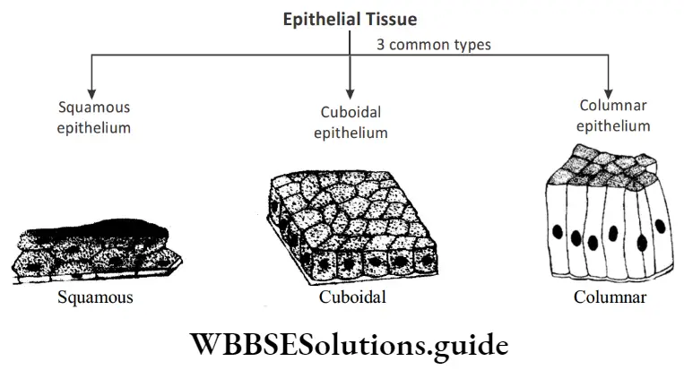
- The cells may be flattened (squamous epithelium), cubical (cuboidal epithelium), or columnar (tall forming a column; columnar epithelium).
“types of tissue “
1. Squamous and stratified squamous epithelium
- Present as thin, delicate, flat-lining.
- Present in the lining of the mouth, esophagus, and skin.
- Skin forms a protective covering on the body surface, it often gets damaged.
- Therefore, It is present in multiple layers. This is called stratified squamous epithelium.
2. Columnar and ciliated columnar epithelium
- When you think of the word column, what structure comes to your mind? Is it a pillar?
- Columnar epithelial cells are pillar-like cells.
- They are present in the inner lining of the intestine and respiratory tract.
- Their functions include the secretion of mucus and absorption of digested food.
- Columnar epithelial cells are often marked by the presence of cilia. Cilia are hair-like projections coming out of cells. Cilia can move freely.
- This helps the mucus to move forward. These cells are called ciliated columnar epithelium.
NEET Biology Class 7 Tissues Notes
3. Cuboidal epithelium
Cuboidal epithelium is composed of cube-shaped epithelial cells.
- They are present in the lining of kidney tubules and ducts of salivary glands.
- Some special cells form the surface of secretory glands and gland cells
- The glandular epithelium is often formed by the inward folding of epithelial tissues.
“tissues and functions “
Functions of Epithelial tissue
- It is a protective tissue. It protects the underlying parts of the body from injury, entry of germs, and desiccation.
- Epithelial cells lining the stomach wall secrete juices. These juices help in digestion.
- Epithelial cells of the skin help in the removal of waste such as sweat.
- Epithelial cells also help in the absorption of digested food as in the case of epithelial cells of the small intestine.

Muscular Tissue
- The human body performs different types of functions. Since different cells perform different functions, the body must have many cells to perform one or more specific functions.
- Tissues are broadly classified into four different groups, namely epithelial, connective, muscular, and nervous tissues.
- Let us explore muscular tissues in detail.
- Movement is an important function in animals. The system called muscular system performs this function. The muscular system is composed of muscular tissues.
- Close your fingers to form a fist and then open the same. Repeat the process and observe the movements of your skin.
- What do you observe? You will observe contractions and relaxations of
muscles. - Muscle cells are elongated cells that contain special contractile proteins to aid this function.
- Some movements are controlled, while some other movements cannot be controlled.
- Therefore, these muscles are categorized into voluntary muscles and involuntary muscles.
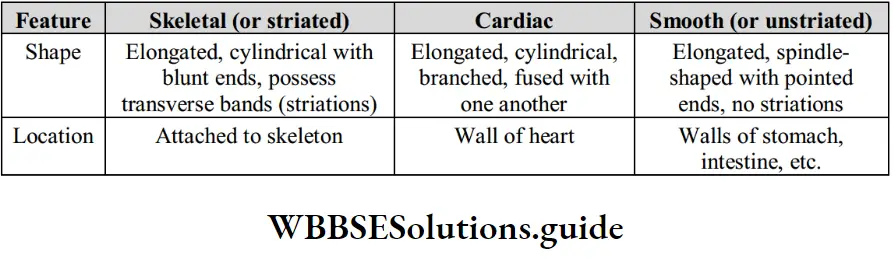
Types of muscular tissues: Based on structure and function, there are three types of muscle tissues
Tissues NEET Notes
Voluntary muscles
- Their movements can be controlled.
- Cells of muscular tissues are elongated with many nuclei; they can be branched or unbranched.
- When observed under the microscope, they appear as alternating dark and light bands.
- Therefore, they are called striated muscles.
Involuntary muscles
- Their movements cannot be controlled.
- Cells are long and pointed. It has a single nucleus.
- They can be found in the alimentary canal, uterus, iris, bronchi of the lungs, etc.
- They are non-striated muscles
Cardiac muscles
NEET Biology Tissues Important Notes
- The tissues present in the heart contract and relax in a rhythmic mode, which forms the heartbeat. These tissues are called cardiac tissues.
- Voluntary muscles, as the name suggests, work at our will. These muscles help in working, writing, running, or doing other jobs.
- Involuntary muscles are not under our control; these work on their own. These muscles take part in breathing movements, digestion, and other activities.
- Cardiac muscles, found only in the heart, expand and contract. These muscles help in pumping the blood to different parts of the body.
Function
Movement of various body parts.
- There are about 639 muscles in our body.
- About two-thirds of the body weight is due to muscular tissues, which we commonly call flesh.
- When you eat meat, you are in fact eating muscles.
“tissue structure “
Nervous Tissue
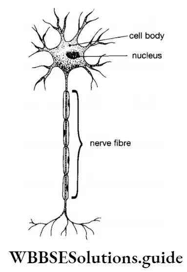
Nervous tissue consists of a group of specialized cells called nerve cells or neurons.
Each nerve cell consists of three parts –
- The cell body or cyton,
- The dendrites which are short branching structures arising from the cell body, and
- The tailor axon is a long tube-like part with fine terminal nerve
endings.
Nerve cells are found in the brain and the spinal cord.
Functions
- Nerve cells are joined end to end forming long nerve fibres.
- The nerve fibers conduct messages from one part of the body to the other.
Best Short Notes for Class 7 Biology Tissues
Connective Tissue
- Connective tissue is composed of cells that are embedded in a non-living medium or fluid.
- Unlike the epithelial, muscular, and nervous tissues where cells are closely packed together, the cells in the connective tissues are separated from one another.
- The space between the cells is filled with solid or liquid materials forming a matrix.
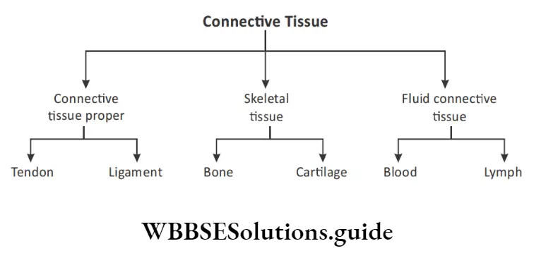
Connective tissues are of three kinds depending upon their structure and function:
“different types of tissues in human body “
- Connective tissue proper: consisting of tendons and ligaments that connect parts of the body, muscles to bones, and bones to other bones.
- Skeletal tissue: consisting of cartilage and bones, provides support to the body.
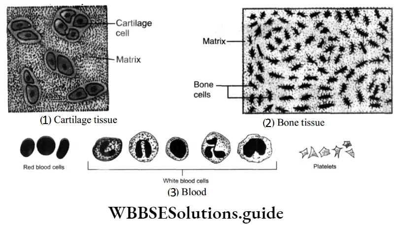
- Fluid connective tissue: consists of blood and lymph.
- Connective tissue is present throughout the body and may be solid or fluid. Blood and lymph are fluid connective tissues. Cartilage, bone tendons, and ligaments are all examples of solid connective tissues.
Cartilage, though a solid connective tissue, is less hard than the bone. It helps in making the organs or tissues flexible and is present in the nose and in the external ear.
Tissues Class 7 NEET Key Concepts and Summary
Bone is a hard connective tissue (harder than cartilage) due to the presence of calcium phosphate in the matrix. The bones perform two functions – support the body and help in the movement of muscles.
Blood is a fluid connective tissue, the matrix consists of a fluid or liquid called plasma. The plasma contains three types of blood cells (called blood corpuscles) red blood cells (RBC), white blood cells (WBC), and platelets.
Lymph
- Lymph is blood lacking the red blood cells.
- While blood flows in blood channels, lymph surrounds the body cells.
Functions
- Connective tissues connect different tissues of the body and provide support to the body.
- Blood, being a liquid connective tissue, connects every part of the body and
transports substances to all body parts.
3. Tissues In Plants
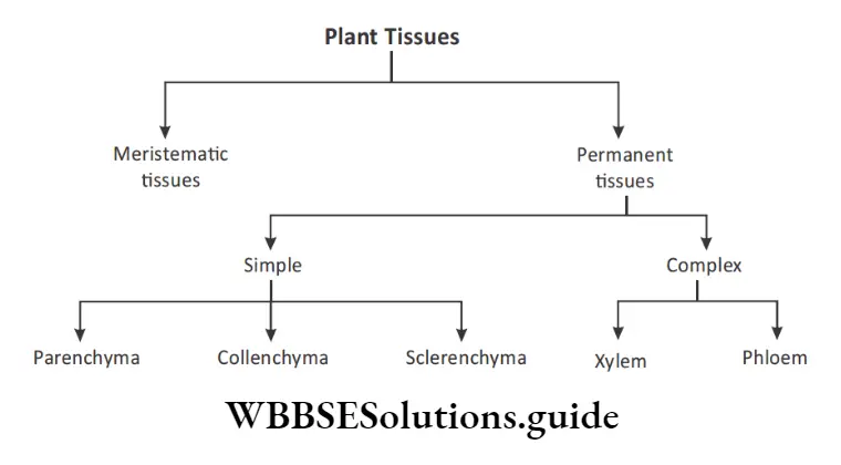
Broadly, there are two types of tissues in plants:
- Meristematic tissues
- Permanent tissues
“classification of tissue chart “
Meristematic Tissues
Meristematic tissues are formative tissues that add new cells to the plant body. They, thus, contribute towards growth in the length and width of the plant.
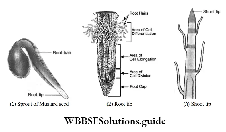
NEET Biology Class 7 Chapter 3 Detailed Notes with Explanation
These tissues are found in the growing regions – the tip of the root and the tip of the stem. The growth in thickness is also due to a meristematic tissue that is present laterally in the plant body.
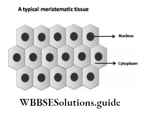
Meristematic cells, composing the meristematic tissue, show the following characteristic features.
- The cells are small.
- The cells are thin-walled.
- The cells are rich in cytoplasm with large prominent nuclei,
- The cells lack spaces between them.
- They divide actively, adding to the growth. Meristematic tissues give rise to permanent tissues.
Types of meristematic tissues
Meristem can be further classified based on the position or locations of meristematic tissues.
They are of three types:
Apical meristem: They are present at the tips of stems, roots, and branches. They are responsible for the axial growth in a plant.
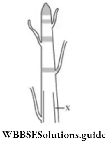
Intercalary meristem: They are present at the base of internodes, and are responsible for the growth of internodal region.
Lateral meristem: They are present on the lateral side of stems and roots. Lateral meristem is responsible for the radial growth of plants. The vascular cambium and cork cambium are examples of the lateral meristem.
“tissues of the human body “
NEET Study Material for Tissues Chapter
Permanent Tissues
Permanent tissues are derived from meristematic tissues. These tissues form the bulk of the plant body.
The cells comprising permanent tissue show the following features:
- Cells may no longer be small and thin-walled.
- Cytoplasm is much less. The nucleus is small.
- Cells do not divide.
Permanent tissues can be classified in two ways:
1. Based on their place or origin, permanent tissues are of three types:
- Dermal tissue
- Vascular tissue
- Ground or fundamental tissue
2. Based on the kinds of cells constituting a permanent tissue, the permanent tissues are of two types :
- Simple tissues
- Complex tissues
Simple tissues consist of only one type of cells while more than one type of cells is present in a complex tissue.
Examples of simple tissue — are parenchyma, collenchyma, and sclerenchyma. Examples of complex tissue xylem and phloem.
Simple Permanent Tissues
Simple permanent tissues are of three types:
Tissues Class 7 NCERT Notes for NEET
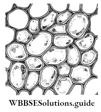
- Parenchyma: Parenchyma is composed of thin-walled cells. It is present in the softer parts of the plant and performs various functions. It stores food, as in potatoes. In leaves, these cells contain chloroplasts and thus help in photosynthesis (manufacture of food).
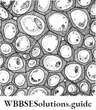
- Collenchyma: Collenchyma has cells that possess thickenings at the corners. It is present in growing stems and leaves, providing mechanical support.
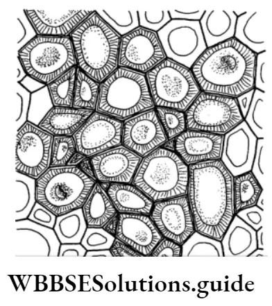
- Sclerenchyma: Sclerenchyma is composed of thick-walled dead cells and is found in hard parts of the plant body. This tissue also provides strength to the plant parts.
“explain the structure function and location of tissues “
Complex Permanent Tissues
Simple permanent tissues are of two types:
- Xylem
- Phloem
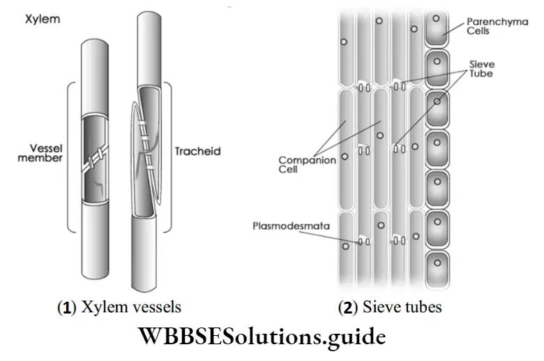
Xylem: Xylem is made of several kinds of cells. The important ones are the thick-walled, tubular cells, often dead at maturity. These are called vessels.
Phloem: Phloem is also made up of several kinds of cells, the most important of which are called the sieve tubes. These are also tubular cells having perforated walls joined end to end.
In addition to sieve tubes, certain specialized thin-walled cells closely: Xylem vessels associated with sieve tubes are present. These cells are called companion cells.
The phloem, unlike the xylem, is a living tissue. Phloem also contains phloem parenchyma and phloem fibers.
Dermal Tissues
Dermal tissue is usually present in the outermost layers of the plant body. Thus, the surface of roots, stems, leaves, flowers, and fruits is made up of dermal tissue, commonly referred to as the epidermis.
Dermal tissue performs various functions:
Plant and Animal Tissues NEET Notes for Class 7
- It protects the plant body and is, therefore, also known as a protective tissue.
- Cells are often thick-walled and develop a waterproof coating on their surface. This helps to reduce the evaporation of water from the leaf surface.
- In the roots, this tissue helps in the absorption of water and minerals from the soil.
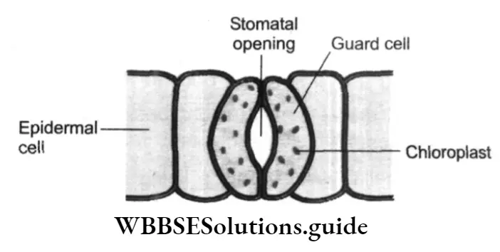
- In the leaves, the dermal tissue helps in the exchange of gases like oxygen and carbon dioxide between the atmosphere and the plant. This exchange takes place through small pores, called stomata.
Vascular Tissue
- This tissue is also known as the conducting tissue, as it transports or conducts water, minerals, and food to different parts of the plant body.
- Vascular tissue consists of two types of tissues – – xylem and phloem.
- Vascular tissue may be compared to the blood vessels of man as a tissue meant for transporting materials through the plant body.
- Movement of water and minerals occurs through the xylem, whereas the food manufactured by the leaves is transported to other parts through the phloem.
Vascular tissues perform two major functions:
- These act as conducting tissues. The xylem transports water and minerals from the roots to various body parts; the phloem transports food from the leaves to all other body parts.
- They also provide mechanical support to the stems and leaves.
Ground Tissue
- Also known as fundamental tissue, is basically a supporting tissue.
- The tissue is present in the root, stem, and leaf, as well as in flowers and fruits.