Chapter 2 Tissues Introduction
- All living organisms are made up of cells. As you have learned, the cell is the fundamental and structural unit of organisms.
- Unicellular organisms like Amoeba and Paramecium have a single cell in their body.
- The single cell performs all basic life activities like movement, intake of food, digestion, respiration, excretion, reproduction, etc.
- However, in multi-cellular organisms, there are millions of cells. Each cell is specialized to perform different functions.
- They show the division of labor. Division of labor has been made possible by the specialization of cells and their grouping with each specialized cluster of cells in a definite place in the body.
- For example, in the animal body, muscle cells help in the movement of body, nerve cells help in the conduction of message and blood cells helps in the transport of materials.
- Likewise, in plants, cells of phloem conduct food from leaves to another part of the plant.
- The specialization is achieved due to differentiation whereby cells occupy a definite shape, size, structure and functions.
- Hence, in multi-cellular organisms with a higher degree of differentiation and specialization, the group of cells aggregates collectively to perform a particular function.
- Such a group of cells are called tissues.
Read And Learn More: NEET Class 9 Biology Notes
What is tissue? Tissue is a group of cells with a common origin, structure and function that work together to perform a particular function.
For example, Blood, bone, and cartilage are some examples of animal tissues while xylem, phloem, parenchyma etc are different types of tissues found in plants.
Let us discuss the importance of tissues in living organism:
- It brings about division of labour in multi cellular organisms. The division of labour increases the survival rate of multi cellular organism.
- Tissues become organized to form organs, which in turn forms organ systems. This increases the effiiency of multi-cellular organisms.
- Tissue decreases the workload of individual cells.
“tissues notes “
Now, Do plants and animals have same types of tissues.? No! Though plants and animals have similar types of life processes but due to difference in their organization, life style and mode of living, they do not have similar types of tissues.
Let us have a look at how plant and animal tissues are different from each other.
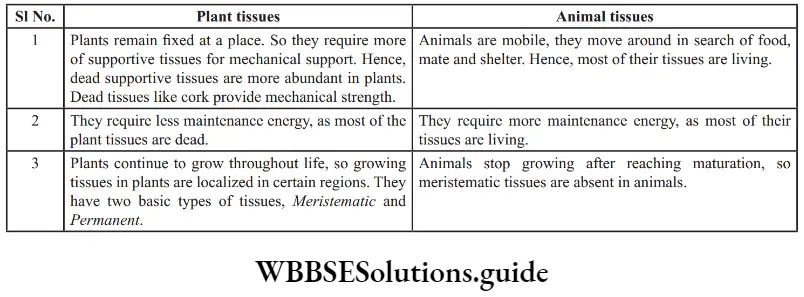
Difference table between plant and animal tissues:
Structural Organization Of Tissues
- As we have learnt, the smallest unit of life in any living organism is cells. The cell has a very complex system of organelles and each organelle is concerned with a particular task.
- Thus, there is a division of labour at the cellular level. As evolution progressed and larger and larger organism appeared with enormous number of cells in the body, it became necessary that the bodies’ function are distributed among group of cells called tissues and even among group of tissues.
- Such higher and higher grouping of cells or tissue is known as levels of organization.

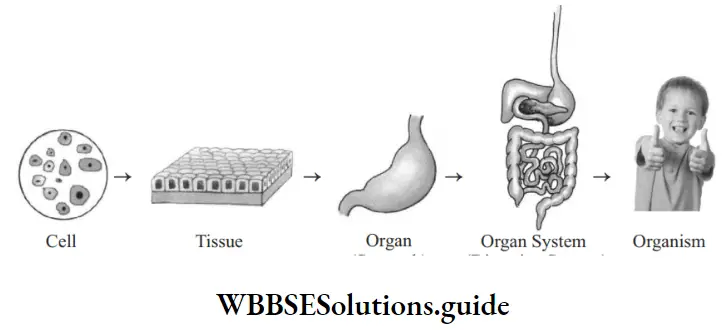
These levels are Cellular level, Tissue level, Organ level, Organ system and Organism.
- Cells group together to form tissue, which in turn are grouped together into larger functional units called organs.
- Tissues are aggregate of cells of same origin and having same function. For example, the surface epithelium of our skin or dividing cells of the root cap of a plant.
- Organ is a group of various tissues that performs a specifi function. For example, stomach and intestine are organs to digest food.
- Similarly, lungs and trachea are organs meant for respiration. All these organs are collections of various tissues like connective, epithelial, muscular and nervous tissue.
- Like wise in plants, stem, root, leaves and flwers are different organs that perform different functions. They are made of different tissues like parenchyma, collenchyma, sclerenchyma, xylem and phloem.
- Various organs group together to form even larger functional units called organ systems.
- Organ system is a combination of a set of organs all of which are usually devoted to one general function.
- For example, Respiratory system (consisting of lungs, trachea, bronchi, diaphragm etc) in man, or the shoot system (consisting of leaves, stem, and branches, etc) in a plant are example of organ system that works in a coordinated way.
- The complete individual comprised of different organ system is known as organism. For example, Man, Dog, Cat or a Mustard plant.
Thus, the different level of organization of the living body is:
Cells → Tissues → Organs → Organ Systems → Organism
NEET Biology Class 9 Tissues Notes
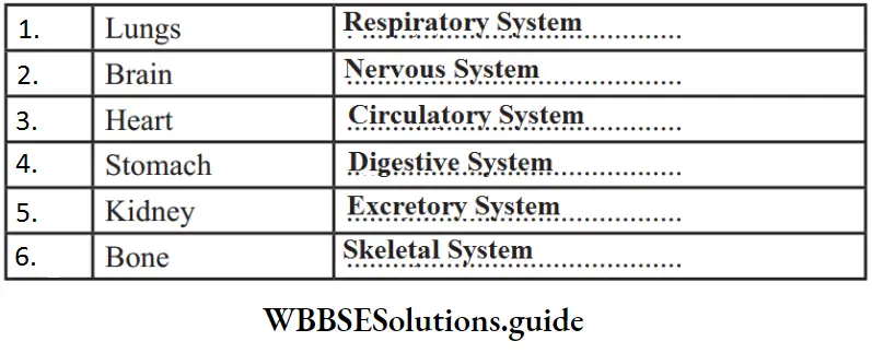
Question 1. The main organs of various system has been given. Find out their respective organ system
Answer:
“class 9th biology chapter tissue “
In this chapter, we will be discussing various kinds of plant and animal tissues, in detail.
Plant Tissues
On the basis of their ability to divide, plant tissues are divided into two types:
- Meristematic tissues (Dividing tissues): It consists of undifferentiated actively dividing cells.
- Permanent tissues (Non-dividing tissues): It consists of differentiated cells that have lost the ability to divide.
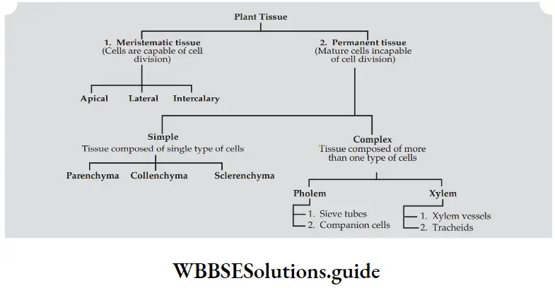
Let us discuss each of these tissues one by one.
1. Meristematic Tissues:
- Meristematic tissues are thin-walled compactly arranged, immature cells that keep on dividing continuously.
- The new cells produced are initially Meristematic. Slowly, they grow, differentiate and mature into permanent tissues.
- Characteristic features of Meristematic tissues:
- The meristematic cells are spherical, or polygonal in shape.
- The cells are compactly arranged without inter-cellular spaces.
- The cell wall is thin, elastic and is made of cellulose.
- Each cell has abundant cytoplasm and prominent nuclei. Vacuoles may or may not be present.
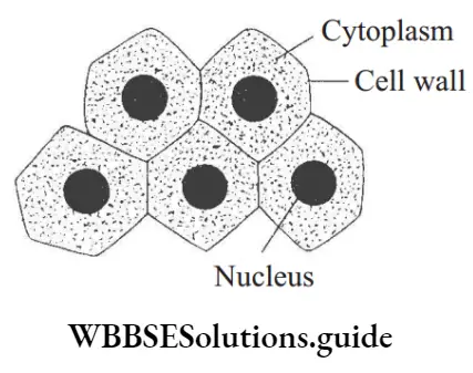
Functions: Meristematic tissue divides continuously to form a number of new cells and help in growth of tissue.
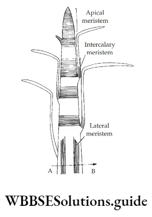
Location: On the basis of position in the plant body, meristematic tissues are divided into three types:
- Apical Meristems: They are found at the growing tips of roots and stems.
Function: It brings about growth in length of root and stem. - Lateral Meristem: It occurs on the sides almost parallel to the long axis of the root and stem.
Function: It increases the width or girth of the stem and root. - Intercalary Meristem: It occurs at the base of the leaves or at the base of internodes.
Function: It increases the length of the internode
NEET Biology Tissues Chapter Important Notes and Formulas
2. Permanent Tissue:
- Permanent tissues are tissues that have lost the ability to divide, and have attained a defiite form and size.
- They are actually derived from Meristematic cells. Different type of permanent tissues is formed due to differences in their specialization.
Characteristic features of Permanent tissue:
- The cells of permanent tissues normally do not divide.
- The cells may be thin walled (living) or thick walled (dead).
- Permanent cells are specialized to perform a particular function.
- The cells have attained defiite shape and size.
“class 9 tissues chapter “
Difference between Meristematic tissue and Permanent tissue:
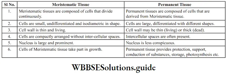
Depending on the type of cells, permanent tissues are divided into two types:
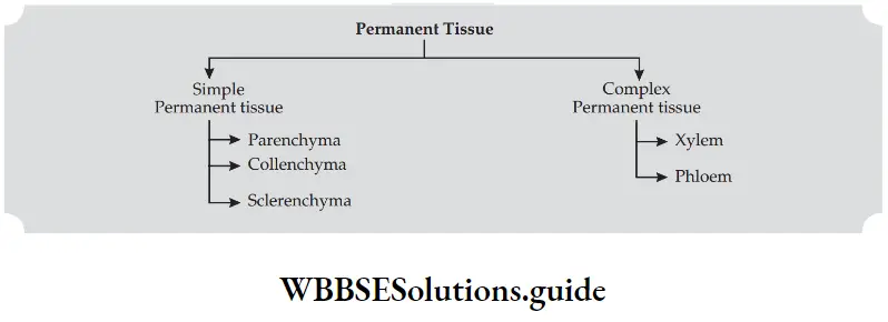
1. Simple Tissue:
Simple tissue is made up of only one kind of cells forming a uniform mass. The cells are similar in structure, origin and function.
Simple permanent tissues are of three types: Parenchyma, Collenchyma and Sclerenchyma
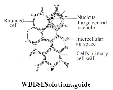
Parenchyma: Parenchyma is widely distributed in plant body such as stems, roots, leaves and flower.
They are found in the cortex of root, ground tissue in stems and mesophyll of leaves.
- Characteristic features:Cells are isodiametric i.e. equally expanded on all sides.
- Cells may be oval, round or polygonal in outline.
- Nucleus is present and hence, living.
- The cell walls are thin and made of cellulose.
- Cytoplasm is dense with a central vacuole.
- Cells are loosely packed with large intercellular spaces between the cells.
- It may contain chlorophyll. Parenchyma containing chlorophyll is called chlorenchyma.
- It is the seat of photosynthesis.
- The parenchyma that encloses large air cavities is known as aerenchyma. Aerenchyma provides buoyancy to aquatic plants.
Functions:
- Parenchyma stores and assimilates food.
- They give mechanical support to the plant body by maintaining turgidity.
- The presence of intercellular spaces in between parenchyma cells helps in the exchange of gases.
- It prepares food if chlorophyll is present.
- It stores waste products like gum, crystal, tannin and resins
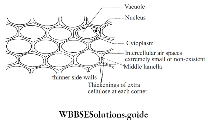
2. Collenchyma: Collenchyma is a strong and flexible tissue that provides flexibility to soft aerial parts.
They are found below the epidermis in leaf stalks, leaf mid-ribs, and herbaceous dicot stems.
Characteristics features:
- Collenchyma cells are elongated cells with thick primary walls.
- Cell wall is unevenly thickened with cellulose at the corners.
- Intercellular spaces are absent.
- Nucleus is present and hence the tissue is living.
- Few chloroplasts may be present in the cells.
Functions:
- Collenchyma provides mechanical support to the stem.
- It provides flexibility to soft aerial parts so that they can bend without breaking.
- Collenchyma cells may contain chloroplasts and thus take part in photosynthesis.
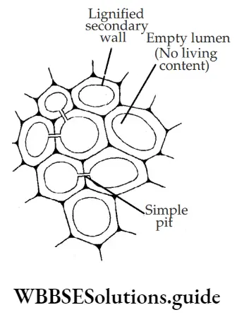
3. Sclerenchyma: Like collenchyma, sclerenchyma is also a strengthening tissue.
It is found in and around the vascular tissue, under the skin i.e. the epidermis in dicot stems.
Characteristic feature:
- Cells are long, narrow, thick and lignified usually pointed at both ends.
- The cell wall is evenly thickened with lignin. Lignin is a waterproof material.
- Intercellular spaces are absent.
- Nucleus is absent and hence the tissue is made up of dead cells.
- Middle lamella i.e. the wall between adjacent cells is conspicuous.
Sclerenchyma cells are of two types:
- Fibres: They are long, narrow and pointed cells.
Location: Fibres are found in and around the vascular tissue. It may also occur below the epidermis. Fibres help in the transportation of water in plants. - Sclereids: The cells are thick-walled, hard and strongly lignified. They are shorter, iso-diametric or irregular cells.
Location: Sclereids are found in the cortex, pith, phloem, hard seeds, nuts and stony fruits.
The flesh of pear and guava are sometimes gritty. Have you ever thought why it is so? It is due to the presence of sclereids. Sclereids give firmness and hardness to the part concerned.
Functions:
- Sclerenchyma gives mechanical support to the plant by giving rigidity, flexibility and elasticity to the plant body.
- It forms a protective covering around seeds and nuts.

Best Short Notes for Class 9 Biology Tissues NEET
2. Complex Tissue:
- Complex tissue is made up of more than one type of cells that work together to perform a particular function.
- Complex tissues are of two types: Xylem and Phloem.
“what is tissue in science “
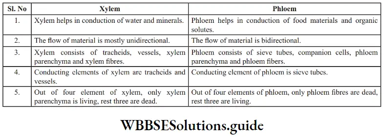
1. Xylem (Greek xylo= wood): The Xylem is a complex permanent tissue that conducts water and minerals upward from root to the plant. It is also known as wood.
Xylem consists of four types of elements:
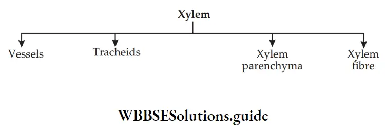
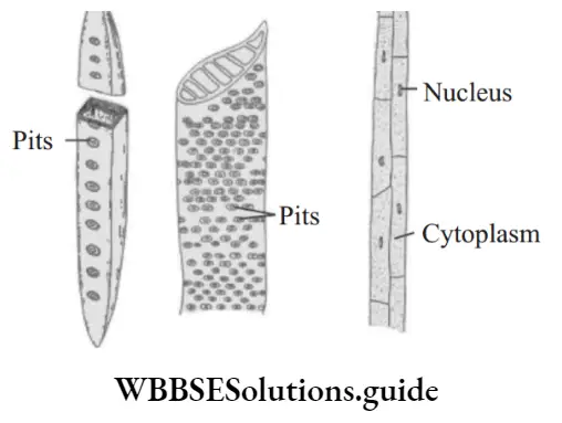
- Tracheids: Tracheids are long, tubular dead cells with wide lumen and tapering ends. The cell wall is thick with lignin. They have pores in their walls.
- Vessels: A vessel is a cylindrical tube like structure that are placed one above the other end to end. It is a non-living cell with lignified walls. They generally possess pits.
Function: Tracheids and vessels both are main conducting elements in the xylem. Vessels are more effiient than tracheids. - Xylem fires: They are long, non-living cells with very thick lignin deposition on the walls. They have a narrow lumen and tapering ends.
Function: Xylem fires provide mechanical support to the plant. - Xylem parenchyma: They are living cells with cellulosic cell wall.
Function: They help in storage of starch and other materials. They also help in lateral conduction of water
2. Phloem (Greek= Phloeis= inner bark): Phloem is a complex permanent tissue that conducts food synthesized in the leaves to different parts of the plant body.
Unlike, xylem, conduction of food occur both in upward and downward directions (From leaves to storage organs and from storage organs to growing organs).
Phloem consists of four types of elements:

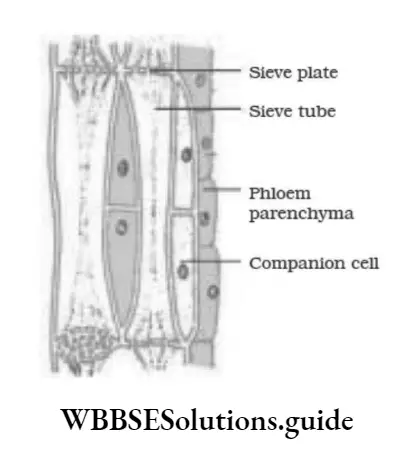
- Sieve tubes: Sieve tubes are elongated, cylindrical tubes with perforated end walls between adjacent sieve tube cells. Sieve tube cells are placed end to end in a linear row. The perforated end walls are called sieve plates. Sieve tube cells have vacuolated cytoplasm and lack a nucleus.
- Companion cells: Companion cells are associated with sieve tubes. They are thin-walled cells that lie on the sides of sieve tube cells. Companion cells have dense cytoplasm and prominent nuclei.
Functions: They help sieve tubes in the conduction of food material by maintaining a proper pressure gradient in the sieve tube cells. - Phloem Parenchyma: The phloem parenchyma cells are thin-walled and living.
Functions: They help in storage and slow lateral conduction of food. - Phloem fires: They are the only non-living (dead) component of phloem. They are thick-walled elongated and spindle shaped cells with narrow lumen.
Functions: Phloem fires provide mechanical support to the tissue. Phloem fires are source of commercial fires. E.g. Jute, Hemp, Flax etc
Protective Tissue:
Protective tissues are the outer layer of cells that protect the plant parts like stem, roots, leaves, flowers and fruits.
They are of two types:
1. Epidermis:
- The epidermis is the outermost protective layer of the plant body. It is usually a single layer.
- The cells are elongated and closely packed without any intercellular spaces between them.
- The outermost layer of cell is covered with a waterproof coating or layer called cuticle. Cuticle is made up of a waxy substance called cutin.
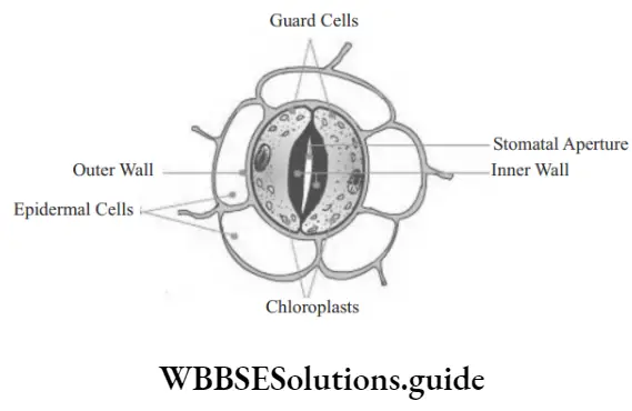
At places, the epidermis is not continuous and bears minute pores called stomata. Stomata consist of an opening called the stomatal opening which is surrounded by two specialized kidney-shaped cells called guard cells. Guard cells are specialized epidermal cells. As guard cells become turgid, they create a pore in between their thick inner walls. Pores help in the exchange of gases. It is also the seat of transpiration.
Tissues In The Lab
- How do epidermal cells help in gaseous exchange? Let us perform an activity.
- Take a freshly plucked leaf of a Rhoeo plant. Stretch it from the upper side and break it by applying pressure. While breaking it, stretch gently so that peel projects out.
- Place this peel in a petri dish filled with water. Add a few drops of safranine stain to it. Observe it under a microscope.
- What did you observe? You will find tiny pores of stomata along with the epidermal cells.
- The stomata are bound by a pair of guard cells. Guard cells are the two curved cells on either side of the pore.
- By changing their shape they can open or close the pore. When guard cells absorb water, they bend outwards, so that the pore between them opens up. When they lose water they go back to a less curved shape, closing the pore between them.
Functions of stomata:
- Stomata help in the exchange of gases, like oxygen and carbon dioxide.
- It also helps in transpiration. Transpiration is the loss of water form the plant into the outside atmosphere.
Activity:
- To show that leaves give out water vapour through transpiration.
- Take a flower pot. Enclose a branch or the whole plant in a transparent plastic bag. Tie the plastic bag tightly around the stem to make sure that air does not enter or leave it. Leave the plant as such for 1-2 hours. What did you observe?
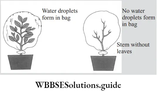
Observation: You will find a few drops of water inside the bag.
Conclusion: Water escapes into the air in the form of water vapor through the breathing pores (called stomata), which are found on the upper or lower surfaces of the leaves.
Functions of epidermis:
- The main function of the epidermis is to protect the plant body from entry of pathogens and pest.
- The presence of cuticle in the epidermis reduces the evaporation of water. It checks the rate of water loss from aerial parts.
- The epidermis of root along with root hairs helps in the absorption of water and minerals from the soil.
- The stomata regulate the exchange of gases and also help in transpiration.
2. Cork:
- Cork is the outer protective tissue of older stems and roots. Cork cells are rectangular in shape and are arranged compactly in several layers.
- They are dead cells and lack intercellular spaces. The walls of cork cells are heavily thickened by the deposition of suberin.
- Suberin makes the cork cells impermeable to water and gases. At places, cork possesses small aerating pores called lenticels.
“what is tissue in science “
Functions of cork:
- Cork prevents the entry of harmful micro-organisms into the plant body.
- It prevents desiccation, by preventing loss of water by evaporation.
- It protects plant body against mechanical injury, extreme temperature and infections.
- Cork possesses lenticels which help in exchange of gases between tissue and the atmosphere.
- Though cork is light, impervious, non-reacting and insulating, it is commercially used in the manufacture of stoppers for bottles, shock absorbers, sports good, insulation board etc.
Question 1. How epidermis protects the plant body against the invasion of parasites?
Answer:
The epidermis does not allow the parasites to gain entry into the internal tissues due to
- Absence of intercellular spaces
- Thick outer walls
- Deposition of cutin wax in the cuticle covering the epidermis
- Deposition of silica
Animal Tissues
- Do you know, the working of animal body is coordinated by tissues and organs present in their body.
- For example, breathing occurs due to contraction and relaxation of certain muscles.
- During the process, the intake of air (inhalation) provides oxygen to blood inside lungs while carbon dioxide present in blood passes into the air via exhalation.
- Thus, the blood carries oxygen and food to all cells. What are these blood and muscles? These are actually types of tissues.
- Blood is a type of connective tissue while muscle is a example of muscular tissue.
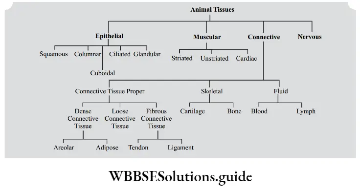
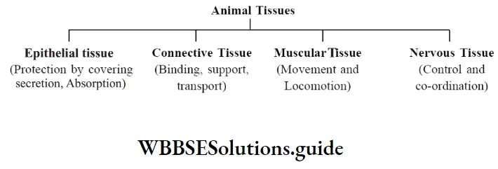
On the basis of structure and function, animal tissues are divided into four types:
Tissues Class 9 NEET Important Questions and Answers
Let us now discuss each of these tissues one by one in detail.
1. Epithelial Tissue:
- Epithelial Tissue is the simplest animal tissue that forms the continuous sheet of closely packed cells that covers all external and internal surface of the animal body.
- Thus, it is also known as covering tissue. The epithelial cells lie close together with little or no intercellular substance.
- The cells are held together by various types of junctions and small amount of cementing materials.
- The epithelial membrane rests over an extra-cellular layer of white, non-elastic collagen fires called basement membrane. This membrane connects the epithelial tissue to the underlying connective tissue.
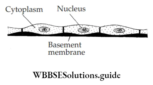
Location: It occurs over the skin, lining of mouth and other parts of alimentary canal, lung alveoli, lining of respiratory tract, kidney tubules, urinary tract, reproductive tract, blood vessels, etc.
Functions:
- It protects the underlying tissues against mechanical injury, dehydration and infection by micro-organisms.
- Epithelium lining the lung alveoli allows exchange of gases between blood and alveolar air.
- Epithelium lining the uriniferous tubules helps in ultrafiltration, secretion and reabsorption to produce urine.
- Glandular epithelium produces secretions. For example, tears, mucus, intestinal juice etc.
- Ciliated epithelium helps in movement of various types of materials. For example, dust particles in respiratory tract, ovum in the oviduct etc.
- Some epithelial cells like that of intestinal mucosa become specialized for absorption.
Types of Epithelial Tissue:
On the basis of arrangement of layers, epithelial tissues are classified into two types:
- Simple epithelium: The epithelial cells arranged in a single layer are known as simple epithelium.
- Stratified epithelium: The epithelial cells arranged in many layers are known as stratified epithelium. It is found in the body, where there is lot of wear and tear. For example, skin, inner lining of cheeks, etc.
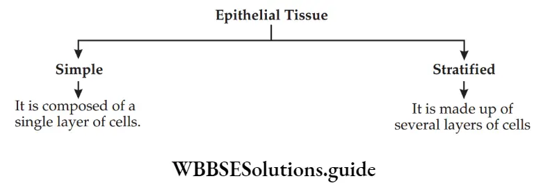
On the basis of cell shapes, the epithelial tissues are classified as:

1. Squamous epithelium: It is made of thin, flat, irregular-shaped cells that fit together to form a compact tissue.
The margins may be smooth or wavy. Squamous epithelium can be single-layered (simple) or multilayered (stratified).
Stratified Squamous epithelium: Unlike Squamous epithelium, cells of this tissue are arranged in many layers.
The basal layer lies in contact with basement membrane, so that new cells can be added on older surface cells as they are torn away.
They are found in skin and cover the external dry surface of the skin. The epithelium is water-proof and highly resistant to mechanical injury.
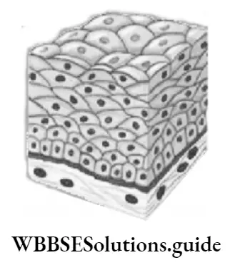
Location:
- Simple Squamous epithelium is found in lung alveoli, Bowman’s capsule, blood capillaries etc.
- Stratified Squamous epithelium lines the cavities and ducts.
Function:
- It protects the underlying structures from mechanical injury and germs.
- As squamous epithelium lines the Bowman’s capsule, it helps in ultrafiltration.
- In blood capillaries, the epithelium helps in exchange of materials between blood and tissue.
- In the alveoli of lungs, epithelium helps in exchange of gases between blood and atmosphere.
- Stratified Squamous epithelium provides protection against abrasion by sloughing off the outermost layer.
2. Cuboidal epithelium: The Cuboidal epithelium is made up of cube like cells, which are square in section but they are polygonal in surface view. The nucleus is centrally placed and round in structure.
Microvilli may be present on the free surface which increases the surface area of absorption.
Location: They are found in the uriniferous tubules, thyroid vesicles, salivary and pancreatic ducts.
“what is tissue in science “
Functions:
- The cuboidal epithelium helps in secretion, excretion and absorption.
- It also provides mechanical support to the part where they are found
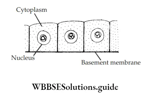
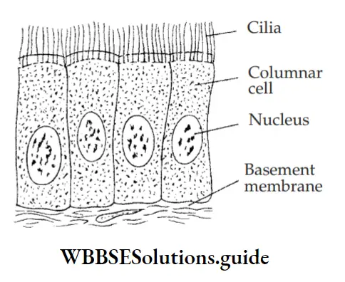
3. Columnar Epithelium: Columnar epithelium is tall and pillar-like. The nucleus is oval and lies at the base. The free surface bears a number of tiny figer-like projections called microvilli. Microvilli increase the surface area for absorption.
Location: They are found in the lining layer of stomach, intestine and their glands. They are also present in the salivary glands, sweat glands, tear glands, and covering of epiglottis.
Functions:
- Columnar epithelium lines the intestine and is specialized to absorb nutrients.
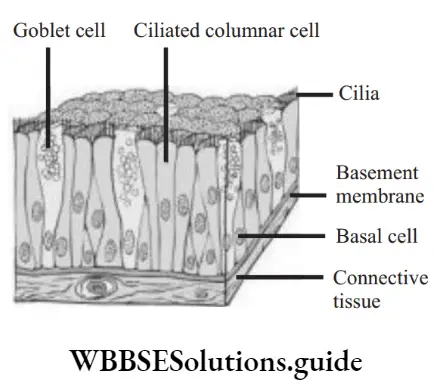
- Goblet cell is a modified columnar cell, which produces mucus.
- It also provides protection to the underlying tissues.
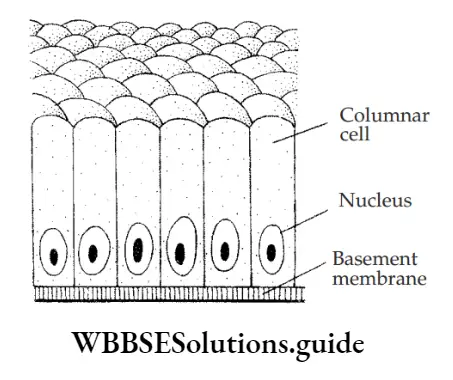
On the basis of specific functions, the epithelial tissue is classified into ciliated and glandular epithelium:
4. Ciliated epithelium: Ciliated epithelium is Cuboidal or columnar cells that bear cilia at their free surface.
Location:
Ciliated Cuboidal epithelium is found in sperm ducts and uriniferous tubules.
The ciliated columnar epithelium is found in the inner lining of the respiratory tract (trachea or windpipe) and oviducts.
Functions:
The beating of cilia helps to keep the unwanted particles from entering into the lungs. Cilia also help in pushing the ovum in oviduct.
5. Glandular epithelium: Glandular epithelium is actually a modification of columnar epithelium. The epithelium is infolded to form multicellular glands.
Location: The glandular epithelium is found lining the intestine and glands.
Function: The glands are specialized for the secretion of specific chemicals.
For example sweat glands secrete sweat, similarly oil is secreted from oil glands, enzymes from digestive glands, hormones from endocrine glands, mucus from mucus glands, etc.
2. Connective Tissue:
Connective tissue is a fundamental animal tissue that has scattered living cells embedded in matrix.
The matrix and cells are different in different connective tissues. Matrix is the ground substance and is secreted by the living cells of the connective
tissue. It may be jelly-like, flied or solid.
It is the most abundant tissue of the animal body. It helps in connecting, binding, packing and supporting different structures of the animal body. Thus, it helps the body to function as an integrated whole.
Functions of connective tissue:
- It helps in binding the different structures of the body. For example, muscle with bone, bone with bone, and muscle with skin.
- It forms the packing material in different organs.
- Skeletal connective tissue like bones and cartilage forms supportive framework of the body.
- Fluid connective tissue like blood forms an internal transport system of the body.
- The cells present inside connective tissue protect the body against microbes and toxins.
- It also forms shock-absorbing cushions around organs like the eye, heart and kidneys.
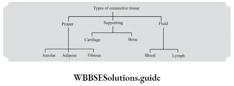
Based on the nature of the matrix, connective tissue is divided into three types:
- Connective Tissue proper (the matrix is Jelly-like, i.e. less rigid)
- Skeletal Tissue (matrix is solid i.e. rigid)
- Vascular Tissue (matrix is a flied called plasma)
Types of Connective Tissues:
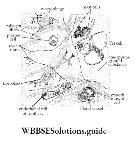
1. Connective Tissue Proper: It is type of connective tissue that has a jelly-like matrix and three types of fibers- white collagen, yellow elastin and reticular fires. The living cells present may include fibroblasts, mast cells, plasma cells, Macrophages and lymphocytes.
It is of two types:
- Loose connective tissue proper- It has fewer fires and more of matrix. Examples: Areolar tissue and Adipose tissue.
- Dense connective tissue proper- It has more of fires and less amount of matrix.
Examples: Tendon, Ligament
Areolar Tissue: It is the most widely spread connective tissue in the body. The non-living intercellular matrix contains irregular shaped cells and two kinds of fires.
The cells forming the tissue are:
- Fibroblasts: form the yellow fires, made of elastin and white fires, made of collagen in the matrix.
- Macrophages: help in engulfing the bacteria and micro-organisms.
- Mast cells: secrete heparin. Heparin helps in clotting of blood.
Location: Areolar tissues are found inside organs, around blood vessels, muscles and nerve. It also occurs below sub-cutaneous tissue and structures like muscles and skin.
Functions:
- It helps in binding skin with underlying parts.
- It provides packing material in various organs.
- It provides material for repair of injury.
- Macrophages present in tissue feed on microbes, produce antibodies to fiht against infection.
- Mast cells in tissue are involved in allergic reactions
NEET Biology Class 9 Tissues MCQs with Solutions
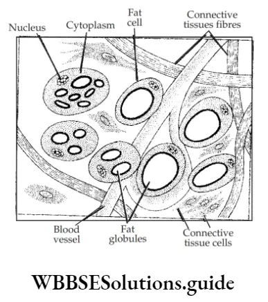
2. Adipose Tissue: It is a type of connective tissue that is specialized to store fat called adipose cells.
The fats are stored inside cells called adipocytes. Adipocytes are large cells with one or more globules of fat and peripheral cytoplasm with nucleus at one end.
Like areolar tissue, the adipose tissue also has soft jelly like matrix, living cells like fibroblasts, macrophages, mast cells etc and two types of fibers called collagen and elastin.
Location: The tissue is found below the skin, around internal organs and inside yellow bone marrow.
Functions:
- Adipose tissue acts as storage tissue that stores fat in reserve for use when required.
- It acts as shock absorbing cushion around certain organs.
- It forms an insulating layer below the skin. It keeps the body warm.
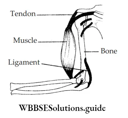
3. Fibrous Tissue: Fibrous tissue is mainly made of firoblasts. Fibroblasts form tendons and ligaments.
- Tendon: Tendon is a tough, non- fibrous, dense, white fibrous connective tissue. It has great strength but limited flexibility.
Function: It joins a skeletal muscle to a bone, thereby helping the bone to move on contraction and relaxation of the muscle. - Ligament: Ligament is a dense yellow fibrous connective tissue. It has considerable strength and high elasticity.
Function: The ligament binds a bone with another bone, thereby allowing bending and rotation movements over a joint.
Difference between tendon and ligament:

2. Supportive Connective Tissue: It is a connective tissue in which the matrix is rigid and the living cells occur in fluid-filed spaces called lacunae.
It is also known as skeletal tissue and is of two types.
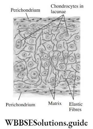
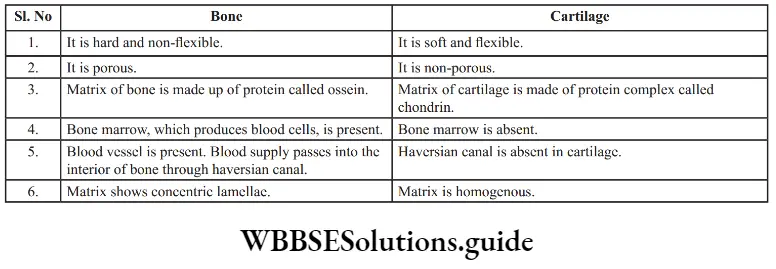
1. Cartilage: Cartilage is a non-porous, firm but flexible supportive tissue. It has a solid matrix which is composed of chondrin.
Chondrin is secreted by the chondrocytes. Chondrocytes lie in the matrix singly or in groups of two or four surrounded by fluid-filed space called lacunae.
Cartilage is usually covered by a tough fibrous membrane called perichondrium.
Location: Cartilage is found in the nose tip, ear pinna, epiglottis, larynx, rings of trachea and bronchi, sternal ends of ribs, and tips of several bones.
Function: It provides support and flexibility to various parts of the body
2. Bone: Bone is a strong, rigid, and non-flexible tissue. Bone is the hardest tissue of the body.
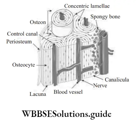
- It consists of a solid matrix with flied-filed lacunae having osteocytes or bone cells.
- Matrix is composed of a collagenous protein complex called ossein and mineral matter like salts of calcium, phosphorus, and magnesium. The hardness of bone is due to the deposition of mineral matter.
- The matrix in mammalian bone like in thigh bone, is arranged in concentric rings or lamellae around nutrient-filed haversian canals.
- The osteocytes lie on the lamellae and give out branched processes that join with those of the adjoining cells.
- Some bones have a central cavity that contains a tissue that produces blood cells.
- The soft connective tissue present in the bone cavity is known as bone marrow.
- The sheath of bone is called the periosteum. A layer of osteoblasts or bone-forming cells lie below it.
“what is tissue in science “
Bones are of two types:
- Spongy bone, in which bone cells are irregularly arranged. Such bones are found at the ends of the long bones.
- Compact bone, in which cells are arranged in circles or lamellae around a central canal, haversian canal
Location: Bones are found all around the body. It forms the supportive framework of the body.
Function:
- It forms the supportive framework of the body.
- It provides a surface for attachment to many muscles.
- It forms joints that take part in body movement and locomotion.
- The red bone marrow of bones forms blood cells.
- Bone is a reservoir of calcium, phosphorus, and other minerals.
Question 1. Which tissue smoothens bone surfaces at the joint?
Answer: Cartilage
Fluid Connective Tissue: It consists of cells and matrix without fibers. Plasma is the extra cellular flied of matrix, the ground substance. Blood and Lymph are two types of flied connective tissue.
Blood:
Blood is a bright red coloured flied connective tissue. It is a complex of straw-coloured flied plasma in which various kinds of cells are embedded. Plasma contains large number of proteins like Fibroblast, Albumin and Globulin.
The blood cells embedded inside plasma are:
- Erythrocytes (Red Blood cells): Red blood cells are coloured enucleated living cells. It contains a red colour pigment called haemoglobin that takes part in transport of oxygen. It also carry small amount of carbon dioxide.
- Leucocytes (White blood cells): They are colourless nucleated cells that have the ability to change their shape like Amoeba. White blood cells protect the body from diseases and help in healing of wound.
- Thrombocytes (Blood Platelets): Blood platelets are non-nucleated, oval to rounded colourless cells. It helps in clotting of blood
Tissues In The Lab
Take a drop of blood in a clean slide and observe the slide under a microscope. What did you observe? You will fid different types of blood cells in it. Try to identify them and write their functions.
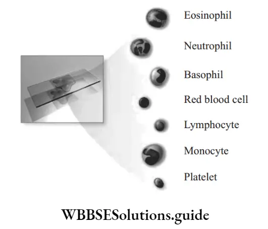
White Blood Cells/Leucocytes:
White blood cells are colorless, nucleated cells. Their amoeboid nature helps them to squeeze through the walls of the blood vessels in order to capture and engulf bacteria. They lack hemoglobin and hence are colorless. They are actually larger than red blood cells.
Based on the shape of nucleus and structure of the cytoplasm, leucocytes are broadly classified into two types.
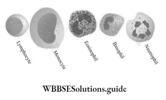
1. Granulocytes: They are irregular shaped cells, with distinct and lobed nucleus. Their cytoplasm is granular.
It is further classified into three types:
- Basophils: Their cytoplasm is large but with less number of granules. The nucleus is two or three-lobed. They develop into mast cells of connective tissue.
- Eosinophils: They are more numerous in blood than basophils. Nucleus is bilobed. They are generally found in the wall of alimentary canal and blood capillaries. Eosinophils are thought to protect the body against allergies.
- Neutrophils: The nucleus is formed of two or more lobes that are attached with each other.
- They are about 67% of the total leucocytes in blood. Their function is phagocytosis of bacteria.
2. Agranuloctyes: Their cytoplasm is devoid of granules. Nucleus is large.
Based on their size, agranulocytes are of two types:
- Lymphocytes: They are small in size but with large nucleus. It comprises about 28% of the total leucocytes in the blood. Their functions are phagocytosis and antibody- production. Phagocytosis is the process of engulfig solid particles.
- Monocytes: Monocytes have kidney shaped nucleus. They function as tissue macrophages feeding on dead tissues.
NEET Study Material for Tissues Chapter Class 9
Location: Blood flows continuously all over the body inside the blood vessels- Arteries, Veins and Capillaries.
Functions:
- Blood transports gases like oxygen and carbon dioxide.
- It also transports food materials like glucose, amino acids and fatty acids.
- Blood regulates body temperature by conducting heat within the body.
- Blood transports excretory products like urea and uric acid to the kidneys.
- White blood cells fiht against infection and protect the body from foreign agents. They are basically soldiers of the body.
- Blood platelets help in clotting of blood.
2. Lymph:
Lymph is a light, yellow coloured flied connective tissue, consisting of plasma and white blood cells. It is devoid of red blood cells and blood platelets.
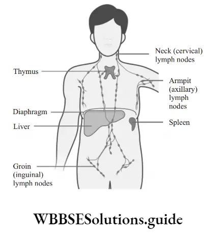
Location: Lymph flows through lymph vessels and lymph capillaries. Most of the tissues and organs pour their secretions and excretions into lymph instead of blood. However, lymph is ultimately passed into blood.
Functions:
- It helps in the exchange of materials between blood and tissue flied. Actually, it acts as a middleman between tissues and blood.
- It brings carbon dioxide and wastes from tissues to blood and nutrients, oxygen etc from blood to tissues.
- The lymph nodes and lymphoid organs (like the spleen) function as traps for microbes.
- Lacteal, a special type of lymph vessel present in intestinal villi helps in the absorption of fat from the intestine.
- The lymph also maintains the blood volume by removing or adding excess plasma.
Difference between Blood and Lymph:

Question 1. Why blood is considered to be connective tissue?
Answer:
Blood is considered fluid connective tissue because:
- Blood consists of living cells scattered in an abundant matrix. The matrix is liquid or plasma in blood.
- Blood connects all parts of the body. It circulates throughout the body, receiving, and providing materials to all tissues and organs.
Question 2. What will happen if lymph is not returned to blood?
Answer:
The blood volume will decrease and passage of substances from tissues to blood and blood to tissue would be disturbed.
3. Muscular Tissue:
- Muscular tissue is a contractile tissue that occupies more than 40% of total weight of the body.
- Each and every movement, every breath, every mouthful you chew-all these actions are carried out by the body’s muscle cells.
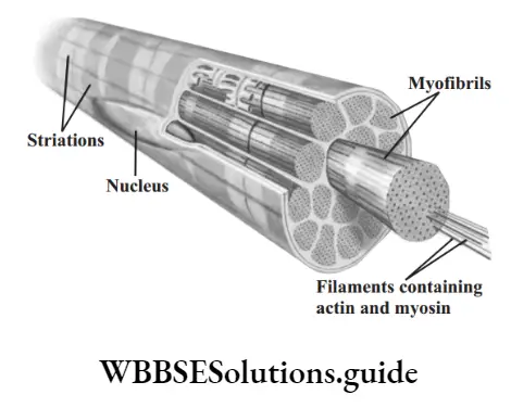
Structure of Muscular tissue: Muscular tissue is composed of contractile proteins inside cells.
- Cells of muscular tissue are elongated and are known as muscle fires. Because of their elongated shape, muscle cells are known as muscle fires.
- The group of muscle fires is known as muscles. The muscle fire is covered by a sheath of membrane called Sarcolemma.
- The cytoplasm of muscle fire contains a large number of fie longitudinally running firils called myofirils.
- Myofirils are actually the contractile elements of muscle fires. Each myofiril has two types of protein fiaments called thicker myosin and thinner actin.
- The actin and myosin fiaments slide past each other to shorten the firils causing the whole muscle to contract.
- The cytoplasm is called the sarcoplasm.
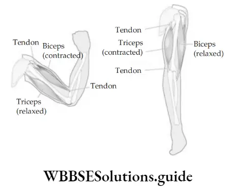
Working of Muscles: Muscles are attached to the bones of the skeleton by a cord called a tendon. If you want to lift your forearms, the brain sends a signal to your arms muscles through nerves. As a result, the muscle contracts (becomes smaller) and pulls the bones of forearm up.
Voluntary And Involuntary Muscles:
There are two kinds of muscles:
- Voluntary Muscles are those muscles, that function as per the direction of conscious will. The brain can stop or start then. For example, skeletal muscles come into use when we walk.
- Involuntary muscles are those muscles, that function on their own, independent of conscious will. Brain cannot stop or start them. For example, breathing in and out of air.
1. Differentiate the following activities on the basis of voluntary and involuntary muscles.
Question 1. Jumping of frog
Answer: Voluntary muscle
Question 2. Movement of food in your intestine
Answer: Involuntary muscle
Question 3. Writing with hand
Answer: Voluntary muscle
Question 4. Pumping of heart
Answer: Involuntary muscle
Types of Muscle Fibres:
On the basis of their location, structure and function, there are three types of muscle fires.
- Striated muscle fire
- Unstriated muscle fire
- Cardiac muscle fire
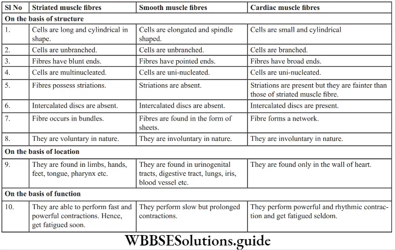
1. Striated Muscle Fibres: (Also known as striped, skeletal or voluntary muscle fires):
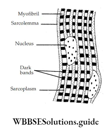
- Each striated muscle fire is long, cylindrical, unbranched with multinucleated condition.
- It bears striations in the form of alternate light and dark bands.
- The fires have blunt ends.
- A number of oval nuclei occur peripherally in each fire below the sarcolemma.
- The muscle has the ability to contract rapidly and thus is responsible for quick movements.
- The muscles are also known as voluntary because their contraction is under the control of will.
- They get fatigued soon
Location: They are found in the limbs, face, neck and body wall.
Function:
- Striated muscle attached to bones helps in body movement.
- It controls the breaking, chewing and swallowing of food.
- It helps in breathing activity.
- The muscles also control the blinking of eyes.
Tissues Class 9 NCERT Biology NEET
2. Unstriated Muscle Fibres: (Also known as Non-striated, Smooth or involuntary muscle fires)
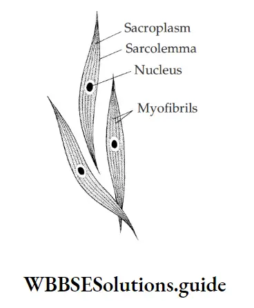
- Each smooth muscle fire is spindle-shaped, unbranched and uni-nucleated cell.
- The muscle fires do not show striations.
- The fires have pointed ends.
- The muscles contract and relax very slowly.
- They are known as involuntary muscles as their movement is not controlled by the mind.
- Fatigue is rare in smooth muscle fires.
Location: They are found in the walls of organs like stomach, intestines, blood vessels, ureters, bronchi, lungs, urinary bladder, iris etc.
Functions:
- In intestine, smooth muscle fire helps to push the food down along the alimentary canal by peristalsis.
- Emptying of urinary bladder occurs due to contraction of smooth muscles.
- Involuntary movement of iris and ciliary body of eye helps in automatic adjustment of amount of light and focusing of object based on distance.
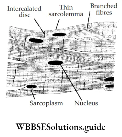
3. Cardiac Muscle Fibres: (Also known as involuntary muscles)
- They are small, cylindrical, branched, and involuntary muscle fire.
- The fires have broad ends.
- They have transverse striations with light and dark bands. However striations are fainter than those of striated muscle fires.
- Special electrical junctions called intercalated discs are present at intervals in the fires.
- Cells are uninucleated. Nucleus is centrally placed.
- The muscles show rhythmic contractions.
- They are involuntary muscle fires. They are not under the control of one’s will.
- They seldom get fatigued. They keep on performing their function throughout life.
Location: Cardiac muscle fires are found in the walls of the heart.
Function: The rapid contraction and relaxation of cardiac muscle fires helps in pumping of blood through heart.
Difference between Striated, Smooth and Cardiac muscle fires:
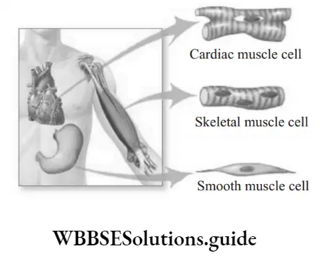
Functions of Muscle fires:
- Excitability: They can respond to stimuli.
- Extensibility: They have the ability to get stretched.
- Contractibility: They can contract.
- Elasticity: They can move back to the original position
4. Nervous Tissue:
- Nervous tissue is specialized to transmit messages in our body. They can receive, integrate and transmit stimuli to various parts of the body.
- It is devoid of matrix. Its cell is surrounded by a special connective tissue cell.
- Nervous tissue contains two types of cells: Neuron and neuroglial cells.
Neuron: It is the functional unit of nervous tissue. It is also known as nerve cells. They are the longest cells of the body reaching upto a metre in length.
Each neuron is made of three parts:
- Cell body (Cyton): It is a broader nucleated part of a neuron. Its cytoplasm is called Neuroplasm. Neuroplasm contains two special structure called neurofirils and Nissl granules.
- Neurofibrils are fie fibrils involved in the transmission of impulses.
- Nissl granules are ribosome containing structures. They are made of RNA and protein.
- Dendrons: Dendrons are small, branched protoplasmic outgrowths of cell body. Like cyton, dendrites also possess neurofirils and Nissl granules. Dendrons further branch into many thin dendrites.
Function: Dendrites receive impulses and transmit the same towards cyton. - Axon: Axon is a single, long, fire like process generally arising singly from the cell body of a neuron. It is devoid of Nissl granules. However, it contains neurofirils. Axon is surrounded by a sheath called neurolemma of a special connective tissue called Schwann cells.
The axon forms five branches at its terminal end called nerve endings. The nerve ending has knobbed ends in contact with muscles, glands, skin etc for providing an impulse for activity. Each such junction is called synapse. Synapse is meant for the transmission of impulses from one neuron to another.
Plant and Animal Tissues NEET Notes Class 9
Function: Axon carries impulses towards the cell body.
The transmission of impulses is usually carried out with the help of a neurotransmitter like acetylcholine.
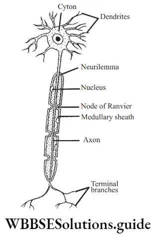
Difference between Axon and Dendrite:

Synapse:
- Synapse is a junction between two neurons. The terminal knobbed branch end of an axon are connected with dendrite branches of an adjacent neuron.
- This gap junction helps in transmission of impulse from one neuron to the next.
- The transmission of impulse is generally carried out with the help of a neurotransmitter chemical like acetylcholine.
Transmission of Nerve Impulse:
- As stimulus is passed on, the branching dendrites receive the stimulus and transmit through the cyton to the axon.
- From axon, the impulse is transmitted through its variously branched end into either a muscle (in order to contract) or to a gland (in order to secrete).
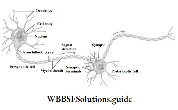
Question 1. What is the direction of the “flw of impulse” within a nerve cell?
- Dendrite to axon
- Axon to dendrites
Answer:
The branching dendrites receive the stimulus and transmit through the cyton (cell body) to axon, which fially transmits it through its branched end, either to muscle or gland.
Location of nervous tissue: They are present in brain, spinal cord and nerves.
Functions:
- It picks and conducts messages from one part of body to another.
- They also receive all types of sensations like sight, sound, smell, pain, touch etc. from the outside environment and send the message to the brain and the spinal cord.
- In turn, impulses from the brain and spinal cord are carried to the various organs.
- Nervous tissue provides responses to all types of stimuli.
- It exerts control over entire body activities by coordinating the functioning of different body parts.
