
Structural Organization In Animals Animal Tissue
You see different kinds of animals around you. All these animals carry out the same basic processes to sustain life.
Although these basic processes like digestion, respiration, reproduction, etc., are carried out by common organ systems, the complexity of the systems varies from animal to animal.
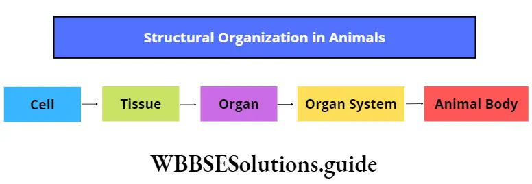
This variation is due to the difference in their structural organization. The cell is the basic unit of this organization.
Several cells performing similar functions organize to form tissues, that further constitute organs.
| Class 11 Biology | Class 11 Chemistry |
| Class 11 Chemistry | Class 11 Physics |
| Class 11 Biology MCQs | Class 11 Physics MCQs |
| Class 11 Biology | Class 11 Physics Notes |
Several such organs work together to form an organ system. Altogether, these are the different levels of structural organization of the body.
In the previous chapter, you have learned about the structural organization of plants. Now in this chapter, you shall learn about the structural organization in animals.
Read and Learn More: WBCHSE Notes for Class 11 Biology
Cells of animal bodies that are similar in origin, structure, and function, are organized to form structures, known as animal tissues. Cells in the embryo are organized into three specific layers called germ layers. Each germ layer has a specific name.
“structural organization in animals notes for class 11”
The outermost layer is called the ectoderm, the middle layer is called the mesoderm, and the innermost layer is called the endoderm. All the tissues and organs of the body arise from either of these three layers.
Body parts originating from different germ layers
- From ectoderm: Epidermis of skin, nails, hair, sweat gland, sebaceous gland, scales of fish, feathers of a bird; brain and spinal cord, lens and cornea of eyes; epithelium of nasal cavity and mouth cavity; mammary gland; enamel present in teeth, etc.
- From mesoderm: Muscles; connective tissue, blood vessels, lymphatic vessels, blood cells; ducts within the excretory and reproductive system; adrenal cortex; bones, cartilages, tendons, ligaments; testes, ovary; kidney, spleen, etc.
- From endoderm: Epithelium of digestive and respiratory tract; liver, pancreas, prostate gland, thymus gland, thyroid gland, parathyroid gland; tympanic membrane of ear and epithelium of auditory canal, etc.
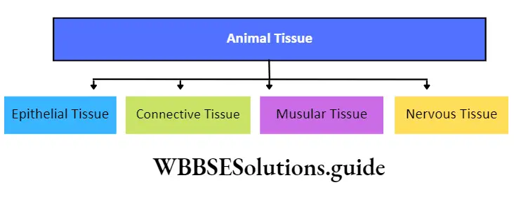
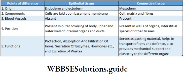
Types of tissues: The bodies of all vertebrates and most of the invertebrates are made of a variety of tissues.
However, all the tissues may be grouped into four main types. These are epithelial tissue, connective tissue, muscular tissue, and nervous tissue.
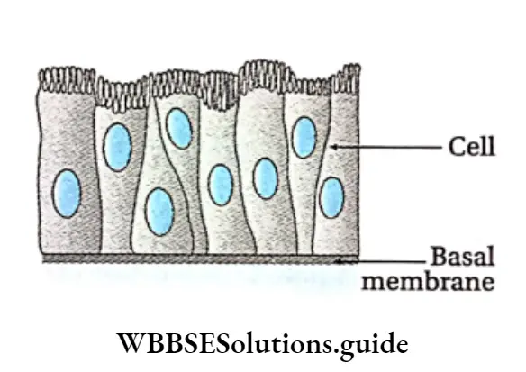
Pseudostratified Epithelial Tissue Definition: The epithelial tissue which has a single layer of cells but appears multi-layered, due to the irregular shape of the cells, is known as pseudostratified epithelial tissue.
Pseudostratified Epithelial Tissue Position: It is found in the trachea, nasal passage, epididymis, large duct of salivary gland, male urethra, etc.
“detailed notes on structural organization in animals”
Pseudostratified Epithelial Tissue Structure:
- All the cells touch the basement membrane but all do not reach the free surface. As a result, it appears that more than one layer of cells is present.
- The nuclei of the cells lie in different planes. This again makes the epithelium appear as stratified.
- Longer cells are columnar in shape with oval nuclei.
- Shorter cells have round or cone-shaped nuclei.
- These shorter cells can replace the degenerated longer cells.
- The free surface of longer cells may or may not have cilia. (For example, the inner surface of the nasal passage has ciliated pseudostratified epithelium while in salivary glands, the pseudostratified epithelium has no cilia.)
class 11th chapter 7 biology notes
Some other types of cells belonging to epithelial tissue
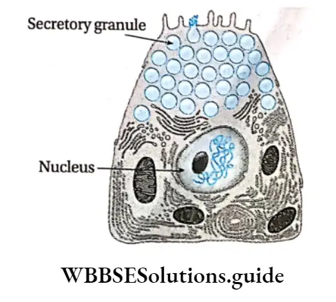
Serous cells: Serous cells are present at the distal end of certain glands such as salivary glands, acinus of the pancreas, etc.
These cells contain a large spherical nucleus and secretory granules called zymogen granules.
“tissues, organs, and organ systems in animals notes”
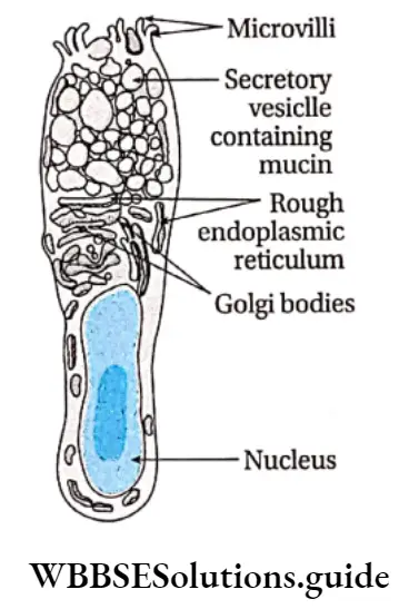
Mucus-secreting cells: The most predominant mucus-secreting cells are goblet cells that are glandular in nature.
These are present within the digestive tract, along with the simple columnar epithelial cells.
These cells are highly polarised with the nucleus and other cellular organelles are situated towards the base of the cells.
The apical portions contain secretory vesicles which secrete a glycoprotein named mucin. Mucin can transform into mucus under mechanical stress. Mucus helps in lubrication and defense as well.
“structural organisation in animals short notes “
Myoepithelial cells: These cells are usually found in glandular epithelium as a thin layer above the basement membrane.
They are found in various exocrine glands such as sweat glands, mammary glands, lacrimal glands, and salivary glands. They can contract and release the secretions of the respective exocrine glands.
Function: Secretion of saliva, reabsorption of ions, and protection of visceral organs are the main functions of this tissue.
Connective Tissue
Connective Tissue Definition: The tissues that are derived from embryonic mesoderm, present throughout the body to support and bind other tissues in vertebrates are called connective tissues.
Connective Tissue Origin: Connective tissue develops from mesoderm.
“animal tissue types and their functions explained”
Maintains structure of the body: It forms a supporting framework of cartilage and bones in the body.
Coordination: It connects different organs and systems of the body.
” class 11 structural organisation in animals”
Support: It fills up spaces within the organs and tissue layers, hence providing support to the organs.
Temperature regulation: Connective tissue such as blood, helps to regulate the temperature of the body.
Storage: It also stores several nutrients such as glucose, certain proteins, salts of different ions, etc.
Protection: It protects the body from pathogens (bacteria, fungus, flu, etc.).
Structural Components
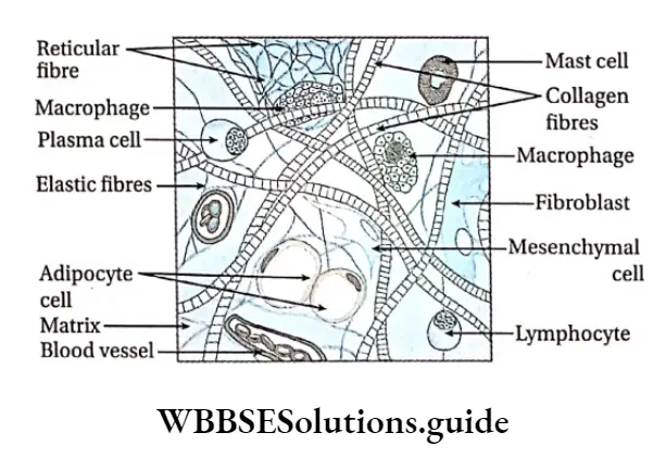
Three main components of connective tissue are—cells, intercellular medium or matrix, and fibers. Apart from these, blood vessels are also present in this tissue.
The matrix is made up of proteoglycans associated with mucopolysaccharides like chondroitin, hyaluran, etc. The three components of connective tissue are described below.
Cells: The constituent cells of connective tissue are different in terms of structure, function, and origin. Some of the connective tissue cells are described as follows.
” structural organisation in animals new ncert pdf”
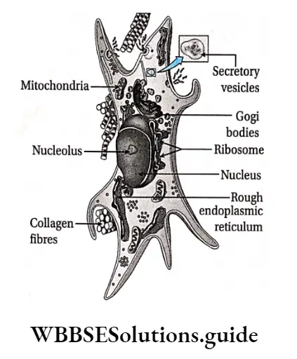
Fibroblast or Fibrocytc Definition: The spindle-shaped, branched cells of connective tissue that help in the formation of the matrix and fibers are called fibroblasts or fibrocytes.
Fibroblast or Fibrocytc Characteristics:
- Cells are large, flat, star-shaped, and branched,
- Cytoplasm is distinct with large and oval nuclei,
- Small granules and fat droplets are present in the cytoplasm.
Fibroblast or Fibrocytc Function: These cells mainly secrete collagen and elastin proteins. These proteins form white collagen fibers and reticular, elastic fibers respectively.
Some important facts about fibroblast tissue are called adipocytes or fat cells.
- Fibroblast cells do not undergo cell division. However, if the number of fibroblasts required for growth or regeneration is high, then these cells divide by mitosis to form more cells.
- Fibroblast cells play a major role in wound healing.
- Myofibroblast is a special type of cell that has the characteristics of both, smooth muscles and fibroblasts. It plays an active role in wound healing.
Mast cell Definition: The polygonal and calculated secretory cells of the connective tissue that secrete heparin, serotonin, histamine, etc., are called mast cells.
Mast cell Characteristics:
- Cells are large, round, or oval with dense, granular cytoplasm,
- The nucleus is small and oval in shape,
- Cells are capable of forming pseudopodia-like projections.
Functions:
- Heparin secreted by the mast cells, prevents blood coagulation within blood vessels.
- Serotonin secreted by mast cells helps in maintaining blood pressure by constriction of the blood vessels,
- Histamine secreted by these cells dilates blood vessels during allergic reactions.
Plasma cell Definition: The round or oval cells of the connective tissue that produce antibodies and contain an eccentric (away from the center) nucleus, are called plasma cells.
Plasma cell Characteristics:
Cells are oval or round with non-granular cytoplasm,
The Chromatin material of the nucleus is oriented in the form of the spokes of a cartwheel. Hence, these are also called cartwheel cells.
Plasma cell Functions: These cells synthesize 7-globulin protein (a plasma protein made up of immunoglobulins acting as antibodies) that help in defense.
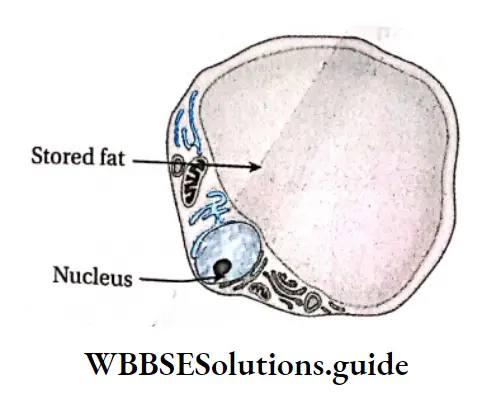
Adipocyte Or Fat Cell
Adipocyte Characteristics:
- Cells are large, round, or oval with a single distinct nucleus,
- They store fat droplets within the cell cytoplasm which push the nucleus to the periphery. This gives the cell a signet ring-like appearance
Adipocyte Functions:
- The cells store food in the form of fat that can be used for energy production,
- They help in thermoregulation.
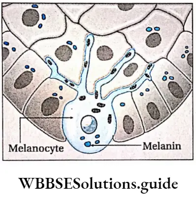
Pigment Cell Or Melanocyte Definition: The pigment-containing cells of connective tissue which determine the color of skin and hair, are known as pigment cells or melanocytes.
They are also called chromatophores.
“structural organization in animals NEET notes PDF”
Pigment Cell Or Melanocyte Characteristics:
- Cells are irregular with cytoplasmic processes,
- Cells contain yellow, brown, or black pigment granules.
Pigment Cell Or Melanocyte Functions:
- They produce melanin pigment, that imparts color to skin and hair,
- This pigment also provides protection from harmful UV rays.
Macrophage Or Histiocyte Definition: The irregular-shaped cells of connective tissue that contain lysozyme and have phagocytic functions are known as macrophages or histiocytes.
Macrophage Or Histiocyte Characteristics:
- Cells are small, irregular in shape with short protoplasmic processes,
- The cytoplasm is granular and vacuolated with a single oval nucleus.
- Cells contain Golgi bodies and lysosomes.
Macrophage Or Histiocyte Functions:
These cells have phagocytic and pinocytic properties. They engulf foreign particles and microbes by their phagocytic action and destroy them by secreting proteolytic enzymes.
They remove dead and damaged cells from wounds etc., by digesting them
Mesenchymal cell Definition: The immature cells of the connective tissue that transform into other types of connective tissue cells like osteoblasts, myocytes, adipocytes, etc., when required, are known as mesenchymal Cells.
Mesenchymal cell Characteristics:
Cells are star-shaped with cytoplasmic processes. These cytoplasmic processes continue with the adjacent cells to form syncytium (multinucleated structure) with reticular fibrils (small fibers made up of reticulin protein).
These cells occur in embryonic connective tissue.
Mesenchymal cell Function: Mesenchymal cells do not have a specific function. However, they act as reserve cells that transform into other connective tissue cells when required.
Wandering cell or hematogenous cell: Different kinds of cells of the circulatory system such as erythrocytes, and leucocytes are found in the special fluid connective tissue. They move from one part to another with the circulatory fluid and perform various functions.
Mononuclear phagocyte system
The mononuclear phagocyte system or MPS (also called the Reticuloendothelial system or Macrophage system) is a part of the immune system.
It consists of phagocytic cells of reticular connective tissue (a type of connective tissue made up of fibers called reticulin).
The phagocytic cells primarily include monocytes and macrophages, which are important for the immune system. They accumulate in lymph nodes and spleen.
Matrix Definition: The amorphous, viscous, colorless, transparent ground substance of the connective tissue is known as a connective tissue matrix.
Components: The matrix is made up of three chemical components. These are—
- Glycosaminoglycan (acid mucopolysaccharide),
- Proteoglycan, And
- Glycoprotein.
Matrix Functions:
- The matrix gives protection and support to the connective tissue.
- It is present in the intercellular spaces and acts as a lubricant, helping in the easy movement of the cells.
- It also serves as a medium for the exchange of metabolites between the cells.
Fibre Definition: The proteinaceous, elastic, reticular components of connective tissue that provide structural support are called fibers.
“earthworm, cockroach, and frog morphology and anatomy notes”
Fibre Functions: They provide mechanical support to the connective tissue cells.
Fibre Characteristics:
- The fibers are yellow, branched, elastic, and present as single units
- Elastic fibers are made up of three types of proteins—elastin, melanin, and oxytocin. These fibers make up the elastic fiber system. Out of the three types of fibers, elastin fibers are the predominant one.
- A single fiber branches out. Several such branches connect to form a reticular or net-like structure,
- The bundles of fibers are straight and not wavy.
Functions:
- They provide elasticity to the tissue.
- These fibers are present in blood vessels and help maintain blood pressure through constriction and dilation of blood vessels.
Reticular fibre
Reticular fiber Definition: The proteinaceous, branched, non-elastic fibers of connective tissue that are arranged to form a mesh is called reticular fibers.
Reticular fiber Characteristics:
- The fibers are short, thin, and branched and form a network or mesh,
- They are composed of reticulin protein,
- The fibers are non-elastic in nature.
Reticular fiber Functions:
- They provide support to the organs and protect them from mechanical shocks,
- They also form the cytoskeleton of some structures of the body (such as the lymphatic gland),
- They hold the cells in position within the matrix.
Classification of connective tissue
Connective tissue is categorized into two types— connective tissue proper and specialized connective tissue. They are described as follows—
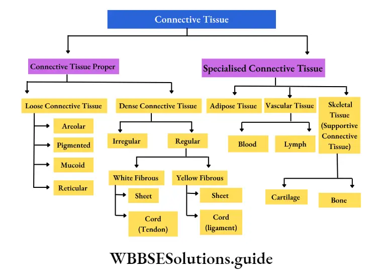
Connective Tissue Proper Definition: Connective tissue proper is a fibrous tissue consisting of cells, fibers, and intercellular ground substance or matrix which constitute the framework of many organs.
Connective tissue proper is characterized by a soft matrix.
It is of two types—
Loose connective tissue and dense connective tissue.
Loose connective tissue: The type of connective tissue that has less loosely arranged fibers and soft, gel-like ground substance or matrix is called loose connective tissue.
It may again be divided into the following types—
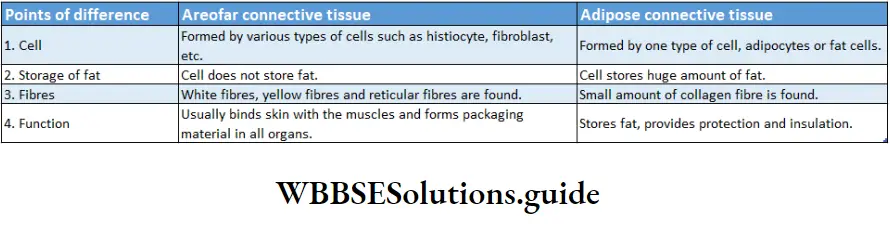
Areolar connective tissue: The white, translucent, loose connective tissue which consists of many connective tissue cells in between the meshwork of fibers in its matrix is called areolar connective tissue.
Areolar connective Position: It is the most abundant type of connective tissue and is present surrounding the nerves and blood vessels, between muscular and nervous tissues, beneath the skin.
Areolar connective Structure: It is a soft, transparent tissue consisting of numerous spaces or cavities.
“difference between epithelial, connective, muscular, and nervous tissue”
It contains different types of cells like—fibroblasts, macrophages, mast cells, plasma cells, pigment cells, and some fat cells. It contains white collagenous and yellow elastin fibers.
The spaces between the fibers are called areolae, hence called areolar connective tissue. Cells and fibers remain embedded within the matrix.
Areolar connective Functions:
- It serves as the binding material as it helps in binding various structures of the body like skin with the muscles, etc.
- It is present around the blood vessels and hence, holds them in position.
- Different types of cells present in it perform different types of functions which are already mentioned earlier.
- For example, macrophages and plasma cells, present within this tissue, play a defensive role against infection, fibroblasts play an important role in fibrosis (formation of excess fibrous connective tissue in an organ or tissue) during tissue repair, etc.
Pigmented connective tissue: The loose connective tissue which is similar to areolar tissue and consists of a large number of pigment cells called chromatophores is called pigmented connective tissue.
Pigmented connective tissue Position: It is present in the dermis of the skin, iris, and choroid of the eye, etc.
Pigmented connective tissue Structure: The cells are irregular in shape. Pigment cells are of two types—melanocytes or melanophores and lipophores.
The melanocytes are branched and synthesize melanin and lipophores synthesize lipochrome.
Pigmented connective tissue Functions:
- The pigments absorb harmful solar radiations, hence protect the skin,
- Pigments present in the choroid prevent internal reflection of light in the eye, etc.
Mucoid connective tissue: The jelly-like connective tissue that contains an excess matrix, containing hyaluronic acid, and very less fibers is called the mucoid connective tissue.
Mucoid connective tissue Position: Mucous connective tissue forms the main component of the umbilical cord. This tissue is commonly called Wharton’s jelly. It is present in the pulp of milk teeth. It is present in the vitreous humor of the eye.
Mucoid connective tissue Structure: It is composed mainly of a ground substance with few cells and fibers. Cells present are mostly large fibroblasts. A few macrophages and lymphocytes are also found. The matrix is largely made up of mucopolysaccharides (hyaluronic acid and chondroitin sulfate).
Mucoid connective tissue Functions:
- It provides insulation and protection to the umbilical cord. It also generates new cells that help to form the internal structure of the umbilical cord,
- It regulates the internal pressure of the eye.
Reticular connective tissue: The fibrous connective tissue in which fibers are anastomosing (attached) and arranged in an irregular manner forming a network, is called reticular fibrous connective tissue.
Reticular connective tissue Position: It is present in lymph nodes, liver, spleen, thymus, and bone marrow.
Reticular connective tissue Structure: This tissue comprises star-shaped reticular cells called stellate cells. The cytoplasmic processes of the stellate cells form a network. The fibers are fine, and branched, forming a network made up of protein, reticulin. The ground substance, the matrix, fills up the intercellular spaces.
Reticular connective tissue Functions: It provides structural support to organs like the liver, lymph nodes, etc. Reticular cells play a defensive role due to their phagocytic nature.
Dense connective tissue. The type of connective tissue that has a greater number of fibers and is mainly responsible for providing mechanical strength is called dense connective tissue.
It is of two types—
- Dense irregular connective tissue, containing bundles of fibers arranged irregularly.
- Dense regular connective tissue, containing bundles of fibers arranged parallel or along the same plane. Dense regular connective tissue can be of the following types
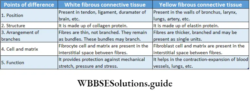
White fibrous connective tissue: The dense, regular, fibrous connective tissue, primarily made up of white collagenous fibers is known as white fibrous connective tissue.
White fibrous connective tissue Position: It is present in the tendons, ligaments, cornea of the eye, dura mater of the brain and spinal cord, etc. It is also present in the periosteum and perichondrium (of bone and cartilage respectively).
White fibrous connective tissue Structure:
- The tissue is tough and appears shiny. It is made up of more or less regular bundles of wavy white fibers.
- Fibers are made up of collagen protein.
- The bundles may be branched but the individual fibres are unbranched.
- Relatively few fibroblast cells and matrix are present in between the bundles.
White fibrous connective tissue Functions: It provides protection to various organs against mechanical stretch, pressure, and stress. The organs can withstand pressure due to the elastic properties of these fibers.
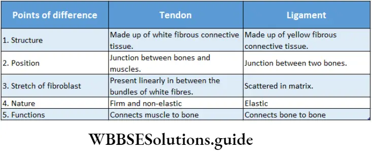
White fibrous connective tissue Types: Two types of white fibrous connective tissue are sheets and tendons. In Sheet form, the white fibrous bundles are arranged in a criss-cross manner. The fibroblasts are present in the areolae.
Sheets of white fibrous connective tissue form tough coverings. In the tendon, the bundles of white fibers run parallel to one another.
The fibroblasts are present in single rows between the bundles. Tendons connect skeletal muscles to bones.
Sheets of white fibrous connective tissue form tough coverings. In the tendon, the bundles of white fibers run parallel to one another. The fibroblasts are present in single rows between the bundles. Tendons connect skeletal muscles to bones.
Yellow fibrous connective tissue: The dense, regular, fibrous connective tissue almost entirely made up of yellow elastic fibers is known as yellow fibrous connective tissue.
Yellow fibrous connective tissue Position: It is present in the larynx, bronchi, lungs, arteries, ligaments, etc.
Yellow fibrous connective tissue Structure: The tissue is made up of a branched network of yellow elastic fibers. These fibers do not have wave-like structures. These fibers are made up of elastin protein.
Fibroblasts and matrix are present in between the elastic fibers. The yellow fibers join to form a reticular structure. The fibers are individually branched and are arranged linearly.
Yellow fibrous connective tissue Functions:
The tissue provides maximum elasticity and extensibility to the structures where it is present,
In the arteries, it helps to maintain normal circulation of blood and blood pressure due to their contraction-expansion property. In the lungs, it helps in the expansion and contraction of the lungs.
Yellow fibrous connective tissue Types: Two types of yellow fibrous connective tissue are found. They are—sheets and ligaments. Sheets are commonly found in yellow fibrous connective tissue.
“important points on structural organization in animals for exams”
These are found in various organs like walls of bronchi, bronchioles, lungs, etc. They help in undergoing stretching.
Ligaments appear as thick cords and fibroblasts are scattered. Ligaments connect bones to bones and help in the movement of the neck, fingers, etc.
Specialized connective tissue Definition: The tissue formed by special cells of the connective tissue that have undergone a transformation to carry out specific functions is called specialized connective tissue.
Specialized connective tissue is of three types—
- Adipose tissue,
- Vascular tissue and
- Skeletal tissue
Adipose tissue Definition: The specialized connective tissue that consists of abundant fat-storing cells, on a framework of loose fibers and the excess matrix is known as adipose tissue.
Position: It is found around the heart, folds of the peritoneum, the kidney, in orbits behind eyeballs, beneath the skin, mammary glands, and in the mesenteries (the structure that attaches the body wall to the organs) around the viscera.
Adipose tissue Structure: This type of connective tissue contains distended, oval, or round, flat cells called adipocytes. Adipocytes can be monolocular when fat is present in the center as a large globule or can be polylocular when fat is present as several fat globules.
The peripheral nucleus and cytoplasm form a signet ring-like structure due to the deposition of fat bodies at the center of the cell. White fibers are present as bundles. A vasculated thick matrix is present.
Adipose tissue Functions: It protects the internal organs from mechanical injury, and maintains the proper shape of organs. It mainly stores fat and prevents the loss of heat from the body.
Adipose tissue is called a special tissue because it performs the following special functions
Stores energy: In terms of storage of energy (in the form of triglyceride), adipose tissue plays the most important role.
The muscles and liver also help in storing energy (in the form of glycogen). Triglyceride has a more calorific value than glycogen.
Adipose tissue Insulator: The adipose tissue layer beneath the skin insulates the body (because fat is a bad conductor of heat). This property helps to maintain body temperature.
Structural component: Adipose tissue is an important structural component of the body.
In a normal male, adipose tissue constitutes about 15-20% of body weight.
While in a normal adult female, this value is 20-25% of body weight.
Shock absorber: A thick layer of adipose tissue is found beneath the skin. This deposition of adipose tissue acts as a shock absorber and protects the body from physiological damage.
Formation of organ system: This tissue fills the gaps between other tissues at the time of organ formation.
Formation of organ system Types: Based on the position, structure, color, and other features, adipose tissues are of two types— Unilocular adipose tissue and multilocular adipose tissue.
Vascular Tissue Definition: The type of mobile connective tissue that consists of a fiber-free fluid matrix and specialized living cells and is responsible for transport in the body, is called vascular connective tissue.
Vascular connective tissue is of two types—blood and lymph.
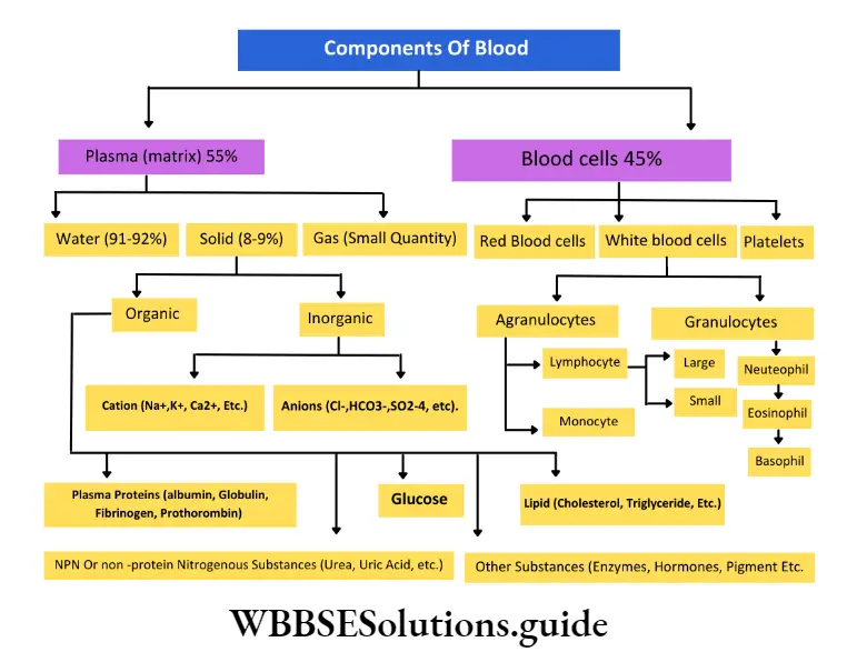
Vascular Tissue Blood: The red-colored, viscous, slightly alkaline, opaque, fluid connective tissue that flows through the heart and blood vessels is called blood.
Vascular Tissue Position: Blood is present within the vessels and chambers of the heart in vertebrates. In invertebrates, it is found in the members of phyla annelida, arthropoda, mollusca, etc.
Vascular Tissue Structure: It consists of blood cells suspended in a pale yellow fluid matrix called plasma.
The matrix is viscous and colloidal in nature and present in excess quantity.
Blood is mainly red in color due to the presence of hemoglobin protein (Haemoglobin is red when oxygenated and blue-red when deoxygenated).
On the other hand, the blood of crustaceans is blue due to the presence of a protein called hemocyanin (Haemocyanin is blue when oxygenated and colorless when deoxygenated). Insects have colorless blood since their blood lacks any pigment.
Vascular Tissue Functions:
- The blood maintains homeostasis within the body,
- It transports nutrients and oxygen throughout the body,
- It removes wastes from tissues,
- Certain cells (lymphocytes, monocytes, neutrophils, etc.) present in the blood provide immunity.
Vascular Tissue Components: Components of blood.
Lymph: The colorless fluid which is made up of plasma and white blood cells, flows through lymph vessels and ultimately drains into venous blood is called lymph.
Lymph Position: It is present mainly in the lymph vessels and also between the blood capillaries and tissues.
Lymph Structure: It is formed of two parts—Plasma—the matrix of lymph and is fluidity in nature.
It contains less proteins, calcium, and phosphorous but more glucose than blood. Leucocytes—derived from either lymph nodes or blood capillaries by the process of diapedesis.
Lymph Functions: Lymph carries food and oxygen from the blood to the tissues and excretory products, hormones from the cells to the blood.
Skeletal connective tissue Definition: The connective tissue in which the matrix is dense, solid, and mineralized and which forms the endoskeleton of vertebrates is called skeletal connective tissue.
Skeletal connective tissue is divided into two types—
- Cartilage and
- Bones
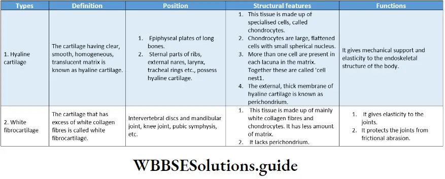
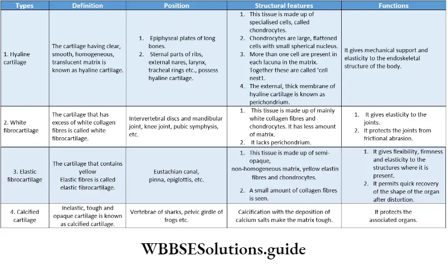
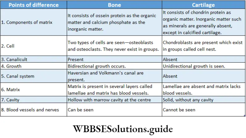
Skeletal connective tissue Cartilage: The firm, elastic, non-vascular skeletal connective tissue comprising cells and solid matrix is known as cartilage.
Skeletal connective tissue Position: It is found in the tip of the nose, external ear or pinna, tracheal rings, intervertebral discs, bony joints, endoskeleton of cartilaginous fish, etc.
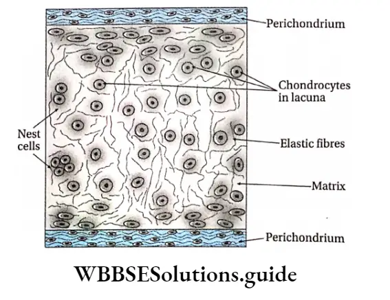
Skeletal connective tissue Structure: The cartilage is covered by a tough, dense layer of cells and fibrils, called the perichondrium. Cartilage is mainly composed of cells called chondrocytes.
The other components of cartilage are fibers and intercellular ground substance or matrix. The cells are large, oval, or round with small cytoplasmic processes and a round or oval nucleus.
The chondrocytes are released into the matrix from the perichondrium. They are present in groups of two or four and remain embedded in the lacunae of the matrix. Each group of cells is enclosed within a capsule.
The matrix is tough, transparent, and is called chondrin. It is made up of chondromucoid (complex formed by chondroitin sulfate and collagen protein) and chondroalbuminoid (complex formed by chondroitin sulfate and albumin protein).
The matrix also contains a very fine network of elastin and collagen fibers.
Skeletal connective tissue Functions:
- It provides mechanical support and elasticity to delicate structures of the body such as the knee, hips, etc.
- It also prevents mechanical erosion at the joints and the intervertebral discs.
- In cartilaginous fish, it forms the endoskeleton of the body.
Skeletal connective tissue Types: Based on the type of cells, matrix, and fibers, it is of four types.
These are described in the following table—
Bone: The hardest connective tissue comprising of i calcified matrix and which forms the skeleton of the body is known as bone.
Bone Position: Bones are found throughout the endoskeleton of the vertebrates.
Bone Structure: The bone is covered by a tough, fibrous sheath called the periosteum. It is composed of bone cells, collagenous fibers, and intercellular ground substance or matrix.
The cells are large, oval, or round with cytoplasmic processes. They are found in the lacunae of the matrix and have a round or oval nucleus. The matrix is made up of 40% organic and 60% inorganic substances.
The organic content of bones consists of a collagen-like protein called ossein, a mucoid derived from ossein protein called osseomucoid, and osteocollagenous fibers. Vitamins J A, D, and C are also present in the bones.
The inorganic content of the bones includes—
- Calcium salts such as calcium carbonate, calcium phosphate, calcium fluoride and
- Magnesium salts such as magnesium phosphate and a small amount of magnesium sulfate and hydroxides.
Bone Functions:
- Bones form the endoskeleton of vertebrates. They provide mechanical support to the structural framework of the body,
- They provide protection to delicate organs like the brain, heart, lungs, kidneys, etc.
- They act as a storehouse of minerals, mainly calcium and phosphorus,
- Bones are associated with muscles. The muscles act as a lever system for the movement of bones at the joints,
- They produce reticuloendothelial cells to protect the body from various diseases.
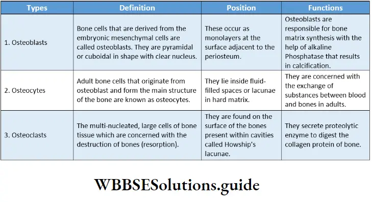
Types of bone cells: Based on their position, function, and structure, three types of bone cells are found.
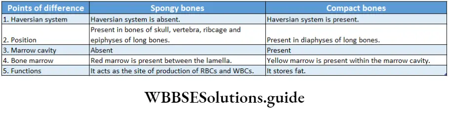
Types of bones: On the basis of density and compactness, bones are two types—compact bones and spongy bones.
The histological structure of compact bone has been discussed under a separate head later.
Histological structure of compact bones: Under the electron microscope, the following structures of a compact bone are seen.
Haversian system: The system formed by the numerous longitudinal canals, along with the Haversian lamellae, lacunae, canaliculi, and osteocytes, is called the Haversian system or Osteon of mammalian bones.
The haversian system is the structural and functional unit of compact bone. Its components are discussed below.
“structural organization in animals short notes for quick revision”
Haversian canal: Each Haversian system has a central canal or fine duct that runs lengthwise through the compact bone.
This canal is called the Haversian canal. It contains blood vessels, nerves, lymphatic vessels, and marrow cells.
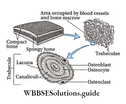
Lamellae: Each Haversian canal is surrounded by 6-18 concentric layers of the matrix. These concentric layers are called Haversian lamellae.
Lacunae: Between two consecutive lamellae as well as within the lamellae, small irregular fusiform (spindle-shaped) spaces are found, arranged in circles. These spaces are known as lacunae.
Canaliculi: Very fine wavy branched channels that radiate out from the periphery of lacunae are called canaliculi.
Osteocytes: Each lacuna has a branched bone cell or osteocyte. Its cytoplasmic processes or appendages extend through the canaliculi.
Cementing line of Ebner: The thin boundary of an osteon or Haversian system is called the cementing line of Ebner. The term was coined by Von Ebner.
Volkmann’s canals: In addition to the Haversian canal, another set of transversely (slanting) or horizontally arranged small canals are present in the periosteum.
These are called Volkmann’s canals. These canals contain blood vessels and nerves and help to interconnect the Haversian canals of adjacent Haversian systems.
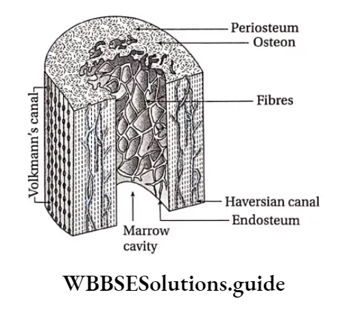
Ground or Interstitial lamellae: The lamellae found between the Haversian systems are known as the ground or interstitial lamellae.
Periosteal or Circumferential lamellae: The lamellae that are concentrically arranged below the periosteum and outside the endosteum are called periosteal or circumferential lamellae.
Endosteal lamellae: The lamellae that are present concentrically around the endosteum are known as endosteal lamellae.
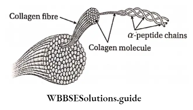
Collagen
It is the most abundant protein of connective tissue. Collagen contains amino acids such as glycine, proline, hydroxyproline, and arginine.
These amino acids are arranged in different patterns, forming the triple helix.
The most common arrangements in the amino acid sequence of collagen are glycine-proline-X and glycine-X-hydroxyproline, where X is any amino acid other than glycine, proline, or hydroxyproline.
Structural Organisation In Animals Notes
- Autonomic nervous system: Part of the nervous system; responsible for the regulation of involuntary physical processes within the body.
- Collagenous: Made up of collagen protein.
- Central nervous system: Part of the nervous system, comprising the brain and the spinal cord.
- Ectodermal: Originated from ectoderm (outermost germ layer).
- Oastln: Highly elastic protein found in connective tissue.
- Elaunin: Component of elastic fibers formed from deposition of elastin protein.
- Hyaluronic acid: An anionic, non-sulfur-containing mucopolysaccharide found in connective, epithelial, and neural tissues.
- Keratin: A type of protein found in nails, hair, etc.
- Lacinia and Galea: The Apex of each stipe has two lobes—the inner one is called Lacinia and the outer one is called Galea.
- Lacunae: Fluid-filled spaces within the matrix of connective tissue.
- Microvilli: Minute finger-like projections arising from the cell membrane, that increase the surface area.
- Mucopolysaccharides: Long chains of unbranched polysaccharides.
- Mucoproteins: A glycoprotein composed mainly of mucopolysaccharides.
- Olfactory mucosa: Epithelial tissue present in the upper region of the nasal cavity.
- Oxytalan: A type of fibers found in the dermis, tooth sockets, etc.
- Periplanetin: A type of peptide (a compound containing two or more amino acids) found in insects.
- Phagocytic: Cell eating.
- Phenotype: Characteristics of an organism that can be observed externally.
Pinocytic: Cell drinking. - Podical plates: Ventrolateral plates arising from the tenth abdominal segment of an insect.
- Proteoglycans: Compounds formed by binding proteins with mucopolysaccharides, found mainly in connective tissue.
- Proteolytic enzymes: Enzymes that bring about proteolysis (breakdown of proteins)
- Protopodite: Basal segments of a biramous limb or appendage.
- Pulsatile: Having periodic vibrations or pulses
- Regenerative medicine: It is a branch of medical science, concerned with the restoration of the structure and function of damaged tissues and providing solutions for organs that have become permanently damaged.
- Reticuloendothelial: Related to non-circulating and circulating phagocytic cells (macrophages and monocytes) associated with the immune response.
- Sclerotin: A type of protein found in the cuticle or tough outer covering of the insects.
- Sclerites: Chitin or calcium-containing plates that form the exoskeleton in insects.
- Segmental arteries: Arteries present in each segment of the body of an insect.
- Sloughed off: Allowed to fall off or degenerate.
“structural organization in animals notes PDF download”
Points of Remeber
- The bodies of all higher animals are made of four types of tissue—epithelial tissue, connective tissue, muscle tissue and nervous tissue.
- Epithelial tissue forms the outermost surface of the body. lt also forms the outer covering of the internal organs.
- This tissue is of two types—simple epithelium and compound or stratified epithelium. Besides, there is another type of epithelial tissue called the pseudostratified epithelial tissue.
- Fibroblasts are large, flat, and spindle-shaped cells that are found in areolar connective tissue.
- Histiocytes are irregular, large cells that are found in all areolar connective tissue.
- These cells are phagocytic in nature and engulf the microbes, thereby protecting the body. So, they are known as macrophages.
- Tough connective tissue containing calcium-rich matrix is called bone.
- Bone is formed of three types of cells—osteoblasts, osteoclasts, and osteocytes.
- All bones are covered by an outer layer of vasculature connective tissue. This outer layer is known as periosteum.
- Each layer of bone or the region between two layers of bone is occupied by small cavities. These are called lacunae.
- Thin, wavy tubules originate from each lacunae and are called canaliculi.
- The connective tissue which is elastic and softer than bone is called cartilage.
- The structure which is inelastic, composed of white fibers, and which connects the muscles with bones, is called a tendon.
- The connective tissue which is elastic, composed of yellow fibres, and connects bone with bone, is called ligament.
- White blood corpuscles can move like Amoeba and so can come out of blood vessels. This property of white blood cells is called diapedesis.
- The membrane surrounding muscular cells is called sarcolemma. The cytoplasm of muscle cells is called sarcoplasm.
- The endoplasmic reticulum in the cytoplasm of muscle cells is called sarcoplasmic reticulum.
- Four proteins of muscle cells are—actin, myosin, tropomyosin, and troponin.
- The numerous thin fibers that are arranged parallel in the sarcoplasm are called myofibrils.
- Myofibrils are made up of smaller fibers that are visible under a microscope. These are called myofilaments.
- The main components of nervous tissue are—nerve cells or neurons and neuroglia.
- Neuroglia divide by mitosis. Dead neurons are replaced by another type of supporting cell called neuroglia.
- The part of the central nervous system where myelinated axons are seen is called the white matter. Cell bodies are not located in this region. The white matter is located in the central part of the brain and the peripheral part of the spinal cord.
- The part of the central nervous system that contains cell bodies, dendrites, and the initial segment of the axon is called the grey matter. It is located in the periphery of the brain and the central part of the spinal cord.
- The island-like structures formed by clusters of nerve cell bodies, scattered in the white matter of the central nervous system are called nuclei.
- The cell body of numerous neurons of the central nervous system forms a cluster that is covered by a membrane to form a structure outside the CNS. The structure formed is called a ganglion.
- 30 species of cockroaches are found in human habitats. Among them, 4 species have been designated as pests.
- The most abundant cockroach in the world is the German cockroach. It is followed by the American cockroach.
- The head of the cockroach is hypognathous. It means that the head remains bent ventrally.
- The head bears a pair of antennae, compound eyes, and fenestrae. The mouth is surrounded by movable mouth parts—one labrum, a pair of mandibles, a pair of maxillae, one labium, and a hypopharynx. The adductor and abductor muscles help in chewing.
- Thorax has three segments, each segment bears a pair of jointed appendages that help in locomotion.
- It also contains 1 pair of large wings and 1 pair of small wings. The large wings keep the small wings covered. The small pair of wings help in flying.
- The coelom of the cockroach is not covered by the peritoneum but is filled with a fluid called hemolymph. Hence, the coelom is called hemocoel.
- Respiration in cockroaches is carried out by hollow, branched tubules called trachea. The trachea opens outside the body with ten pairs of spiracles. Branches of the trachea are called tracheoles.
- Spiracles are covered by ring-like sclerite called peritreme.
- The colorless fluid in hemocoel that helps in circulation in cockroaches is called hemolymph.
- Haemolymph lacks respiratory pigment. The colorless granule present in hemolymph is called hemocyte. The cockroach has an open circulatory system.
- The heart of a cockroach is neurogenic. It has 13 chambers.
- Beneath the antennae, two accessory hearts are present in cockroaches that produce periodic vibrations. These are known as pulsatile hearts.
- The main excretory organ of cockroaches is the Malpighian tubule, the excretory substance is uric acid.
- The sense organs of cockroaches are compound eyes, bristles of antennae, etc.
- The endocrine glands of cockroaches are the intercerebral gland, prothoracic gland, corpora cardia, and corpora allata. These endocrine glands secrete hormones that control various physiological functions.
- A cockroach is a uricotelic animal.
- A cockroach undergoes 12 moultings to transform into a developed cockroach from a nymph.
- Cockroaches undergo incomplete metamorphosis.