Erythropoietin
It is a glycoprotein hormone which controls erythropoiesis. It is produced in the interstitial fibroblast cells closely associated with peritubular capillary and proximal convoluted tubule in the kidney (90%), liver and other organiser:
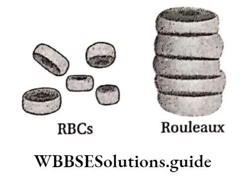
Rouleaux formation
Sometimes RBCs are stacked on each other and form a structure like stacked dinner plates. This condition is known as rouleaux formation.
These stacked structures can move easily through broad blood vessels but cannot move through narrow blood vessels, and sometimes become barriers in blood circulation.
erythropoietin definition
Site of rupture: After completing their lifespan, RBC rupture in the liver and spleen. The spleen is known as the graveyard of RBC.
Haemolysis and fate of RBCs: At the end of their lifespan, the plasma membrane of the RBC ruptures and releases their content including haemoglobin in the surrounding fluid. This process is known as haemolysis.
“WBCHSE class 11 biology notes for erythropoietin”
Functions of RBC: These are as follows—
Transportation of O2 and CO2: RBCs collect oxygen from the lungs and release it to the cells and tissues.
They take up carbon dioxide from the cells and tissues and release it to the lungs.
| Class 11 Biology | Class 11 Chemistry |
| Class 11 Chemistry | Class 11 Physics |
| Class 11 Biology MCQs | Class 11 Physics MCQs |
| Class 11 Biology | Class 11 Physics Notes |
erythropoietin meaning
Haemoglobin found in the red blood cells helps in the transport of oxygen and carbon dioxide between the lungs and the cells.
Regulation of acid-base balance: Haemoglobin present in RBC acts as a buffer and regulates the carbonate ion concentration.
Regulation of viscosity of blood: RBCs help to regulate the viscosity of the blood.
Maintenance of ionic balance: In the RBCs, carbon dioxide either binds to haemoglobin or reacts with water.
In reaction with water, it produces carbonic acid, which when dissociates, produces hydrogen ions. The deoxygenated haemoglobin is a stronger base and is capable of taking up these hydrogen ions. Hence, they are buffered by haemoglobin.
Other functions: RBCs carry the blood group antigens and Rh factor.
Erythrocyte sedimentation rate (ESR) The erythrocyte sedimentation rate (ESR) is the rate at which red blood cells settle down in a tube containing blood under standardized conditions and is a measure of inflammation or abnormal proteins in the body.
ESR commonly increases with any condition that gives rise to inflammation, such as infection, arthritis, or cancer.
“detailed notes on erythropoietin and its function WBCHSE”
It plays a very important role in the detection of any disease. According to the Wintrobe method, the normal ESR value is 0-9mm/h for males and 0-20mm/h for female
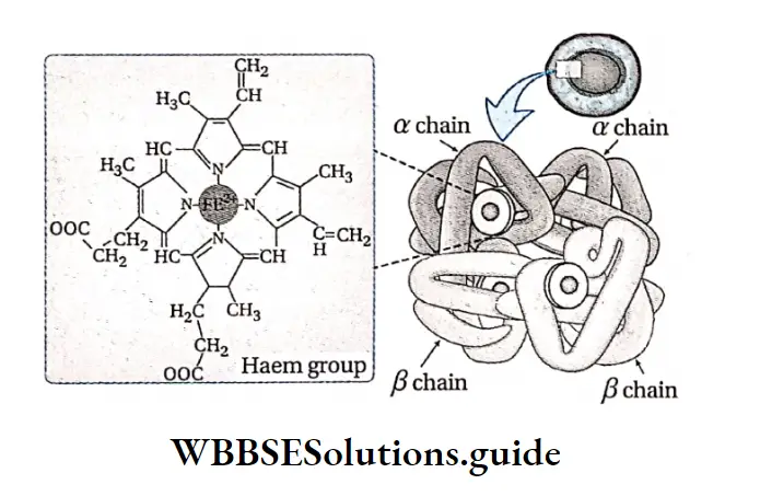
Haemoglobin: The red, oxygen-carrying, iron-containing pigment, comprising of a haem group and globin protein, that is found in the red blood cells of vertebrates is called haemoglobin.
Structure of haemoglobin: Haemoglobin (Hb) is a molecule made up of four subunits. Each subunit contains a haem moiety conjugated to a polypeptide. There are two pairs of polypeptides in each haemoglobin molecule.
Haem is an iron-containing porphyrin derivative. The polypeptides are collectively referred to as globin.
“role of erythropoietin in red blood cell production”
Instruments for measuring haemoglobin
The concentration of haemoglobin can be measured by using different instruments and methods. This includes Helden’s haemoglobinometer and Gauhar’s haemoglobinometer. Nowadays, a spectrophotometer is used to measure the concentration of haemoglobin.
RBC ghost
After hypotonic haemolysis of RBCs, all the cytoplasm along with the haemoglobin comes out from the cells. If these cells are observed under a microscope, then a spectrin-rich endoskeleton is found in these cells. These cells are known as RBC ghosts.
Location: Haemoglobin is found in the RBCs of human beings and other vertebrates.
Types: Normally there are two types of haemoglobin found in human beings, these include
HbA and HbF.
HbA: HbA (Adult haemoglobin) is found in adult human beings. It has 2 alpha and 2 beta polypeptide chains and comprises 97% of normal adult haemoglobin.
HbA is again of two types: HbA2 and HbA2 The globin portion of HbA-! consists of four polypeptide chains—2ar chains and 2/3 chains.
Each chain contains 141 amino acid molecules. Each chain contains 146 amino acid molecules.
Thus, HbA is designated as α2δ2. Not all the haemoglobin in the blood of normal adults is HbA HbA2 consists of two a and two 6 chains.
Thus, HbA2 is designated as α2δ2. The β chains also contain 146 amino acid molecules, but 10 individual residues differ from those in the chains.
hormone erythropoietin
About 98% of the total haemoglobin in adults is HbAi and 2-2.5% is HbA2.
HbF: HbF is normal foetal haemoglobin. It has 2 alpha and 2 gamma polypeptide chains mainly found in the embryonic stage.
“mechanism of erythropoiesis regulated by erythropoietin”
Variants of normal haemoglobin are formed when there are genetic changes in the globin genes.
Amount: The amount of haemoglobin present in 100 ml of blood is 14-17g in healthy adult males and 12-16g in healthy adult females.
Chemical nature: Haemoglobin is formed of iron-containing pigment haem (4%) and globin protein (96%). Haem consists of Fe2+ or ferrous ions in the centre of heterocyclic organic compounds, called porphyrins.
The molecular weight of haemoglobin is 68000 Da.
Fate of haemoglobin: Old RBCs are destroyed by the macrophages. Breakdown of haemoglobin occurs by the action of the enzyme haem oxygenase.
This generates haem and globin molecules. The haem is then converted to bilirubin and biliverdin. These pigments get released in the bile. The iron group of the haem portion is reused in the formation of haemoglobin.
Functions of haemoglobin: The functions of haemoglobin are discussed below.
“importance of erythropoietin in blood formation class 11 WBCHSE”
Helps to transport respiratory gases: During respiration in vertebrates and in higher animals, the inhaled oxygen combines with haemoglobin to form oxyhaemoglobin. This oxyhaemoglobin moves through blood capillaries and reaches the tissues.
Similarly, the carbon dioxide produced in the tissues combines with haemoglobin and form carbaminohaemoglobin, which reaches the alveoli of the lungs through the blood capillaries.
In this way, haemoglobin helps in the transportation of oxygen and carbon dioxide.
Maintains the acid-base balance in blood: Haemoglobin acts as a buffer and maintains the acid-base balance of the body.
Produces pigment materials: The haemoglobin found in dead RBCs forms pigments such as biliverdin, bilirubin, sercobilinogen, etc.
These give colour to the faeces. Haemoglobin also produces the pigment urochrome, which gives a straw colour to the urine.
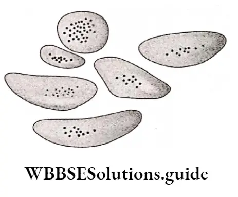
White Blood Corpuscle or Leucocyte or WBC
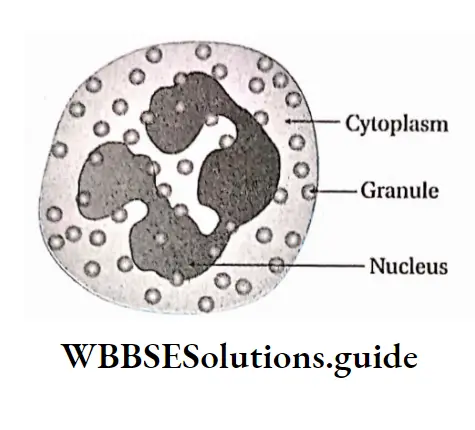
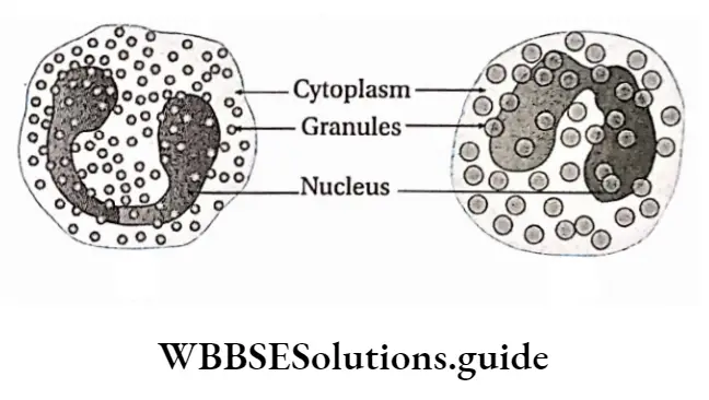
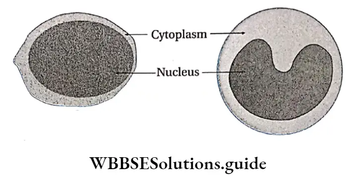
White Blood Corpuscle or Leucocyte or WBC Definition: White blood cells (WBCs) are colourless nucleated blood cells which lack haemoglobin and participate in reactions against invading pathogens or foreign particles in the body.
Structural characteristics:
- WBC do not contain any pigment so, they are colourless.
- They contain cytoplasm and a persistent nucleus. The cytoplasm of WBC can be granular or smooth (agranular). The nucleus can be of different shapes, such as spherical, kidney-shaped, etc.
- All the white blood cells are capable of amoeboid movement.
- WBCs are rich in nucleoprotein. Their cytoplasm consists of lipids, glycogen, cholesterol, vitamin C and different enzymes.
- Mitochondria, Golgi apparatus and other organelles are present in the cytoplasm of WBCs.
Size: WBCs do not have a fixed diameter. Their average diameter is 8-18 pm.
Lifespan: The average lifespan of WBC is 1-15 days.
Number: The total WBC count is normally about 4,000 – 11,000 per mm3 of blood.
Origin of WBC: WBC originates in bone marrow, spleen and lymph nodes. White blood cells are produced from pluripotent haemopoietic stem cells—myeloblast, lymphoblast and monoblast.
Types: Depending on the presence and absence of granules in the cytoplasm, WBCs are categorised into two main groups— Granulocytes (including neutrophils, eosinophils and basophils) and Agranulocytes (including lymphocytes and monocytes).
This classification depends on whether granules can be distinguished in their cytoplasm using a light microscope and conventional staining methods. WBCs can also be divided into two broad groups—the phagocytes and the immunocytes.
“how erythropoietin is produced and its effects on the body”
Granulocytes, which include three types of cells— neutrophils, eosinophils and basophils, together with monocytes comprise the phagocytes.
The lymphocytes, their precursor cells and plasma cells make up the immunocyte population. The types of WBCs (granulocytes and agranulocytes) are discussed under separate heads.
Granulocytes: The white blood cells that contain granules in their cytoplasm are known as granulocytes.
According to the characteristics of granules, granulocytes are categorised into three types— neutrophil, eosinophil and basophil.
The granulocytes have lobed nuclei which have varying shapes from cell to cell. Hence granulocytes are also known as polymorphonuclear leucocytes.
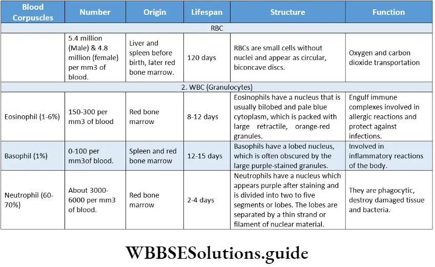
Neutrophil: The granular WBCs which can be stained with neutral stain are called neutrophils.
Structural features:
The cytoplasm of the neutrophils consists of numerous granules and they can be stained with neutral stain. They get stained with both basic (methylene blue) and acidic (eosin) dyes. Hence, they appear purple in colour.
- The nucleus appears purple due to staining and is divided into two to five segments or lobes.
- The lobes are divided into two to five segments or lobes.
- The lobes are connected by a thin strand or filament of nuclear material,
Neutrophils have 3 types of granules—
- Azurophilic or primary granules,
- Specific or secondary granules,
- Gelatinase-containing granules or tertiary granules.
Diameter: 10-12 pm
Lifespan: A few hours to a few days (2-4 days).
Number: Neutrophils are the most abundant type of WBCs in our body. They make up 50-70% of the total amount of WBCs. There are about 3000-6000 neutrophils present per mm3 of blood.
Functions: Neutrophils act as the first line of defence against pathogens. They are motile and phagocytic (destroy damaged tissues). They contain enzymes like protease, elastase, metalloproteinase, NADPH oxidase and antibody-like substances, defensins.
These antimicrobial peptides are active against bacteria and fungi. They secrete Platelet Aggregation Factor (PAF) which accelerates the aggregation of platelets at the injured site of blood vessels.
Reasons for the increase in the neutrophil count
The following reasons are responsible for the increase in the neutrophil count—
- Infection,
- Destruction of cells and tissues due to burns, myocardial infarction, etc., © leukaemia,
- Excess smoking, taking contraceptive pills, etc.
Eosinophil: The granulated WBCs where the granules can be stained with acidic dye (eosin) are known as eosinophils.
- Structural features:
- Usually, eosinophils have a bilobed nucleus. The cytoplasm is packed with large, coarse, retractile, orange-red granules containing histamine. The granules are referred to as eosinophilic because they take up the acidic dye, eosin.
- Eosinophils are produced in the bone marrow and circulate in the bloodstream for about 6 hours before migrating to the tissues.
- Diameter: 10-12 pm
- Lifespan: A few days (8-12 days).
- Number: There are 150-300 eosinophils per mm3 of blood. They constitute about 1-4% of the total WBCs.
- Functions:
- Eosinophils respond to chemotactic stimuli and are phagocytic. They can kill ingested organisms,
- Eosinophils also take part in allergic reactions. They also produce histaminase and aryl sulphatase B. These are the enzymes that inactivate two inflammatory agents released by mast cells,
- Eosinophils take part in dissolving blood clots in blood vessels.
Basophil: The granular WBCs where the granules are stained with basic dyes (methylene blue) are known as basophils.
- Structural features:
- The nucleus of basophil is 2-3 lobed. The cytoplasm has fewer number of coarse granules,
- The granules are called basophilic because they take up basic stains (such as methylene blue). Upon staining the granules take up bluish purple color against pale blue cytoplasm.
- Diameter: 8-10 pm.
- Lifespan: 12-15 days.
- Number: They are the rarest type of white blood cell, making up only 0.4% of the total white blood cells, found in a blood smear. 0-100 basophils are present per mm3 of blood.
- Functions: They have a role in allergic and inflammatory responses.
Basophil granules release some important substances such as—
- Proteases: Exaggerate inflammation,
- Hyaluronic acid: Necessary for deposition of ground substances in the basement membrane,
- Heparin: Prevents intravascular blood clotting,
- Histamine: Mediate’s acute hypersensitivity reaction—vascular changes, increased capillary permeability
- Serotonin: Acts as vasoconstrictor.
Agranulocytes: The white blood cells that do not have any granules in their cytoplasm are known as agranulocytes.
“short notes on erythropoietin for WBCHSE board exams”
Agranulocytes are mainly of two types—
- Monocytes and
- Lymphocytes.
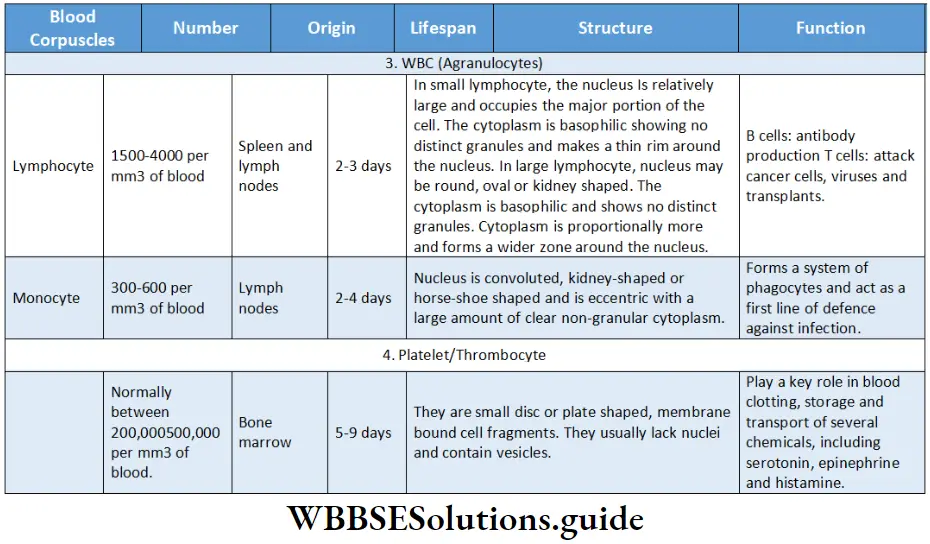
Monocyte: The large agranulated cells, capable of phagocytosis are known as monocytes.
- Structural features:
- Monocytes are amoeboid in appearance. The nucleus is round or oval-shaped when cells are young.
- But as they grow older, the nucleus becomes convoluted, kidney-shaped or horse-shoe-shaped.
- The cells have a large amount of dear non-granular cytoplasm, maybe, with vacuoles in it.
- These cells are mainly formed in the bone marrow.
- Diameter: About 16 – 18 pm in diameter.
- Lifespan: About 2-4 days.
- Number: 300-600 monocytes per mm3 of blood.
- Functions:
- Monocytes in the circulation are precursors of tissue macrophages that are actively phagocytic.
- Monocytes circulate in the blood for 1-3 days and then migrate to body tissues, where they transform into macrophages.
- They destroy dead cells and bacteria by the process of phagocytosis,
- They secrete some signalling proteins like interleukin 1 (IL-1), which increase the body temperature during infections and help in the production of T-lymphocytes.
“clinical significance of erythropoietin in anemia treatment”
Lymphocyte: The granulated white blood cells which produce antibodies are known as lymphocytes.
- Structural features:
- Lymphocytes form an essential component of the immune system and are derived from haemopoietic stem cells.
- On the basis of nuclear size, the lymphocytes are of two types. These are—
- Small lymphocytes: These are more common in normal blood. They have a large, dense, round nucleus which occupies the major portion of the cell.
- Large lymphocyte: The nucleus may be round and indented. The cytoplasm is basophilic and thicker than that of the small lymphocyte and forms a wider zone around the nucleus.
- Diameter: A small lymphocyte is slightly larger than a red blood cell and the diameter is about 7.5pm. The diameter of large lymphocytes is about 12 p.m.
- Lifespan: About 2-3 days.
- Number: These are the second most common WBC (25-35%), and are easy to find in blood smears 1500-4000 lymphocytes are present per mm3 of blood.
- Functions:
- They produce immunoglobulins or antibodies,
- Lymphocytes are mainly concerned with the production and transport of antibodies and they take an active part in the response to a wide range of antigens,
- They attack viruses, cancer cells, etc.
- Certain lymphocytes are part of the generalised response of the innate immune system, responding quickly to threats. Others act against specific pathogens or infected cells and are part of the adaptive immune response,
- Lymphocytes of the adaptive immune system are specific to a particular antigen—substances foreign to the body
General functions of WBCs
- Destruction of germs: The neutrophils and monocytes destroy the ingested pathogens and foreign materials by phagocytosis. They help to protect the body from harmful agents.
- Production of antibodies: The lymphocytes increase the defensive mechanism of the body by producing antibodies.
- Prevention of allergies: Eosinophils play an important role in allergic reactions by producing histamine.
- Secretion of heparin: Basophils prevent intravascular blood coagulation by secreting heparin.
- Trephone production: From plasma proteins, the monocytes produce a chemical substance, known as trephone. It provides nourishment to the tissues.
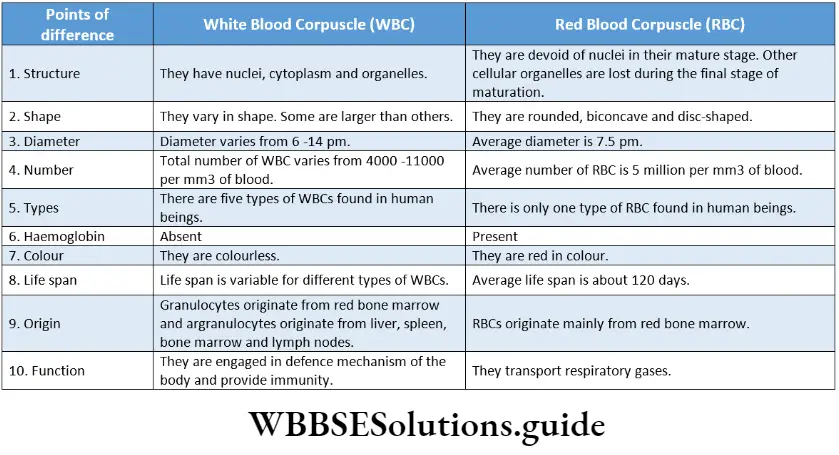
Thrombocyte Or Platelet
Definition: Platelets or thrombocytes are small, granulated and anucleated blood corpuscles, which are involved in blood clotting. (Greek. Thrombos= lump or clot and cyta= vessel; the word cute is derived from it and used to name these cells)
Platelets are membrane-bound fragments of cells that form when larger cells in the bone marrow break apart. Platelets do not contain nuclei and help in coagulation and clotting of blood at the wound.
Structural features:
- Platelets are actually not true cells but merely circulating fragments of cells.
- They are non-nucleated, round or oval, plate or disc-shaped structures, which is the reason behind their nomenclature.
- Platelets are formed from the terminal end of the pseudopodia of megakaryocytes (large bone marrow cells will lobed nuclei).
- They are surrounded by selectively permeable membranes.
- They appear somewhat smaller in the microscope. This is because their cytoplasm is divided into two zones—an outer hyalomere, which hardly stains, and an inner granulomere, which contains granules, that appear bluish in colour when stained.
Size: The mean diameter of platelets varies in different individuals, ranging from approximately 2-4 m.
Lifespan: These break down in the blood within 7 to 10 days after their formation.
Number: The platelet count in the circulating blood is normally between 2,00,000 and 5,00,000 per mm3 of blood. Newborn babies have a slightly lower level of platelets, which normally attain the adult range within three months from birth.
“difference between erythropoietin and other blood growth factors”
Functions: These are as follows—
Blood clotting and coagulation: Platelets play a key role in blood clotting, which prevents excessive blood loss after an injury.
Storage of important biomolecules: Platelets also act as the storage for several substances, including serotonin, epinephrine, histamine, and thromboxane and also help in their transportation.
Repairing of wounds: Upon activation, they release specific molecules which initiate constriction of local blood vessels, which facilitates clot formation.