Plant Kingdom – Classification and Characteristics Notes

Plant Kingdom Introduction To Play Kingdom

The kingdom Plantae or plant kingdom includes all eukaryotic chlorophyll-containing autotrophic organisms which are commonly called plants. Some of the general features of the plants are as follows

Habitat: This kingdom comprises the following groups—Algae, Bryophyta, Pteridophyta, Gymnosperms and Angiosperms. The algae thrive in aquatic habitats. Members of the Bryophyta and Pteridophyta include both aquatic and terrestrial members.

The gymnosperms and angiosperms are mostly terrestrial. However, they also include several aquatic species as well, for example, Hydrilla sp., Vallisneria sp., etc.

The aquatic species are found in freshwater, brackish water or seawater. The terrestrial members may live in different places such as mountains, deserts, marshlands, river basins, etc.

Read and Learn More: WBCHSE Notes for Class 11 Biology

Structural organisation: All the members of this kingdom are eukaryotic in nature. Many members of primitive groups, i.e., algae and bryophytes, have a thallus-like body structure. Such a body is not differentiated into roots, stems and leaves.

The body of the higher plants, i.e., pteridophytes, gymnosperms and angiosperms, are generally differentiated into true root, stem and leaves.

Nutrition: Most plants are autotrophic in nature, they can synthesise their own food. Some may be partially heterotrophic such as insectivorous plants or parasitic plants. Bladderwort and Venus flytrap are examples of insectivorous plants. Cuscuta sp. is an example of a parasitic plant.

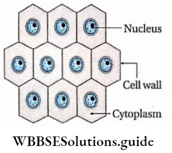
Cell covering and vacuoles: The plant cells have a eukaryotic structure with a prominent cell wall. The cell wall is mainly made up of cellulose. Each mature cell may have a large central vacuole.

Plastids: All plants have plastids in their cells. The green parts of the plant have chlorophyll-containing chloroplastids. The coloured parts have chromoplastids, that contain pigments other than chlorophyll. The parts of the plant that store food, contain leucoplastids, which are without any pigment.

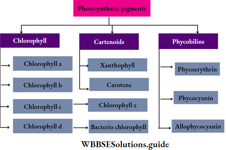
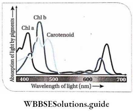
Pigments: Chlorophyll a is present in the chloroplasts of almost all plants. Chlorophyll b, c, and d are present in addition to Chlorophyll a, in different plant groups. Other than chlorophyll, carotenoids (orange), xanthophyll (yellow), phycocyanin (blue-green), phycoerythrin (red), etc., are also present.

Reserve food: Starch is the major reserve food in plants. Other than this, fat or oil droplets, some proteins and crystals of different salts are also found in plant cells as reserve food.

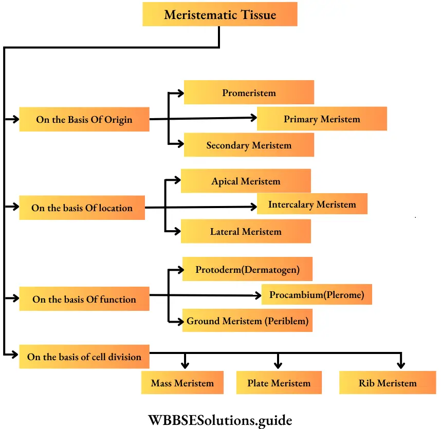
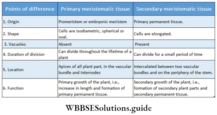
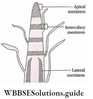
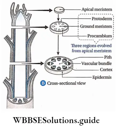
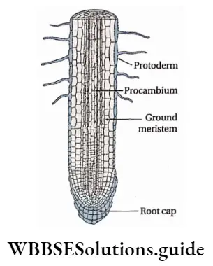
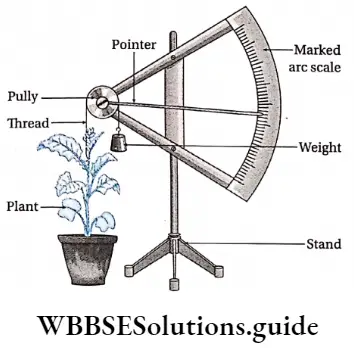
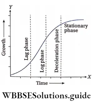
Growth: Except in some unicellular plants, all other plants show unregulated growth. Meristematic tissues are the tissues that form various plant parts and keep the plant growing. Some higher plants also show secondary growth besides primary growth.

Movement and locomotion: Only some unicellular green algae are able to show locomotion. The rest of the plants are static in one place and spread their branches from there. However, some plants (such as algae, bryophytes, pteridophytes and some gymnosperms) produce motile gametes (reproductive units or cells).

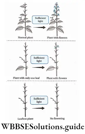
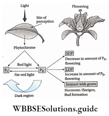
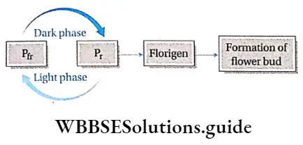
Response to stimuli: Despite the absence of a nervous system, plants respond to external stimuli. They produce several growth-regulating chemicals to respond to environmental stimuli. Movement or growth toward stimuli like gravity, light, water, etc., is known as tropism. Shoots tend to grow or move toward light source (phototropism) and roots grow towards gravity (geotropism).

The male gametes of bryophytes show chemotropism (stimulus for such movement is a specific biochemical substance) towards female gametes.

If the response to stimuli is independent of the direction of stimuli, then such response is called nastic movement. Insectivorous plant Venus flytrap rapidly closes its laminar halves when an insect sits on its leaf.

Presence of vascular tissues: Except for algae and bryophytes, all plants have vascular tissues to transport water and nutrients across the cells. In some bryophytes, there are special tissues, called hydroid and leptoid, that are similar to vascular tissues.

Reproduction: Reproduction in plants is of three types—vegetative, asexual and sexual. Plants reproduce asexually by different types of asexual reproductive units or spores. In angiosperms, vegetative reproduction occurs through underground stems, roots, leaves, etc.

During sexual reproduction, plants produce male and female gametes. These gametes may or may not be morphologically similar Union of gametes leads to the formation of zygotes which develop into new plants.

Alternation of generations and life cycle: Life cycle patterns of the members of the plant kingdom show two distinct phases—the diploid (2n) or sporophytic phase and the haploid (n) or gametophytic phase—that alternate with each other throughout the life cycle.

This phenomenon is called the alternation of generations. The duration of each phase, and their dependence on each other, vary, among different groups of plants.

Classification Of Plant Kingdom Into Major Groups

The history of plant classification is fascinating. To understand the concerns that affected the classification system let us consider classification within Angiosperms.

Classification is a logical system of organisation of several categories or taxa, each containing one or more organisms. Plants are also classified into different taxas by different authors. These classifications are categorised into three types—artificial, natural and phylogenetic.

Artificial system: It is the earliest system of classification of plants, which was based on one or a few morphological features such as habit, colour, number ar|d shape of leaves, floral characters, etc.

Theophrastus (370-285 B.C.), the father of botany,c|assjfjed a|| p|ants on the basis of form and texture. John Ray (1628-1705) broadly divided plants into two groups -Herbae, i.e., herbaceous and Arborae, i.e. woody. The Herbae were divided into two groups Imperfectae (non-flowering plants or cryptogams) and Perfectae (flowering herbaceous plants).

The group Arborae contains the flowering trees. He classified the group perfectly and airborne into monocots and dicots. Carolus Linnaeus (1936), also known as the father of taxonomy classified the then existing plants based on number, union and length of stamens.

Natural System: In this system, the plants are grouped on the basis of both external and internal features, like ultrastructure, anatomy, and embryology. Classification propounded by George Bentham and sePh Dalton Hooker is one of the most popular natural systems of classification.

This was published in the book Genera Plantarum (1862-1883). In Bentham and  Hooker’s system of classification, plants are grouped into two sub-kingdoms, Cryptogamia (non-flowing plants) and Phanerogamia (flowering plants).

Phylogenetic system: In this system, plants are classified on the basis of their course of evolution, and genetic and phylogenetic relationships. Some examples of a phylogenetic system of classifications are those propounded by A. W. Eichler (1875), A. Engler and Karl A. E. Prantl (1936), John Hutchinson (1959), etc.

In some modern phylogenetic systems of classification information available from various fields of study, such as palaeobotany, biochemistry, cytology, molecular biology, anatomy, etc., are used along with morphological features of the plants.

Classifications propounded by A. Takhtajan (1980), Arthur Cronquist (1981) and Thorne (1983) are examples of modern phylogenetic systems.

The outline classification of plants which is widely accepted among the biologists is shown below in tabular form.

Plant Kingdom Plant kingdom

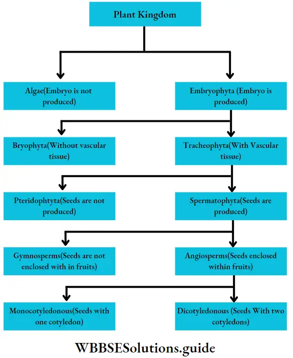
Each group mentioned in this classification has been discussed separately, later in this chapter.

Phaeophyceae (Brown algae)

Phaeophyceae Definition: Phaeophyceae (Greek: phaios= brown; phyton = plant) are the multicellular, mainly marine, brown-coloured group of algae.

This group of algae is commonly known as brown algae, as they appear golden brown in colour due to the presence of a xanthophyll pigment—fucoxanthin. The fucoxanthin partially masks the chlorophylls and carotenes. Thus gives the characteristic brown colouration.

It is a large group of algae consisting of 240 genera and over 1500 species. Among these 32 genera and 93 species have been reported from India.

Phaeophyceae Distribution: They are mostly marine in habitat and a few are freshwater. The freshwater members are Pleurocladia spv Heribaudiella sp., Lithoderma sp. and Sphacelaria sp. Pleurocladia lacustris grow both in freshwater and marine habitats. They are generally found attached to the marine substratum, in the colder regions of the sea.

Phaeophyceae General features:

  1. They range from simple microscopic (1 mm) heterotrichous filament (Ectocarpus sp.) to the largest alga (60-90 metres) (Macrocystis pyrifera).
  2. Giant brown algae are called kelps. The plant body is non-motile, multicellular and highly differentiated, both externally and internally. Their shapes may range from ribbon-like, filamentous, leaf-like, bush-like, fern-like or some may be branched like trees. Unicellular, colonial and unbranched filamentous forms are completely absent.
  3. The plant body is differentiated into holdfast, short or elongated stipe and an expanded leaf-blade or lamina. The lamina performs photosynthesis and bears reproductive structures. Many species can float due to the presence of air bladders.
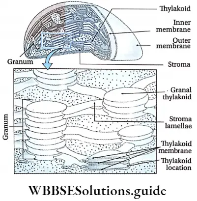
  4. The photosynthetic plastids present in Phaeophyceae are called phaeoplasts. The phaeoplasts contain three thylakoid-containing lamellae. The photosynthetic pigments include chlorophyll a, chlorophyll c, beta-carotene, luteins and xanthophylls. The pigment fucoxanthin gives it a distinct brown colour
  5. The growth pattern may be apical (Fucales, Dictyotales), intercalary (Laminariales) or trichothallic (Ectocarpales).
  6. The cell wall is differentiated into outer mucilaginous and inner cellulose layers. The outer layer contains succinic and alginic acids. The cell wall is covered by a layer of gelatinous hydrocolloid called algin. This protects the cell during unfavourable conditions.
  7. The cells usually have many small vesicles and white granules. The granules are called fucosan granules.
  8. Single, stalked pyrenoides are present.
  9. Motile zoospores and gametes have two laterally inserted unequal flagella. The larger one is tinsel or pantonematic and the smaller one is whiplash or acronematic type.
  10. The reserve foods are commonly laminarin and mannitol. Sucrose and glycerol are also present in some members.
  11. Reproduction takes place vegetatively (mainly by fragmentation and special propagules), asexually (asymmetrical flagella-containing zoospores or non-flagellate tetraspores) and sexually (isogamy, anisogamy and oogamy).
  12. In most of the members, fertilisation is external. The zygote does not undergo meiotic division and on germination, it develops into a diploid thallus.
  13. The life cycle is diplohaplontic.
  14. The alternation of generations is isomorphic or heteromorphic in nature.

Plant Kingdom Life cycle ofa brown alga

Embryophyta

Embryophyta Definition: The embryophytes are the most familiar sub-kingdom of green plants that form embryos from the zygote after fertilisation.

Embryophyta Definition General features:

  1. Embryophyta is mainly characterised by the presence of multicellular sporophytes, cuticles, archegonia, antheridia and sporangia, as well as the presence of sporopollenin in spore walls.
  2. They are a terrestrial species of plants. They produce embryos but do not produce accessory spores.
  3. The gametangia are covered by a jacket of sterile cells.

Embryophyta Definition Classification: According to the presence of conducting tissue, embryophyta can be divided into two groups—Bryophyta (without vascular tissue), and Tracheophyta (with vascular tissue). These two groups have been discussed under separate heads.

Tracheophyta Tracheobionta

The term tracheophyta came from two Greek words ‘trachea’ (vascular strand) and ‘phyton’ (plants).

Tracheophyta Definition: Tracheophyta are the major terrestrial plant group, with conducting tissues.

The members of this group are also called vascular plants. Vascular plants include the club mosses, ferns, gymnosperms and angiosperms.

Tracheophyta General features:

  1. The tracheophyta is the largest and most advanced group of plants.
  2. The sporophyte (2n) is the dominant generation in the life cycle of tracheophytes.
  3. The sporophyte is independent and autotrophic and possesses specialised vascular tissues.
  4. The plant body is differentiated into true roots, stems and leaves.
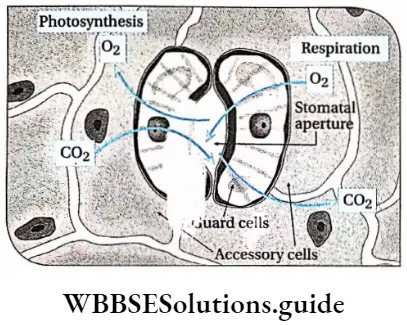
  5. Leaves are broad, containing a large amount of chlorophyllous tissues. Stomata are present on the leaves, for the exchange of gases.
  6. Reproduction is mostly vegetative and sexual. Formation of the embryo takes place.
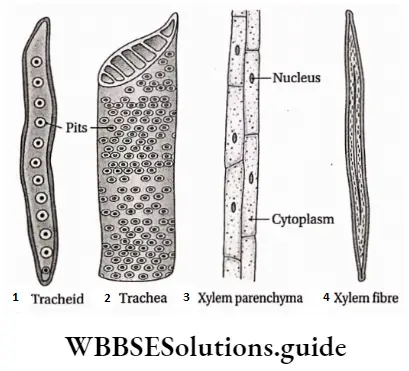
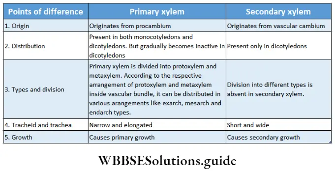
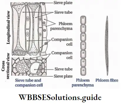
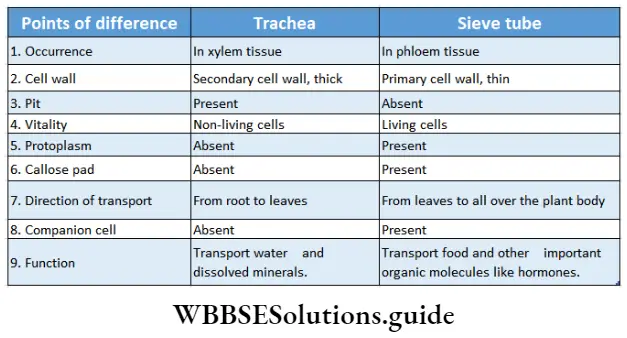
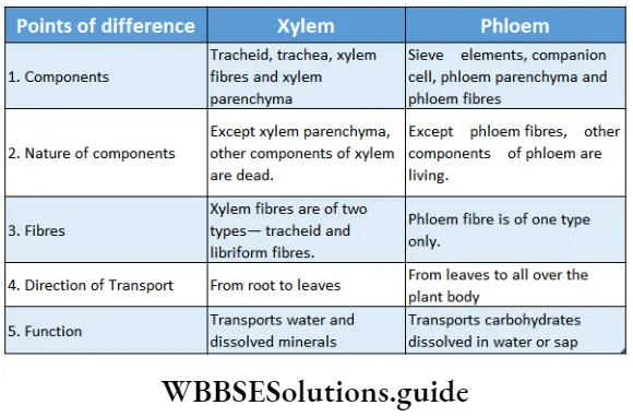
  7. They contain vascular or conductive tissues—xylem and phloem. The xylem transports water and minerals from soil to leaves. The phloem transfers photosynthetic products from leaves to all other parts of the plant.
  8. Their size may range from a few millimetres to several hundred metres.
  9. Their life cycle extends from about some weeks to several years.

Classification of Tracheophyta:

Tracheophytes are classified into two groups—

  1. Pteridophyta (seeds are not produced) and
  2. Spermatophyta (seeds are produced). These have been discussed below under separate heads.
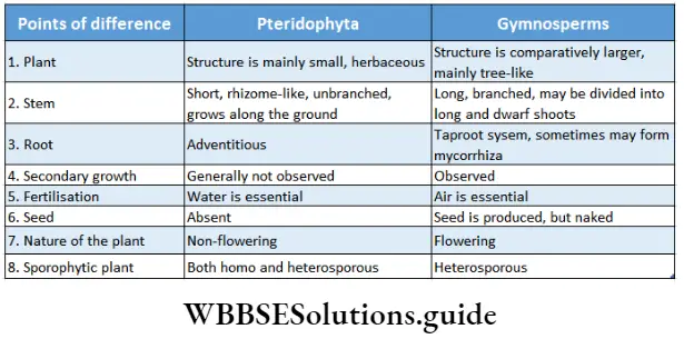
Pteridophyta

Pteridophytes (Greek: pteron-feather, phyton-plants) are non-flowering vascular plants. Hence they are also known as ‘vascular cryptogams’.

Pteridophytes Definition: Pteridophyta is the large group of primitive land plants, with true body differentiation and conducting tissues but does not produce seeds.

They are represented by about 400 genera and about 10,500 species including both living and fossil plants.

Pteridophytes Distribution: Most of the pteridophytes are terrestrial in nature. They are mainly found in damp, shady parts of mountains, damp walls, tree trunks, etc. Aquatic plants like Azolla sp. and parasitic plants like Polypodium sp. are also included in this group.

Pteridophytes Life cycle: Pteridophytes show mainly two types of life cycle—sporophytic and gametophytic.

Characteristics of sporophytes:

  1. Pteridophytes are the first true land plants. The predominant generation is the sporophytic plant body, differentiated into true root, stem and leaves. The sporophyte is independent and autotrophic.
  2. The stem is aerial or rhizomatous. The stem is generally branched—either dichotomous or monopodial. Stem sometimes is densely covered with brown scales.
  3. The primary root is short-living. The primary root is replaced by a dichotomously branched, adventitious root.
  4. Conducting tissues (xylem and phloem) are present. But tracheids and companion cells are absent in the xylem and phloem respectively.
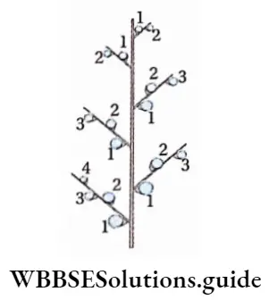
  5. Leaves are of two types— brown-coloured scale leaves and green-coloured sporophylls bearing sporangia, Sporophylls may be small microphyllous and large megaphyllous. The young leaf tips of ferns show circinate ptyxis (arrangement of immature leaf coiled form, which unfolds with maturity).
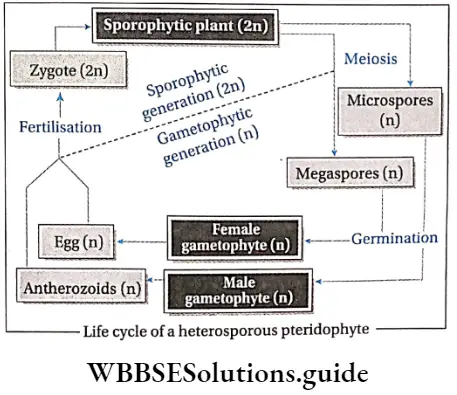
  6. Haploid (n) spores are produced within sporangia. In some species (for example Dryopteris) all the spores are similar in structure. They are called homosporous pteridophytes. In some species (for example Marsilea) spores are dimorphic (i.e., larger female spores or megaspores and smaller male spores or microspores). Such plants are known as heterosporous pteridophytes.
  7. In the case of heterosporous plants, megasporangia are borne on megasporophyll, while microsporangia are borne on microsporophyll.
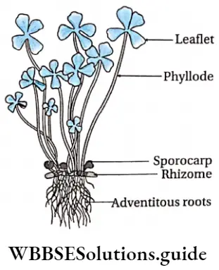
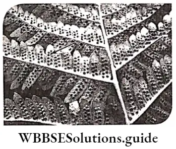
  8. Some pteridophytes bear a cluster of sporangia called sori (singular: sorus) on either the lower surface of leaves (example Dryopteris sp.) or inside special structures called sporocarps (example Marsilea sp.) or in spike(for example Ophioglossum sp.).
  9. The sori are covered by a bilayered, smooth membrane, called indusium.
  10. These spores germinate to form gametophytic plants.

Plant Kingdom Pteridophyte—Marsileasp

Plant Kingdom Sori ofDryopteris sp

Characteristics of gametophytes:

  1. The gametophytic plant is haploid, autotropic, independently living, dorsiventral, heart-shaped thallus. It is called prothallus.
  2. Prothalli are rather simple structures and do not have vascular tissues. They may be attached to the substrate by fine multicellular rhizoids. They resemble thallose liverworts.
  3. In the case of homosporous plants, the spores germinate to produce monoecious prothallus, which produces both antheridia and archegonia.
  4. In the case of heterosporous plants, antheridia are produced on male prothallus developed from microspores and archegonia are produced on female prothallus developed from megaspores.
  5.  Antheridia are small club-shaped structures, that produce motile sperms or antherozoids. They show chemotactic movement towards archegonia. Archegonia are flusk-shaped structures, with a cup-shaped venter and a long neck. Inside the venter an egg cell is present.
  6. The zygote is produced after fertilisation and quickly undergoes repeated mitotic divisions to form an embryo. The embryo develops the sporophytic plant.
  7. Flowers, fruits and seeds are not produced.

Some common examples of Pteridophytes are— Psilotum sp., Lycopodium sp., Selaginella sp., Isoetes sp., Pellia sp., Equisetum sp., Ophioglossum sp., Pteris sp., etc.

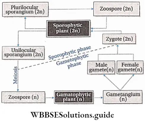
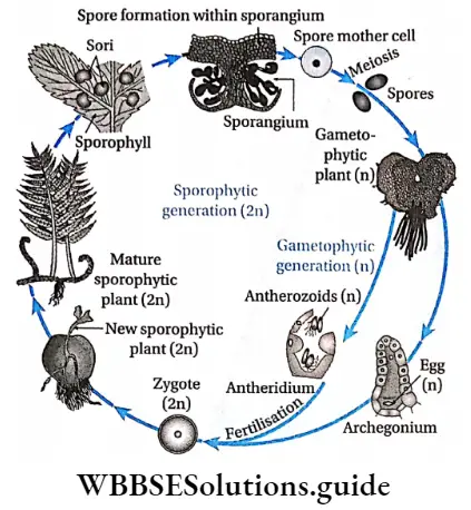
Alternation of generations: All pteridophytes show a diplohaplontic life cycle, heteromorphic alternation of generations.

Plant Kingdom Life cycle ofhomosporousfern

Plant Kingdom Life cycle ofheterosporousfern

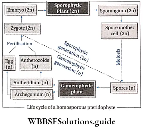
The alternation of generations in the life cycle of a typical pteridophyta (Dryopteris sp.) is shown in the figure below.

Plant Kingdom Alteration ofgenerations in life cycle ofDryopteris sp.

Commercial importance of pteridophyta: Many species of pteridophytes are used for various purposes.

Source of Food: The young leaves or fronds of Ampelopteris proliferate, leaf tips of Matteuccia struthiopteris and leaves of Diplazium esculentum, Marsiiea sp., are also used as vegetables.

Source of fodder: Dry fronds of many ferns are used as the livestock feed for cattle. The leaves of Marsiiea sp. are used as fodder for animals. The rhizome of many ferns such as Pteris sp., which is rich in starch, is used as animal food. The corm (modified stem) of Isoetes sp. is consumed as food by pigs, ducks and other animals.

Source of medicines: Lycopodium sp., Equisetum sp., Selaginella sp., Marsiiea sp., etc., are some pteridophytes that are used to treat neural disorders.

The spores of Lycopodium sp. have been widely used in pharmacies as a protective powder for tender skin and also as water-repellants. The foliages of Lycopodium sp. are used as tincture, powder, ointment and cream and used as a diuretic too.

Equisetum sp. (Horsetail) is rich in silicic acid and silicates. Potassium, aluminium and manganese, along with fifteen types of flavonoid compounds, have been reported from Equisetum.

The flavonoids and saponins are assumed to cause the diuretic effect. The silicon is believed to exert connective tissue-strengthening and anti-arthritic action.

Several ferns are used as herbal medicine. Oil extracted from Aspidium rhizome is used as a vermifuge (medicine against worms). The leaf extract of Polypodium is used to cure stomach and liver problems.

The root extract of Osmunda regatis is used for the treatment of jaundice. The root extract of Osmanda vulgaris is used to heal wounds.

Aesthetic uses: Many species of pteridophytes are cultivated for their aesthetic value. Some epiphytic species of Lycopodium sp. are grown on hanging baskets.

Several species of Selaginella are used in decoration during festive occasions due to their decent foliage and colour. Several ferns such as Angiopteris sp., Aspienium sp., Marattia sp., etc., have beautiful soral arrangements, hence used for ornamentation.

Polishing purpose: Accumulation of silica in the stem cells of Equisetum sp. gives it rough structures. Thus, it can be used as a scrubber for polishing purposes.

Preparation of fire-crackers: Since the spores of Lycopodium sp., are highly inflammable, hence they can be used to prepare fire-crackers.

Biofertilisers: Azolla sp., is a free-floating water fern which can grow very quickly through vegetative propagation. They live symbiotically with a nitrogen-fixing cyanobacteria—Anabaena sp., etc.

The alga provides nitrogen to the plant and to the growing aquatic medium. Thus, Azolla sp., growing in rice fields serves as a green manure for better crop yield.

Metal indicators: Equisetum sp., accumulates minerals, especially gold, in their stem. The rate of accumulation even reaches up to about 4.5 ounces per ton.

They may be referred to as gold indicator plants, which help in identifying a region with gold ore deposits. Similarly, Aspienium adulterinum is an indicator of nickel and Actinopteris australis is a cobalt indicator plant.

Fossil fuels (coal): During the carboniferous period, the flora of the earth was dominated by huge pteridophytes (Lycopods), ferns and other large leafy plants. The coal formed after their death had become sources of different fossil fuels.

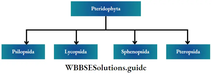
Classification of pteridophyta: Pteridophytes have been classified into four major classes by Doyle (1971)—Psilopsida, Lycopsida, Sphenopsida and Pteropsida.

Plant Kingdom Pteridophyta

Fossil fuels General features:

  1. They are mainly terrestrial pteridophytes, found in warm climates.
  2. The plant body is a rootless sporophyte that differentiates into a subterranean (just below the earth’s surface) rhizome and an aerial erect shoot.
  3. Branching is dichotomous in both rhizome and shoot.
  4. The large rhizoids borne on the rhizome, absorb water and nutrients from the soil.
  5. The aerial shoots bear spirally arranged scale-like (for example Psilotum sp.) or leaf-like appendages (for example Tmesipteris sp.).
  6. Stele is protostelic or siphonostelic with sclerenchymatous pith.
  7. Cambium is absent Hence, secondary growth is absent.
  8. Bi or trilocular sporangia are borne in the axils of leaf-like appendages.
  9. The mode of sporangial development is of eusporangiate (more than one cell initiates sporangial development)type.
  10. They are homosporous.
  11. The gametophytes or prothalli are independent, colourless, cylindrical, branched and subterranean. They grow as saprophytes with an associated endophytic fungus.
  12. Antherozoids are spirally coiled and multi-flagellated. Examples: Psilotum sp. (living); fossil members are- Rhynia sp., Cooksonia sp., Zosterophyllum sp etc.

Lycopsida (Lycopods)

Lycopsida General features:

  1. Thin and small structured pteridophyta, found mostly in forests and mountains.
  2. The sporophytic plant body is differentiated into definite root, stem and leaves.
  3. The sporophytes are dichotomously branched.
  4. The leaves are usually small and microphyllous. The leaves are either isophyllous or dimorphic. In some species, ligules (tongue-shaped membranous structures) may present towards the leaf apex.
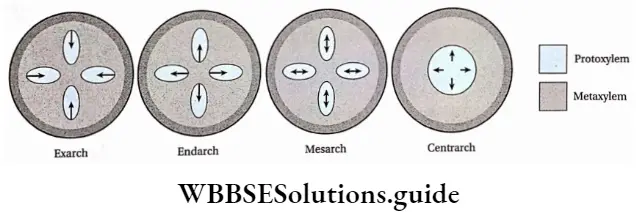
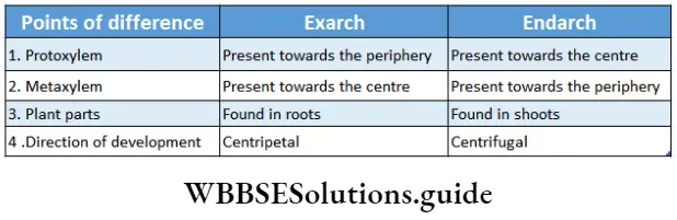
  5. The xylem arrangement in the stem is exarch.
  6. Stele may be protostele, siphonostele or polystele in nature
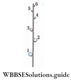
  7. Sporangia are borne singly on the adaxial (upper) surface of the sporophylls. In most cases, sporophylls join to form cones or strobili.
  8. The spores are homosporous (for example Lycopodium sp.) or heterosporous (for example Selaginella sp.).
  9. The spores develop into independent gametophytes.
  10. Antherozoids are flagellated.

Examples: Lycopodium sp. and Selaginella sp. are living genera and there are 14 extinct genera—Asteroxylon sp., Baragwanathia sp., etc.

Sphenopsida (Horsetails)

Sphenopsida General features:

  1. The living members of this group are found in damp and shady regions of mountains, forests and marshlands.
  2. The stems and branches are differentiated into nodes and internodes. Internodes are with longitudinal ridges and furrows.
  3. Branches arise in whorls.
  4. The leaves are extremely reduced and borne in whorls at the nodes of aerial branches and stems.
  5. Stele is Protostele or siphonostele in nature.
  6. The sporangia develop on an elliptical pallet appendage called sporangiophore. Sprorangiophores are arranged into a cone shaPed strobilus (P|ural: strobila)-
  7. Most of the members are homosporous including Equisetum sp. However, some extinct forms were heterosporous (for example Catamites Hashana).
  8. The gametophytes are exosporic (develop outside the spore) and green.
  9. Antherozoids are multiflagellate.

Examples: This class is represented by a single living genus and 18 extinct genera. The living genus is Equisetum sp. and the fossil genera are Catamites sp. Annularia sp., etc.

Pteropsida

This group is commonly known as ferns. The pteropsida differs from other classes in having large leaves (fronds). This is the largest and highly evolved group of pteridophytes and is represented by about 9,000 species which show a wide range of distribution. Pteropsida are known from as far back as the Devonian period of the Paleozoic Era.

Pteropsida General features:

  1. Members of Pteropsida are found in shady and damp forests, high altitudes of mountains, marshlands and moist soil of plains. Some members are aquatic and some are epiphytic.
  2. The sporophytes are usually perennial in nature and differentiated into roots, stems and spirally arranged leaves.
  3. The stem is rhizomatous, mostly short and stout. Adventitious roots grow from it.
  4. The rhizome is covered with brown scales.
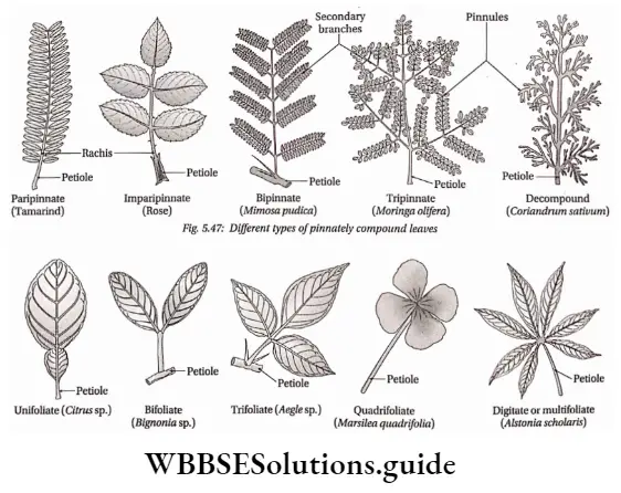
  5. The leaves are large, simple (Ophioglossum sp.) or pinnately compound (Dryopteris sp.) and are called fronds. In Adiantum sp., the rachis is dichotomously branched and bears a fan-shaped leaflet.
  6. The rachis is covered with tiny brown hairs called ramenta.
  7. Young fronds show deracinate ptyxis, except Ophioglossum sp.
  8. The stele can be of different types, for example protostele, siphonostele, etc.
  9. Vegetative reproduction fragmentation, adventitious buds and bulbils (fleshy globose structures). In Adiantum sp., new plants, are produced from leaf tips when they come in contact with the soil.
  10. Matured vegetative leaves bear sori on their lower surface. They are then known as sporophylls. Sori bear a number of sporangia. Sporangial clusters are sometimes enclosed by a shield-like structure called indusium.
  11. These plants are homosporous in nature. Haploid spores are formed through meiosis from diploid spore mother cells.
  12. Monoecious gametophytes or prothalli are produced by the germination of spores. Rhizoids present on prothallus.
  13. Antherozoids are ciliated and coiled.

Examples: Ophioglossum sp., Pteris sp., Dryopteris sp., etc.

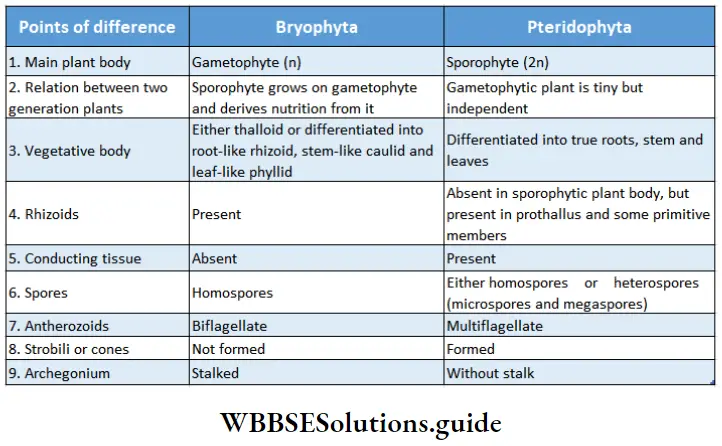
Similarities between bryophyte and Pteridophyta

  1. Both are non-flowering and terrestrial.
  2. Both have multicellular and multiflagellate reproductive organs.
  3. Both their antheridia and archegonia are similar in structure, with a layer of sterile cells covering them. Antheridia are club-shaped and archegonia are flask-shaped in both.
  4. Antherozoids are coiled and motile in both.
  5. Both have a definite alternation of generations.
  6. Asexual reproduction takes place by spores.
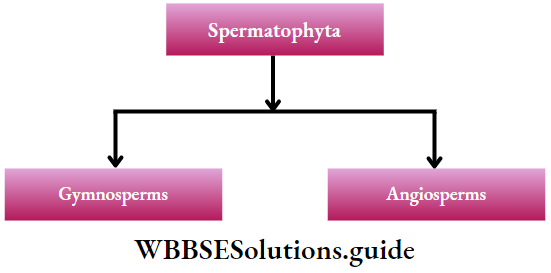
Plant Kingdom Differences between bryophyta and pteridphyta

Spermatophyta

Spermatophyta Definition: Spermatophyta is the higher group of flowering land plants where seeds are produced and embryo development takes place.

Spermatophyta Distribution: They are found all over the land and sometimes in water as well.

Spermatophyta General features:

  1. The body is differentiated into roots, stems and leaves. Vascular tissues (xylem and phloem) and mechanical tissues, both are present.
  2. The plants are heterosporous with microspores and megaspores.
  3. Gametophytic plants are dependent on sporophytic plants.
  4. The reproductive organs are multicellular. Instead of being released, the megaspores remain within the ovules where one or more female gametes are formed.

Classification of the Spermatophyta: The spermatophytes are further classified into two groups

Plant Kingdom Classification of the Spermatophyta

These topics are discussed under separate heads.
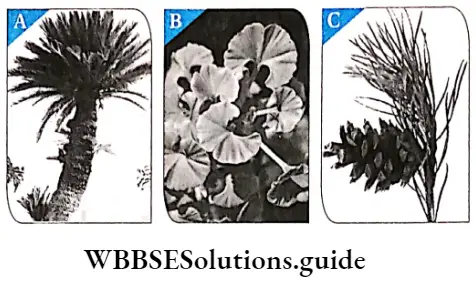
Cycadopsida

Members of the class Cycadopsida produced the most dominating flora during the mid-Mesozoic Era.

Cycadopsida General features:

The general features of Cycadopsida are as follows—

  1. Plants generally with stout trunks having manoxylic wood. They have large pith regions but have woody cortexes.
  2. Leaves are large and frond-like. They are basically pinnately compound in form or in venation.
  3. Annual rings are absent.
  4. Presence of ciliated sperms.
  5. These plants are usually dioecious with the reproductive organs borne on specialised leaves (sporophylls).
  6. The pollen grains (microspores) form within the microsporangia on the sporophylls of the male cones.
  7. The ovules (megaspores) develop without protective coverings on the sporophylls of the female cones.
  8. The pollination is carried out by wind or beetles.
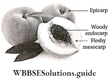
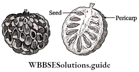
  9. After fertilisation, the seed develops without a protective pericarp. The seed is radially symmetrical. The seed has an outer fleshy layer and the embryo has two cotyledons.
  10. The life cycle is diplohaplontic, alternation of generations is heteromorphic. The life cycle of cycads has two distinct phases. The gametophyte stage is microscopic and enclosed within the microspores and megaspores.

Examples: Cycas sp., Ceratozamia sp., Zamia, etc.

Ginkgopsida

The class Ginkgopsida includes a single order of Ginkgoales—now represented by a single living member Ginkgo biloba. The members of this group first appeared in the Permian period, but gradually depleted from the earth.

The leaf impressions of G. biloba have been identified from the Permian rocks and probably from the Carboniferous rocks. G. biloba has escaped extinction and still exists today being the oldest living seed plant Thus, Ginkgo biloba is referred to as a ‘living fossil’.

Ginkgopsida General features:

The general features of Ginkgopsida are as follows—

  1. The members of this group are mostly tall, woody, deciduous and branched trees. They have both long and dwarf shoots.
  2. Secondary wood is pyroxylin i.e., has a large number of thick-walled tracheids and fibres.
  3. Leaves are leathery, strap-shaped or fan-shaped, often with dichotomous venation.
  4. These plants are dioecious, i.e., both male and female gametophytes are present on the same plant. Produce both male and female cones.
  5. Male fructifications are axillary, unbranched catkin-like bearing microsporangiophores.
  6. Each microsporangiophore bears 2-12 pendulous microsporangia.
  7. Ciliated sperms are present.
  8. Ovules are 2-10 in number, terminal on axillary branching or almost unbranched axes.
  9. Seeds are large showing radial symmetry.
  10. Seeds have with fleshy outer layer and a stony middle layer.

Examples: Ginkgo biloba

Coniferopsida

The Coniferopsida includes four orders—Cordaitales, Voltziales, Coniferales and Taxales. They probably appeared during the Upper Devonian and were the most dominating taxa during the Upper Carboniferous to Triassic. At present, this class is represented by 57 living genera.

Coniferopsida General features:

The general features of Coniferopsida are as follows—

  1. Mostly tall, woody, evergreen plants which are profusely branched.
  2. Secondary wood is pyroxylin.
  3. They have needle-shaped, paddle-shaped or fan-shaped foliage leaves. The arrangement of leaves is whorled or a|ternate. The )eaves are covered with a thick cuticle.
  4. Long and dwarf shoots are present.
  5. Stele in the stem are endarch, pith and cortex are small, with large conducting tissues. The woody part is more in these plants.
  6. Resin canals are often present in leaves and stems.
  7. The plants are usually dioecious. The reproductive organs are borne on specialised leaves (sporophylls), which are compactly arranged in cones or strobili.
  8. Male cones or microcstrobili are smaller and survive for less number of days. Female cones or megastrobili are large, and long and survive for longer periods of time.
  9. The gametophyte stage is represented by microspores or pollen.
  10. Male gametes are represented by the male nucleus only.
  11. The pollen forms within the microsporangia present on the scales (microsporophylls) of the male cones. They are often winged.
  12. The ovules are borne on the sporophylls of the female cones and the pollen is usually transferred to the ovules by wind.
  13. After fertilisation, the seed develops directly on the female sporophylls.
  14. Seeds are bilaterally symmetrical. The embryo has two too many cotyledons.

Examples: Pinus sp., Picea sp., Abies sp., Cedrus sp., etc.

Gnetopsida

The class Gnetopsida is considered to be the highly evolved group of gymnosperms. The class comprises three distinct orders—Ephedrales, Welwitschiales and Gnetales, with a monogeneric family in each. However, some scientists prefer to retain them within a single order, Gnetales, with a monogeneric family in each.

Gnetopsida General features:

The general features of Coniferopsida are as follows—

  1. The members of this group are small trees or shrubs or lianes in nature.
  2. The plants bear opposite or whorled leaves, with reticulate venation.
  3. Vessels are found in the secondary xylem. For that reason, they are considered to be ancestral to angiosperms.
  4. Resin canals are absent.
  5. Flowers are monoecious. Male and female cones are borne separately.
  6. Compound male and female strobili resemble the inflorescence of angiosperms.
  7. Male flowers consist of a stalk bearing two or more sporangia. They may bear bract and perianth.
  8. Female flowers consist of ovules only.
  9. They show unique fertilisation features i.e., a pollen tube grows in order to fertilize the egg. Thus, the sperms themselves are not motile, like other gymnosperms.
  10. The embryo contains two cotyledons.

Examples: Gnetum ula, Ephedra sp., Welwitschia sp., etc

Plant Kingdom Different types ofgymnosperm

Similarities between Pteridophyta and gymnosperms

  1. Both have a major sporophytic phase and a less important gametophytic phase.
  2. Both have sporophytic plant bodies, differentiated into roots, stems and leaves.
  3. Both have vascular tissues, without vessels in the xylem (exception: Gnetopsida) and companion cells in the phloem.
  4. Pteridophytes are heterosporous and have motile male gametes (reproductive cells) like many gymnosperms (for example Cycas sp.).
  5. Both show definite alternation of generation.
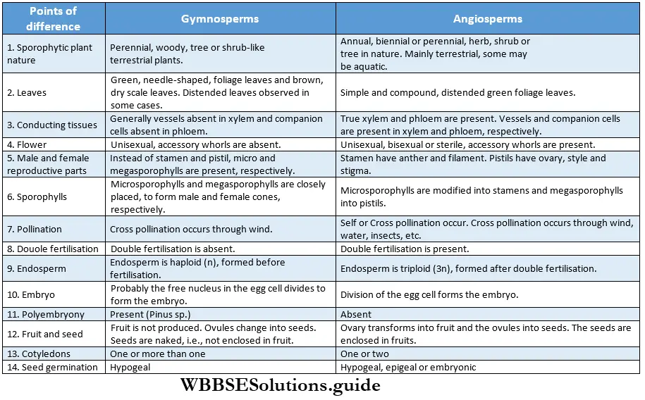
Plant Kingdom Differences between pteridophyta and gymnosperms

Angiosperms

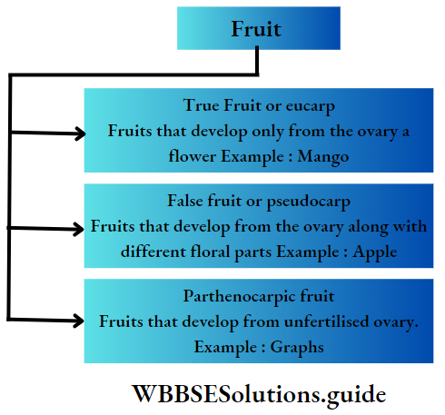
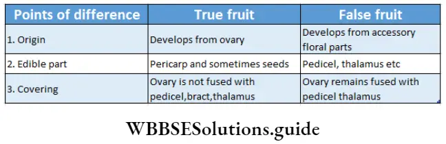
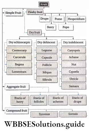
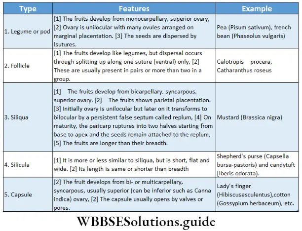
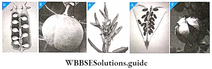
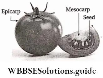
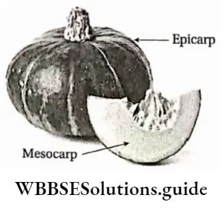
Angiosperms Definition: Angiosperms are flowering plants that produce fruits, that enclose the seeds.

Angiosperms are the highest-order plants in the plant kingdom. They are the largest group of plants, that are flowering and contain seeds in special structures, called fruits.

Angiosperms Distribution: Angiosperms originated quite late on earth, yet they occupy the majority of it. Most of them are terrestrial, and only a few are aquatic. They are found under the ocean, as well as at 6000m above sea level.

Angiosperms General features:

  1. The main plant body is sporophytic in nature. The sporophytic stage is independent and diploid (2n). The gametophytic phase is haploid (n) and dependent on the sporophytic phase.
  2. Angiosperms are trees, shrubs or herbs in nature. The trees and shrubs are generally perennials, while the herbs are annuals, biennials or perennials.
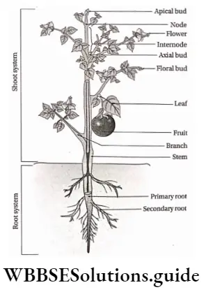
  3. An angiospermic plant consists of different organs—root, stem, branches, leaves, flowers, fruits and seeds.
  4. The body of an angiosperm plant consists of two systems— the root system and the shoot system.
  5. The roots may be taproot or adventitious.
  6. The stem may be branched or unbranched. In some cases, they may be climbers or lianes.
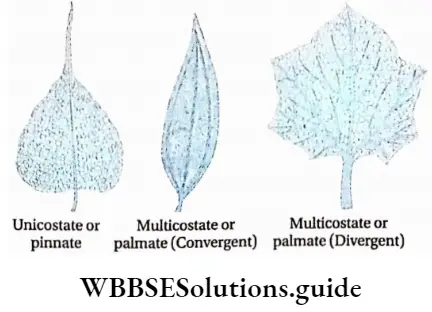
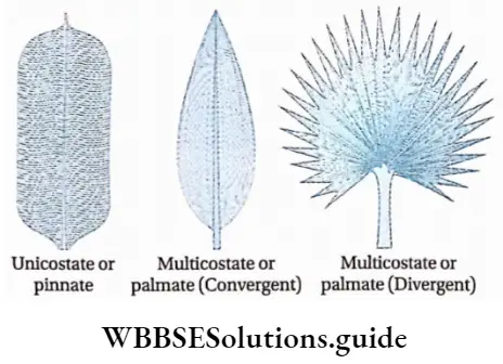
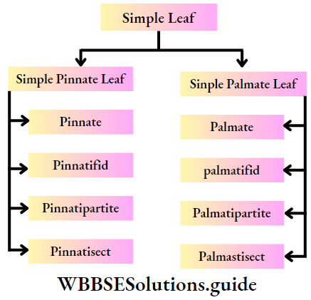
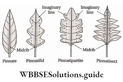
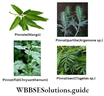
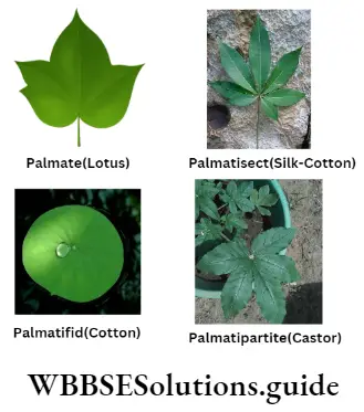
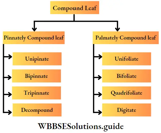
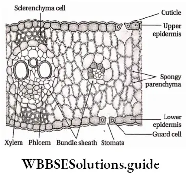
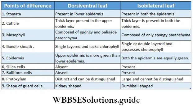
  7. The leaves are simple or compound, dorsiventral or isobilateral.
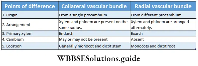
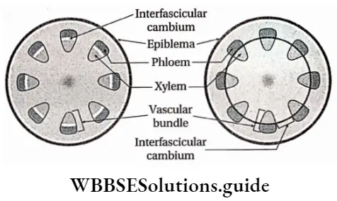
  8. The conducting tissues are the xylem and phloem. The vascular bundle of roots are radial and exarch, while that of the stem is collateral or collateral, free or enclosed and endarch. Secondary growth is observed in dicots.
  9. Angiospermic plants contain chlorophyll in their leaves and thus are able to photosynthesise.
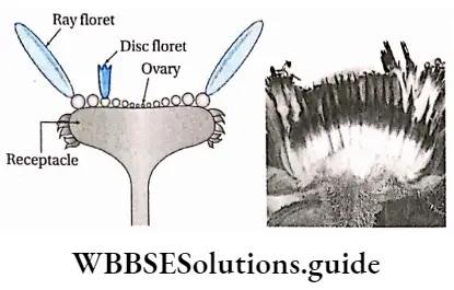
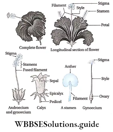
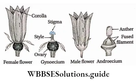
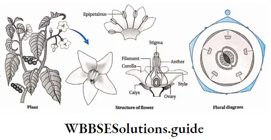
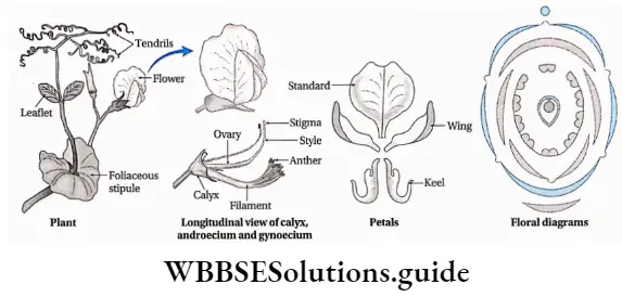
  10. Flowers may be complete (all four floral whorls are present), incomplete, bisexual, unisexual (male or female), or sterile in nature. They have accessory whorls (sepals and petals), along with reproductive whorls (androecium and gynoecium).
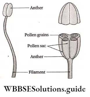
  11. Androecium has an anther and filament. The anther produces pollen grains.
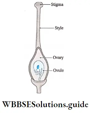
  12. The gynoecium has an ovary, style and stigma. One or more ovules are produced within the ovary.
  13. The male gametophytes form pollen tubes. They contain two nuclei.
  14. The archegonia are not produced within female gametophytes. Instead, the female gametophytes get reduced to four megaspores.
  15. Only one of the megaspores develops into an embryo sac which contains an egg nucleus. Double fertilisation, occurs in an egg angiosperms. Apart from a zygote, a triploid endosperm is also produced from this.
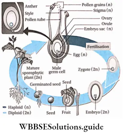
  16. After fertilisation, the ovule transforms into seed and the ovary into fruit.
  17. The zygote develops into an embryo by repeated cell division. After germination, it grows into an adult plant.

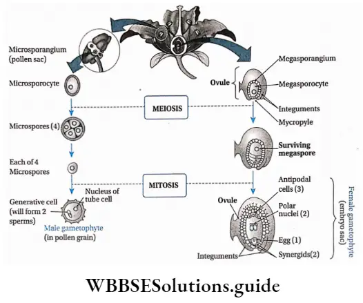
Sexual Reproduction in Flowering Plants: Various steps of the sexual reproductive procedure of flowering plants are discussed below.

Microsporogenesis: Microspore i.e., the pollen grain is the first cell of the male gametophyte, which contains only one haploid (n) nucleus. During the early stage of development, four microspores or pollens (n) are formed from each microsporocyte or pollen mother cell through meiosis.

Pollination: Transfer of pollen grains to the stigma takes place by several agents, such as wind, water, insects, etc. This process is called pollination. It takes place by two means—self-pollination (within the same flower or two different flowers of the same plant) and cross-pollination (between two different flowers of two plants).

Development of male gametophyte: Firstly, the pollen cell (n) undergoes mitotic division and forms a small generative cell and a large vegetative or tube cell. The generative cell further divides and gives rise to two male gametes. This is known as the 3-celled stage containing a vegetative cell and two male gametes.

The division of the generative cell may take place either in the pollen grain or in the pollen tube. The pollen tube comes out through germ pores after pollination. The nucleus of the vegetative cell, commonly known as the tube nucleus, usually degenerates with the maturation of the generative cell.

Plant Kingdom Development ofmale andfemale gametophyte in angiosperms

Development of megaspore mother cell: The ovule, inside the ovary, develops as a multicellular placental outgrowth including the epidermal and numerous hypodermal cells. This structure develops into a mass of tissue called nucellus, with one or two integuments.

One of the hypodermal (just below the outermost layer) cells of the nucellus enlarges in size and has a dense cytoplasm and a conspicuous nucleus. This is called an archesporial cell, which subsequently divides transversely and forms an inner primary sporogenous cell and an outer primary parietal cell.

The inner primary sporogenous cell functions as a megasporocyte or megaspore mother cell and the primary parietal cell through repeated vertical divisions forms layers of parietal cells. Sometimes, the archesporial cell does not divide and directly functions as a megaspore mother cell.

Megasporogenesis: The diploid megaspore mother cell meiotically divides to form four haploid (n) megaspores, within the ovule. Two subsequent transverse divisions form four, linearly arranged, haploid megaspores (linear tetrad).

Out of four megaspores, only one moves towards the chalazal end (mostly towards the stalk) and becomes the functional megaspore.

The other three, remain at the micropylar end (the small opening of the integument), where they gradually disintegrate. The functional megaspore enlarges to form the female gametophyte i.e., the embryo sac.

Development of female gametophyte: Megaspore (n) is the first cell of the female gametophyte. The functional megaspore enlarges and forms the embryo sac. The nucleus of this embryo sac through three successive divisions forms eight nuclei.

Only one of these eight nuclei forms the egg cell (n). Four of these eight nuclei reside towards the micropylar end and the remaining four towards the chalazal end. A single nucleus from each pole then moves towards the centre and forms a pair of polar nuclei, which then fuse to form a diploid nucleus, called the definitive nucleus (2n).

This is also called a polar fusion nucleus or secondary nucleus. The three nuclei at the micropylar end (one egg cell and two synergids) form the egg apparatus and the rest three at the chalazal end, are called antipodal cells.

In the egg apparatus, each nucleus remains surrounded by a viscous mass of cytoplasm without any wall. The m‘dd*e largest one is called egg or ovum or oosphere and the rest two on each side of the egg are the synergids. The antipodal cells have a viscous mass of cytoplasm, each covered by a cellulosic wall.

Fertilisation: Fertilisation is a process where male and female gametes fuse with each other to form a zygote, which later on forms the multicellular embryo. In angiosperms, the embryo sac remains embedded into the ovarian cavity.

The pollen grains reach the stigma through pollination. The pollen grain germinates on the stigma and forms a pollen tube which grows through the style and reaches the ovule. Here it releases the male gametes near the egg.

One of these male gametes fuses with the egg, to form the diploid zygote (2n). The other male gamete fuses with the definitive nucleus (2n) to form the endosperm nucleus (3n). This gives it the name, double fertilisation.

Plant Kingdom Life cycle ofangiosperms

Germination of pollen grains

The pollen grain usually develops a single pollen tube (monosiphonous), but occasionally in some cases, it may; develop more than one tube (polysiphonous). Sometimes the single pollen tube may be branched.

The commercial importance of angiosperms:

Human civilisation is dependent on plants. The three major necessities of human beings— food, clothing and shelter, are fulfilled mainly by plants.

Food source:

Plants yield various types of food such as—

  1. Cereals, i.e., rice, wheat, maize, etc.
  2. Millets, i.e., small-grained food such example, jowar, bajra, ragi, etc.
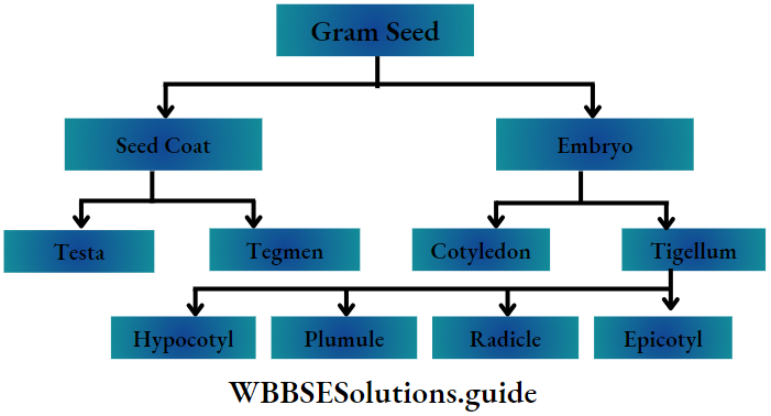
  3. Pulses, like, gram, pea, pigeon pea, lentil, mung, etc.
  4. Vegetables, like, radish, carrots, sweet potatoes, etc.
  5. Fruits, like, orange, banana, apple, guava, mango, etc.

Food adjuncts: Plants yield spices, flavouring materials, beverages, etc.

  1. Spices and other flavouring materials like ginger, turmeric, cinnamomum, cloves saffron, pepper, etc.
  2. Beverages like tea, coffee, etc.

Production of medicines and drugs:

  1. Medicinal plants such as Cinchona sp., Ipecac sp„ Rauvolfia sp. etc., are sources of various alkaloids. These alkaloids are used to produce medicines.
  2. Many angiosperms are sources of fumitories and masticatories, for example, tobacco, °P‘um’ marijuana (ganja), etc.

Industrial use of plants and plant products: Plants yield fibres (for example jute, cotton, hemp, etc.), timber (for example teak, sal etc.), and essential oils (for example Eucalyptus, Citronella, etc. sugars (for example sugarcane, palm), cellulose products etc. Each of these has different uses in our daily lives.

Fuel: Dead remains of plants and woody plant parts are used as fuel.

Similarities between gymnosperms and angiosperms

  1. Both have major sporophytic phases. The gametophytic phase was dependent on the sporophytic phase.
  2. Both have their body differentiated into roots, stem and leaves. Both produce flowers.
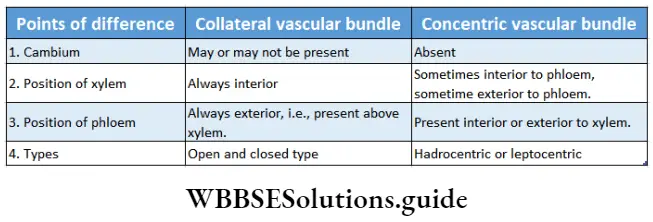
  3. Both have open or closed collateral vascular bundles.
  4. Both show the growth of pollen tubes.
  5. Both are heterosporous plants in nature.
  6. In both of them, female spores remain within sporangia or ovules.

Plant Kingdom Differences between gymnosperms and angiosperms

Classification Of Angiosperms

Bentham and Hooker’s System: Bentham and Hooker worked together for about 25 years and jointly published their work in the book Genera Plantarum (1862-1883) in three volumes.

They divided the Angiospermae into two major classes—

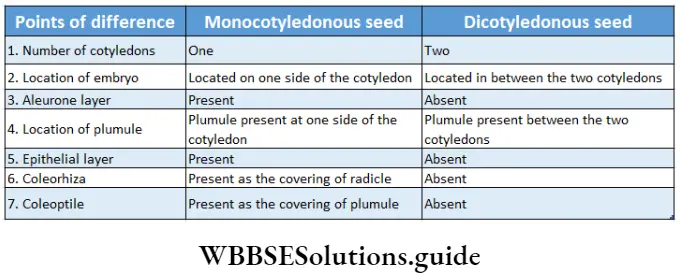
  1. Monocotyledonae (comprises plants having single cotyledon in seed, i.e., monocotyledonous plants) and it was divided into seven series. The Series were further divided into cohorts (= orders), and cohorts into natural orders (= families). Natural orders include genera and finally, each genus includes species.
  2. Dicotyledonae (comprises plants having two cotyledons in seed, i.e., dicotyledonous plants). It was further divided into three sub-classes—Polypetalae, and Monochlamydeae. Polypetalae was divided into three series; Gamopetalae was divided into three series and Monochlamydeae into eight series. These groups are discussed under separate heads.

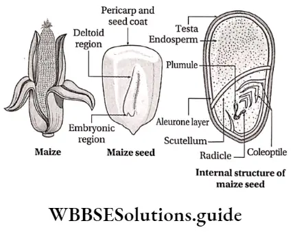
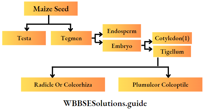
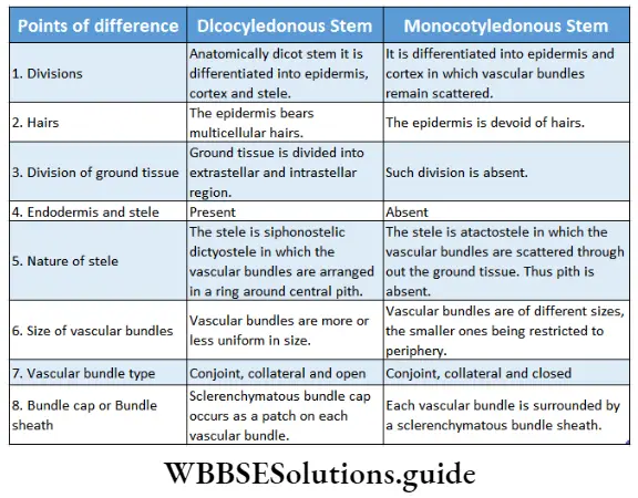
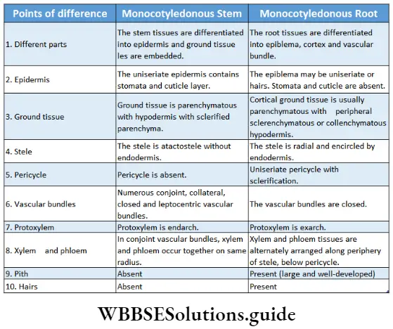
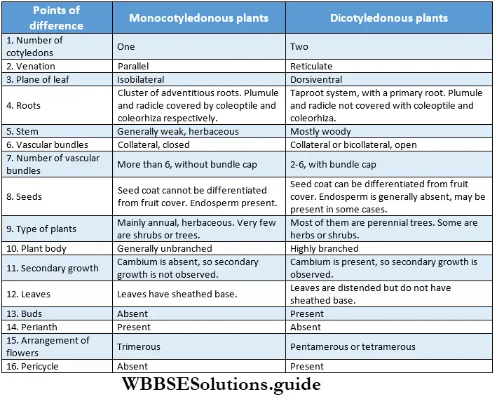
Monocotyledonous Plants

Definition: Monocotyledonous plants are those angiosperms that have a single cotyledon in their seeds.

Monocotyledonous plants General features:

  1. Generally, they are annual plants, but the trees are perennial.
  2. Plants are usually herbaceous, with weak stems (for example rice, wheat, grass, etc.). Some may be shrubs and trees (palm, date palm, coconut), while some may be climbers (orchids).
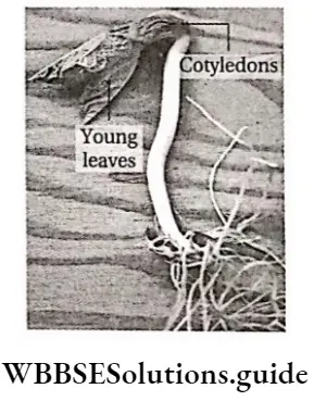
  3. The embryo contains one cotyledon. The endosperm is well-developed and store food for future use.
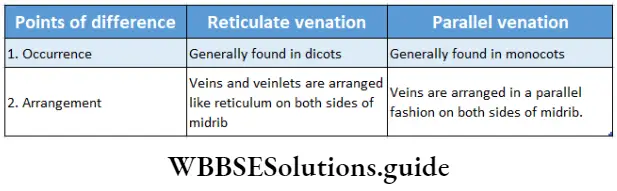
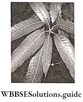
  4. The leaves may be simple or compound. They show parallel venation and sheathing leaf bases.
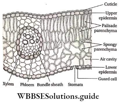
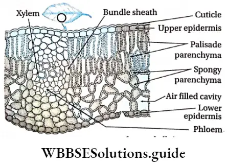
  5. Leaves are isobilateral, mesophyll tissue is not differentiated into spongy and palisade parenchyma.
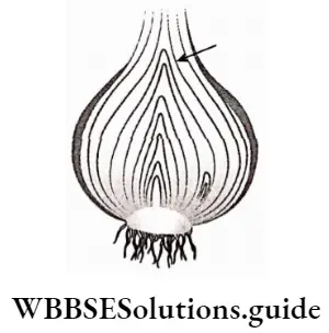
  6. Stems are generally unbranched, with nodes and internodes. In paddy and bamboo, the internodes are hollow, while in maize and sugarcane, those are solid. In some plants like ginger, onion, etc., stems are underground.
  7. At the early stage of development, plants lose their tap roots and fibrous root systems develop.
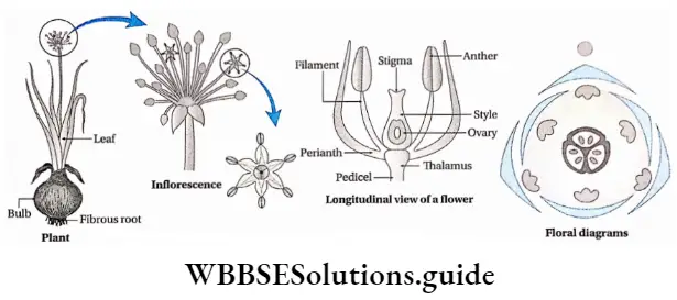
  8. The flowers are trimerous (the number of whorls present is 3 or in multiples of 3).
  9. Calyx and corolla fuse to form the perianth.
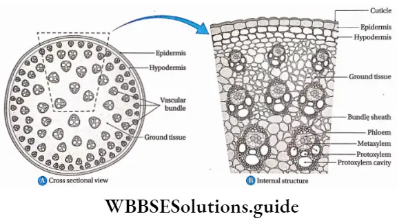
  10. Stem vascular bundles are conjoint, collateral and closed. The bundles remain scattered throughout the cortex. No stele is formed.
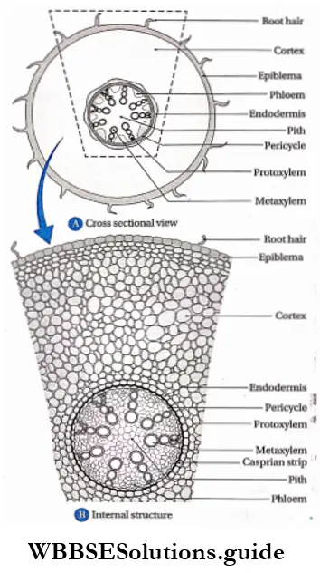
  11. The number of vascular bundles in the root is usually six or more.
  12. Due to the lack of cambium in vascular bundles, no secondary growth is observed in the stem and roots.
  13. The plumule is present near the embryo axis. Coleoptile and coleorhiza are present in seed.
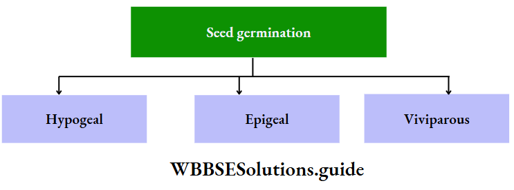
  14. The seeds show hypogeal germination.

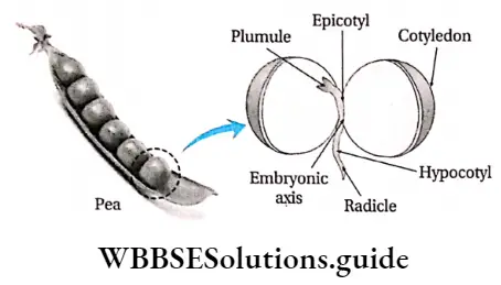
Examples: Pisum sativum, Mangifera indica, Hibiscus rosa-sinensis, etc.

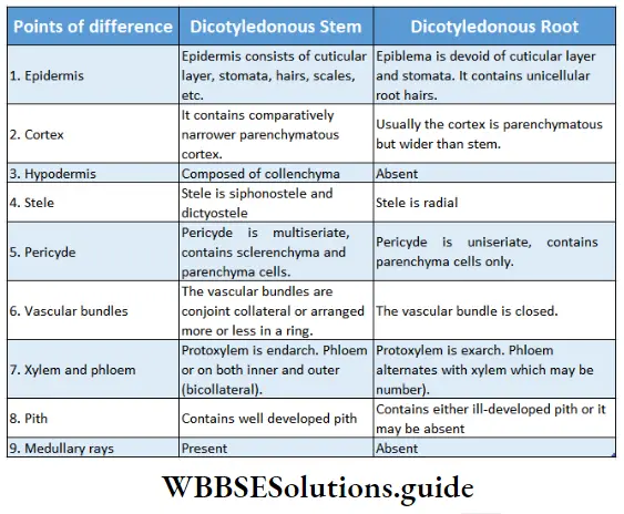
Dicotyledonous Plants

Dicotyledonous plants Definition: Dicotyledonous plants are those angiosperms that have two cotyledons in their seed.

Plant Kingdom Differences between monocotyledonous and dicotyledonous plants

Plant Life Cycles And Alternation Of Generations

A life cycle is a sequence of events that occur from birth to death of an organism. Generation is any entity with distinct morphological features that represent a particular event or phase in the life cycle.

Based on chromosome number an organism can be haploid or diploid. In plants, haploid gametes are produced by gametophytes while the diploid structures that produce spores by meiosis are called sporophytes.

In their life cycles, plants have the gametophytic or sporophytic phase as the dominant one, in which they pass most of their life span. Depending upon these dominant phases, life cycles are mainly of three types.

Those types are discussed below—

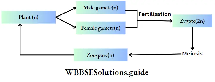
Haplontic or haplobiontic life cycle:

  1. In this type of life cycle, the gametophyte (haploid) stage is dominant throughout the life cycle and the sporophyte (diploid) stage is represented only by the zygote.
  2. The gametophytic plant develops haploid gametes inside the gametangia.
  3. The fusion between these two gametes forms the zygote.
  4. The zygote immediately undergoes meiotic division and forms four haploid meiospores.
  5. These meiospores develop into haploid plants. The alternation of generations can be interpreted by changes in the chromosome number.

Example: This type of life cycle is observed in Volvox sp., Chlamydomonus sp., Spirogyra sp., etc.

Plant Kingdom Plant

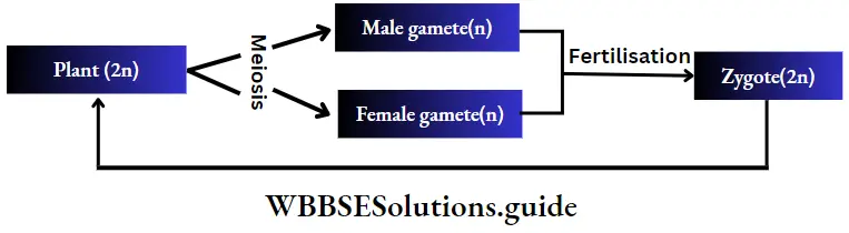
Diplontic or diplobiontic life cycle:

  1. In this type of life cycle, the plant body is sporophytic and diploid and the haploid gametophytic phase is represented only by the gametes.
  2. Sporophytic plants bear gametangia inside which gametes are produced by meiosis.
  3. The gametes upon fertilisation form diploid zygote.
  4. The zygote gives rise to a new sporophytic plant body.

Example: This type of life cycle is observed in Cladophora sp., Fucus sp., etc., algae and in all seed-bearing plants i.e., gymnosperms and angiosperms.

Plant Kingdom Fertilisation

Haplo-diplobiontic life cycle:

  1. In this type of life cycle, both the haploid and diploid phases are prominent.
  2. They differ in chromosome number and reproductive function. The haploid gametophytic plant reproduces sexually, i.e., by producing gametes.
  3. The gametes produce diploid zygotes through fertilisation. The zygote gives rise to the diploid sporophyte.
  4. The sporophytic plant reproduces by asexual processes, i.e., by producing haploid spores.
  5. In this life cycle, alternation of two generations occurs by sporogenic meiosis and fusion of gametes.

Plant Kingdom Gamatophytic plant

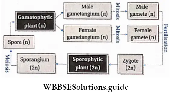
  1. Isomorphic or homologous types, i.e., gametophyte and sporophyte are morphologically similar. Example: This type of life cycle is observed in Ectocarpus sp., Polysiphonia sp., etc.
  2. Heteromorphic or heterologous type, i.e., the gametophyte and sporophyte are morphologically dissimilar. Example: This type of life cycle is observed in bryophytes and pteridophytes.

Plant Kingdom Notes

  • Alkaloid: A class of nitrogenous organic compounds of plant origin having physiological actions on humans.
  • Cilium (plural—cilia): An organelle found in eukaryotic cells. In some organisms, cilia are the means of motility.
  • Coenocytic: A multinucleate thallus/body with a continuous mass of protoplasm enclosed by one cell wall.
  • Coleoptile: A sheath protecting the tip of the plumule in monocots.
  • Coleorhiza: A sheath protecting the tip of a radicle in monocots.
  • Dichotomous branching: A pattern of branching where an equal division of an apical bud or apical growth point forms two equal branches.
  • Dorsiventral: An organ or body of an organism having dissimilar upper (dorsal) and lower (ventral) surfaces.
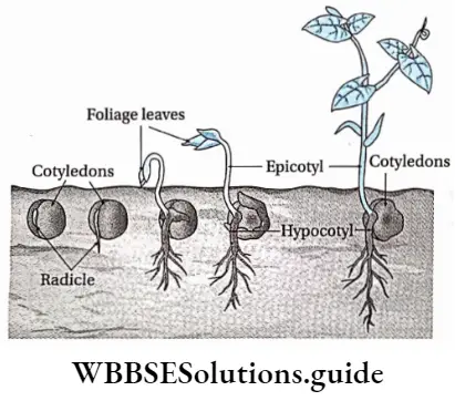
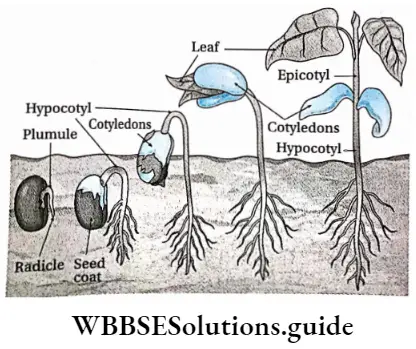
  • Epigeal: Germination of seeds with one or more seed leaves or cotyledons appearing above the ground.
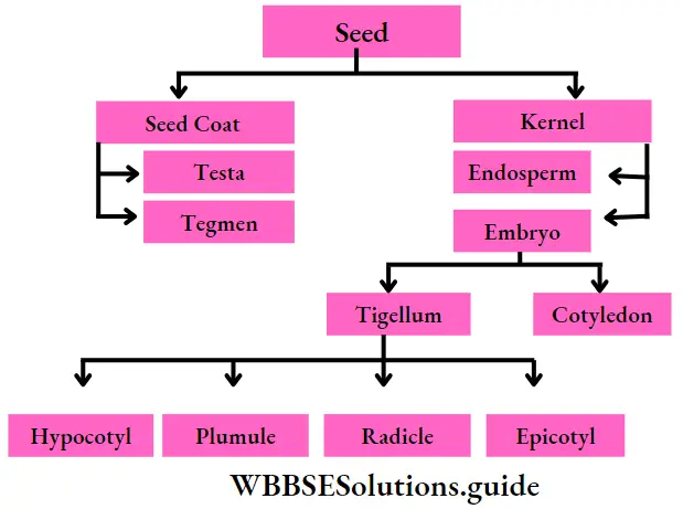
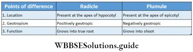
  • Hypogeal: Germination of seeds with cotyledons appearing above the ground. Plumule: Part of the embryo which will grow into the shoot.
  • Radicle: Part of the embryo which will grow into root.
  • Sporopollenin: It is an inert biopolymer found in the tough outer wall of spores and pollen grains.
  • Tinsel flagellum: A type of flagellum which has fine minute hairs along the axis.
  • Whiplash flagellum: A type of flagellum which has a smooth surface.
  • Xylan: A type of polysaccharide found in the cell walls of plants and algae.

Points To Remember

  1. Linnaeus first used the term algae.
  2. The branch of science that deals with the study of algae is called phycology.
  3. Body of algae is a simple thallus, that consists of parenchymatous cells.
  4. Large marine algae are known as seaweeds or kelp.
  5. The longest phase in the life cycle of algae is its gametophytic phase.
  6. Stored food in green algae are starch and oil.
  7. Red algae appear reddish due to the presence of the pigment phycoerythrin.
  8. The pigment responsible for the brownish colour of brown algae is fucoxanthin.
  9. The main stored food of Phaeophycean algae is laminarin.
  10. Algae mostly exist as haplobiontic, diplobiontic and triplobiontic.
  11. The cell wall of Chlamydomonas sp. lacks cellulose.
  12. Volvox sp. is a freshwater green algae that forms colonies.
  13. Volvox sp. was first observed by Antonie van Leeuwenhoek in 1700.
  14. Chlorella sp. is a unicellular green algae that is used as a food.
  15. Chlorella sp. is a source of antibiotic chlorellin.
  16. The common name of Spirogyra sp. is pond silk.
  17. The algae Spirogyra sp. has three types of reproduction—vegetative, asexual and sexual.
  18. Algae are consumed by various organisms in the aquatic ecosystem.
  19. lodin is obtained from some phaeophycean algae such as Laminaria digitatia, Ecklonia sp., etc.
  20. Bryophytes are known as amphibians of the plant kingdom.
  21. Alternation of generations is observed in mosses.
  22. Although bryophytes are terrestrial, they require water during the process of fertilisation.
  23. The sporophytic phase of the life cycle in bryophytes depends on the gametophytic plant body for food.
  24. The common name of bryophytes of the Class Anthocerotopsida is hornworts.
  25. Apospory can be seen in some species of Anthoceros.
  26. Seedless, vascular plant fern has approximately 10,000 species.
  27. Salvinia sp., Azolla sp., and Ceratopteris sp., are aquatic ferns.
  28. Pteridophytes have a distinct vascular system made up of xylem and phloem tissues. They are called vascular cryptogams.
  29. Generally, four types of ferns are seen—Psilopsida, Lycopsida (clubmoss or ground pine), Sphenopsida (horsetail) and Filicopsida or Pteropsida (ferns).
  30. Lycopsids are of two types-(a) homosporous (like Lycopod/t/m sp.) and (b) heterosporous (like Selaginella sp.).
  31. The aquatic fern Azolla is used as organic manure.
  32. Lycopodium sp. is used to cure skin diseases.
  33. Gymnosperms are intermediates between ferns and angiosperms.
  34. Psilotum sp. is known as a primitive fern. It lacks true roots. Leaves are small and lack veins.
  35. Sequoia sempervirens is the largest gymnosperm (366 ft), Zamia sp. is the smallest gymnosperm (25 cm), and Sequoiadendron gigantia is the oldest gymnosperm (3,500 years based on the annual ring count).
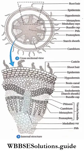
  36. The vascular bundle of gymnosperms is conjoint, collateral and open.
  37. Gnetum sp. is the intermediate between gymnosperm and angiosperm.
  38. The wood obtained from gymnosperms is called softwood.
  39. The endosperm of gymnosperms are produced before fertilisation, so the endosperms are haploid (n).
  40. Polyembryony is observed in gymnosperms, like, Pine.
  41. The largest ovum, ovule and antherozoa are present in Cycas sp.
  42. The gymnosperm Ginkgo biloba (maidenhair tree) is known as a living fossil.
  43. Cycas revoluta is the scientific name of the plant sago palm.
  44. Pine tree bears numerous pollens which are yellow in colour. These pollens have wing-like structures and are dispersed by air. When these are carried in the air, it appears like a yellow cloud. This is known as a ‘shower of sulphur’ or a ‘shower of golden dust’.
  45. Fertilisation in pine trees may occur one year after pollination. Polyembryony is seen in pine trees. However, they produce one embryo after maturation.
  46. Angiosperms are vascular plants, that form fruits, containing seeds.
  47. Angiosperms are classified into two divisions— monocotyledons and dicotyledons
  48. Angiosperms are of three types—herbs, shrubs or trees. They can also be annual, biennial or perennial.
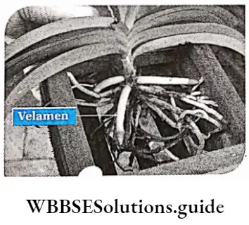
  49. Some species of P|ants on|Y grow other plants but do not obtain food from the host. These are known as epiphytes. Aerial roots of epiphytes absorb water from air through the spongy cells, called velamen, present at the tips.
  50. Endosperms of angiosperms are produced by double fertilisation (fusion of a male gamete to a diploid secondary nucleus). Thus, endosperm cells have a triploid (3n) set of chromosomes.
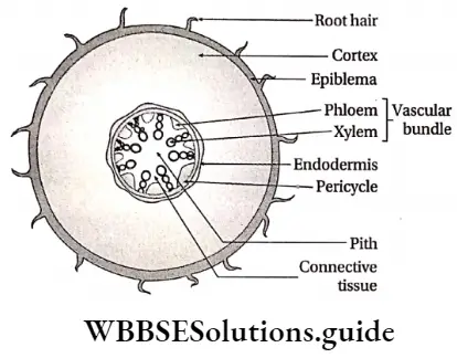
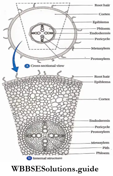
  51. The vascular bundle in the roots of angiosperms is radial and exarch. Number of the vascular bundles is more than 6 in monocots and 2-6 in dicots.
  52. The vascular bundle in the stem of angiosperms is conjoint, collateral or collateral, open or closed and endarch.
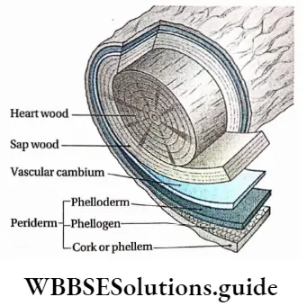
  53. Secondary growth is seen in dicot plants but it is not seen in monocot plants.
  54. The largest flower is Rafflesia Arnoldi, whose diameter is 1 meter and weighs approximately 15 kg. The smallest flower and smallest angiosperm is Wolffia microscopic (duck weed).
  55. Largest angiosperm in Eucalyptus regnance.

Animal Kingdom Notes

Animal Kingdom Basis Of Classification

Both plants and animals show a high degree of diversity. In the previous chapter, we have studied the classification of plants. Plants constitute the plant kingdom. Similarly, all animals belong to the kingdom Animalia or animal kingdom.

Even under the same kingdom, each species may exist in different forms. Classification helps us to deal with this enormous diversity. The study of the classification of the animal world is known as systematic zoology.

The animals are classified into different categories according to their general characteristics. These characteristics include body organization, body symmetry, presence of germ layer, etc.

Read and Learn More: WBCHSE Notes for Class 11 Biology

General Characteristics Of Animals

The general characteristics of animals, on the basis of which they are classified, are discussed below.

Habitat

The geographical region and environment, where an animal lives, is called its habitat.

According to the habitat, the animals are classified into three types—

  1. Aquatic,
  2. Terrestrial, and
  3. Aerial.

Aquatic animals: Animals that live in water are called aquatic animals.

These animals are further classified into the following types—

Zooplankton: Microscopic organisms that float on water are called zooplankton. Example Microscopic animals and larvae of some higher animals.

Nektons: Organisms that can swim freely in water are called nektons (derived from the Greek word, nekton, meaning swimming). For example, Fish, prawns, etc.

Benthon: Organisms that are present in the lower region of the sea are called benthons (derived from the Greek word —benthos, meaning depths of the sea). Example Corals, sponges, etc.

Littoral: Organisms that live near the shores are called littoral. Example Small fish.

Neritic: Organisms that live in shallow regions of the water bodies are called neritic. Example Snail.

Neuston: Organisms that are found just below the water surface or on the water surface are called neuston (derived from the Greek word, neustos, meaning floating). Example Mosquito larvae.

Lcntic and lotic: Organisms that are found in still water (such as ponds or lakes) are called lentic. Example Small fish. Organisms that are found in flowing water (such as rivers) are called lotic. Example Big fish.

Terrestrial animals: Animals that live on land are called terrestrial animals. They are further divided into several types. Some of them are discussed below.

Amphibians: Animals that live on both land and water are called amphibians. Example Common Asian toad.

Arboreal: Animals that Live on trees are called arboreal animals. ExampleMonkey.

Cursorial: Animals that can run very fast are called cursorial animals. Example Horse.

Subterranean: Animals that live in underground holes are called subterranean. Example Earthworms.

Scansorial: Animals that can climb trees are called scansorial animals. Example Squirrel.

Fossorial: Animals that dig holes or burrows and live in them, are called fossorial animals. Example Rabbits, armadillos, etc.

Aerial animals: Animals that can fly in the sky, are called aerial animals. According to their adaptation, they are of two types—primary and secondary aerial animals.

Primary aerial animals: Animals that fly in the sky throughout most part of their life, are called primary aerial animals. The ability to fly in such animals is due to their ancestors who were adapted to flight. Example Birds.

Secondary aerial animals: Animals that can fly for certain intervals of time, but are not aerial in nature are called secondary aerial animals. Example Bats.

Food habit

Different types of food habits are found in the animal kingdom.

According to food habits, the classification of animals is as follows—

Animal Kingdom Classification of animals according to food habits

Structural organization

Different kinds of structural organizations are found in multicellular organisms. These may be at the cellular, histological, or organ system level. These have been discussed below.

Cellular organization: In this case, several cells together form a body, but do not form any tissue. Members of the phylum Porifera have this kind of body organization.

Histological organization: In this case, several ceÿs together form tissues, and several tissues together form the body. Members of phyla Cnidaria and Platyhelminthes have this kind of body organization.

Morphological organization: In this case, several cells form tissue, several tissues form an organ, and several organs form an organ system. Several such organ systems form the body. Members of phylum Nemathelminthes and other higher phyla show this kind of body organization.

Body plan

Different multicellular animals show different types of body plans. The types of body plan are cell aggregate, blind sac, and tube within tube plan.

Cell aggregate plan: In the case of animals without tissues (cellular organization), several cells aggregate to form the body. This type of body plan is called the cell aggregate plan. Animals belonging to the Phylum Porifera show this type of body plan.

Blind sac plan: In the case of animals with tissues, a common opening (acting as both mouth and anus) is present, along with a blind sac-like structure or cavity. This type of body plan is called a blind sac plan. Animals belonging to the Phyla Cnidaria and Platyhelminthes, show this type of body plan.

Tube within tube plan: In the case of higher animals that have organ systems, the body plan is that of a tube within another tube. The outer tube is the body wall, while the inner tube is the alimentary canal.

This type of body plan is called a tube-within-tube body plan. All the animals from Phylum Nemathelminthes to Phylum Chordata, show this type of body plan. This body plan is further divided into two types—protostome and deuterostome.

Protostome: When the blastopore in the embryo develops into the mouth, the body plan is called the protostome type. This implies that the mouth develops before the anus. This type of body plan is found in the members of the phyla Platyhelminthes, Nemathelminthes, Annelida, etc. These animals are known as protostomia.

Deuterostome: When the blastopore in the embryo develops into the anus, the body plan is called deuterostome type. This implies that the anus develops before the mouth. This type of body plan is shown by members of the Phyla Echinodermata and Chordata. These animals are known as deuterostomia.

Animal Kingdom Different types ofbody plan

Symmetry Of The Body

Multicellular organisms show both symmetry and asymmetry of the body.

Symmetry: The similarity in the arrangement of organs along an axis that, divides the body into two equal halves, is called symmetry. Such organisms that show symmetry are called symmetrical. Different types of symmetry are discussed below.

Bilateral symmetry: In this type, the body of an organism can be divided into two equal halves along a longitudinal plane of division. Such organisms are called bilaterally symmetrical. In these organisms, both halves of the body appear as mirror images of each other (with respect to shape, structure, and color). Example Some invertebrates (Phyla Annelida, Arthropoda, etc.) and phylum Chordata (Fish, amphibians, reptiles, birds, and mammals).

Radial symmetry: The type of symmetry in which the body of an organism can be divided into equal halves along any vertical plane passing through the central axis of the body, is called radial symmetry. Such organisms are called radially symmetrical. Examples are Hydra, starfish, sea cucumber, etc.

Radial symmetry is further divided into two types—

  1. Biradial and
  2. Pentaradial symmetry.

Biradial symmetry: In this case, a body can be divided across the central axis, along two vertical planes. For example, Animals that belong to phylum Ctenophora.

Pentaradial symmetry: In this case, a body can be divided across the central axis, along five vertical planes, into five equal parts. Example Certain animals such as starfish, under Phylum Echinodermata. The larvae of echinoderms are bilaterally symmetrical, but the adult forms are pentaradially symmetrical.

Spherical symmetry: In this type, a spherical body, can be divided into two equal halves, along any plane. Such organisms are called spherically symmetrically. Example Noctiluca.

Asymmetry: This occurs when the body of the animal can never be divided into two or more equal parts. Such animals are called asymmetrical in nature. Example Amoeba.

Animal Kingdom Different types ofsymmetry

Body axis and body surface

Different characteristics are observed in different animals, according to body axis and body surface. These include the ends of the body and different types of P|anes that Pass through it. These have been discussed below.

Anterior end: The part of the body, carrying the mouth, that is the first to move forward, during locomotion, is called the anterior end.

Posterior end: The part of the body, (opposite to the anterior end), that is the last to move forward, during locomotion, is called the posterior end.

Aboral end: The end of the body that lies opposite to the mouth, is called the aboral end.

Oral end: The end of the body, which bears the mouth, is called the oral end.

Proximal end: The part of any organ, that remains closer to the main body, is called its proximal end.

Distal end: The part of any organ, that remains at a distance from the main body is called its distal end.

Ventral plane: It is the plane of the body which remains closer to the ground. In the case of vertebrates, the plane that contains the reproductive organs and lies opposite to the spinal cord is called the ventral plane. This plane lies in front of the alimentary canal.

Dorsal plane: It is the plane of the body which remains away from the ground. In the case of vertebrates, it is the plane that contains the spinal cord. This plane lies behind the alimentary canal.

Lateral planes: The two planes on both sides of the body are called lateral planes.

Planes of Division

Different planes across which an animal body may be divided, are called planes of division. These planes of division have been discussed below.

Frontal or coronal plane: The plane extending along the longitudinal axis, parallel to the dorsal and ventral plane, is called the frontal or coronal plane.

Transverse plane: The plane extending perpendicular to the longitudinal axis, is called the transverse plane. The section of the body along the transverse plane is called the transverse section. The section of the body along the longitudinal or transverse plane, that passes through the vertical regions of the body, is called the vertical section.

Sagittal plane: The plane that divides the animal body into left and right parts, is called the sagittal plane. It extends over a longitudinal and dorsal-ventral axis. The section of the body along the longitudinal axis, is called longitudinal or sagittal section.

Metamerism or Segmentation

The process by which an animal body can be divided, both externally and internally, into linearly arranged, repeating, identical segments, is called metamerism or metamerisation. Each segment is known as a metamere or somite. Metamerisation is observed in some animals under the phylum Annelida, phylum Arthropoda, and phylum Chordata. Metamerism is of two types—homonymous and heteronomous.

Homonymous metamerism: This type of metamerism is characterized by similar types of segments or metameres. Such type of metamerism is found in animals under Phylum Annelida. Example Nereis sp.

Heteronomous metamerism: This type of metamerism is characterized by different types of metameres. Out of these different metameres, the similar ones together perform similar functions. Such type of metamerism is found in animals under the phylum Arthropoda, Class Insecta. Example Hornet.

Animal Kingdom Types ofmetamerism

Tagmatisation

The process by which several adjacent segments of an animal body are grouped into larger specialized functional units or stigmas is called tagmatisation. These are observed in animals under the phylum Arthropoda.

Coelom

The cavity that is covered both externally and internally by the peritoneum, extending up to the alimentary canal and body wall, is called coelom. Coelom is formed from mesoderm. Hence, it can be considered as an intermediate structure lying between somatic mesoderm (parietal layer of mesoderm) and splanchnic mesoderm (visceral layer of mesoderm). It is found in triploblastic organisms.

Types of coelom

The two types of coelom based on its origin, are described below.

Schizocoelic coelom: The coelom that has developed from a mesodermal chord is called schizocoelic coelom. This type of coelom is found in members of Phyla Annelida, Arthropoda, and Mollusca.

Enterocoelic coelom: The coelom that has developed from a sac-like structure (mesodermal pouch), produced from a primitive alimentary canal, is called enterocoelic coelom. It differentiates mesoderm and endoderm. This type of coelom is found in members of the phylum Echinodermata and Chordata. According to the presence of coelom, animals are of three types— acoelomate, pseudocoelomate, and coelomate.

Animal Kingdom Acoelomate, coelomate and pseudocoelomate organisms

Animal Kingdom Classification of animals according to presence of coelom

Functions of coelom:

  1. It separates the muscles in the alimentary canal from those in the body wall. This makes the circulation easier within the muscles. It also allows the internal organs to maintain their position within the body.
  2. In some animals, excretory products and matured germ cells are collected within the coelom.

Coelomic fluid: The fluid present within the coelom, is called the coelomic fluid. This fluid functions as a hydrostatic endoskeleton (found in phylum Annelida) or circulatory medium for the transport of gases, nutrients, and wastes.

Coelomoduct: The duct produced from the mesoderm membrane, that connects the coelom to the external environment in some invertebrates, is called a coelomoduct. This duct is responsible for carrying the germ cells or excretory wastes out of the body.

Haemocoel: The cavities that carry blood or other fluids, within the body of invertebrates with the open circulatory systems, are called hemocoel. The fluid that fills the hemocoel is called haemocoelic fluid. Such animals are called haemo coelomates. This type of hemocoel is found in members of Phyla Arthropoda and Mollusca. They are also found in some members of phylum Annelida such as leeches.

Germ Layers or Germinal Layers

The cellular layers of the embryo, from which different organs are formed, in the gastrula stage of the embryo, are called germ layers.

Types: Germ layers are of three types—

  1. ectoderm,
  2. endoderm, and
  3. mesoderm.

Ectoderm: The outermost layer of the gastrula, that forms the epidermis and epidermal outgrowths (such as ectodermal scales, feathers, nails, horns, enamel of teeth, etc.) is called ectoderm. Most of the nervous system and a part of the alimentary system also develops from the ectoderm.

Mesoderm: The intermediate layer, within the external and the internal layers of the gastrula, is called the mesoderm. Later, this layer forms the muscles, blood vessels, internal parts of the skin, etc.

Endoderm: The innermost layer of the gastrula, which later on forms the alimentary canal and its corresponding glands, is called the endoderm. A part of both the excretory and respiratory systems also develop from this layer.

Classification of animals according to germ layers: According to the types of germ layers, animals are divided into three groups—monoblastic, diploblastic, and triploblastic.

Animal Kingdom Classification of animals according to germ layers

Digestive system

The organ system that helps to take in food, digest it, and absorb some of it, while ejecting the rest of it is called the digestive system. The central tube-like structure that is present in the animal body, to which other digestive organs remain associated, is called digestive organs remain associated, is called the digestive tract or alimentary canal.

Generally, there are two types of alimentary canals complete and incomplete.

Complete digestive tract: In this type of digestive tract, separate openings as the mouth and anus are present. Animals under Phyla Aschelminthes, Chordata, etc., have this type of digestive tract.

Incomplete digestive tract: In this type of digestive tract, only one opening is present, that serves as both mouth and anus, i.e, they are not separate Animals belonging to Phyla Cnidaria, Ctenophora, Platyhelminthes, etc., have this type of digestive tract.

Circulatory System

The system that transports important constituents to the specific cells and their released products to the specific organs, both through a liquid medium, is called the circulatory system.

Circulatory systems are of two types—

  1. open and
  2. closed.

Open circulatory system: In this type of circulatory system, the circulating fluid does not flow through the blood vessels. Instead, it is released into the lumen. Animals under Phyla Arthropoda, Mollusca, etc., have this type of circulatory system.

Closed circulatory system: In this type of circulatory system, the circulating fluid flows through the blood vessels. It is not released into the lumen. Animals under Phyla Annelida, Chordata, etc., have this type of circulatory system.

Respiratory system

The organ system that helps in the exchange of gases and in respiration, is called the respiratory system. With the rise in the complexity of the structural organization of the animals, the respiratory system is also modified. Some of them are discussed below.

Animal Kingdom Types of respiratory (aerobic) organs with corresponding phylum

Excretory system

The organs that help to carry out excretion are called excretory organs. Together they constitute the excretory system. Different animals have different types of excretory organs. Some of such organs have been mentioned in the table below.

Animal Kingdom Types of excretory organs with corresponding phylum

Animal Kingdom kidney

The most primitive type of vertebrate kidney is pronephros. It is functional in early larvae of fishes and amphibians.

Nervous system

The system by which animals regulate and coordinate their various physiological processes, due to the effect of external and internal stimulants, is called the nervous system. Types of nervous systems found in animals of different phyla have been mentioned in the table below.

Animal Kingdom Types of nervous system found in animals belonging to different phyla

Skeletal system

The system which forms the skeleton of the animal body is called the skeletal system. There are two types of skeletal systems.

They are as follows—

  1. endoskeleton and
  2. exoskeleton.

Endoskeleton: The skeleton formed inside the body, in the case of vertebrates is called the endoskeleton.

Exoskeleton: The skeleton formed outside the body, in the case of invertebrates and some vertebrates, is called an exoskeleton. Some of these have been listed in the given table.

Animal Kingdom Types of exoskeleton with corresponding animals

Fertilisation

Fertilization is a stage in sexual reproduction, where the sperm and ovum fuse to form the zygote which forms the embryo.

The different types of fertilization are—

Types according to the site of occurrence There are two types of fertilization according to the site of occurrence.

  1. External fertilization: This type of fertilization takes place outside the body. This is seen in the members of Class Pisces and Amphibia.
  2. Internal fertilization: This type of fertilization takes place inside the body. This is seen in the members of Class Reptilia, Aves, and Mammalia.

Types according to the nature of fertilization There are two types of fertilization according to its nature.

  1. Self-fertilization: This process takes place as the egg and sperm, produced in the same animal, fuse to form the zygote. It occurs mostly in bisexual animals. Example Tapeworm.
  2. Cross-fertilization: This process takes place as the egg and sperm, produced by two different animals, fuse to form the zygote. It occurs mostly in unisexual animals Example cockroaches, frogs, human beings, etc. Even though roundworm is bisexual, it shows cross-fertilization.

Reproduction

The process by which an organism produces its offspring is called reproduction. Different types of reproduction are discussed below.

Vegetative reproduction: This process by which an animal produces its offspring from a part of its own body, by cell division. Found in Protozoans.

Parthenogenesis: By this process, an animal produces its offspring from an unfertilized egg. Found in male wasps, male bees, etc.

Asexual reproduction: By this process, an animal produces its offspring by division, regeneration, or spore formation. Found in Planaria.

Sexual reproduction: By this process, an animal produces its offspring by fusion of its gametes. Found in all vertebrates and higher invertebrates.

Body temperature

According to body temperature, animals are of two types—poikilotherms and homeotherms.

Poikilotherms: The animals in which the body temperature varies with the external environment, are called poikilotherms or ectotherms. Example Amphibians, reptiles, etc.

Homeotherms: The animals that have their body temperatures constant irrespective of changes in the temperature of the external environment, are called homeotherms. The homeotherms that have an internal regulation system for body temperature are called endotherms. Examples, Birds and Mammals.

Sex organs

The animals that can reproduce sexually have reproductive or sex organs. These organs also help in determining the sex of the animals. They are of two types

Unisexual: The animals that have only one type of reproductive system (either male or female), are called unisexual. Examples Human beings, cockroaches, etc.

Bisexual: The animals that have both types of reproductive systems (male and female), are called bisexual. Example Roundworms, tapeworms, etc.

Classification Of Animals

Scientists have classified animals according to different criteria. R.H. Whittaker has classified the animal kingdom into 26 animal phyla, consisting of more than 1 million known animal species. Each phylum includes animals with similar characteristics.

Out of these, there are 11 major phyla (phylum Hemichordata, though classified as a major phylum, is considered to be a part of phylum Chordata. Hence, it has not been shown in the chart). The phylogenetic tree of the animal kingdom showing the major phyla only is given below.

Animal Kingdom Kingdom Animalia

Other than these major phyla, there are certain minor phyla within the classification. These minor phyla have not been explained to avoid the complexity of the chapter.

Subkingdom Classification

The Kingdom Animalia is further divided into subkingdoms. They are— Protozoa and Metazoa

Subkingdom Protozoa

[Latin proto: first; zoom animal]

Protozoa is a group of simple, unicellular, microscopic organisms with different types of locomotory organelles. The term ‘Protozoa’ was coined by Goldfuss (1820). The group comprises about 80,000 species.

General features:

  1. Simple, unicellular, microscopic animal, either free-living or symbiotic or parasitic.
  2. The nucleus is single, vesicular (nucleus contains more nucleoplasm but less chromatin), or compact (nucleus contains less nucleoplasm but more chromatin) in majority. Few protozoa are multinucleated.

Phylum Porifera

(Latin porus: pore;/erre: to bear)

Porifera is a group of multicellular animals, sedentary in habit. They have numerous pores on their body surfaces. They have distinct canal systems lined internally by specialized cells called choanocytes. They are multicellular without the development of organ systems, division of labor, and specialization at the cellular level —cells function more or less independently. This phylum comprises about 10,000 species.

General features: The general features of members of the phylum Porifera are discussed below.

Nature:

  1. Most of them are aquatic, sedentary, and sessile inhabit.
  2. They may remain solitary or in colonies.
  3. The body is multicellular, elongated, cylindrical, sometimes branched, and irregular in shape.
  4. They are diploblastic with an outer ectoderm (pinacoderm) and an inner endoderm (choanoderm) separated by a non-cellular, jelly-like, loose mesenchyme layer. True body cavity or gut is absent.
  5. The body is asymmetrical or radially symmetrical.

Pores: The body is perforated with numerous minute pores, called ostia. Water enters through the ostia into a cavity called spongocoel or paragastric cavity. This water ‘released body, through a single, large aperture, at one end of the body, called an osculum. Separate mouth and anus are absent in the body.

Canal system: They possess a branched network of canals known as the canal system. Water containing food particles and oxygen enter through the Ostia. The canal system helps to circulate the water throughout the body.

Animal Kingdom Different types ofcanal system in members ofphylum Porifera

Choanocytes and other cells; The canal system is lined by a type of flagellated cells, called choanocytes. The movement of their flagella creates a water current which helps in the movement of the cells. These cells also help in the ingestion of food and the movement of sex cells, etc.

There are other cells like amoebocytes, pinacocytes, etc. Amoebocytes possess pseudopodia which helps in the movement of the cells, while pinacocytes provide contractibility.

Animal Kingdom Choanocyte

Organ system: Though they are multicellular, they lack an organ system.

Endoskeleton: The body is provided with a large number of minute, pointed calcareous or siliceous spicules and spongin fibers, that form the internal skeletal framework.

Reproduction: Reproduction occurs asexually by budding, fragmentation, and by formation of reproductive bodies, called gemmules. The sexual mode of reproduction occurs through male and female gamete formation. They possess the ability to regenerate lost body parts.

Animal Kingdom Body wall and gemmule in sponges

Animal Kingdom Some members of phylum Porifera

Phylum Cnidaria

(Greek Cnidos: thread)

Cnidarians are a group of diploblastic, radially symmetrical animals. They possess special sensory cells (cnidoblast cells) and a gastrovascular cavity (coelenteron) with a single opening, called a mouth. In this phylum, the tissue grade of organization is observed for the first time. Cnidarians are considered true multicellular animals.

General features: The general features of the members of the phylum Cnidaria are discussed below.

Nature:

  1. Cnidarians are mostly marine while few are freshwater.
  2. They are multicellular and acoelomate in nature.
  3. The body is radially symmetrical, diploblastic with jelly-like, non-cellular mesoglea in between the outer ectoderm and an inner endoderm.
  4. They may remain solitary or form colonies.
  5. They may be unisexual or bisexual.

Cnidoblasts: They possess special sensory cells called cnidoblasts or nematoblasts, within the tentacles at the oral end. The cnidoblasts contain a sac called a nematocyst, which contains a tube. This tube may contain structures such as barbs and barbules, that help in offense, defense, and food capture. The cnidoblasts burst and release the barbs when touched or sensitized.

Coelenteron: The body has a central cavity, called coelenteron or gastrovascular cavity, lined with endodermal cells. It has a single opening, the mouth at the oral end.

Mouth: The mouth is present on the hypostome, encircled with tentacles in one or more whorls.

Animal Kingdom Structure ofbody wall in Cnidaria

Digestion and organ systems: Digestion is of two types—extracellular, within the gastrovascular cavity, and intracellular, within the endodermal cells. Respiratory, circulatory, and excretory systems are absent. A primitive type of nervous system is present with a diffused network of nerve cells.

Reproduction: Asexual reproduction takes place through fission and budding, while sexual reproduction takes place through gamete formation.

Life cycle: The life cycle has two phases, hence polymorphism is distinct. It has an asexual phase called polyp (cup-shaped) and a sexual phase called medusa (umbrella-shaped). They also show alternation of generations and metagenesis. Planula larva is found in the life cycle.

Animal Kingdom Polyp and Medusa

Polymorphism: The occurrence of structurally and functionally different forms within the same animal during its life history is known as polymorphism. In colonial Cnidarians, such as Obelia, polyp, and Medusa occur in different forms performing different functions.

Metagenesis: The interconversion of asexual and sexual phases is known as metagenesis.

Animal Kingdom Differences between polyp and medusa

Animal Kingdom Some members of phylum Cnidaria

Animal Kingdom Differences between Protozoa and Porifera

Phylum Ctenophora

(Greek Ktenos: comb; photos: bearing)

Ctenophorans are commonly known as sea walnuts or comb jellies. Ctenophorans represent a small group of animals with only 50 known species, distributed abundantly in the coastal waters. The first description of Ctenophora was given by Martens (1671). Hatschek (1839) placed Ctenophora under a distinct phylum.

General features: The general features of the phylum.

Ctenophora are discussed below.

Nature:

  1. Ctenophorans are exclusively marine animals.
  2. The members may exist as single units or in colonies, devoid of any skeleton-like structures.
  3. The body is transparent, flat, and biradially symmetrical.

Locomotion: They swim freely with the help of eight vertical ciliated comb plates. These plates are present throughout the body, at regular intervals.

Colloblasts: The tentacles, if present, occur in pairs and opposite to each other. Special adhesive cells called colloblast cells or lasso cells are present on the tentacles. Other than these, smooth muscle cells, nerve cells, amoebocytes, etc., are present. Nematocysts are absent.

Germ layers: The body is diploblastic with mesenchyme in between the outer ectoderm and inner endoderm, containing muscle cells and amoebocytes.

Sensory organ: Statocyst is a sensory organ, present at the aboral end (the end of the body opposite to that of the mouth).

Digestion: The gastrovascular system or coelenteron is complete with a mouth and a highly branched digestive tract. The digestive system can perform both extracellular and intracellular digestion.

Organ systems: Respiration takes place by diffusion. The nervous system is underdeveloped but the sub-epidermal network of nerves is well developed beneath the comb plates.

Life cycle: The members of this phylum are hermaphrodites. Sexual reproduction is common and fertilization is external. No alternation of generation is seen in the life cycle. Cydippid larva is a ciliated spherical type of larva, found in their life cycle.

Animal Kingdom Some members of phylum Ctenophora

Similarities between Cnidaria and Ctenophora

There are some similarities between the phylum Cnidaria and Ctenophora.

They are as follows—

  1. Both are diploblastic in nature.
  2. Both have coelenteron.
  3. Both have tentacles.

Animal Kingdom Differences between phylum Cnidaria and Ctenophora

Animal Kingdom Differences between coelenteron and coelom

Animal Kingdom Member ofphylum Ctenophora

Phylum Platyhelminthes

(Greek Platy: flat; helminth: worm)

Phylum Platyhelminthes is a diverse group comprising 25,000 living species.

General features: The general features of the members of the phylum Platyhelminthes are discussed below.

Nature:

  1. The body is thin, dorsoventrally flattened, and bilaterally symmetrical.
  2. The body is ribbon-like or leaf-shaped, covered by a syncytial membrane.
  3. The true body cavity is absent, hence, acoelomate (a = without; coelom = body cavity).
  4. The members may be parasitic or free-living.

Germ layers: Triploblastic organization with ectoderm, endoderm, and mesoderm. Mesoderm is present between ectoderm and endoderm.

Segmentation: Metameric segmentation is absent.

Organ systems: Alimentary canal is incomplete. The mouth is present, but the anus is absent. Excretory organs are protonephridia or numerous flame cells or solenocytes. Circulatory and respiratory systems are absent. Gaseous exchange occurs by diffusion. The nervous system is simple, ladder-like with a brain and a double ventral nerve cord. The organ system is absent or reduced in parasitic forms.

Animal Kingdom Nervous system ofPlatyhelminthes

Reproduction: They are hermaphrodite in nature. A complex reproductive system is generally present. Asexual reproduction occurs by transverse fission. Free-living Planaria has the power of regeneration.

Life cycle: The life cycle is completed through one or two hosts. Different larval forms (like cysticercus cellulose in pig tapeworm) are usually present.

Animal Kingdom Some members of phylum Platyhelminthes

Phylum Nemathelminthes or Nematoda

(Greek Nematos: thread; eidos: form)

Nematodes are a diverse group of organisms comprising 15,000 living species. The members of this phylum were initially included in the phylum Aschelminthes(Grobben, 1910).

General features: The general features of the members of the phylum Nemathelminthes or Nematodes are discussed below.

Nature:

  1. The body is bilaterally symmetrical, thread-like, cylindrical, elongated, unsegmented, tapering at both ends, covered with cuticle.
  2. They are mostly parasitic, while some are free-living.

Germ layers: They are triploblastic and pseudocoelomate (body cavity is present, but not mesodermal in origin), with pseudocoelomic fluid.

Body wall: Longitudinal muscles are present in the body wall, but circular muscles are absent.

Digestive system: Alimentary canal is simple, straight with a distinct mouth and anus at opposite ends.

Excretory system: The Excretory system comprises a canal called protonephridial canal, without flame cells. In some cases, a type of cells called rennet cells, carry out excretion.

Nervous system: Nervous system comprises a brain in the form of a nerve ganglia surrounding the gut. This nerve ganglion is called the cricopharyngeal nerve ring.

Other organ systems: Respiratory and circulatory systems are absent.

Reproduction: Sexes are separate with distinct sexual dimorphism. Males are smaller, having a curved tail end with a pair of pineal setae, while females have a straight pointed tail end. Fertilisation is internal. Development is generally direct but in parasitic forms, the life cycle passes through infective larval stages like microfilariae, rhabditiform, etc.

Animal Kingdom Some members of phylum Nemathelminthes

Phylum Annelida

(Latin annulus: ring; eidos: form)

Phylum Annelida exhibits great diversity of body form, comprising approximately 17,000 species. Lamarck (1802) coined the term ‘Annelida’ due to their ring-shaped body segments.

General features: The general features of members of phylum Annelida are discussed below.

Nature:

  1. The body is elongated, bilaterally symmetrical, and metamerically segmented, into several ring-like structures called somites or metameres.
  2. The body surface is covered with a thin cuticle.

Body cavity: The body is triploblastic and coelomate with a true body cavity lined by mesodermal cells.

Body wall: Both circular and longitudinal muscles are present in the body wall.

Germ layers: Triploblastic (ectoderm, mesoderm and endoderm are present).

Digestive system: Alimentary canal is complete. It is a muscular tube-like structure, with a separate mouth and anus at opposite ends.

Respiratory system: Respiration generally takes place through the skin; in some forms gills are present.

Circulatory system: Well-developed closed circulatory system with blood vessels. Hemoglobin or haemoerythrin remains dissolved in the plasma. Distinct heart-like structures and blood vessels appear for the first time in Annelida.

Excretory system: It comprises segmentally arranged nephridia.

Nervous system: The nervous system in annelids, comprises of brain or cerebral ganglion, circumoesophageal ring, and a double ventral nerve cord with a segmental ganglion (nerve ganglia arising from each segment). Statocysts, tentacles, etc., are present as sensory organs.

Locomotory organs: Locomotory organs are setae (stiff bristles on the body, that help in attachment, found in earthworms), parapodia (simple, unjointed, locomotory appendages, found in Nereis) or suckers (structures present at both anterior and posterior ends of the body, found in leech).

Reproduction: Most animals are hermaphrodite but some unisexual forms are also found. They show sexual reproduction.

Life cycle: Development may be direct or indirect. A free-swimming, ciliated, marine larva called trochophore larva is found in annelids, during indirect development.

Animal Kingdom Some members of phylum Annelida

Animal Kingdom Some members ofphylum Annelida

Animal Kingdom Differences between Nematoda and Annelida

Phylum Arthropoda

(Greek Arthron: jointed; photos: foot)

Arthropoda is the biggest phylum with respect to the number of species, presently including approximately 9,00,000 species. They are one of the oldest and the most biologically adapted phylum. They can inhabit practically all spheres of earth—terrestrial, aquatic (sea and freshwater), and air.

General features: The general features of the members of the phylum Arthropoda are discussed below.

Nature:

  1. The body is triploblastic, bilaterally symmetrical, and coelomate.
  2. The body is segmented, but the segments are not separated internally by septa.
  3. Each segment contains a pair of jointed appendages.
  4. These organisms have a chitinous exoskeleton and a haemocoelomic body cavity.

Body: The body is divided into three parts, thorax, and abdomen. In some cases, the head and thoracic regions are fused together, known as cephalothorax.

Exoskeleton: The body is enclosed in a thick and tough exoskeleton formed of a nonliving substance, called chitin. The exoskeleton is periodically shed off, being replaced by a new one. This process is called molting or ecdysis.

Animal Kingdom Body plan ofan arthropod

Digestive system: Alimentary system is complete and complex with the mouth and anus at opposite ends.

Circulatory system: The circulatory system’ is the open type with a dorsar heart and blood vessels opening into irregular blood-filled spaces, called the sinuses.

Respiratory system: The respiratory organs are gills (in aquatic forms like prawns), book gills (Limulus), trachea (in terrestrial insects), or book lungs (in scorpions) or the body surface which helps in respiration in certain forms.

Excretory system: Excretion occurs through Malpighian tubules (coiled tubules, found in insects), coxal g|and (a type S that can co,, etc and excrete urine, found in scorpions) or green gland (a type of gland that acts like kidneys in terms of function, found in Prawn).

Nervous system: Nervous system is well developed. It is composed of a brain or cerebral ganglion and a pair of solid ventral nerve cords with thoracic and abdominal ganglia.

Sense organs are well-developed like antennae or feelers (for tactile sensations), simple or compound eyes (photoreceptors), statocysts (balancing organ), taste receptors on insect feet, and sound receptors in crickets.

Ommatidium: Compound eyes are made up of many similar units, called ommatidia. Each of these units has its own lens and help in the formation of several images (mosaic image).

Life cycle: Prominent sexual dimorphism is seen. Fertilisation is internal. Metamorphosis may be direct without a larval stage or indirect with one or more larval stages (like a caterpillar, grub, etc.).

Animal Kingdom Some members of phylum Arthropoda

Animal Kingdom Differences between Annelida and Arthropoda

Phylum Mollusca

(Latin mollis: soft)

Mollusca is the second largest invertebrate phylum, after Arthropoda. It contains about 1,00,000 described living species and 35000 known fossil species.

General features: The general features of members of the phylum Mollusca are discussed below.

Nature: The body is soft, unsegmented, triploblastic, and asymmetrical or bilaterally symmetrical.

Body parts: A distinct head with a mouth, eyes, and tentacles are present. Buccal mass contains radula (except Bivalvia).

Animal Kingdom Body plan ofa mollusc

Shell: The body is generally enclosed by a calcareous shell. Mostly, the shell is external except for Loligo sp., Sepia sp., and Octopus sp., which have internal shells.

Mantle: The visceral mass is enclosed within a thick muscular fold of the body wall, called the mantle. The mantle is present below the shell and secretes substances that help in the formation of the shell.

Ventral muscular feet: Most of the species have a ventral, thick, muscular foot for locomotion, except cephalopods (Octopus, Sepia, Loligo). In cephalopods, the foot is modified into a circle of tentacles or arms.

Respiratory organs: Respiration occurs by ctenidia or gills in aquatic forms and lungs or pulmonary sacs in terrestrial forms.

Excretory organs: Excretory organs are kidneys or Organ of Bojanus. Other than these, in some cases, a gland named the pericardial gland or Keber’s gland acts as an accessory excretory organ.

Circulatory system: It is open lacunar type with a dorsal heart and a few blood vessels. Haemocyanin is a respiratory pigment.

Nervous system: The nervous system is well-developed with distinct brains, different ganglia, connectives, and commissures.

Sense organs: Sense organs are mainly eyes and tentacles. Other than this, sensory organ like osphradium (chemoreceptor) is also found. Osphradium can sense the change in the chemical nature of water.

Radula: They have a type of muscular, rasping organ, called radula, that helps in acquiring food.

Reproduction: Sexes are usually separate. Some are hermaphrodite. Fertilization is internal or external.

Life cycle: Development may be direct or through veliger or glochidium larva.

Animal Kingdom Some members of phylum Mollusca

Animal Kingdom Some members ofphylum Mollusca

Animal Kingdom Differences between Arthropoda and Mollusca

Phylum Echinodermata

(Latin chinos: spiny; derma: skin)

The phylum contains about 7000 living species and 20,000 fossil species. This group was established by Jacobklein (1734) who coined the term ‘echinodermata’for sea urchin.

General features: The general features of the members of the phylum Echinodermata are discussed below.

Nature: The members are exclusively marine, triploblastic, and coelomate.

Skin and endoskeleton: The body is covered with calcareous ossicles or spines. These ossicles together form an endoskeleton.

Symmetry: Adults are pentamerous radially symmetrical and larvae are bilaterally symmetrical.

Animal Kingdom Bodyplan ofan echinoderm

Body ends: Anterior and posterior ends are missing. Distinct oral and aboral surfaces are present.

Tube feet: Ambulacral groove (a tube-like structure with grooves) and tube feet (small flexible appendages, used for locomotion) remain on the oral surface. The tube feet are arranged in rows on both sides of the ambulacral groove. They are responsible for locomotion. A perforated plate called madreporite is present on the aboral surface that permits the entry of water into the water vascular system.

Water vascular system: A characteristic water vascular system consists of various canals such as radial canals, ring vessels, and tube feet. This type of vascular system helps in grasping objects, locomotion, food capture, and respiration.

Organ systems: The Alimentary canal is usually a coiled tube. Excretory and respiratory organs are absent in their body. Respiration occurs mainly by papulae, present on the skin. The nervous system is of reduced primitive type with circumoral ring and radial nerves. The brain is absent.

Reproduction: Sexes are separate. Fertilization is usually external. Development is indirect through different types of larvae (bipinnaria, brachiolaria, and doliolaria in Star Fish, echinopluteus in Sea Urchin, and auricularia in Sea Cucumber) in the life cycle.

Animal Kingdom Some members of phylum Echinodermata

Animal Kingdom Members ofphylum Echinodermata

Animal Kingdom Differences between Mollusca and Echinodermata

Phylum Hemichordata Or Stomochordata

(Greek hemi: half; Latin chorda: cord)

General features: The general features of the members of phylum Hemichordata or stomochordata are discussed below.

Nature: The members are mostly marine and exist either as single units or in clusters.

Body: The body possesses true coelom. Worm-like unsegmented body, divisible into proboscis, collar and trunk. Post-anal tail is absent.

Oral diverticulum: A hollow outgrowth called oral diverticulum arises from the roof of the buccal cavity and extends up to the proboscis. Previously, it was regarded to be equivalent to notochord. It is now called stomochord.

Pharyngeal gill-slits: The members of this group have pharyngeal gill-slits (except in Rhabdopleura).

Life cycle: It is completed through a free-swimming tornaria larval stage.

Animal Kingdom Some members of phylum Hemichordata

Animal Kingdom Member ofphylum Hemichordata

Phylum Chordata

(Greek Chorda: string/rod)

There are controversies and doubts among evolutionary biologists and taxonomists regarding the classification of chordates. The animals having the rod-like structure—’notochord’ are included in the phylum Chordata. It was established by Balfour in 1880. It includes about 48,000 living species.

General features: The general features of members of the phylum Chordata are discussed below.

Nature: The body is coelomate, triploblastic, and bilaterally symmetrical.

Notochord: There is a flexible, rod-like, slender notochord lying dorsal to the coelom and beneath the dorsal nerve cord. It is either persistent throughout life or present during early embryonic life.

Dorsal nerve cord: A dorsal, hollow, tubular nerve cord, lying above the gut and dorsal to the notochord, is present along the length of the body.

Pharyngeal gill-slits: Paired pharyngeal gill slits are found either during the early embryonic life or throughout the organism’s life span.

Tail: Post-anal tail is found in a majority of chordates.

Similarities between members of Echinodermata and Chordata: Both have triploblastic embryonic layers. Both have true coelom and body cavities. Both are unisexual and undergo sexual reproduction. Though members of the phylum Echinodermata are radially symmetrical, their larvae are bilaterally symmetrical like members of the phylum Chordata.

Animal Kingdom Differences between non-chordates and chordates

The classification of chordates has been done mainly on the basis of the scheme of classification by J.Z. Young.

According to this classification, phylum Chordata is divided into four subphyla—

Hemichordata or Adelochordata, Urochordata or Tunicata, Cephalochordata or Acraniata and Vertebrata or Craniata. The above-mentioned scheme of classification is discussed below.

Animal Kingdom phylum chordata

Hemichordata, Urochordata, and Cephalochordata are known as ‘Protochordates’ (means primitive chordates) as they possess a very simple type of body organization. They are also known as ‘invertebrate chordates’ as they lack vertebral columns.

Subphylum Hemichordata or Adelochordata

According to Barnes et al. (1993), the subphylum comprises nearly 100 known species. Hemichordates are considered as half-chordates because they possess at least half of the main features of the chordates, like pharyngeal gill slits and dorsal nerve cord.

As a result, a separate non-chordate phylum, Hemichordata, has recently been created by scientists, following a new system of classification. This is the reason why phylum Hemichordata has been placed earlier.

Earlier, scientists assumed a buccal diverticulum of Hemichordates as a notochord but recent research proves no similarity of notochordal cells with the cells of the oral diverticulum of Hemichordates.

Thus, they discarded the idea of the existence of notochord in Hemichordates. The scientists now call the diverticulum a stomochord.

Subphylum Urochordata or Tunicata

(Greek Uro: tail; Latin chorda: rod/string)

Urochordata is a group of animals with a notochord and nerve cord confined to the tail region in the larval stage. The adult lacks notochord. These animals are also called sea squirts.

General features: The general features of the subphylum Urochordata are discussed below.

Nature: The members are sessile, mostly living at the bottom of the sea. The body is sac-like, covered by a; covering called a tunic or test. The tunic is made up of I protein tunicin and polysaccharide cellulose.

Oral and atriopore: The body has two openings— mouth or oral pore and atriopore.

Pharynx: Pharynx is sac-like and perforated with numerous gill slits or stigmata.

Notochord: Larvae are free-swimming tadpoles with a nerve cord and a notochord in the tail region. In adults, the nerve cord is reduced to a ganglion.

Life cycle: The larvae undergo retrogressive metamorphosis (a more developed larva changes into a degenerated adult) and lose the tail and notochord.

Animal Kingdom Some members of subphylum Urochordata

Animal Kingdom Ascidia sp.

Subphylum Cephalochordata or Acraniata

[(Greek Cephalo: head; chords: rod) (o= without; cranial brain box)]

Cephalochordata is a group of marine animals, having fish-like elongated bodies. The body is provided with a notochord that extends throughout the entire length of the body up to the head region.

General features: The general features of the members of the subphylum Cephalochordata are discussed below.

Nature: The body is fish-like, laterally flattened. It is tapering at the anteroposterior ends, with unpaired dorsal, ventral, and caudal fins. This type of shape of the body is called lanceolate.

Myotome muscles: ‘V’ shaped segmental myotomes are present along the length of the body, on its lateral sides.

Notochord: It extends throughout the length of the body. Cranium is absent.

Nervous system: A dorsal hollow nerve cord is present. Brain and spinal cord, both are absent.

Animal Kingdom Body plan ofa cephalochordate

Oral hood: Presence of oral hood with thin tentacle-like strands called buccal cirri. It is surrounded by a transverse, muscular velum, in the posterior wall of the buccal cavity. The velum has a hole with an adjustable diameter. The walls of the buccal cavity in front of the velum, bear several ciliated grooves. These grooves make up the wheel organ that creates a vortex of water current that helps in filter-feeding.

Pharynx: Large, sac-like pharynx perforated with numerous gill slits.

Endostyle: A well-developed endostyle is present on the floor of the pharynx.

Excretory system: The excretory system comprises ciliated nephridia with solenocytes (long, narrow, flagellated cells that help to excrete nitrogenous wastes).

Circulatory system: The circulatory system is without a heart.

Reproduction: Gonads are without ducts. Members are unisexual.

Animal Kingdom Some members of subphylum Cephalochordata

Animal Kingdom Comparisons between Hcmichordata, Urochordata and Cephalochordata

Animal Kingdom Differences between protochordates and vertebrates

Animal Kingdom Differences between notochord and nerve cord

Subphylum Vertebrata or Craniata

Vertebrata is a group of chordates with a well-developed vertebral column. In this group, the notochord is replaced by a bony vertebral column. Hence, they are called Vertebrata. The vertebrates are highly advanced due to their adaptability under various environmental conditions. Subphylum Vertebrata is the dominant group among the chordates. It comprises about 90% of all chordate species.

General features: The general features of the members of the subphylum Vertebrata are discussed below.

Vertebral column: The notochord is replaced by a bony vertebral column in adults. It is composed of several segmented vertebrae.

Cranium: The brain is enclosed within a well-developed, bony, or cartilaginous covering, called a brain box or cranium.

Endoskeleton: The endoskeleton is made up of bones or cartilage.

Brain: The dorsal tubular nerve cord is specialized anteriorly into a complex brain. The rest of the nerve cord forms the spinal cord, extending from the brain to the posterior end of the body along the mid-dorsal line.

Respiratory organ: In aquatic vertebrates, gills act as respiratory organs, while in the case of terrestrial vertebrates, lungs serve the function.

Circulatory system: The circulatory system is of closed type with a ventral muscular heart. The heart shows increasing structural complexity from two-chambered (fish) to four-chambered (birds and mammals) in order to prevent the mixing of deoxygenated and oxygenated blood. Haemoglobin, the respiratory pigment is confined within the red blood corpuscles (RBC).

Excretory system: One pair of kidneys is the primary excretory organ. It is important for nitrogenous waste excretion and osmoregulation.

Nervous system: It is well developed with cranial and spinal nerves.

Locomotory organs: Paired lateral appendages in the form of fins or limbs are present.

Tail: Most vertebrates have post-anal tails. Though it is an extension of the body it is devoid of coelom.

Reproduction: Sexes are separate. They reproduce sexually.

Animal Kingdom Differences between invertebrates and vertebrates

Animal Kingdom Differences between Acraniata and Craniata

Classification: The subphylum Vertebrata is classified into two superclasses—

  1. Agnatha and
  2. Gnathostomata

Animal Kingdom Subphylum Vertebrata

Both these superclasses are discussed under separate heads.

Superclass Agnatha

(Greek A: without; gnathos: jaw)

Agnatha is a group of fish-like aquatic animals, that do not have jaws surrounding their mouth.

General features: The general features of the members of superclass Agnatha are discussed below.

Jaws: The mouth is not bounded by jaws, hence called Agnatha.

Notochord: Notochord persists throughout life

Paired appendages: Paired appendages are absent.

Skin: Skin is soft and slimy without an exoskeleton.

Semicircular canal: One or two semicircular canals are present in the internal ear.

This superclass includes the class Cyclostomata with two orders—Petromyzontiformes and Myxinoidea.

Class Cyclostomata

(Greek Cyclos: circular; stoma: mouth)

Cyclostomata is a group of jawless vertebrates.

General features: The general features of the members of class Cyclostomata are discussed below.

Body: The body is elongated and cylindrical in shape.

Mouth: The members have a circular and suctorial mouth without a jaw.

Endoskeleton: Endoskeleton is cartilaginous. The notochord is persistent.

Nasal aperture: Unpaired, median nasal aperture.

Skin: Skin is glandular, smooth, and without scales.

Locomotory organs: Median fin is present, paired fins are absent.

Respiratory organs: 5-16 pairs of sac-like gill pouches are present. Hence, Marsipobranch (Greek Marsipos: pouch; bronchia: gills).

Nervous system: Sub-epithelial nerve plexus (branched network of intersecting nerves), ganglionic cells, and neurites (projections from the cell body of the neuron) are present. Presence of lateral line sense organ (a system of sense organs in aquatic vertebrates, used to detect movement and vibration in the surrounding water).

Animal Kingdom Some members of subclass Agnatha.

Animal Kingdom Member ofclass Cyclostomata

Superclass Gnathostomata

(Greek Gnathos: jaw; stoma: mouth)

Gnathostomata is a group of vertebrates with a pair of jaws surrounding the mouth.

General features: The general features of members of the superclass Gnathostomata are described below.

Jaws: The mouth is bounded by jaws.

Vertebral column: Notochord is replaced by vertebral column.

Appendages: Paired appendages—fins or limbs are present.

Skin: Skin is soft and slimy or dry and covered with scales, feathers, or hairs.

Animal Kingdom Differences between Agnatha and Gnathostomata

Class Chondrichthyes or Elasmobranchii

(Greek Chondros: cartilage, ichthyic: fish)

Class Chondrichthyes is a group of vertebrates with jaws and comprises of approx 600 living species.

General features: The general features of the members of class Chondrichthyes are described below.

Nature: Marine, carnivorous and cold-blooded animals.

Endoskeleton: The Endoskeleton is cartilaginous.

Scales: Body covered with microscopic placoid scales, that are dermal in origin.

Fins: Paired and unpaired fins supported by cartilaginous rays. The tail fin is heterocercal in nature, i.e., the segments of the tail are unequal. Pelvic fins are modified into clasper in males.

Mouth: The shape of the mouth is like a half-moon and it is ventrally placed.

Pharyngeal gill-slits: 5-7 pairs of lamelliform septal gill which open directly outside by 5-7 pairs of gill slits and not covered by operculum.

Swim bladder: Air bladder or swim bladder is absent.

Reproduction: Sexual reproduction is observed Fertilisation occurs internally. These animals may be oviparous or ovoviviparous.

Animal Kingdom Some members of class Chondrichthyes

Animal Kingdom Some members ofclass Chondrichthyes

Animal Kingdom Different types of scales—their occurrence and description

Class Osteichthyes or Teleostomi

(Greek Osteon: bone; ichthyic: fish)

Class Osteichthyes Is a group of vertebrates with jaws, comprised of about 25,000 living marine and freshwater species.

General features: The general features of the members of the class Osteichthyes are discussed below.

Nature: The members of this class are generally cold-blooded and live in fresh or saline water. They may be herbivorous or carnivorous.

Endoskeleton: The Endoskeleton is bony.

Scales: The body is covered with dermal scales that may be cycloid, ctenoid, or ganoid.

Fins: Paired and unpaired fins supported by bony rays. The tail fin is generally homocercal or diphycercal*. In some cases, the tail fin is heterocercal.

Operculum: Gills are present in 4 pairs, covered by a bony operculum.

Mouth: Mouth is placed anterior to the head.

Swim bladder: Air bladder or swim bladder is present.

Sense organs: Lateral line sense organ is present.

Heart: The heart is two-chambered with one auricle and one ventricle. Conus arteriosus and sinus venosus are present.

Excretory organs: The kidneys are the primary excretory organs.

Reproduction: Sexual reproduction is observed, Males do not have clasper (a pair of appendages in male sharks, insects, etc., for clasping the female during copulation). Fertilization occurs externally.

Lungfish

Lungfishes are freshwater cartilaginous fishes belonging to the subclass Dipnoi under class Osteichthyes. They have air bladder modified into lungs enabling them to breathe air. Today the three surviving genera of lungfishes live only in the southern hemisphere of the earth confined to Africa (Protopterns), South America (Lepidosiren), and Australia (Neoceratodus).

Coelacanth

The coelacanths belong to a rare order of fish that includes two extant species of the genus Latimeria: the West Indian Ocean coelacanth and the Indonesian coelacanth. They are the oldest living fish that have survived on the earth, from the Devonian period, and have retained their primitive features. The coelacanth is considered a Tiving fossil’ due to its apparent lack of significant evolution over the past millions of years.

Animal Kingdom Some members of class Osteichthyes.

Animal Kingdom Member ofclass Osteichthyes

Animal Kingdom Differences between Chondrichthyes and Osteichthyes

Class Amphibia

(Greek Amphi: both sides; bios: life)

Amphibia is a class of vertebrates that spend some part of their life cycle on land and the remaining part in water. They comprise approximately 5000 living species.

General features: The general features of the members of class Amphibia are described below.

Nature: During the larval stage, they live mostly in water. During the adult stage, they change their habitat and start living on land. They are cold-blooded animals.

Skin: Body covered with moist, glandular, naked skin. Generally, scales are absent. If present, they remain deeply embedded within the skin.

Body: The body is divided into a head and a trunk. The neck is absent.

Appendages: Tetrapod (2 pairs of limbs) with four digits in the forelimb and five in the hindlimb, digits without claws. Webs are present between the toes.

Respiration: Respiration occurs through the lining of the buccopharyngeal cavity, lungs, or moist skin (in adults) or gills (in larvae).

Heart: The heart is three-chambered with two auricles and an undivided ventricle (mixed heart). In most cases, accessory chambers like sinus venosusm, and conus arteriosus are present in the heart.

Excretory organs: The primary excretory organ is the kidney. The kidney is mesonephric, and the animals are ureotelic.

Cranial nerves: 10 pairs of cranial nerves are present.

Tympanum: Distinct tympanum present, external pinna absent.

Occipital condyle: Skull with double occipital condyle (two rounded knobs present at the base of the skull, which articulates with the first vertebra).

Reproduction: Sexes are separate. They are oviparous and fertilisation takes place in water. A chamber receiving fecal matter and urinogenital product, called cloaca, is present. This chamber opens outside through an opening called the cloacal aperture. Development occurs through an aquatic larval stage, tadpole.

Animal Kingdom Some members of class Amphibia

Animal Kingdom Frog—a member ofclass Amphibia

Class Reptilia

(Latin Reptilis: creeping)

Reptilia is a group of terrestrial vertebrates with dry skin covered with horny epidermal scales and clawed pentadactyl limbs. It comprises 6000 living species.

General features: The general features of members of Class Reptilia are described below.

Nature: The members of this group are terrestrial and cold-blooded animals.

Skin: Skin is dry, rough, and non-glandular covered with epidermal scales or spines or scutes.

Body: The body is divided into a head, neck, trunk, and tail. A developed post-anal tail is present.

Skull: Skull with single occipital condyle (monocondylic).

Teeth: Teeth are homodont, pleurodont and polyphyodont.

Respiratory organs: A pair of lungs acts as the respiratory organ.

Heart: The heart is incompletely four-chambered with two auricles and an incomplete ventricle. Sinus venous is present, but conus artists have split into 3 arches.

Locomotory organs: Tetrapod with pentadactyl limbs, digits ending in claws.

Cloaca aperture: Cloaca is present which opens to the outside through transverse cloacal aperture.

Excretory organs: Primary excretory organs are kidneys. These are of metanephros type. The members are uricotelic.

Cranial nerves: 12 pairs of cranial nerves are present.

Sense organs: Lateral line sense organs and external ears are absent. However, the middle and internal ears are present. Jacobson’s organ or Camaro-nasal organs are also present.

Reproduction: Sexes are separate. They are oviparous or ovoviviparous. Fertilization is internal and development is direct. Eggs are large, shelled, yolky, and macrolecithal. Foetal membranes—amnion, allantois and chorion are present.

Animal Kingdom Some members of class Reptilia

Animal Kingdom Some members ofclass Reptilia

Dentition of Reptiles

Polyphyodont dentition is the type of dentition in which there is replacement of teeth from time to time. This signifies that jaws are never left without teeth.

Pleurodont dentition is a type of dentition in which teeth are attached to the inner and upper sides of the jawbone. This brings a larger surface area of teeth in contact with the jawbone and hence attachment is stronger, as in lizards.

Homodont dentition is the type of dentition in which all teeth are functionally and anatomically of the same type, although size may vary. It is found in the majority of vertebrates such as fish, amphibia, and reptiles. Sometimes functionally some teeth may be specialized as fangs, found in snakes.

Animal Kingdom Differences between Amphibia and Reptilia

Class Aves

(Latin avis: bird)

Aves is a class of highly evolved, bipedal, feathered vertebrates, adapted for aerial mode of life.

Birds are regarded as ‘Glorified reptiles’, comprising about 9000 living species.

General features: The general features of members of class Aves, are discussed below.

Nature: They are warm-blooded animals.

Body: The body is streamlined and covered with an exoskeleton, made of feathers. Hindlimbs are covered with scales.

Locomotion: Forelimbs are modified into wings for flight. Hindlimbs are armed with claws for bipedal locomotion.

Beak: Jaws are replaced with beak or bill

Skin: Glands are absent in the skin except the oil gland. Oil glands are also known as the uropygial or preen glands. They are located at the base of the tail. This gland is a holocrine gland, that secretes oil.

Sense organs: Eyes are large, with specialized comb-like structures, called pecten, for acute vision.

Syrinx: A sound-producing organ, syrinx is present

Heart: The heart is four-chambered with two auricles and two ventricles. The right aortic arch is present.

Air sacs: Lungs are provided with nine air sacs for performing double respiration.

Skeleton: Skull with a single occipital condyle, sternum with keel, and pneumatic bones with air cavities (that help in flight) are present.

Excretory system: The kidney is metanephros in nature. These animals are uricotelic (uric acid is the excretory product). The urinary bladder is absent to reduce body weight.

Reproduction: Sexes are separate. Right ovary and oviduct absent. They are oviparous, eggs produced are large, yolky, and macrolecithal with a hard shell. Fertilisation is internal. Foetal membranes—amnion, allantois and chorion present. Development is direct.

Animal Kingdom Some members of Class Aves

Animal Kingdom Members ofclass Aves

Class Mammalia

(Latin mammals: breast)

Mammalia is a class of highly evolved vertebrates adapted to a variety of habitats. This class has several specialized characteristics like mammary glands, left aortic arch, corpus callosum in the brain, pinna, etc.

The term ‘Mammalia’ was given by Linnaeus (1758). The class comprises about 4500 living species including man.

General features: The general features of members of class Mammalia, are described below.

Nature: The members of this group are warm-blooded and their bodies are bilaterally symmetrical.

Body: The body is covered with epidermal hairs (Exception: Hair is absent in whales).

Mammary glands: Special glands that secrete milk, called mammary glands, are present. The young ones are nourished by milk secreted by these glands. Other than these, glands like sweat glands, and sebaceous glands are also present on the skin.

Ear pinna: Presence of external ear, pinna, and middle ear with three ear ossicles.

Diaphragm: The thoracic and abdominal cavities are separated by a muscular partition, called the diaphragm.

Heart: The heart is four-chambered; the left aortic arch is present. RBCs are non-nucleated, biconcave, and circular in nature.

Brain: A transverse band of nervous tissue, called corpus callosum, joins the two cerebral hemispheres. Optic lobes are divided into four parts, forming corpora quadrigemina.

Teeth: Teeth are thecodont (implanted in the sockets of the jaws), heterodont (different types), and diphyodont (two sets—milk set and permanent set).

Endoskeleton: The Endoskeleton is made of bones. Skull with double occipital condyle; vertebrae with aqueous centrum; cervical vertebrae seven in number; lower jaw composed of a single bone, dentary; double-headed ribs—capitulum and tuberculum, for articulation with the thoracic vertebrae.

Excretory system: The primary excretory organ is a pair of kidneys.

Reproduction: Testes are extra-abdominal, enclosed in scrotal sacs. Generally, the animals are viviparous, except for egg-laying mammals (Monotremes). The developing embryo is attached to the uterine wall by a membrane-like structure called the placenta, which provides nourishment and oxygen to the embryo.

Animal Kingdom Some members ofclass Mammalia

Classification: Class Mammalia has been divided into the following two subclasses—

  1. Subclass Prototheria with one order—
    • Monotremata (egg-laying mammal).
  2. Subclass Theria is further divided into two infraclasses—
    • Infraclass Metatheria (Marsupial mammal with a skin pouch) and
    • Infraclass Eutheria (Placental mammals)

Subclass Prototheria

(Greek Protos: first; therion: wild animal)

General features: The general features of the members of subclass Prototheria are as follows—

  1. They are oviparous mammals—lay eggs from which young ones are hatched.
  2. Mammary glands are devoid of teats.
  3. Ear pinna is either absent or degenerating.
  4. Presence of cloaca, but absence of urinogenital apertures.
  5. Absence of teeth but beak is present.
  6. Testes are present within the abdominal cavity, the uterus is absent and the placenta is not formed.
  7. They have webbed toes.

Monotremes constitute the only order under this subclass. They are considered as connecting links between mammals and reptiles because they exhibit features that are common to both reptiles and mammals.

Animal Kingdom Some members of subclass Prototheria.

Subclass Theria

(Greek Therion: wild animal)

General features: The general features of the members of this subclass are as follows—

  1. They are viviparous mammals—give birth to young ones.
  2. Mammary glands are with teats.
  3. Ear pinna is present.
  4. Teeth present.
  5. Cloaca absent.
  6. After birth, testes remain lodged within a sac-like structure called the scrotum.

Classification: Subclass Theria is further divided into two infraclasses—Metatheria and Eutheria.

Infraclass Metatheria: (Greek Meta = nearby; therion = wild animal) This infraclass has one order— Marsupialia.

General features: The general features of the members of infraclass Metatheria are as follows—

  1. Females with a skin pouch or sac on the ventral abdominal surface, are called marsupium.
  2. The young ones are born in an immature condition and are kept in the marsupium (a pouch-like structure outside the body) for complete development.
  3. Mammary glands open inside the marsupium and young ones are nourished by the milk.
  4. The uterus and vagina, are both nourished by the milk.
  5. The uterus and vagina, both are two in number.
  6. The true placenta is absent.

Classification: Subclass Theria is further divided into two infraclasses—Metatheria and Eutheria.

Infraclass Metatheria: (Greek Meta = nearby; therion = wild animal) This infraclass has one order— Marsupialia.

General features: The general features of the members of infraclass Metatheria are as follows—

  1. Females with a skin pouch or sac on the ventral abdominal surface, are called marsupium.
  2. The young ones are born in an immature condition and are kept in the marsupium (a pouch-like structure outside the body) for complete development.
  3. Mammary glands open inside the marsupium and young ones are nourished by the milk.
  4. The uterus and vagina, both are two in number.
  5. The true placenta is absent.

Animal Kingdom Some members of infraclass Metatheria

Infraclass Eutheria:

(Greek Eu: developed; therion: wild animal)

General features: The general features of members of the infraclass Eutheria are as follows—

  1. Complete development of the offspring occurs within the mother’s body. The gestation period is variable. Marsupium absent.
  2. Ear pinna is developed.
  3. Anus and the urinogenital apertures are separate.
  4. Young ones are nourished by the placenta.
  5. Single vagina and single uterus present. Subclass Theria

Animal Kingdom Some members of infraclass Eutheria

Animal Kingdom Comparisons between Prototheria, Metatheria and Eutheria

Animal Kingdom Notes

  • Asexual Reproduction: the process by which an organism produces its own offspring by division, regeneration, or spore formation
  • Blastopore: It is an opening through which the cavity of the gastrula (an embryonic stage in the developmental process of animals), communicates with the exterior
  • Contractile: capable of contraction
  • Cuticle: hard outer covering of the body in some animals
  • Distal: away from the point of reference
  • Exoskeleton: the external covering of the body
  • Endoskeleton: the internal skeleton of the body
  • Hypostome: any part of the mouth
  • Hermaphrodite: The sex of the organism is not differentiated as male or female. So, the organism has both male and female reproductive organs and produces both male and female gametes in the same body
  • Macrolecithal: egg with a large amount of yolk
  • Obligatory Parasite: unable to complete its life cycle without a host
  • Oviparous: animals that lay eggs that hatch out the produce young ones
  • Ovoviviparous: animals producing young ones within eggs which are hatched inside their bodies
  • Proximal: nearer to the point of reference
  • Peritoneum: the lining of the abdominal cavity
  • Pneumatic: containing air
  • Pentadactyl: having five digits in limbs
  • Sessile: fixed, immobile
  • Sexual Dimorphism: having separate sexes of the same species
  • Sexual Reproduction: the process by which an organism produces its offspring by fusion of its gametes
  • Viviparous: animals that give birth to their young ones

Points To Remember

  1. Greek philosopher and naturalist Aristotle is known as the father of the animal kingdom.
  2. Organisms belonging to the animal kingdom are classified on the basis of their characteristic features such as habitat, embryonic germ layer, coelom, body organization, body symmetry, etc.
  3. The cellular layer of the gastrula from which organs of the body develop is called the germ layer. Higher organisms have three germ layers—endoderm, mesoderm, and ectoderm.
  4. The process of generation of the head in the anterior part of the body is called cephalisation.
  5. The cavity within the animal body which is present between the somatic layer and visceral layer of the mesoderm, and is surrounded by the peritoneum is called coelom.
  6. The organisms that have coelom derived from the splitting of the embryonic mesodermal cord are called schizocoelic. The organisms that have coelom derived from the gut of the embryo as a mesodermal pouch are called enterocoelic.
  7. The body cavity of arthropods and mollusks is filled with blood. This kind of body cavity which is filled with blood is called haemocoel.
  8. The organisms in which the mouth develops before the anus from the blastopore, at the time of embryonic development are called protostomes.
  9. The organisms in which anus develops before the mouth from the blastopre, at the time of embryonic development are called deuterostomes.
  10. The regular pattern following which organs of the body are arranged in it is called body symmetry.
  11. When a spherical body is divided into two equal halves in any plane, along the central axis, the symmetry is called spherical symmetry.
  12. When a body is divided longitudinally into two equal halves along the central axis, the symmetry is called bilateral symmetry.
  13. The process by which the body of an organism, is divided into several segments which can be identical or non-identical is called segmentation.
  14. The organisms whose bodies are made up of more than one type of cells are called metazoa. The multicellular organisms in which the tissue system is not formed are called parazoa. The multicellular organisms in which the tissue system is present are called enterozoa.
  15. In diploblastic (organisms with two germ layers, namely ectoderm, and endoderm) organisms, a gel-like acellular layer is present between ectoderm and endoderm. This gel-like layer is called mesoglea.
  16. In some organisms, a fluid-filled cavity consisting of mesodermal cells is present between the epidermis and visceral organs which is called a pseudocolor.
  17. Small pores are seen throughout the body of organisms belonging to the phylum Porifera. These pores are called ostia. A large exhalant aperture, osculum, is present.
  18. A network of canals connecting the Ostia with the spongocoel forms the canal system. The central cavity of the canal system is called paragastric or spongocoel.
  19. Different types of cells are seen in organisms belonging to porifera. these are choanocyte, amoebocyte, pinacocyte,sclerocyte, etc.
  20. The body of Poriferans bears spike-like structures called spicules. These spicules are calcareous, siliceous, or made of spongin fibers.
  21. The body cavity of organisms belonging to the phylum Cnidaria is called coelenteron or gastrovascular cavity.
  22. Cnidarians have a special type of cell called cnidoblast which consists of a toxic structure called nematocyst.
  23. Organisms belonging to the phylum Ctenophora have eight comb plates in their body.
  24. Colloblast or lasso cells are present in the body of ctenophores instead of cnidoblast.
  25. Platyhelminths bear a special type of cell called flame cell, as an excretory organ.
  26. Some organisms of phylum Nematoda (such as— Ascaris sp., Wuchereria sp., etc.) live as parasites in the human body and cause various diseases (such as ascariasis, filariasis, etc.).
  27. The locomotory organ of organisms of phylum Annelida is called seta.
  28. Each segment of the body of annelids bears a pair of excretory organs. These are called nephridia.
  29. The life cycle of annelids has a trochophore larval stage.
  30. The respiratory organs of some arthropods (like—prawns, crabs, etc.) are book gill. However, some other members of the phylum Arthropoda have book lungs as their respiratory organs (like—scorpions, spiders, etc.).
  31. A green gland is the excretory organ of some members of Arthropoda (like—prawns, crab, etc.). Again, some members of Arthropoda have the coxal gland as their excretory organ (like—scorpion, Limulus, etc.).
  32. The organ of Bojanus is the excretory organ of members of the phylum Mollusca.
  33. The life cycle of mollusks has free-swimming, ciliated larvae, called trochophore larvae, followed by another larval stage called the veliger stage.
  34. Some members of the phylum Echinodermata have grooves on their oral surfaces. These are called ambulacral grooves.
  35. A complex circulatory system is seen in the echinoderms.
  36. The longitudinal, elastic part present between the alimentary canal and nerve cord in the dorsal surface of organisms belonging to the phylum Chordata is called notochord or chorda dorsalis.
  37. The cavity present in the nerve cord of chordates is called neurocoel.
  38. The paired openings at both the lateral sides of the pharynx at any stage of the life cycle in chordates are called pharyngeal gill slits.
  39. The tornaria larva stage is seen in animals belonging to the subphylum Hemichordata.
  40. Embryos of some vertebrates are covered by a membrane called amnion. These organisms are called amniotes.
  41. The birds which cannot fly but can run are called ratitae or running birds.
  42. The birds which can fly in the sky are called carinatae or flying birds.
  43. A disc-like structure is present in the body of roundworms which is called trochal disc. With the help of this disc, these worms move in a circular pattern and so are called wheel animalcules.
  44. A thin, elastic cell membrane is called a pellicle. It is seen in Euglena sp.
  45. Living fossils of invertebrates are Peripatus and Limulus. Living fossils of vertebrates are Sphenodon (reptile), and Coelacanth (fish).


Morphology Of Flowering Plants Notes

Morphology Of Flowering Plants Introduction

As we look around, we see different kinds of plants. They are of different forms and structures. Each of them has certain structural features that distinguish them from others. The branch of biology dealing with the study of these different forms and structural features of plants is called plant morphology [Greek: morphe = form and logos = discourse].

Importance Of Studying Plant Morphology:

  1. The study of morphology is important for understanding phytogeography, phylogeny, and evolution of plants.
  2. Plants are usually identified and classified on the basis of their morphological characters.
  3. Modification of different parts of a plant can be identified by comparing the morphological characters.
  4. It also helps in the study of plant breeding, genetics, crop production, etc. This chapter deals mainly with the morphology of angiosperms or flowering plants.

Read and Learn More: WBCHSE Notes for Class 11 Biology

Different Parts Of An Angiosperm

Angiosperm or flowering plants are the plants in which the seeds are embedded within the fruits. These types of plants appeared on Earth many years ago. These plants are most diverse and are found all over the world.

Morphology Of Flowering Plants Different parts of an angiosperm Tomato plant

The body of an angiosperm is divided into two systems—the root system (the underground portion) comprising roots and their branches and the shoot system (the aerial or sub-aerial portion) comprising stem, branches, leaves, flowers, and fruits.

Root system: The root system usually consists of a main axis—the tap root, and its lateral branches—the branch roots. This system helps in water and mineral absorption from the soil. It also anchors the stem to the soil.

Shoot system: The shoot system consists of the main axis of the plant body—the stem. It bears the branches and the leaves. This system helps in reproduction as it bears flowers and fruits. Shoot system also helps in the transportation of water and minerals, food production, gaseous exchange, transpiration, etc.

Stipules

Stipules Definition: The lateral outgrowths from both sides of the leaf base are known as stipules.

Stipules  Location: Present mainly in pairs at the leaf base.

According to their presence or absence, leaves are of the following types—

Stipulate leaves: These leaves bear stipules. example china rose.

Exstipulate leaves: These leaves do not bear any stipule. example mango, Psidium guajava, etc.

Morphology Of Flowering Plants Stipulate leaf

Morphology Of Flowering Plants Exstipulate leaf

Stipules Function:

  1. Stipules protect the axillary buds from mechanical injury at the young stage.
  2. They protect the leaves as bud scales.
  3. Photosynthesis occurs in foliaceous stipule.
  4. Spiny stipule provides protection to the plant.
  5. Tendrillar stipule helps the plants to climb.

Stipules Types: On the basis of life span, shape, location, and special functions, stipules are of different types. Those are discussed under separate heads.

According to life span

Caducous: The stipules that fall off before leaf maturation, are called caducous stipules. example Ficus benghalensis, Michelia champaca, etc.

Deciduous: The stipules that fall off soon after the maturation of the leaf, are called deciduous stipules. example Dillenia indica.

Persistent: The stipules that persist throughout the life span of a leaf, are called persistent stipules. example, Rose.

According to position

Free-lateral: When two distinct small stipules grow on two sides of the leaf base, it is called the free-lateral type. example china-rose.

Adnate: When the stipules grow and remain attached on both sides of the petiole up to a certain distance, then they are called adnate type. example Rosa centifolia (rose).

Intrapetiolar: When two stipules, occurring on both sides of opposite leaves, unite together by their inner margins and remain at the axils of leaves, then it is known as intrapetiolar stipule. example Paederia foetida and Gardenia jasminoides, etc.

Morphology Of Flowering Plants Types ofstipule

Interpretiolar: Weakhen the stipules, occurring on both sides of opposite leaves, fuse along their outer margins, and remain on both sides of the stem between the petioles of opposite leaves, then it is known as interpetiolar stipule. Examples are Anthocephalus indicus, Ixora coccinia, Strychnos nux-vomica, etc.

Ochreate: In this type, two stipules emerge from a single leaf base and fuse along both of their margins to form a tube-like structure around the lower portion of the internode. Examples are Polygonum orientate, Rumex vesicarius, etc.

Modified Stipule

Sometimes the stipules are modified for various special functions.

Modifications in stipules are as follows—

Foliaceous: These stipules are modified into leaf-like structures that perform the functions of foliage leaves. example wild peas (Lathyrus aphaca) and peas (Pisum sativum).

Tendrillar: These stipules are modified into tendrils and help in climbing. example Smilax macrophylla.

Spiny: These stipules are modified into spines and protect the plants from herbivorous animals. example Ziziphus mauritiana, Acacia arabica, etc.

Convolute or bud scales: These stipules are membranous and remain a protective covering of the buds. They fall off as the bud matures. example Ficus religiosa (peepal) and F. benghalensis (banyan), etc.

Winged: These stipules are modified into an expanded wing-like structure. example Crotalaria aiata.

Morphology Of Flowering Plants Modified stipule

Venation

Venation Definition: The arrangement of veins in leaves is known as venation.

The arrangement of veins varies with plant species.

Usually, two types of venations are observed in leaves—

  1. Reticulate and
  2. parallel (striate).

Reticulate venation

In this type of venation, the main vein or midrib of the leaf divides into numerous branches which again branch repeatedly to develop a network throughout the lamina.

Depending on the number of main veins, reticulate venation may be of two types

  1. Unicostate and
  2. Multicostate.

Unicostate or pinnate: In this type of venation, leaves bear one midrib. This runs centrally to the lamina and develops branches laterally resembling the pattern of a feather. These branches again form veinlets which form a network. example Artocarpus heterophyllus, Mangifera indica, etc.

Multicostate or palmate: in this type of venation, leaves bear many main veins. These develop from the tip of the petiole and run either towards the apex or towards the margin of the lamina.

Venation  is of two types—

  1. Convergent and
  2. Divergent.

Convergent type: In this type of venation, the main veins initially diverge out in different directions from the base of the lamina, and gradually converge at the apex. example Ziziphus mauritiana, cinnamon, etc.

Divergent type: In this type of venation, the main veins diverge in different directions from the base of the lamina towards the margin. example Cucurbita maxima, Gossypium herbaceum, etc.

Morphology Of Flowering Plants Leaves with different types ofreticulate venation

Parallel or striate venation

In some leaves, all the veins grow parallel to each other, either vertically or horizontally without forming any network. This type of arrangement of veins is known as parallel venation. Based on the number of mid veins,

It may be of two types—

  1. Unicostate and
  2. Multicostate,

Unicostate or pinnate: Leaves of some plants only bear a single midrib at the center of the lamina. Several parallel lateral branches arise from the midrib, on both sides. The lateral branches are perpendicular to the midrib. example Zingiber officinale, Canna indica, etc.

Multicostate or palmate: Leaves of some plants bear many major parallel veins. These develop from the base of the lamina and run either towards the margin or towards the apex.

It is further divided into two types—

  1. Convergent and
  2. Divergent.

Convergent type: In this type of venation, all the major veins run parallel to each other from the base of the lamina and converge at the apex of the lamina. example rice, bamboo, etc.

Divergent type: In this type of venation, all the major veins arise from the base of the lamina and diverge towards the margin in a parallel fashion. example Borassus flabellifer.

Morphology Of Flowering Plants Leaves with different types ofparallel venation

Morphology Of Flowering Plants Differences between reticulate venation and parallel venation

Types Of Leaves

Based on the origin, nature and incision of lamina leaves are of different types.

On the basis of nature and origin

Foliage leaves: These are the green, expanded leaves that perform physiological functions such as photosynthesis, respiration transpiration, etc. These are the lateral appendages of the aerial shoot growing at the nodes.

Seed leaves or cotyledons: The embryonic leaf, present within the seed, is known as seed leaf or cotyledon. Dicotyledonous plants contain two seed leaves and monocotyledonous plants contain only one seed leaf.

Usually, cotyledons are swollen and fleshy due to the reserved food stored in them. During germination, they provide food to the growing embryo.

Morphology Of Flowering Plants Foliage leaves of mango plant

Morphology Of Flowering Plants Seed leaves of pea plant

Morphology Of Flowering Plants Scale leaves of onion

Scale leaves or cataphylls: These leaves are usually achlorophyllous, scaly or membranous and usually brown in color. In some cases, these leaves become fleshy due to food reserved in them. Sometimes they are chlorophyllous, such as, in aerial shoots of young bamboo, Casuarina, Asparagus, etc. The scale leaves are reduced forms of foliage leaves.

Prophylls: The first few leaves of a branch which modify into different structures other than the foliage leaves, are known as prophylls. Generally, prophylls are modified into one (orange) or two (wood apple) spines or tendrils (Cucurbita sp.).

Bract leaves or hypsophylls: These are the reduced form of foliage leaves and bear floral buds at their axils. They can be colored or colorless. They may be leafy (Acalypha indica), petaloid (Bougainvillea spectabilis), spathe (Colocasia antiquorum), glume {Triticum aestivum), epicalyx i.e., present below calyx (Hibiscus rosa-sinensis), involucre or cupule (Quercus spicata, Betula bhojpatra).

Morphology Of Flowering PlantsBract leaves ofBougainvillea

Floral leaves: These are the specialised leaves of a typical flower which constitute sepals, petals, stamens, and carpels. They are discussed in detail while discussing the flower.

Homophyllous and heterophyllous plants

On the basis of the shape of leaves, plants are of two types—

Homophyllous plant: When a plant bears leaves of similar shape, then it is known as homophyllous plant. example Hibiscus rosa-sinensis.

Heterophyllous plant: When a plant bears leaves of more than one shape, it is known as a heterophyllous plant. example, Brassica campestris.

Sporophyll: Some leaves of gymnosperms and ferns, which bear spore-producing structures called sporangia, are known as sporophylls. They mainly produce and protect the sporangia.

On the basis incision of the lamina

On the basis of the incision of the lamina,

The leaves may be divided into two groups—

  1. Simple leaf and
  2. Compound leaf.

Simple Leaf

Simple leaf Definition: When a leaf is formed of a single lamina with usually entire or incised margin, but the incision never touches the mid-rib, the leaf is referred to as a simple leaf.

Simple leaf Characteristics:

  1. These leaves have entire or incised margins.
  2. The incision does not touch the midrib.
  3. They may have axillary bud in their axil and stipules at their base.

Simple leaf Types: On the basis of incision, simple leaves are of two types-

  1. Simple pinnate leaf and
  2. Simple palmate leaf

Morphology Of Flowering Plants Simple Leaf

Simple pinnate leaf: In this leaf, the direction of the incision is towards the midrib. According to the extent of incision they are of different types.

  1. Pinnate: In this type, the leaf blade is entire, i.e., the leaf does not have an incision. example mango leaves.
  2. Pinnatifid: In this type, the incision extends halfway towards the midrib. exampleChrysanthemum coronarium.
  3. Pinnatipartite: In this type, the incision extends more than halfway towards the midrib. example, Argemone mexicana.
  4. Pinnatisect: In this type, the incisions almost touch the midrib but the lamina is not separated into leaflets. Example Tagetes patula.

Morphology Of Flowering Plants Diagram ofsimple leafwith imaginary divisions

Morphology Of Flowering Plants Different types ofsimple pinnate leaf

Simple palmate leaf: In this leaf, the incision is directed towards the petiole from the margins. According to the extent of incision they are of different types.

Palmatifid: In this type, the incision extends halfway towards the petiole from the margins. example Gossypium herbaceum.

Palmatipartite: In this type, the incision extends more than halfway towards the petiole from the margins. Example Ricinus communis.

Palmatisect: In this type, the incision almost touches the tip of the petiole. example Ipomoea paniculata.

Morphology Of Flowering Plants Different types ofsimple palmate leaf

Compound Leaf

Compound Leaf Definition: When the lamina of a leaf becomes completely incised and the incision reaches up to the midrib or petiole forming separate leaflets, then the leaf is known as a compound leaf.

Compound Leaf Characteristics:

  1. these leaves do not have any apex or apical buds.
  2. they have axillary buds in their axil and stipules at their base.
  3. leaflets do not have any axillary bud or stipule.

Compound Leaf Types: According to the arrangement of leaflets,

Compound leaves are of two types—

  1. Pinnate compound leaf and
  2. Palmate compound leaf.

Morphology Of Flowering Plants Compound leaf

1. Pinnately compound leaf: In this type, the incision of the lamina extends towards the rachis (main midrib), and the leaflets are arranged on both sides of it or on its branches.

On the basis of the arrangement of leaflets on the rachis, leaves are of four types—

  1. Unipinnate: In this type, the leaflets are directly attached on both sides of the rachis, as in feathers.
    It is of two types—

    • Paripinnate: In this type, the leaflets are arranged in pairs on opposite sides of the rachis. The terminal end of the rachis contains an even number of leaflets. example, Tamarindus indicus.
    • Imparipinnate: In this type, the leaflets are arranged in pairs on the rachis, and the terminal end of the rachis contains an odd or unpaired leaflet. example Azadirachta indica.
  2. Bipinnate: In this type, the rachis gives rise to secondary branches laterally. The leaflets are arranged on both sides of these secondary branches. These leaflets are known as pinnules. example , Mimosa pudica, Caesalpinia pulcherrima.
  3. Tripinnate: In this type, the secondary branches of rachis further divide to produce tertiary branches, The leaflets are laterally arranged on these tertiary branches. example Moringa oleifera, Oroxylon sp.
  4. Decompound: In this type, the tertiary branches are again branched in an indefinite manner. The J branches become flat and the pinnules become highly suppressed. example, Daucus carota, and Coriandrum sativum.

2. Palmately compound leaf: In this type of leaf, the incision of the leaf lamina extends towards the petiole. As a result, all leaflets seem to be attached to the apex of the petiole.

It does not consist of any rachis and may be of the following five types—

  1. Unifoliate: In this type, only one leaflet is attached to the apex of the petiole. example lemon, orange.
  2. Bifoliate or bipinnate: In this type, two leaflets are attached to the apex of the petiole. example of Bignonia grandiflora.
  3. Trifoliate or ternate: In this type, three leaflets are attached to the apex of the petiole. example Vitex negundo.
  4. Quadrifoliate or quadrinate: In this type, four leaflets are attached to the apex of the petiole. example, Marsilea quadrifolia.
  5. Multifoliate or digitate: In this type, more than four leaflets are attached to the tip of the petiole. example Bombax ceiba.

Morphology Of Flowering Plants Different types ofpalmately compound leaves

Morphology Of Flowering Plants Differences between simple leaf and compound leaf

Morphology Of Flowering Plants Differences between pinnately and palmately compound leaf


Racemose (Indefinite Or Ndeterminate) Inflorescence

Racemose Definition: The inflorescence, where the rachis grows indefinitely by producing lateral flowers acropetally or centripetally is known as racemose inflorescence.

Racemose Characteristics:

  1. The floral axis increases indefinitely in length.
  2. It is terminated by a bud.
  3. Sessile or stalked flowers are borne acropetally or centripetally on the floral axis. It means the mature flowers remain at the lower region of the axis and immature flowers at the upper region.
  4. Sometimes the rachis is condensed and develops into a round structure known as a receptacle. The flowers open from periphery J to center, centripetally.

Racemose Types:

The racemose inflorescence is divided into the following groups—

Morphology Of Flowering Plants Racemose inflorescence

Raceme: In this type, the main axis or the rachis grows indefinitely, and bears pedicellate flowers. The flowers grow acropetally on the axis. example Raphanus sativus and Brassica nigra.

Raceme is of two types—

  1. Simple raceme and
  2. In compound raceme or panicle, compound raceme, the floral axis is branched and each branch appears like a simple raceme.

(Numbers in the figures given indicate the gradual growth of the flowers. For example, the flower which is numbered as 1 grows first followed by 2, and so on.)

Corymb: In this type, the floral pedicels or stalk are unequal in length. (The main floral axis is shorter than the axis of the basal flowers. All flowers grow almost at the same plane.) The flowers grow centripetally. example Prunus cerasus (cherry), Cassia sophera, Iberis amara.

Corymb is of two types—

  1. Simple or unbranched and
  2. Compound or branched. In compound corymb, the floral axis is branched and each branch appears like a simple corymb.

Morphology Of Flowering Plants Simple raceme

Morphology Of Flowering Plants Compound raceme

Morphology Of Flowering Plants Corymb

Spike: In this type, the floral axis grows indefinitely and bears sessile flowers. The flowers grow acropetally on the rachis and are bracteate. example Piper longum, Achyranthes aspera, Adhatoda vasica, etc.

Spike is of two types—

  1. Simple or unbranched and
  2. Compound or branched or spikelet. In compound spike, the floral axis is branched and each branch appears like a simple spike.

Morphology Of Flowering Plants Simple spike

Morphology Of Flowering Plants Compound spike

Morphology Of Flowering Plants Umbel

Umbel: In this type, the main floral axis is much reduced and the pedicels of all flowers are of equal length. The flowers grow centripetally.

Flowers are usually bracteate and the bracts unite to form an involucre (covering) at the base of the pedicellate flowers. example Centella Asiatica and Coriandrum sativum of Apiaceae, Prunus cerasus during the young stage.

Umbel is of two types—

  1. Simple and
  2. Compound In a compound umbel, the floral axis is branched and each branch appears like a simple umbel.

Spikelet or Locusta

It is a small spike with one or more flowers on the rachilla (secondary rachis). Usually, many flowers are borne on each inflorescence as in Triticum aestivum but in Oryza sativa, it has a single flower. In Zea mays, the male inflorescence bears a spikelet of two flowers.

In grasses like Panicum sp., two scaly bracts are present at the base of the entire inflorescence. These are known as glumes or empty glumes. Above them, there are one or more fertile glumes. These are known as the flowering glumes or lemmas.

Each lemma contains a single sessile flower in its axil, opposite to which a small glume is present, known as palea.

Catkin: In this type, the sessile, unisexual flowers grow acropetally on a pendulous peduncle. It is a modified compact spike. Example Acalypha hispida.

Morphology Of Flowering Plants Catkin

Morphology Of Flowering Plants Spadix

Spadix: It isCatkina special type of spikeFig. with fleshy rachis having both male and female flowers. The whole inflorescence is surrounded by a large bract called spathe. The spathe is absent in some cases such as in the Acorus calamus.

The female flowers are always borne towards the base of the rachis whereas the male flowers are towards the apex. This inflorescence also bears sterile flowers which are present in between the male and female flowers.

The terminal region does not bear any flowers and is infertile. This region is termed an appendix. example Coiocasia antiquorum.

Capltulum: In this type, the small sessile flowers grow centripetally on the modified thick, fleshy, and flattened rachis, known as the receptacle.

Mostly two types of flowers are found in the capitulum—

  1. Ray florets on the margin of the receptacle and
  2. Disc floret in the center. Each floret is covered with green-colored scaly bracteoles. The whole inflorescence is surrounded by a cover of bracts. The disc florets are bisexual whereas the ray florets are sterile. example Helianthus annuus.

Morphology Of Flowering Plants Capitulum

Cymose Inflorescence

Cymose Inflorescence Definition: The inflorescence, in which the rachis or peduncle grows up to a definite point and is terminated with a flower, is known as cymose inflorescence.

Cymose Inflorescence Characteristics:

  1. The growth of the floral axis is limited.
  2. The first flower grows at the tip of the axis, thus pausing the growth of the peduncle.
  3. The flowers are borne basipetally or centrifugally on the axis.

Cymose Inflorescence Types: Cymose inflorescence is of four types.

Morphology Of Flowering Plants Cymose inflarescence

Solitary: In this type, the terminal or apical bud grows into a single flower. E.g. Hibiscus rosa-sinensis.

Morphology Of Flowering Plants Solitary cymose inflorescence

Uniparous or dichasial cyme: In this type, the main floral axis is terminated by a single flower. The main axis gives rise to a single lateral branch which is also terminated by a single flower.

The other lateral branches grow in the same manner. This type is again divided into the following groups—Scorpioid cyme and helicoid cyme or bostryx.

  1. Scorpioid cyme: In this type, the lateral branches bearing flowers grow alternately on both sides of the main axis forming a zigzag structure. example Hamelia patens, and Commelina benghalensis.
  2. Helicoid cyme or bostryx: In this type, the lateral branches bearing flowers grow successively on the same side forming a curved structure. example, Heliotropium indicum, and Ranunculus bulbosus.

Morphology Of Flowering Plants Uniparous cymose

Biparous or monochasial cyme: In this type, the main floral axis is terminated by the flower. Two lateral branches develop from the same point of the main axis in opposite directions. They are also terminated by flowers. This process continues. example Jasminum sp., Clerodendrum infortunatum.

Morphology Of Flowering Plants Biparous cymose

Morphology Of Flowering Plants Multiparous cyme

Multiparous or polycrystal cyme: In this type, the main axis is terminated by a flower and two or more lateral branches develop from the main axis. They are also terminated by flowers.

The lateral branches also behave like the main axis and the branching process continues. Examples are Caiotropis procera, and Carissa carandas.

Morphology Of Flowering Plants Differences between racemose and fferences between racemose and

Special Inflorescence

Special Inflorescence Definition: The highly modified and condensed cymose inflorescence.

Special Inflorescence Types: they are of the following types

Morphology Of Flowering Plants Special inflorescence

Hypanthodium

Hypanthodium Characteristics:

  1. In this type, the main axis condensed to form a hollow, fleshy, cup-like receptacle,
  2. The receptacle has an apical pore (ostiole) which is guarded by scales.
  3. The cup-like cavity of this inflorescence bears three types of flowers—male flowers in the apical region, female flowers in the basal region, and neutral sterile flowers, in between male and female flowers. The flowers grow on the margins of the receptacles. example Ficus benghalensis, Ficus hispida.

Morphology Of Flowering Plants Hypanthodium

Cyathium

Cyathium Characteristics:

  1. The floral axis condenses and forms a conical receptacle.
  2. There is only one female flower in the central part of the receptacle and it hangs down due to the long pedicel.
  3. This flower is represented by a single pistil.
  4. Surrounding the female flower, several male flowers grow centrifugally on the receptacle.
  5. Male flowers bear a single stamen.
  6. A bright-colored involucre surrounds the whole floral arrangement on the receptacle. Thus, it appears as a single flower. example Poinsettia pulcherrima, and Pedilanthus tithymaloides.

Morphology Of Flowering Plants Cyathium

Verticillaster

Verticillaster Characteristics:

  1. It is a condensed, biparous cymose inflorescence.
  2. This inflorescence occurs at the axil of two opposite decussate leaves surrounding the stem.
  3. At first, the inflorescence is dichasial cyme and then each branch of dichasium is reduced to scorpioid cyme.
  4. Here, the sessile and bilabiate flowers develop in clusters around the stems. example Leonurus sibiricus.

Morphology Of Flowering Plants Verticillaster

Morphology Of Flowering Plants Coenanthium

Coenanthium

Coenanthium Characteristics:

  1. This is like the hypanthodium but here the floral axis becomes a saucer-shaped receptacle, with slightly curved margins.
  2. The small flowers are arranged with cymose inflorescence on the receptacles. For example, found in Dorstenia sp.

Morphology Of Flowering Plants Differences between capitulum and hypanthodium inflorescence

The Flower

The flower is the main organ for sexual reproduction in angiosperms. The axillary or apical bud, from which the flower develops is known as the floral bud. Flower is a modified shoot.

Fruits and seeds both develop from flowers. So it is also known as the reproductive shoot. It is mainly composed of four parts—corolla, calyx, androecium and gynoecium.

The Flower Definition: The highly condensed and modified shoot responsible for reproduction is known as a flower.

Morphological characteristics of a flower:

  1. The flower has definite growth.
  2. This modified shoot is a temporary part of the plant.
  3. Flowers grow at the shoot apex or axils of the leaves or bract.
  4. A typical flower grows on a stalk called a pedicel.
  5. The swollen upper part of the pedicel is known as the thalamus. The thalamus is also differentiated into nodes and internodes. The internodes are highly condensed, thus the nodes are packed closely together.
  6. On the thalamus, the floral parts remain arranged in four concentric whorls.
  7. The outer two whorls, i.e., calyx and corolla, are the accessory whorls. The inner two whorls, i.e., androecium and gynoecium are the essential or reproductive whorls.

Different Parts Of A Typical Flower And Their Functions

The flower that bears all the floral parts, i.e., pedicel, thalamus, corolla, calyx, androecium, and gynoecium with their proper functions is known as a typical flower. example, Hibiscus rosa-sinensis.

Morphology Of Flowering Plants Differentparts ofa typicalflower

Pedicel: The green-colored stalk below the flowers, which bears the thalamus is known as the pedicel. The flowers with pedicel are known as pedicellate flowers (Example Hibiscus -sp.) and the flowers without pedicel are known as sessile flowers (Example Tube rose).

Pedicel Function: It connects the flower with the stem.

Thalamus: The disc-like, flattened structure above the pedicel, which bears all the floral whorls in concentric rings is known as the thalamus. It is also called the torus axis or receptacle. Usually, it is convex or concave and often it is very short or condensed. Sometimes it becomes elongated (axis) and shows distinct internodes.

Thalamus Function: It bears different floral whorls.

Calyx

Calyx Definition: A generally green-colored lowermost or outermost whorl of the flower, formed of sepals is known as calyx.

Calyx Characteristics:

  1. It is the first and outermost floral whorl on the thalamus.
  2. The individual member of the calyx is known as a sepal.
  3. The sepals may be free i.e., polysepalous, or may be partially or completely united i.e., gamosepalous.
  4. Generally, sepals are sessile and green, but sometimes they are of different colors. The number of sepals in the whorl is variable.

Calyx Function:

  1. It protects the other whorls in their bud stage.
  2. Green calyx takes part in photosynthesis.
  3. Colored calyx, helps in reproduction by attracting insects for pollination.

Corolla

Corolla Definition: The white or bright-colored, second whorl of the flower, formed of petals is known as a corolla.

Corolla Characteristic:

  1. It is the second whorl from the outside, situated above the calyx.
  2. The individual member of this whorl is known as the petal.
  3. The petals are mostly bright in color or white and have a sweet smell.

Corolla Function:

  1. Either with their bright color or smell, petals attract insects for pollination.
  2. They protect the essential reproductive whorls i.e., the androecium and the gynoecium.
  3. Usually, the base of the petals contains, nectaries(the nectar-secreting glands).

Androecium

Androecium Definition: Androecium is the essential, male reproductive whorls formed of one or more stamens.

Androecium Characteristic:

  1. It is the first essential and third whorl of the flower.
  2. The structural unit of the androecium is the stamen. They are placed on the thalamus either in a cyclic or in a spiral fashion.
  3. A stamen appears quite different from sepals and petals.
  4. Stamen is generally composed of an elongated narrow stalk called a filament. A sac-like structure, called anther, is present at the tip of the filament.
  5. Each anther usually consists of two anther lobes connected by connective formed by the extension of the filament.
  6. Each anther lobe has two chambers, called pollen sacs or microsporangia.
  7. Each stamen, therefore, consists of four microsporangia and each microsporangia contains a large number of pollen grains or microspores.

Morphology Of Flowering Plants Differentparts ofandroecium

Androecium Function:

  1. Stamens produce pollen grains (male gametophytes) inside the anther lobes.
  2. They help in fertilization.

Gynoecium or pistil

Gynoecium or pistil Definition: The essential, female reproductive whorl of flower that lies innermost and terminates the thalamus.

Gynoecium or pistil Characteristic:

  1. It is the second essential and fourth whorl of the flower.
  2. The gynoecium, also called the pistil, is the most essential reproductive whorl of the flower. The sterile pistil or gynoecium is known as pistillode.
  3. The gynoecium consists of one or more carpels, which is the structural unit of the gynoecium.
  4. Each carpel consists of three parts—ovary, style, and stigma.

Morphology Of Flowering Plants Differentparts ofthe gynoecium

1. Ovary: It is the lowermost swollen, pitcher-like part of the gynoecium. It contains ovules (structures that later form the seeds) inside. It may be made up of single (monocapellary) or many (polycapellary) carpels. It may be of the following types.

2. Style: It is the slender and elongated stalk-like structure formed by gradual tapering of the tip of the ovary. Style development may be apical or terminal (For example Adhatoda vasica), lateral (For example, Mangifera indica), or basal (gynobasic—for example, Ocimum sanctum).

3. Stigma: It is the extreme tip of the style, which receives the pollen grains during pollination. The shape of stigma varies among species.

Stigma Function:

  1. Carpels produce female gametophytes.
  2. After fertilization embryo is produced the ovary matures to form fruits and seeds.

Perianth

In some flowers, the accessory floral whorls can not be differentiated into calyx and corolla. Such an accessory floral whorl is known as perianth. example Polyanthes tuberosa etc. The individual member of the perianth is known as tepal.

Again in some flowers, only one set of these accessory whorls are present. These members of the perianth can be like sepal (sepaloid), for example, Borassus flabellifer and Cocos nucifera, etc., or like petal (petaloid), for example, Michelia champaca, etc.

The flower that contains only one accessory whorl, i.e., either calyx or corolla or perianth, is called monochlamydeous or haplochlamydeous. example Polyanthes tuberosa. Ordinary flowers with both calyx and corolla are called dichlamydeous. example Pisum sativum etc.

Flower is a modified shoot

The vegetative shoot is composed of an elongated stem differentiated into nodes and internodes with leaves arranged at the nodes. The flower is morphologically similar to the shoot.

It has been modified for similar to the shoot. It has been modified for parts in different flowers proving that, flowers are the modified shoots.

The modifications of various parts are as follows

  1. Axis nature of thalamus,
  2. The leaf-like nature of the floral members and
  3. Homology of floral buds.

Axis nature of thalamus: Each flower bears a condensed axis—thalamus, on which the floral leaves remain arranged in concentric whorls. In some exceptional cases, it gets modified into an elongated axis and shows the stem characters.

1. The floral leaves develop from the nodes. The elongated internodal region between petal and androecium is referred to as androphore, as seen in Passiflora suberosa.

The elongated axis between the androecium and gynoecium is referred to as gynophore as in Capparis septaria. Gynandropsis gynandra contains gynandrophore, i.e., both androphore and gynophore.

Anthophore is produced due to the elongation of the internode between the calyx and corolla and is found in Sielene sp.

Morphology Of Flowering Plants Androphore(Passijlora sp.)

Morphology Of Flowering Plants Gynophore(Capparis sp.)

Morphology Of Flowering Plants Gynandrophore(Gynandropsis sp.)

Morphology Of Flowering Plants Anthophore(Silenesp.)

2. The growth of the thalamus usually stops and is terminated by the gynoecium. But sometimes the thalamus develops beyond the gynoecium bearing either a leafy vegetative shoot or a flower above the first one. This growth is known as proliferation or monstrous development. example Found in pears, roses, etc.

3. In some plants, after fertilization the thalamus elongates like an ordinary stem and gives rise to an aggregate fruit. example Michelia champaca and Polyalthia longifolia.

Leaf nature of the floral members: The sepals, petals, stamens, and carpels are the modified leaves. They exhibit the same type of ptyxis and aestivation.

These may be proved from various instances, which are as follows

Gradual transition of floral members: The floral leaves are spirally arranged on the thalamus. The outermost sepals are green with distinct venations and they gradually transform into petals.

The petals gradually become narrow bearing anther at the tip and then are transformed into a typical stamen. It is commonly found in water lily, Nymphaea sp.

Modification of sepal into leaf: In Mussaenda frondosa, out of five sepals one remains as the leaf with prominent venation, but instead of being green, it is colored like petals.

In Mussaenda philippica, all five sepals maintain leaf-like structures and have prominent venations but are colored.

Transformation of leaf to petal: In Paeonia officinalis, a gradual transition of leaves to sepals and sepals to petals can be observed, supporting the leafy nature of perianths.

Morphology Of Flowering Plants Venation in petalsin Mussaenda sp.

Morphology Of Flowering Plants Modifiedsepals of Nymphaea sp.

Leafy petals: In some flowers, sepals, and petals appear as foliage leaves, as in green roses.

Petaloid stamen: In Canna indica, the stamens become petaloid staminodes. However, in some cases, a part of the anther lobe becomes petaloid and the other part remains fertile.

Petaloid and sepaloid carpel: In Zinnia sp., the carpels become petaloid or sepaloid.

Leafy nature of carpel: In Pisum sativum, the gynoecium is formed by the folding of a single leaf along its midrib. The leaf develops a single-chambered ovary containing seeds. The upper elongated part of the leaf develops into the style and its apex forms the stigma.

Homology of floral buds: Floral buds are homologous to some organs. In some cases, the floral buds get transformed into vegetative buds or bulbils. example Agave sp., Allium sativum, Globba bulbifera, etc. Floral buds occupy the terminal or axillary positions, like the vegetative buds.

Types Of Flower

Flowers are of different types based on different characteristics of their whorls.

Classification of flowers based on the presence or absence of the floral whorls:

Flowers based on the presence or absence of floral wholes are of the following types.

Complete flower: The flower that bears all the four floral whorls i.e., calyx, corolla, androecium, and gynoecium, is known as a complete flower. example Datura metel.

Incomplete flower: A flower that lacks one or more floral whorls is known as an incomplete flower. example Polianthes tuberosa

Naked or achlamydeous flower: The flowers which bear either androecium or gynoecium, or both but do not have calyx and corolla, are known as naked or achlamydeous flowers. example Beta vulgaris.

Morphology Of Flowering Plants Completeflower(Datura sp.)

Morphology Of Flowering Plants Incompleteflower(Tuberose)

Classification of flowers on the basis of the absence or presence of essential whorls: Flowers on the basis of presence or absence of essential wholes are of the following types.

Bisexual (perfect or hermaphrodite or monoclinous) flower: The flower that bears both the essential whorls i.e. androecium and gynoecium, is known as bisexual flower. Common examples of bisexual flowers are china-rose, mustard, etc.

Unisexual (imperfect or diclinous) flower: The flower which bears only one essential whorl i.e. either androecium or gynoecium, is known as an unisexual flower.

Morphology Of Flowering Plants Unisexualflower

Thus they are called—pistillate or female flower and staminate or male flower. In Cucurbita maxima, the flower is incomplete and unisexual due to the absence of either androecium or gynoecium.

Sterile flower: The flower in which both the androecium and gynoecium are either absent or non-functional, is known as a sterile or neutered flower. example Amorphophallus companulatus.

Classification of flowers on the basis of symmetry: Flowers on the basis of symmetry are of the following types.

Actinomorphic or regular flower: The flower that can be divided into two equal and symmetrical halves if cut through any vertical plane passing through the axis, is known as actinomorphic or regular flower. example Vinca rosea, china rose.

Morphology Of Flowering Plants Actinomorphic flower (Vinca sp.)

Morphology Of Flowering Plants Zygomorphicflower(Clitoria sp.)

Zygomorphic or irregular) flower: The flower that is equally and symmetrically divisible only through a single vertical plane passing through the axis, is known as a zygomorphic or irregular flower. example Pisum sativum, Clitoria ternatea, etc.

Asymmetrical flower: There are some flowers, that can not be divided into two equal halves through any vertical plane are known as asymmetrical flowers. example Canna indica.

Morphology Of Flowering Plants Asymmetricalflower (Canna sp.)

Classification of flowers on the basis of arrangement of floral whorls on the thalamus:

Flowers on the basis of the arrangement of floral whorls on the thalamus are of the following types.

Cyclic flower: When the sepals, petals, stamens, and carpels are arranged on the thalamus in separate whorls, then the flower is termed a cyclic flower. Most angiosperms have cyclic flowers. example Hibiscus rosa-chinensis (china rose), Brassica nigra, etc.

Acyclic flower: When all the floral leaves of a flower are spirally arranged on the thalamus but not in distinct whorls, then it is termed an acyclic flower. example Nelumbo nucifera, Michelia champaca, etc.

Morphology Of Flowering Plants Cyclic flower(China rose)

Morphology Of Flowering Plants Acyclic flower(Michelia sp.)

Spirocyclic (hemicyclic flower: When some floral leaves are arranged in whorls and some are arranged spirally, then the flower is called a spirocyclic or hemicyclic flower. example, Nymphaea stellata, rose, etc.

Morphology Of Flowering Plants Spirocyclicflower (Rose)

Classification of flowers on the basis of number of the members in floral whorls: Flowers on the basis of number of the members in floral whorls are of the following types.

Isomerous flower: In this type of flower, the number of sepals, petals, stamens, and carpels are of the same number or present in multiples of the same number. It may be of the following types.

  1. Bimerous flower: The number of floral members in each whorl is two or multiple. example Circaea lutetiana.
  2. Trimerous flower: The number of floral members in each whorl is three or multiple. example Tulipa clusiana, Annona squamosa, etc. Monocotyledonous flowers are mostly trimerous.
  3. Tetramerous flower: The number of floral members in each whorl is four or multiple.example radish and mustard, Gynandropsis gynandra, etc.
  4. Pentamerous flower: The number of floral members in each whorl is five or multiple. It is commonly found in dicotyledons. example Hibiscus rosa-sinensis.

Morphology Of Flowering Plants Isomerousflower

Heteromerous flower: The number of floral leaves of different whorls is not the same. example Beilis perennis.

Morphology Of Flowering Plants Heteromerousflower (Beilis sp.)

Classification of flowers on the basis of presence or absence of bract: Flowers on the basis of a number of presence or absence of bract are of the following types.

Bracteate flower: The flower which arises from the axil of a bract is known as a bracteate flower. example Clitoria ternatea.

Ebracteate flower: The flower that does not arise from any bract is known as an ebracteate flower.example Mangifera indica.

Classification of flowers on the basis of insertion of floral leaves on the thalamus in respect to the ovary: Flowers on the basis of a number of insertion of floral leaves on the thalamus in respect to the ovary are of the following types.

Hypogynous flower: The ovary (gynoecium) is seated at the uppermost position on the convex or conical thalamus. The other whorls arise below the ovary. Thus, the thalamus is present below the gynoecium. Hence, the ovary is superior and all other floral members are inferior, by position example Hibiscus rosa-sinensis.

Perigynous flower: The thalamus is cup-shaped or concave, and the ovary remains in the center of the cup. The other floral whorls remain attached to the rim of the cup-shaped thalamus.

The ovary is superior by position in this flower. Sometimes the ovary is said to be half inferior instead of inferior. example pea, rose, Portulaca oleracea, etc.

Epigynous flower: The deep concave cup-like thalamus completely encloses the ovary and fuses with the ovary wall. Here, the other floral whorls remain above the ovary. The ovary in such cases is inferior and the rest of the floral members are superior. Common examples are sunflower, pumpkin, etc.

Classification of plants, on the basis of the presence of male and female flowers

Plants on the basis of the presence of male and female flowers are of the following types.

  1. Monoecious plant: When both the male (staminate) and female (pistillate) flowers are borne on the same plant, then the plant is known as a monoecious plant. example Trichosanthes dioica and Cucurbita maxima.
  2. Dioecious plant: When male and female flowers are borne on different plants, then such plants are known as dioecious plants. example Borassus flabellifer (palm) and Carica papaya.
  3. Trioecious plant: If the male, female, and bisexual flowers are borne on different plants, then they are known as trioecious plants. example Silene sp.
  4. Polygamous plant: If unisexual, bisexual, and sterile flowers are borne on the same plant, then the plant is known as a polygamous plant. example Mangifera indica.


Placentation

The ridge of soft parenchymatous tissue on which the ovules grow by means of funicles (stalk) is known as the placenta.

Placentation Definition: The arrangement of the placenta bearing the ovules inside the ovary is known as placentation.

Types of placentation

Morphology Of Flowering Plants Placentation

Marginal: This type of placentation is found in the monocarpellary, and unilocular ovary. The placenta develops on one side of the ovary. Ovules are present at the margins of the carpel in one or two rows. Example Dolichos lablab.

Axile: This type of placentation is found in polycarpellary, and multilocular ovary. The carpels remain joined together to form an axis. The placenta with ovules develops around this axis. Example Citrus limon.

Parietal: This type of placentation is found in the polycarpellary, and unilocular ovary. Several false partition walls are formed in the ovary due to the fusion of carpels, known as replum. The placenta occurs along the wall of the ovary. Example Brassica nigra.

Free central: This type of placentation is found in polycarpellary, and unilocular ovary. The placenta with ovules develops on the central axis. Example Scoparia dulcis.

Basal: This type of placentation is found in the monocarpellary, and unilocular ovary. The placenta grows at the base of the ovary. Example Helianthus annuus.

Morphology Of Flowering Plants Differents types ofplacentation

Superficial: This type of placentation is found in the polycarpellary, and multilocular ovary. The placenta with ovules is present all around the inner wall of the ovary. Example Nymphaea sp.

Morphology Of Flowering Plants Differences between axile and margina placentation

The Fruit

After fertilization, the floral parts, except the ovary, dry up and fall off. The ovary enlarges and the ovules get modified into seeds. This enlarged ovary with seeds is the fruit. Besides this, fruits may also develop from a whole inflorescence. Sometimes different parts of a flower may also develop into fruits.

The Fruit Types:

Fruits can be of various types—

Morphology Of Flowering Plants Fruit

Structure Of A True Fruit

A true fruit consists of mainly two parts

  1. Seeds and
  2. Pericarp.

Morphology Of Flowering Plants True Fruit

Pericarp: The pericarp can be thin or thick and fleshy or dry.

A well-developed pericarp is differentiated into three layers—

Epicarp or exocarp: It is the outermost thin layer of the fruit, i.e., the skin of the fruit.

Mesocarp: It is the thick, fibrous, or fleshy middle layer of the fruit. It is present just below the epicarp and forms the pulp. In most of the fruits, this part is edible.

Endocarp: It is the innermost layer of the fruit and encloses seeds or seeds. It may be membranous or hard.

Morphology Of Flowering Plants True fruit (Mango)

Seed: Fruits contain one or more seeds. After fertilization, the ovules are transformed into seeds. The seed coat may remain attached or separated from the inner wall of the fruit.

Morphology Of Flowering Plants Differences between true and false fruit

Structure of a true Function of fruit: it protects the seeds and helps in seed dispersal.

Classification Of Fruits

Based on origin, texture, and dehiscence, fruits are mainly of three types—

  1. Simple fruit,
  2. Aggregate fruit and
  3. Composite or compound or multiple fruit.

Morphology Of Flowering Plants Fruit flow chart

Simple fruits

The fruit, which develops from the ovary of a solitary pistil is known as a simple fruit. Example Pisum sativum (pea), and Oryza sativa (rice).

These types of fruits are further classified into two categories—

  1. Dry fruits and
  2. Succulent or fleshy fruits.

Dry fruits: The fruit in which the pericarp is simple, dry, and cannot be differentiated into three layers, i.e.,ectocarp, mesocarp, and endocarp, is known as dry fruit.

The dry fruits are further divided into the following three types

  1. Dry dehiscent fruit,
  2. Dry indehiscent three and
  3. Dry schizocarpic fruit.

Dry dehiscent fruits: In this type of fruit, the pericarp ruptures at maturity, and then the seeds are dispersed. These fruits contain numerous seeds. These fruits are further divided into five types.

Dry indehiscent fruits: In this type of fruit, the pericarp does not rupture even after maturity or ripening. The seeds remain inside the fruits. These fruits mostly contain single seed. These fruits can be divided into six types as given in the.

Dry schizocarpic fruits (splitting fruits): In this type, the ripe fruits are divided into two or more indehiscent segments. These segments are called mericarps. Each mericarp contains only one seed. These fruits are divided into four types as given in the.

Morphology Of Flowering Plants Types of dry dehiscent fruits

Morphology Of Flowering Plants Different types ofdry dehiscentfruit

Morphology Of Flowering Plants Types of dry indehiscent fruits

Morphology Of Flowering Plants Different types ofdry indehiscentfruit

Morphology Of Flowering Plants Types of dry schizocarpic fruit

Morphology Of Flowering Plants Different types ofdry schizocarpic fruit

Succulent or fleshy fruits: These fruits become succulent and juicy after ripening. The pericarp of the fruit is differentiated into three layers—epicarp, mesocarp, and endocarp.

They have thick, fleshy, or fibrous mesocarp. The fruits are indehiscent. Hence, the seeds are released only after the decay of the fleshy tissue enclosing them.

These fruits are of the following types—

Drupe (Stone fruit): The fruits develop from the monocarpellary, superior ovary. They are generally one-seeded. The pericarp is differentiated into an outer exocarp or epicarp, a middle fleshy mesocarp, and an inner hard (stony) endocarp. Examples are mango, peach (Prunus persica), etc.

Pome: The fruits develop from syncarpous, bi- or multicarpellary, inferior ovary. They are mostly false fruits. The edible part of this fruit is the fleshy thalamus which surrounds the true fruit. Seeds are surrounded by a thin ovarian wall. For example pear(Pyrus communis) and apple (Malus sylvestris), etc.

Morphology Of Flowering Plants Drupe

Berry or Bacca: The fruits develop from the multicarpellary, syncarpous, superior, or inferior ovary. The seeds are embedded freely in the massive pulp from by mesocarp and endocarp. The epicarp remains as the outer skin of the fruit. Example brinjal (Solanum melongena), tomato (Lycopersicon esculentum), etc.

Morphology Of Flowering Plants Pome

Berry or Bacca: The fruits develop from the multicarpellary, syncarpous, superior, or inferior ovary. The seeds are embedded freely in the massive pulp from by mesocarp and endocarp. The epicarp remains as the outer skin of the fruit. Example brinjal (Solanum melongena), tomato (Lycopersicon esculentum), etc.

Morphology Of Flowering Plants Berry

Date palm—berry or drupe

An example of a one-seeded berry is the date palm, (Phoenix sylvestris). In the case of date palms, the endocarp is thin and paper-like. But, it is also considered as a type of drupe.

Pepo: The fruits developed from tri carpellary, syncarpous, and inferior ovary. The seeds are firmly attached to the placenta. The exocarp is tough and leathery. Example cucumber (Cucumis sativa); pumpkin (Cucurbita maxima), etc.

Morphology Of Flowering Plants Pepo

Hesperidium: The fruits are multi-chambered and developed from the multicarpellary, syncarpous, superior ovary. In these fruits, the epicarp and mesocarp remain fused together and form the skin.

The endocarp projects inwards forming distinct chambers or lobe-like structures. The inner part of the endocarp contains unicellular juicy hairs. Example sweet orange, (Citrus sinensis) lemon (Citrus aurantium), etc.

Morphology Of Flowering Plants Hesperidium

Balausta: The fruits are many-chambered and develop from the polycarpellary, syncarpous, and inferior ovary. They contain many seeds. The seeds have seed coats known as outer fleshy testa and inner hard tegmen. The fleshy testa is the edible part.

Seeds are irregularly arranged inside the fruit. The pericarp is rough and leathery with persistent calyx. Example pomegranate (Punica granatum).

Amphisarca: The fruits are many-chambered and develop from the polycarpellary, syncarpous, superior ovary. They contain many seeds scattered within the fruit.

They have hard epicarp fleshy mesocarp and endocarp. The mesocarp, endocarp, and swollen placenta are the edible parts. For example wood apple (Aegle marmelos) and Feronia limonia.

Aggregate Fruit

The fruit that develops from a single flower containing polycarpellary, and apocarpous ovary (many carpels and ovary) is known as aggregate fruit. This type of fruit is composed of many small fruits, known as etaerio of fruitlets.

These fruits are divided into the following four types—

Etaerio of follicles: In this type, each free carpel grows into a follicle and remains arranged together on the enlarged thalamus. This scenario may be composed of two follicles as in Calotropis procera or many as in Magnolia grandiflora.

Etaerio of achenes: In this type, the fruits are achenes and remain aggregated on the thalamus. Many such achenes are arranged in different forms. In lotus (Nelumbo nucifera), the thalamus becomes spongy and achenes are embedded inside it. In Naravelia zeylanica, the hairy achenes are aggregated on the thalamus.

Morphology Of Flowering Plants Etario offollicle(Magnolia sp.)

Morphology Of Flowering Plants Etario ofachene (Clematis sp.)

Etaerio of drupes: In this type, the fruits are drupes. Many small drupes are aggregated on the fleshy thalamus. Example strawberry (Fragaria vesca) raspberry {Rubus idaeus), etc.

Morphology Of Flowering Plants Etario ofdrupes (Strawberry)

Etaerio of berries: In this type, the fruits are berries. Many such small berries are arranged on the sides of the fleshy thalamus. The apical part is fused with each other forming a common rind. For example Artabotrys odoratissimus and custard apple (Anona squamosa), etc.

Morphology Of Flowering Plants Etario ofberries (Custard apple)

Composite or compound or multiple fruit

The fruits that develop from the complete inflorescence are known as composite compounds or multiple fruits. These are also called infructescences or syncarps.

These fruits are of the following two types—

Sorosis: These fruits develop from a spike, spadix, or catkin inflorescence, where the axis and the ovaries are fused together to form a single fruit. Example pineapple (Ananas comosus), and jackfruit (Artocarpus heterophyllus).

Morphology Of Flowering Plants Sorosis

Syconus: These fruits develop from entire hypanthium or coenanthium inflorescence, where the receptacle contains many seeds. Example fig (Ficus hispida), banyan (F. benghajensis), etc.

Morphology Of Flowering Plants Syconus

Morphology Of Flowering Plants Differences between simple and aggregate fruits

Morphology Of Flowering Plants Differences between aggregate and compound fruits

Morphology Of Flowering Plants Edible parts of some common fruits

The Seed

The Seed Definition: A seed is a fertilized matured ovule, consisting of an embryo enclosed by protective seed coats.

Generally, all flowering plants bear seeds. It contains a fully developed embryo in it. This embryo develops into a seedling during germination.

Formation of seeds after fertilization: After fertilization, different parts of the ovule modify to form different parts of the seed. Integuments are modified into testa and tegmen. Funiculus is modified into the stalk of the seed.

Micropyle and hilum of ovule become that of the seed. Egg (n) cells are modified into a zygote (2n) while the definitive nucleus (2n) is modified into the endosperm (3n) of the seed.

Different Parts Of A Typical Seed

A typical matured seed of angiosperms consists of the following parts

Morphology Of Flowering Plants Seed

Types of seeds: Depending on the number of cotyledons and the presence of endosperm, seeds are of various types. Based on the number of cotyledons, seeds are of three types- monocotyledonous, dicotyledonous, and polycotyledonous and polycotyledonous based on the presence of endosperm seeds are of two types endospermic and non-endospermic.

Morphology Of Flowering Plants Structure ofa seed (Pea)

On the basis of the number of cotyledons

  1. Monocotyledonous: The seeds with one cotyledon, are known as monocotyledonous seeds. These types of seeds are found in rice (Oryza sativa) wheat (Triticum aestivum), maize (Zeo mays), etc.
  2. Dicotyledonous: The seeds with two cotyledons, are known as dicotyledonous seeds. These types of seeds are found in mango (Mangifera indica), gram (Cicer arietinum), pea (Pisum sativum), castor (Ricinus communis), gourd (Cucurbita maxima), etc.
  3. Polycotyledonous: The seeds that bear more than two cotyledons are known as polycotyledonous seeds. These types of seeds are found in pine and in most conifers.

On the basis of the presence of endosperm

Exalbuminous (non-endospermic): In these seeds, the food is stored in the cotyledons and the endosperm is inconspicuous. This type of seeds is found in both dicot (Pisum sativum) and monocot (Alisma sp.) plants.

Albuminous (endospermic): In these seeds, the food is stored in a separate tissue, called the endosperm. The cotyledons of these seeds are thin papery structures without reserve food. This type of seed is found in both dicotyledonous (Ricinus communis, Carica papaya) and monocotyledonous (Zea mays, Oryza sativa) plants.

Structure of some important seeds

The structures of some seeds are discussed below.

Structure of non-endospermic dicotyledonous seed:

Gram (Cicer arietinum), mango (Mangifera indica), and pea (Pisum sativum) are examples of dicotyledonous, non-endospermic seeds The structure of a gram seed is discussed below.

Morphology Of Flowering Plants Gram seed

The two main parts of this seed are—

  1. Seed coat and
  2. kernel.

Seed coat: The outer covering of the seed is known as the seed coat. It has two parts—

Testa: It is the brown-colored, thick, leathery outer covering of the seed.

Tegmen: It is present below the testa. It is a thin white-colored membrane that remains attached to the testa. The pointed part of the seed has an oval scar on its surface, known as a hilum. The seed remains attached to the placenta inside the ovary, through this hilum.

A small opening, called micropyle is present near the hilum. The lens-shaped scar present on the middle of the testa is known as strophiole or chalaza.

Morphology Of Flowering Plants Structure ofdicot non-endospermic seed

Kernel: After removal of the seed coat a thick, fleshy, spherical part can be seen within the seed. It is known as the kernel.

It is the embryo. It has two parts—

  1. Cotyledons: These are the thick, fleshy, pale yellow-colored hemispherical structures. They store food for the embryo.
  2. Tigellum or embryo axis:
    • This is a hook-like structure, present in between the cotyledons.
    • The tigellum remains attached to the cotyledons at a point. This point of attachment is known as the cotyledonary node or nodal zone.
    • The upper growing region of the embryo axis is known as the plumule and the lower growing region is known as the radicle,
    • The region between the plumule and the cotyledonary node is known as epicotyl.
    • The region between the radicle and cotyledonary node is known as hypocotyl.

Structure of the endospermic dicotyledonous seed: Castor (Ricinus communis), cotton (Gossypium herbaceum), and jute (Corchorus olitorius) are examples of endospermic dicotyledonous seeds. The structure of castor seed is described below.

Morphology Of Flowering Plants Structure ofdicot endospermic seed (Riccinus seed)

Morphology Of Flowering Plants Riccinus seed

The seed is flat and oval in shape. Its two main parts are—

  1. Seed coat and
  2. Kernel.

Seed coat: These seeds are composed of thick, hard, and fragile testes. Tegmen is absent in these seeds. Testa has black, brown, and white colored ornamentations. The white-colored fleshy outgrowth on the narrow region of the seed is known as a caruncle.

This structure covers the micropyle and hilum. A ridge that runs from hilum to the strophiole of the seed, is known as raphe. It helps to absorb water into the seeds.

Kernel: It is the spherical fleshy structure present below the seed coat. It has three parts. They

  1. Perisperm: The thin transparent membrane, that covers the endosperm, is known as the perisperm.
  2. Endosperm: The thick, whitish-flat region below the perisperm is known as the endosperm. The endosperm provides nutrients to the embryo.
  3. Embryo: It is the part that consists of two cotyledons and a tigellum or embryo axis.
    • Cotyledons: Cotyledons of this seed are thin leaf-like structures with veins and veinlets.
    • Embryo axis or tigellum: The minute rod-shaped structure attached to the cotyledons is known as the embryo axis or tigellum. The upper growing region of the embryo axis is known as plumule and the lower growing region is known as radicle. Both the radicle and plumule of these seeds are very small. Thus, the epicotyl and hypocotyl regions are not clearly visible.

Structure of endospermic monocotyledonous seed: Rice {Oryza sativa), wheat (Triticum aestivum), maize (Zea mays) are examples of endospermic monocotyledonous seeds. The structure of maize seed is discussed as follows.

Morphology Of Flowering Plants Structure ofmonocot endospermic seed

Morphology Of Flowering Plants Maize seed

Covering layer: The outer covering of the maize seed is formed by the combination of pericarp and seed coat. This is translucent, thick, and golden yellow in color.

Kernel: The part below the outer coat is known as the kernel. It has two parts—endosperm and embryo.

  1. Endosperm: 75% of the kernel is the endosperm. This region stores a large amount of starch. Due to the presence of endosperm, this seed is known as endospermic seed. The endosperm is surrounded by a proteinaceous layer known as the aleurone layer.
  2. Embryo: The embryo is present at the triangular swollen region, below the endosperm. The embryo is very small and formed of two parts.
    • Cotyledons: Maize contains only one cotyledon, known as scutellum. It is present between the embryo axis and endosperm. The scutellum supplies food from the endosperm to the embryo.
    • Embryo axis: A small rod-shaped embryo axis is present beside the scutellum. The upper part of the embryo axis is known as plumule and the lower part is known as radicle. The plumule is covered with a protective covering known as coleoptile. The covering of the radicle is known as coleorhiza.

Morphology Of Flowering Plants Differences between radicle and plumule

Morphology Of Flowering Plants Differences between monocotyledonous and dicotyledonous seed

Functions Of Different Parts Of A Typical Seed

The function of seed coat:

  1. It protects the kernel from the external environment.
  2. It helps in the absorption of water and oxygen.
  3. It also provides the passage for the germ tube to come out during germination.

Function of cotyledons:

  1. They provide nutrition to the embryo and help it to grow.
  2. Cotyledon protects the embryo axis.
  3. It also holds the embryo tightly.

The function of the embryo axis:

  1. It joins the two cotyledons.
  2. It helps in the formation of root from the radicle and shoot from the plumule.

Function of endosperm:

  1. It provides nutrients to the embryo in the endospermic seed.
  2. It helps in the development of the embryo.

Dispersal Of Fruits And Seeds

A plant bears numerous fruits and seeds. If all these fruits or seeds fall and germinate in the same place then all of them will compete with each other for necessary requirements, such as water, minerals, sunlight, etc., for survival.

Thus, they must be dispersed away from the mother plant to avoid overcrowding and unwanted struggle for existence. Fruits and seeds can not move by their own.

So, they depend on different agents such as water, animals, wind, etc., for dispersal. In this regard, they produce varied external outgrowths on bpsis of different dispersal agencies.

Dispersal Of Fruits And Seeds Definition: Seed or fruit dispersal is the process of carrying the seeds or fruits away from the parent plant for proper germination under suitable conditions.

Mechanisms of fruit and seed dispersal

Fruits and seeds are dispersed in various ways.

Morphology Of Flowering Plants Seed and fruit dispersal

Dispersal by wind: The fruits or seeds that are dispersed by wind bear certain modified structures. Due to these structures, they can easily carried away by the wind. Some of these structures are discussed below.

Parachute mechanism: Some hairy appendages are found in certain fruits and seeds. These appendages help them to float in the air for a long time. These seeds are dispersed to greater distances.

These are of the following types—

Ucture is formed by the cluster of persistent hairy calyx. Pappi (plural) are present at the upper region of the fruits and help the fruit to float in the air. These are found in Mikania cordata, Helianthus annuus, etc.

Comma: The tuft of hairs is the outgrowth of the testa and is found on both sides of the seed. These tufts help them to float in the air just like a parachute. These are found in Calotropis procera, Alstonia scholaris, etc.

A hairy outgrowth of seeds: In some seeds, long hairy outgrowths develop from the testa and help them to float in the air. These hairy outgrowths are known as lint. These are found in Gossypium herbaceum, Bombax ceiba, etc.

Morphology Of Flowering Plants Hairy outgrowth of seed (Cotton)

Persistent feathery style: Some seeds contain persistent feathery styles, which help them to float with the wind. These are found in Clematis gouriana.

Balloon-like structure: In some plants, some floral parts become air-filled, swollen, balloon-like structures. These help the seeds to remain in the air for a longer period of time. These modifications are found in Cardiospermum halicacabum (inflated fruit), Physalis minima (inflated persistent calyx), etc.

Morphology Of Flowering Plants Balloon-like structure (Physalis sp.)

Light and minute seeds: The seeds of some grasses and orchids are minute and lightweight. So, they can float in air very easily.

Seeds and fruits with wings: Seeds and fruits of many plants have wing-like appendages. They can be easily blown away In the air. Winged fruits are found In Shorea sp., Acer sp., and winged seeds are found In Moringa sp. (drumstick), Cinchona sp.

Morphology Of Flowering Plants Wingedfruit(Maple)

Morphology Of Flowering Plants Winged seeds(Drumstick)

Censer mechanism: Fruits-like capsules of some plants have minute pores, from which a few seeds are dispersed when are being shaken by air. This mechanism is observed in Papaver somniferum (poppy), and Argemone maxicana.

Morphology Of Flowering Plants Capsule (Poppy)

Dispersal by water: The fruits and seeds of the plants, that grow near water bodies, are mainly dispersed through water. Such fruits and seeds must be provided with a waterproof, salt-resistant coat that will make the fruits and seeds buoyant.

Spongy thalamus: The thalamus of lotus (Nelumbo nucifera) is soft and spongy containing clusters of fruits. This helps the thalamus to remain afloat.

Water current carries it to a certain distance. The thalamus degenerates and the fruits are released. They settle down on the soil at the bottom of the water body and germinate.

Morphology Of Flowering Plants Spongy thalamus containing seeds in lotus

Morphology Of Flowering Plants Fibrous mesocarp(Coconut)

Fibrous mesocarp: The fibrous mesocarp of coconut (Cocos nuclfera) fruit can entrap air. Its outer coat Is impermeable to water and keeps the fruit afloat for longer periods. Water current carries it to a certain distance.

Dispersal by animals: Some seeds and fruits are also dispersed by animals, birds, and human beings. There are different methods of fruit and seed dispersal by animals.

Some of these are described below—

Spiny outgrowth or appendages: Hard spiny outgrowths on the outer surface of the fruits are found in some plants such as Aristida sp., Chrysopogon aciculatus, etc. These fruits are attached to the animal’s body by the spiny outgrowths and are dispersed by them.

Hook-like appendages: Hooked appendages are found on the outer surface of the fruits in some plants such as Xanthium strumarium, Aerva aspera, etc. These appendages get anchored to the animal’s fur and are dispersed as the animals go to different places for grazing.

Secretion of sticky juice: Fruits of some plants such as Boerhavia repens, Gynandropsis gynandra, etc., secretes sticky juice from the glands present on their outer surface. This sticky substance helps the fruits to attach to the bodies of the grazing animals and get dispersed with them.

Remains of the food: The fruits of Azadirachta indica, and Ficus benghalensis, are usually eaten by animals and birds. But they cannot digest the seeds, as they have hard testa.

So, these seeds are dispersed through the excreta of animals or birds, many miles away from the place of their origin. In many cases, animals only eat the fleshy part of the fruit and throw away the seeds. Under favorable conditions, these seeds germinate.

Morphology Of Flowering Plants Different mechanisms for dispersal by animal

Mechanical dispersal of fruits and seeds:

  1. The fruits of Oxalis corniculata, Impatiens balsamina burst open suddenly when touched or due to air current and the seeds dispersed far away from the parent plant
  2. The fruits of some plants such as Ruellia tuberosa Andrographis paniculate, and burst into two valves when get soaked in rain or dew thereby ejecting the seeds.
  3. In Luffa aegyptiaca, a pore develops on the ripe fruits through which the seeds get dispersed out in the outer environment.
  4. The fruits of Clitoria ternatea etc., twist after their pericarp bursts open, thus dispersing the seeds.

Morphology Of Flowering Plants Mechanical dispersal offruits and seeds—

Semi-Technical Description Of Typical Flowering Plants

The following factors are important to describe a flowering plant—

  1. Hierarchical position of the plant,
  2. Habit and habitat,
  3. The morphological features of root, stem, leaves, flower, fruits, etc.

Some other factors that are also important for describing a flowering plant are discussed below—

Dissection of a flower: A vertical and transverse section of a flower as well as its bud provide the shape of the thalamus and the arrangement of floral whorls on it,

  1. Shape and size of the different floral whorls,
  2. Adhesion or cohesion between different floral whorls,
  3. Type of placentation,
  4. The number of ovules in the locules of the ovary.

Floral formula: The representation of the number of floral whorls and their interrelationship by using digits, letters, and various symbols, is known as its floral formula.

By this formula, we get to know about the number of floral whorls, the number of bracts, the position of the floral whorls, the adhesion, and cohesion of different floral whorls, sexuality, and other characteristics of a flower.

Symbols used in floral formula: The following symbols are used in the floral formula—

Floral diagram: The floral diagram is the diagrammatic representation of the number and their relative arrangement of different floral whorls as observed in the transverse section of a flower bud, with respect to the mother axis (stalk of the flower).

Examples of floral formula and floral diagram: A sample floral formula and floral diagram are given below

Floral formula:

Morphology Of Flowering Plants floral formula

Description of the flora! formula:

  1. The flower is ebracteate, complete, actinomorphic, and bisexual.
  2. Calyx is polysepalous with 4 sepals arranged in two whorls. Hence, 2 sepals are present in each round.
  3. The corolla is composed of 4 free petals.
  4. There are six stamens, arranged in two whorls, 2 in one whorl and 4 in another.
  5. The two groups of stamens may be of different lengths.
  6. Gynoecium is bi-carpellary and fused
  7. The ovary is superior.

Morphology Of Flowering Plants Floral diagram

Taxonomic Description Of Some Plants Of Selected Angiospermic Families

Descriptions of some selected angiosperm families are given below along with their economic importance. One member from each family is also described.

Description of Solanaceae Family

Distinguishing Characteristics

  1. Plants are mostly terrestrial. Some are aquatic (For example Solanum tampicense).
  2. Plants are mostly herbs, some are shrubs, and rarely soft woody trees (For example Solanum grandiflorum). They are either annual or perennial.
  3. Plants have a tap root system.
  4. Aerial stem is herbaceous or woody, erect or prostrate (For example Solanum surattense), climbing (For example Solanum dulcamara), branched, with solid internodes, hairy or with prickles. The underground stem is tuberous (For example Solanum tuberosum).
  5. Leaves are either simple or pinnately compound, exstipulate, entire or incised, petiolate or sessile, alternate or opposite, unicostate type of reticulate venation.
  6. The inflorescence is either solitary or axillary cyme helicoid cyme or cymose pannicle.
  7. Flowers are bracteate or ebracteate bisexual, actinomorphic or zygomorphic (Example Schizanthus sp., sp.), hypogynous, and pentamerous. Floral whorls are arranged in a tetracyclic pattern.
  8. Calyx is made of five, green colored sepals. It is gamosepalous, rotate, tube or bell-shaped, aestivation valvate. Sepals are hairy, persistent, or accresent (Example Physalis sp.).
  9. Corolla is usually made of five petals. It is gamapetalous, may have hair, rotate or campanulate or tubular or infundibuliform in shape. Aestivation valvate or imbricate.
  10. Stamens are five and epipetalous. Anthers are pithecoid, basifixed, or dorsifixed and dehiscence by an apical pore or longitudinal slit.
  11. Gynoecium bicarpellary and syncarpous. Nectarine glands are present at the base. Stigma is bifid or capitate and style is simple and terminally placed. Ovary oblique, superior, bi- or trilocular, placentation axile, ovule numerous.
  12. Fruit usually berry, sometimes capsule (Example Datura).
  13. Seeds are dicotyledonous and with endosperm.

Economic Importance:

The economic importance of the Solanaceae family is as follows—

  1. There are many food-yielding plants belonging to the family Solanaceae. Examples Solanum tuberosum (potato), Solanum melongena (brinjal), Capsicum annum (chilli), Lycopersicum esculentum (tomato), etc.
  2. Tobacco is obtained from leaves and branches of Nicotiana tabacum and Nicotiana rustica.
  3. Various alkaloids are obtained from Datura stramonium, anoxia, and Hyoscymous niger.
  4. Several medicinally important plants belong to this family. Examples are Datura, Withania somnifera, Solanum xanthocarpum, Atropa belladonna (yield atropine), etc.
  5. Cestrum nocturnum, Petunia sp., etc are some of the ornamental plants belonging to this family.

Description of a member of the Solanaceae family

Scientific name: Solanum nigrum

Common name: Indian nightshade

Systematic position:

Class—Dicotyledonae

Sub-class—Gamopetalae

Series—Bicarpellatae

Order—Polemoniales

Family—Solanaceae

Genus—Solanum

Species—nigrum

Description of the plant

  1. Habit and habitat: Annual, wild herb, that grows mainly in shaded regions.
  2. Root: Tap root.
  3. Stem: Herbaceous, aerial, erect, cylindrical, hard and branched.
  4. Leaves: Simple, exstipulate, unicostate reticulate venation, petiolate, oval-shaped, acute apex, entire leaf margin, smooth leaf surface, and arranged in alternate phyllotaxy.
  5. Inflorescence: Cymose, scorpioid, uniparous with 5-8 flowers (rhipidium).
  6. Flower: Bracteate, pedicellate, complete, bisexual, actinomorphic, hypogynous, white-colored, valvate aestivation.
  7. Calyx: 5 sepals, gamosepalous, persistent
  8. Corolla: 5 petals, gamopetalous, white-coloured.
  9. Androecium: 5 stamens, polyandrous, epipetalous stamens, basifixed, long filament, dehiscence of anther occurs through terminal pores.
  10. Gynoecium: Bicarpellary, syncarpous, obliquely placed superior ovary, thick placenta, containing multiple ovules, axile placentation, elongated pistil, flattened stigma.
  11. Fruit: Fleshy, indehiscent, berry type with persistent stalk.
  12. Floral formula: 


Description of floral formula: Ebr—Ebracteate;

Morphology Of Flowering Plants Actinomorphic—Actinomorphic flower;

Morphology Of Flowering Plants BisexualBisexual, K(5)— 5 Sepals and gamosepalous;

Morphology Of Flowering Plants Epipetalous—Epipetalous stamen where the number of petals is 5, gamopetalous, 5 free stamens;

Morphology Of Flowering Plants —2 pistils—2 pistils, joined and ovary superior.

Morphology Of Flowering Plants Solanum nigrum ofSolanaceaefamily

Description of Fabaceae family

The description of the Fabaceae family is given below along with its economic importance.

Distinguishing Characters

  1. Plants are mostly terrestrial, but some are aquatic (For example Neptunia natans).
  2. Plants are mostly herbs, some are shrubs or woody trees. They are either annual or perennial.
  3. Most plants have a tap root system. Roots bear nodules which carry symbiotic nitrogen fixing bacteria Rhizobium sp.
  4. Stem erect, twinning or climbing, herbaceous or woody, hair may present.
  5. Leaves are simple or pinnately compound, alternately arranged, stipulated, leaf base pulvinus, venation reticulate, whole leaf or upper leaflets of the compound leaf may modify into tendrils.
  6. Inflorescence was racemose or rarely solitary axillary.
  7. Flowers are bracteate or ebracteate, pedicellate or sessile bisexual, complete, zygomorphic, hypogynous or perigynous, cyclic, and pentamerous.
  8. Calyx is made of five, green colored sepals. It is either free or gamosepalous, aestivation imbricate, and often persistent.
  9. Corolla is usually made of five petals. It is polypetalous and may be papilionaceous (For example Pisum sativum), aestivation imbricate (For example Mimosa pudica), or vexillum (For example Clitoria ternate)
  10. The androecium is composed of 10 or more stamens present. Stamens are either free or united (monadelphous or diadelphous), vitreous, and basifixed.
  11. Gynoecium monocarpellary. Stigma is simple or capitate. The style is long and flattened. Ovary superior, unilocular, elongated, placentation marginal, with many ovules.
  12. Fruit legume or pod, rarely lomentum (Example Desmodium sp.)
  13. Seeds are dicotyledonous and non-endospermic.

Economic Importance:

The economic importance of the Fabaceae family is as follows—

  1. There are many members of Fabaceae that yield pulses. Examples are Pisum sativum (pea), Cicer arietinum (gram), Lens culinaris (lentil), Vigna radiata (green mung), etc.
  2. Green young pods of Dolicos lablab, Vicia faba, etc., are used as vegetables.
  3. Glycine max (soybean) and Archls hypogea (groundnut) are important sources of cooking oil. Soybean is used to extract soya milk which Is a substitute for milk.
  4. Fibers are obtained from the stem of Crotolaria juncea, and Sesbania bispinosa. These fibers are used to prepare ropes, canvas, mats, etc.
  5. Timber is obtained from Delbargia sisso, latifolia, Pterocarpus sp., etc.
  6. Roots of Glycyrrhiza glabra (licorice), leaves and seeds of Abrus sp., flowers of Sesbania grandiflora, etc., are known to have different medicinal properties.
  7. Different dyes are obtained from plants like Indigofera tinctoria, Pterocarpus santalinus, Sophora japonica, etc.
  8. Lathyrus odoratus, Clitoria ternatea, etc., are used as ornamental plants.

Description of a member of the Fabaceae family

Scientific name: Pisum sativum

Common name: Sweet pea

Systematic position:

Class—Dicotyledonae

Sub-class—Polypetalae

Series—Calyciflorae

Order—Rosales

Family—Fabaceae

Genus—Pisum

Species—sativum

Description of the plant

  1. Habit and habitat: Annual, climbing herb
  2. Root: Branched tap root, nodules present.
  3. Stem: Herbaceous, weak, branched, hollow, green colored, climber, glabrous.
  4. Leaf: Arranged in alternate phyllotaxy, sessile, foliaceous stipules, compound imparipinnate, leaf apex is transformed into tendrils. Leaves are oval-shaped with unicostate reticulate venation. The entire margin with acute leaf apex.
  5. Inflorescence: Racemose or solitary inflorescence.
  6. Flower: Bracteate, ebracteolate, pedicellate, bisexual, zygomorphic, complete, cyclic, partially perigynous, variously colored.
  7. Calyx: 5 sepals, gamosepalous, valvate aestivation, green-colored, hairy, persistent.
  8. Corolla: 5 petals, papilionaceous, vexillary aestivaion.
  9. Androecium: Stamens 10, diadelphous, 9 stamens fused to form a sheath around the pistil and the tenth stamen remains free. Anther is dithecous, basifixed.
  10. Gynoecium: Monocarpellary, unilocular, superior ovary, marginal placentation with many ovules, style is long and curved, single hairy stigma.
  11. Fruit: Legume or pod.
  12. Floral formula:
    Morphology Of Flowering Plants floral formula
  13. Description of the floral formula: Br—Bracteate; flower; K(5) -5  sepals, gamosepalous; C1+2+(2) Corolla consists of one single large petal (standard), two free petals (wings) and two fused petals (keel), A(9)+1—9 fused stamen and1 free stamen; G(1)—Monocarpellary, superior ovary.

Morphology Of Flowering Plants Pisum sativum ofFabaceaefamily

Description ofLiliaceae family

The description of the Liliaceae family is given below along with its economic importance.

Distinguishing characters

  1. Plants are mostly perennial or annual herbs. Some are shrubs (Example Yucca, Dracaena, etc.) or climbers (Example Smilax sp.). Trees are rare (For example, Xanthorhoea sp.).
  2. Roots are adventitious, generally, fibrous roots and fasciculated tuberous roots may present (Example Asparagus sp.).
  3. Stem aerial or underground. Aerial stems may be erect or climbing. Underground stems may be rhizomes, bulbs or corms. Internodes were solid or fistular. Stem branches may be modified into cladode (Example Aspergus sp.)
  4. Leaves are either simple, stipulate or exstipulate, sessile or petiolate with sheathing bases, cauline or cylindrical, alternate, whorled or opposite, leaves may be fleshy and leathery (Example Yucca sp.), or reduced to scales (Example Ruscus sp.), leaf margin may be entire or spiny, venation mostly parallel.
  5. The inflorescence may be solitary terminal (For example tulip.), solitary axillary (For example glory lily.), racemose (For example Yucca sp.), panini (For example Asphodelus sp.), or umbel (For example Allium sp.)
  6. Flowers are bracteate or ebracteate, bisexual or unisexual, actinomorphic or zygomorphic, hypogynous, incomplete, and trimerous.
  7. Perianth is composed of 4-8 tepals, gamophyllous to form a tube or polyphyllous (For example tulip, glory lily, etc.). Tepals are petaloid or sapaloid, arranged in 2 whorls, aestivation valvate or imbricate.
  8. The androecium is composed of 3-12 stamens. Stamens polyandrous, monadelphous, arranged in two whorls, antitepalous. Anther basifixed or versatile, bithecous and dehiscence longitudinal or by apical pore.
  9. Gynoecium bi- ortricarpellary and syncarpous. Ovary superior, bi- or trilocular, placentation parietal, ovules two or many. Style simple, united, or free. Stigma is often trilobed.
  10. Fruits berry (For example Asparagus sp.) or capsule (For example Asphodelus sp.)
  11. Seeds are monocotyledonous and endospermic.

Economic Importance:

The economic importance of the Liliaceae family is as follows—

  1. Leaves and pedicels of Allium cepa, A. ascolium, tender shoots and roots or Asperagus sp., bulbs of Allium cepa, and A. sativum are used in cooking.
  2. Fibers are obtained from leaves of Yucca sp., Agave sp., Phormium sp., etc.
  3. Several medicinally important plants belong to this family. Examples are Aloe vera, Smilax sp., Asparagus sp., Gloriosa sp., Colchicum sp., etc.
  4. Corms of Colchicum autumnale are used to yield colchicine used to stain chromosomes and to induce polyploidy in organisms.
  5. There are many important ornamental plants that belong to this family. Examples Tulipa (tulip), Lilium (tiger lily), Gloriosa (glory lily), etc.

Description of a member of the LlUiaceac family

Scientific name: Allium cepa

Common name: Onion

Systematic position:

Class—Monocotyledonae

Series—Coronarieae

Order—Liliales

Family—Liliaceae

Genus—Allium

Species—cepa

Morphology Of Flowering Plants Allium cepa ofLiliaceaefamily

Description of the plant

  1. Habit and habitat: Annual, herbs, cultivated plants.
  2. Root: Adventitious, fibrous.
  3. Shoot: Underground tunicated bulb, disc-like stem is present at the lower portion of the bulb. The floral axis is leafless and known as a scape.
  4. Leaf: Leaves bear in whorls, fleshy and scaly leaves arise from small bulbs, aerial leaves green, cylindrical, leaf base, hollow, venation parallel.
  5. Inflorescence: Umbel.
  6. Flower: Ebracteate, pedicellate, ebracteolate, incomplete, bisexual, white in color, cyclic, trimerous.
  7. Perianth: The number of tepals is 6, present in alternately placed two whorls, 3 tepals in each whorl. Tepal is white in color, petaloid.
  8. Androecium: Stamens 6, arranged in two whorls. Each whorl contains 3 stamens. The base of the stamens may be free or attached to the perianth. Anther is dithecous.
  9. Gynoecium: Tricarpellary and fused, superior ovary, trilocular, axile placentation, each locule bears two ovules, short style, and minute stigma.
  10. Fruit: Capsule.
  11. Seed: Monocotyledonous endospermic seed.
  12. Floral formula:
    Morphology Of Flowering Plants Floral formula.
  13. Description of floral formula:
    • Morphology Of Flowering Plants Actinomorphic—Actinomorphic (regular) flower;
    • Morphology Of Flowering Plants Bisexual—bisexual;
    • P3+3—Perianth consists of 6 free tepals, arranged in two whorls, each whorl has 3 tepals;
    • A3+3—6 free stamens, arranged in two whorls;
    • G(3)— Tricarpellary, fused, superior ovary.

Morphology Of Flowering Plants Notes

  • Alkaloids: Any of a class of nitrogenous organic compounds of plant origin that have pronounced physiological actions on humans. These may include drugs or poisons.
  • Canopy: The upper layer or habitat zone, formed by mature tree crowns and biological organisms (For example epiphytes, lianas, and arboreal animals).
  • Definitive nucleus: The diploid nucleus found in the center of the embryo sac, produced after the fusion of the two haploid polar nuclei.
  • Epiphyte: A plant that grows harmlessly upon another plant and drives its moisture and nutrients from the air.
  • Funiculus: A slender rope-like stalk attaching an ovule to the ovary wall.
  • Gynofaasic style: A style that appears to be inserted at the base of the ovary because of the infolding of the ovary wall.
  • Hilum: A scar or mark left on a seed coat by the former attachment to the ovary wall or to the funiculus.
  • Integuments: A tough outer protective layer of the ovule.
  • Line of suture: A fairly rigid joint between two anther lobes, through which anther dehisces.
  • Micropyle: A small opening in the surface of an ovule, through which the pollen tube penetrates the ovule.
  • Nectaries: A nectar-secreting glandular organ found mainly in flower and also on leaf or stem.
  • Staminode: A sterile or abortive stamen, frequently resembling a stamen.

Points To Remember

  1. Assimiiatory roots of Tinospora and epiphytic roots of Venda can synthesize food by photosynthesis.
  2. The flowering plant that lacks root is called epiposium. Example duckweed.
  3. Solanum tuberosum (potato) is a modified stem, but Ipomoea batatus (sweet potato) is a modified root.
  4. Asparagus sp. is a diode.
  5. A rhizome with weak, horizontal, and longer internodes, is called sobole. Example Cynodon dactylon (durba ghas).
  6. When two leaves originate opposite to each other from the same node with a difference in size between them, this condition is called anisophilly. ExampleGoldfussia.
  7. The small, flattened ear-like outgrowths that develop from two sides of the junction of leaf base (hypopodium) and leaf lamina (epipodium) in monocot leaves, is called auricle.
  8. The epipodium of Dischidia rafflesiana gets modified into a pitcher and stores rainwater. This stored water is utilized later as required. This type of modified leaf is called a water reservoir.
  9. Phyllotaxy in Acalypha hispida is of a special type due to its variable length of petiole. This type of phyllotaxy is known as leaf mosaic.
  10. The plants in which bisexual flowers, unisexual flowers, and neuter or neutral flowers grow in a single inflorescence, is called polygamous plant. Example mango.
  11. Bisexual, incomplete flower is found in Polianthes tuberosa.
  12. Coconut sap is the liquid endosperm.
  13. The smallest flowering plant is duckweed (Lemna sp.).
  14. The smallest flower is Wollfia microscopic.
  15. The largest flower is Rafflesia arnoldii.
  16. The world’s largest inflorescence is that of Puya Raimondi (about 32ft).
  17. The dicotyledonous plant that lacks cotyledon is Cuscuta reflexa.
  18. The banana plant is the largest perennial herb.
  19. Double fertilization occurs in angiosperms.
  20. Growth in width occurs in dicot plants but not in monocot plants.
  21. The modified underground stem of potatoes is used as seeds. Underground roots of the stripped gourd is used as plantlets. New plantlets also develop from the stem of rose, berry, etc., and from the leaves of Bryophyllum sp., Begonia sp.
  22. The phenomenon of opening of flower buds is called anthesis.
  23. The junction point of the flagellum and cotyledon is called the nodal zone.
  24. Fleshy scale leaves or cataphylls are present in Allium cepa (onion).
  25. When different types of flowers grow in the same plant, then the condition is known as heterophylly. Example Sagittaria sp., Limnophila sp.


Anatomy Of Flowering Plants Notes

Anatomy Of Flowering Plants Introduction

The living world shows diversity in terms of organisms’ external and internal features. Numerous scientists have descriptions of different organisms based on observations made through the naked eye and/or under the microscope.

The branch of science dealing with the internal structure and organisation of organisms is called anatomy (Greek Ana asunder or into pieces and temnein to cut).

One of the branches of anatomy is histology (Greek Histos tissue and logia= knowledge or study), which includes a study of cellular arrangements into a tissue or higher level of organisation and how such organisation forms an organism.

Like all organisms, plants are also made up of tissues. All these tissues organise together to form vegetative organs of the plants, such as roots, stems, leaves, etc. In this chapter, we shall learn how the different types of tissues originated and organised to form the different organs in plants.

Anatomy Of Flowering Plants

Read and Learn More: WBCHSE Notes for Class 11 Biology

Tissue

Tissue Definition: A tissue is a collection of cells of the same origin and has the same methods of development, performing a specific function in a harmonious way.

The cell is the structural and functional unit of a living organism. All organisms are formed of either a single or a group of cells. For a single-celled organism, all of its biological activities are accomplished within the same cell. In the case of multicellular organisms, the cells are organised into tissues which perform specific functions.

Again, different tissues are organised to form a tissue system. Several tissue systems together form an organ and several organs collectively perform specific physiological activities for the whole organism.

Cell -> Tissue -> Tissue system -> Organ -> Organism

Plant Tissue- Meristematic And Permanent Tissue

The plant tissues may be classified on the basis of different characteristics viz., their position in the plant body, types of constituting cells, functions, the methods of development and origin, etc. However, on the basis of origin and stages of development, the tissues are grouped into

  1. Meristematic tissue and
  2. Permanent tissue.

Meristematic Tissue

Meristematic Tissue Definition: Meristematic tissue or meristem is defined as the tissue, in which the cells continuously divide for an indefinite period to add new cells to the plant body.

In the early embryonic stage, all the cells of the embryo remain actively divisible. But, as the embryo develops into a seedling the dividing property of the cells becomes restricted to some specific regions or zones.

The tissues at these zones are called meristems or meristematic tissues. the word meristem has been derived from the Greek word meristos which means divisible.

Meristematic Tissue Characteristics:

  1. Meristematic cells are living, undifferentiated (not determined to form any specific tissue), isodiametric (having equal diameter) and are usually small and without any intercellular spaces.
  2. Each cell possesses one large and prominent nucleus, and dense cytoplasm with or without small scattered vacuoles, known as pro-vacuoles.
  3. Cells are spherical, oval or polyhedral.
  4. The cell wall is thin, homogeneous and composed of cellulose.
  5. Cells contain proplastids (precursor of plastids), poorly developed endoplasmic reticulums, and mitochondria with fewer cristae. Cells lack ergastic substances.
  6. Cells are capable of dividing for indefinite periods. Meristematic cells which remain active throughout their lifespan are called initiating cells and the cells derived from them are called derivatives.
  7. The rate of respiration is high in meristematic cells. So, the amount of stored food is scanty.

Anatomy Of Flowering Plants Cells ofmeristematic tissue

Meristematic Tissue Function:

  1. Meristems divide continuously to increase the number of cells in the plant body.
  2. This brings about the growth and development of the plant body as a whole through different tissue and organ formation.
  3. The derivatives generated from initiating cells gradually enlarge, change their shape, and ultimately mature with definite shapes and perform specialised functions. They further mature to form permanent tissues. This process of maturation is referred to as differentiation.
  4. These tissues are responsible for the formation of branches, leaves and flowers.
  5. New vascular tissues, which take part in the transportation of water, minerals and food, are formed from specified meristematic tissue called vascular cambium. New vascular tissues bundle up at the core of the plant to develop vascular bundles. Further action of this meristem is to increase the girth of the plants.
  6. Cork is produced from cork cambium to protect the internal structures of the stem and root.
  7. They help to form root hairs too.

Meristematic Tissue Distribution: Meristems are distributed mainly at the growing tips. These regions include the main and lateral shoot apices, root apices, bases of internodes, flower buds and leaf apices.

Meristematic Tissue Classification: Meristematic tissues were classified on the basis of origin and development, location in the plant body, function and plane of division.

Anatomy Of Flowering Plants Meristematic Tissue

Classification of meristem based on origin

On the basis of origin and development, meristems are of the following types—

Promeristem or Primordial meristem

Primordial meristem Definition: The tissue which is present at the tip of growing regions of a plant right from its embryonic stage, is known as primordial meristem.

Primordial meristem Location: Stem and root apices.

Primordial meristem Characteristics:

  1. This meristem develops from embryo cells and is also known as embryonic meristem.
  2. Cells are small, immature and lack vacuoles. The cell wall is very thin.
  3. Intercellular spaces are absent between the cells.

Primordial meristem Function: This tissue divides continuously to form primary meristem, which initiates the formation of new plant parts.

Promeristem -> Primarymeristem -> Apical meristem

Anatomy Of Flowering Plants Location ofpromeristem in shoot apex

Primary Meristem

Primary Meristem Definition: The tissues that originate directly from the embryonic meristem and retain meristematic activity throughout their life span, are called primary meristem.

Primary Meristem Location: Root, stem and leaf apices. Also, present between the internode.

Primary Meristem Characteristic:

  1. Primary meristem develops from the primordial meristem.
  2. This meristem develops in the plants at the embryonic stage and continues to divide throughout the lifespan of the plant.
  3. Cells of this tissue divide anticlinally or periclinally.

Types of cell division on the basis of divisional plane

Anticlinal cell division: The type of cell division where the plane of division is at the right angle to the surface of the plant body is known as anticlinal cell division.

Periclinal cell division: The type of cell division where the plane of division is parallel to the surface of the plant body is known as periclinal division.

Anatomy Of Flowering Plants Cell division at two differentplanes in plants

Primary Meristem Function:

  1. Primary parts of the plant are produced from the primary meristems. Intrafascicular or fascicular cambium produce secondary vascular tissues.
  2. Cells of the primary tissue divide only in one plane and convert into immature permanent tissues.
  3. These immature permanent tissues can no longer divide or develop to form different permanent tissues. These tissues are known as primary permanent tissue.
  4. A group of different types of permanent tissues together forms a tissue system. The tissue systems differentiate and give rise to primary bodies i.e., root, stem and leaf.
  5. This tissue causes the primary growth of the plants.

Secondary meristems

Secondary meristems Definition: Secondary meristems are those meristematic tissues that develop from permanent tissues after they regain their ability to divide.

Secondary meristems Location: This tissue is found in the mature regions with secondary growth of the plant. This type of tissue is known as cambium.

Secondary meristems Characteristics:

  1. The cells of secondary meristematic tissues have vacuoles and thick cell walls.
  2. Cells in these tissues contain a large nucleus and dense cytoplasm.
  3. The cells also contain secretory substances and excretory products.

Secondary meristems Functions:

  1. The secondary meristems add new cells to the primary body forming supplementary tissues during secondary growth.
  2. It thickens the bark and increases the breadth of the tree.
  3. It also gives protection and helps to repair wounds.
  4. Cells of the secondary meristem divide to form secondary permanent tissue. The interfascicular cambium produces secondary xylem, secondary phloem, conjunctive tissues and medullary rays (radially arranged parenchymal cells between two vascular fascicles).
  5. Secondary meristem gives rise to secondary tissue for wound healing.

Secondary meristems Types: There are different types of secondary meristems

Interfascicular cambium: These have originated from primary medullary rays (a primary tissue, extending between vascular bundles). This type of cambium is located between two vascular bundles.

Cork cambium or phellogen: These tissues have originated from the hypodermis, epidermis and outermost layer of cortex (cell layers between epidermis and endodermis). It forms the phellem or cork at the outer side and the phelloderm at the inner side. The phellogen, phellem and phelloderm together are known as periderm.

Wound cambium: This tissue originates injured part and heals the wound.

Accessory cambium: These are present in the lower region of the phloem. In monocotyledonous plants, cambium is generally absent. But in plants, this tissue may be found, then it is called an accessory cambium. These tissues are responsible for abnormal secondary growth in monocot plants, like Dracena, etc.

Anatomy Of Flowering Plants Difference between primary and secondary meristem

Classification of meristem on the basis of location

On the basis of location, there are three types of meristems—apical meristem, lateral meristem and intercalary meristem.

Anatomy Of Flowering Plants Position ofdifferent meristems

Apical meristem

Apical meristem Definition: The meristem which is found at the shoot and root apices of the main and lateral branches is called apical meristem.

Apical meristem Location: Root and shoot apices. Apical meristem includes the pro meristem and primary meristem.

Apical meristem Characteristics:

  1. Cells of the apical meristem are known as apical cells. These cells are always in the terminal (at the shoot apex) or subterminal, i.e., just below the outermost layer (in the root apex).
  2. A single apical cell is found in the apical meristem of the lower group of plants (algae, bryophytes and pteridophytes); but in the case of a higher group of plants (gymnosperms and angiosperms) a group of cells constitute the apical meristem, called apical initials.
  3. This meristem is also known as a growing point as its activity results in plant growth.
  4. It is the origin of primary permanent meristem

Apical meristem Function:

  1. The increase in length of the plant axis is mainly achieved by the apical meristems.
  2. By continuous division, these tissues give rise to permanent tissue. These permanent tissues together form different parts of the plant.
  3. Leaves grow due to the activation of the apical meristem of the shoot apex.

Apical meristem Structural development of apical meristem: Different structural developments are found mainly in the root and shoot apical meristems that are found in the root and shoot apices respectively.

Shoot apex and root apex are discussed under separate heads.

Root apex

Root apex Definition: The root tip that remains protected by the root cap arid contains clusters of primary cells, is known as the root apex.

Root apex Characteristics:

  1. This portion is derived from the radicle of the embryo.
  2. Root apex does not contain branch primordia and leaf primordia.
  3. Meristematic tissues are present in subterminal regions due to the presence of root cap and calyptra.
  4. The root apex does not show any periodic changes in shape and structure.
  5. Root apex not only produces cells towards the axis but also away from it.

Theories related to the structural organisation of root apex: To understand the structure and activity of root apical meristem various theories were proposed by scientists. Some of those related to the structure and activity of root apical meristem are discussed below.

Root apex Histogen theory: In his histogen theory of shoot apex, Hanstein (1868) also included root apex.

The theory explains that the group of primary meristems (histogen) are divided into three regions—

  1. The dermatogen region forms the root epidermis (epiblema) and the root cap (in dicotyledons).
  2. The endodermis and the cortex are formed by the periblem.
  3. The plerome region gives rise to pericycle, central vascular tissues and pith. This theory suggests that the root cap is derived from a separate group of cells, called calyptrogen.

Histogen theory

Anatomy Of Flowering Plants Root apex according to the histogen theory

Korper-Kappe theory: This theory was proposed by scientist Schuepp (1917). According to this theory, the cells of the root cap divide to form Korper and Kappe regions. Characteristic cell division is found in these regions. Cells towards the periphery of the root divide transversely to form Kappe cells and inner cells divide longitudinally to form Korper cells.

Anatomy Of Flowering Plants Root apex showingKorper and Kappe regions

Quiescent centre

Scientist Clowes (1961) named the group of inactive cells, present in the form of a hemisphere or disc, between the root cap and active meristem as a quiescent centre. It is found just behind the root cap region in Zea mays.

Characteristics of quiescent centre:

  1. Cells of this region either remain in G0 phase or show a very slow rate of mitotic division.
  2. The rate of DNA and protein synthesis is also very slow in this region.
  3. This region is the centre of the root apex.
  4. Cells below the quiescent centre are active and give rise to the root cap.

Anatomy Of Flowering Plants Location ofquiescent centre in root apex

Anatomy Of Flowering Plants Differences between shoot apex and root apex

Lateral meristem

Lateral meristem Definition: The laterally situated meristems which are parallel to the surface of the plant body and are composed of a single layer of rectangular cells that produce secondary permanent tissues are known as lateral meristems.

Lateral meristem Location: These tissues run parallel to the axis of the root and stem.

Lateral meristem Characteristics:

  1. Cells of these tissues divide periclinal to produce secondary permanent tissue
  2. Lateral meristem includes both primary and secondary meristem.
  3. The cells of this tissue are rectangular and arranged in a single layer.
  4. The fascicular cambium and the phellogen or cork cambium are examples of this type of meristem.

Lateral meristem Function: By the activity of the lateral meristem secondary growth occurs in the plant. The activity of lateral meristem develops cork, heals wound and the plant body increases in girth or diameter.

Intercalary meristem

Intercalary meristem Definition: The meristems that are located between the regions of permanent tissues during the development of apical meristems are known as intercalary meristems.

Intercalary meristem Location: Intercalary meristems are found in different organs of plants, such as the leaf bases in Pinus, internode bases in the stems of grasses and Equisetum and at the base of the node as in Mentha sp., etc.

Intercalary meristem Characteristics:

  1. Cells of these tissues are elongated but their structures are similar to the primary meristems.
  2. These tissues are found along the axis of the plants.
  3. The life span of the intercalary meristem is short as they get converted into permanent tissues after a short period of time.

Intercalary meristem Function:

  1. The main axis and its branches increase in length by the activity of this type of meristem.
  2. It increases the length of the internodes.
  3. It also increases the length of the leaf base.

Anatomy Of Flowering Plants Comparison between apical, lateral and intercalary meristem

Classification of meristem based on function

After the development of pro meristems in the embryonic stage, they give rise to primary meristems. These meristems generate all types of tissues in plants.

Thus, when scientists categorised meristems functionally, they based their decisions on the functions of different layers of apical meristems of root and shoot.

The different types of meristems according to their functions are as follows—

Types of meristem according to Haberlandt:

Haberlandt classified the meristem into 3 types according to their functions. This was based on his work on apical meristem.

They are as follows—

Protoderm

Protoderm Definition: The outermost cell layer of the primary meristem that gives rise to the epidermis by periclinal division is called the protoderm.

1. Protoderm Characteristics:

  1. It is the outermost layer of the primary meristem.
  2. The cells of this meristem divide periclinal to form the epidermis in root and shoot,
  3. This causes the growth of the dorsal region of the different plant parts and also gives rise to epidermal hairs.

2. Protoderm Function:

  1. Protoderm gives rise to epiblema and epidermis,
  2. It also gives rise to epidermal hairs and epidermal cells.

Procambium

Procambium Definition: The elongated tapering cells, present at the centre of the apical meristem and giving rise to vascular bundle are known as procambium.

Procambium Characteristics:

  1. The elongated and tapering cells present in clusters at the growing region are called procambial strands,
  2. They form a ring in the case of dicot stems and remain scattered in monocot stems,
  3. The procambial strands give rise to vascular bundles, consisting of the primary xylem towards the centre and the primary phloem towards the periphery.
  4. A single procambial strand is present at the centre of the root,
  5. The procambium strands give rise to pericycle in some stems.

Anatomy Of Flowering Plants Divisions and distributions ofprimary meristem

Procambium Function:

  1. It forms vascular bundles in the roots which are radially arranged,
  2. The vascular bundle of the stem originates from the procambium. These vascular bundles of stem may be open or closed type.

Anatomy Of Flowering Plants Distribution ofmeristematic tissue at root tip

Fundamental or Ground meristem

Ground meristem Definition: The part of the primary meristematic tissue other than the procambium and protoderm, is known as ground meristem.

1. Ground meristem Characteristics:

  1. Cells of these tissues undergo anticlinal or peridinal division to form primary meristems.
  2. This tissue is present inside and outside the stellar regions.

2. Ground meristem Function: It forms the hypodermis, cortex, medullary rays, and endodermis, outside the stellar regions. It develops the pericycle and the pith inside the stele.

Anatomy Of Flowering Plants Comparison between protoderm, procambium and ground meristem

Types of meristem according to Hanstein: Hanstein divided meristem into three categories on the basis of their functions—dermatogen, periblem and plerome. This was based on ‘Histogen theory’, on apical meristem, proposed by him in 1868.

Dermatogen

Dermatogen Definition: The outermost layer of the primary meristematic tissue, that gives rise to protoderm is called dermatogen.

Dermatogen Characteristics: Cells divide anticlinally.

2. Dermatogen Function: The epidermis and epiblema are formed by the cells produced by the anticlinal division.

Periblem

Periblem Definition: The layer of primary meristem between the dermatogen and plerome from which components of ground meristem are formed is known as periblem.

Periblem Characteristics: Cells of this layer can divide both anticlinally and periclinally.

Periblem Function: It forms different parts of extrastellar regions (hypodermis, cortex, endodermis) and intrastellar regions (pericycle and pith).

Pierome

Pierome Definition: The innermost layer of the primary meristematic tissue, that gives rise to stele orprocambium is known as plerome.

1. Pierome Characteristics: Cells of this tissue divide periclinal.

2. Pierome Function: It gives rise to stele, by periclinal division

Classification of meristem based on plane of cell division

Based on the plane of cell division, meristems are of three types—mass meristem, plate meristem and rib meristem.

Mass meristem

Mass meristem Definition: The meristem, that divides in all planes and produces an irregular mass of cells, is known as mass meristem.

Mass meristem Characteristics: These cells divide in all planes resulting increase in the volume of the plant body.

Mass meristem Function: Mass meristems give rise to the cortex and pith. This tissue also takes part in the early stages of the development of the embryo, endosperm, sporangia, etc.

Plate meristem

Plate meristem Definition: The meristem, whose cells undergo anticlinal division in two planes and cause a plate-like increase in the surface area of plant parts is known as plate meristem.

Plate meristem Characteristics:

  1. These cells divide anticlinally.
  2. Cells are flat and distributed only in one plane.
  3. This tissue can be uniseriate or multiseriate. However, the number of cell layers does not increase with a further increase in cell number. Thus, it grows in the surface area.

Plate meristem Function:

  1. Single cell-layered plate meristem forms the epidermis.
  2. Several cell-layered thick plate meristem is responsible for the development of leaf blade.

Rib meristem

Rib meristem Definition: The meristem whose cells form columns or rows of cells by repeated anticlinal divisions in one plane is called rib meristem.

Rib meristem Characteristics: Cells are linearly arranged due to anticlinal division. As a result, the cells look like ribs.

Rib meristem Function: This meristem becomes active during the formation of young roots, cortex and pith in young stems. It also helps in the formation of algal filaments, etc.

Collenchyma or Collocate

Collenchyma or Collocate Definition: Collenchyma or collocate is a type of primary, permanent simple tissue consisting of elongated living cells with uneven cellulosic cell walls and angular thickening.

Collenchyma or Collocate Origin: Originates from certain elongated cells resembling procambium, formed in the very early stages of differentiation of the meristem.

Collenchyma or Collocate Distribution: These tissues are found as supporting cells or mechanical tissues in the soft mature parts of the plants. These tissues are found in young leaves, stems and petioles. They are uniformly or non-uniformly distributed just below the epidermis (hypodermis) of dicotyledonous plants.

Collenchyma or Collocate Characteristics:

  1. Younger collenchyma cells show more extensibility and plasticity than the older ones.
  2. Usually, collenchyma cells are polygonal in cross-section.
  3. These are living cells with large vacuoles.
  4. Cell walls are unevenly thickened. Deposition of cell wall material is higher at the corners of the cells.
  5. Collenchyma cells vary in size and shape. The smaller cells resemble parenchyma cells. The older and longer cells resemble fibres as they have overlapping tapering ends.
  6. Cells of this tissue may contain chloroplasts and carry out photosynthesis.
  7. In some cases, collenchyma cells store tannins as secondary metabolites.
  8. The cell wall consists of cellulose, a high amount of hemicellulose, and pectic materials. However, lignin is completely absent.
  9. The collenchyma cells can undergo reversible changes and regain the divisional property.
  10. Primary pit fields can be distinguished in the walls of collenchyma cells.
  11. Collenchyma cells may or may not have intercellular spaces. Often intercellular space is filled with cell wall materials.

Anatomy Of Flowering Plants Collenchyma tissue

Collenchyma or Collocate Types:  According to cell wall thickening, four main types of collenchyma are recognised.

They are as follows—

  1. Angular collenchyma: In these cells, the cell wall material depositions or thickening are localised at the corners or angles of the cells to form a compact tissue. It is found in the stems of Datura, Dahlia, Cucurbita, Solanum tuberosum, Atropa belladonna, etc., and in the petioles of the leaves of Vitis, Begonia, Coleus, Cucurbita, Beta, Morus, etc.
  2. Lacunar collenchyma: In these cells, thickenings appear around the intercellular spaces. This type of collenchyma is also called tubular collenchyma. It is found in the leaf petioles of Salvia, Malva, Althaea, Asclepias and in the members of Compositae.
  3. Plate or Lamellar collenchyma: In this type of collenchyma, cells are compactly arranged without intercellular spaces. Thickenings occur in various patterns mainly on the tangential walls of the cells. This type of collenchyma tissue is found in stems of Sambucus nigra and Rhamnus, etc., and in the petiole of Cochlearia armoracia.
  4. Annular collenchyma: In this type of tissue, the cell wall materials are uniformly deposited towards the centre, which provides a ring-like structure to the cells. Angular collenchyma sometimes transforms into annular collenchyma due to the uniformity of deposition. This collenchyma is found in carrot leaves.

Anatomy Of Flowering Plants Different types ofcollenchyma tissues

Collenchyma or Collocate Functions:

  1. It functions as the supporting tissue.
  2. Collenchyma cells give protection and mechanical support to the growing plant parts.
  3. These cells impart flexibility and elasticity to the plant parts,
  4. Photosynthesis takes place in the chloroplast containing collenchyma cells.
  5. It can also store food.

Sderenchyma

Sderenchyma Definition: The tissue, composed of elongated dead cells with very thick, hard and lignified secondary walls and without any intercellular spaces is called sclerenchyma tissue.

Sderenchyma Origin: Originates from protoderm, ground meristem and procambium.

Sderenchyma Distribution: Present in the pericycle, bundle cap, hypodermis, etc. This tissue is also present in the seed coat of peas, green beans, etc., and the endocarp of apples, etc.

Sderenchyma Characteristics:

  1. Cells of the sclerenchyma tissue differ in shape, structure, origin and development. Cells can be tapered, elongated, star-shaped or oval in shape,
  2. Mature cells of the sclerenchyma tissue are dead with an almost obliterated cell cavity or lumen.
  3. Several unthickened or non-lignified areas called simple pits were found. Sometimes these pits have a border or rim of cell wall materials, thus called bordered pits.
  4. The thick cell wall is composed of cellulose, hemicellulose and lignin.
  5. Cell wall materials are even deposited inside the cell lumen and intercellular spaces.

 Sclerenchyma tissue

Sderenchyma Types: According to the shape and size, sclerenchyma is of two types—sclerenchyma fibre and steroids or sclerotic cells. These are discussed under separate heads below.

1. Sclerenchyma fibre: These are much elongated and narrow, spindle-shaped cells with tapered ends.

  1. Sderenchyma Origin: Fibres originate from meristematic cells of protoderm or ground meristem.
  2. Sderenchyma Distribution: Fibres remain distributed in different organs of the plant body. In the leaflets of Cycas, they occur singly as idioblasts. They may occur in separate strands in the cortex or as sclerenchymatous patches or bundle caps above the vascular bundles or in the vascular bundle as components of the xylem and the phloem.
  3. Sderenchyma Characteristics:
    • Cell walls are uniformly thickened and highly lignified with simple pits.
    • Cell lumen is reduced due to much thickened secondary wall and deposition of cell wall materials inside the lumen,
    • The fibres are always dead at maturity,
    • They appear polygonal in cross-sectional view and elongated and tapered at both ends in the longitudinal view,
    • In certain cases, the fibre walls are cellulosic and non-lignified. Some fibres have mucilaginous walls.
    • These fibres remain overlapped over one another.

Anatomy Of Flowering Plants Longitudinal view ofsclerenchyma

4. Sderenchyma Types: Based on the positions in the plant body, fibres are classified into different types

  1. Xylary fibres or wood fibres refer mainly to the sclerenchyma fibres that are associated with the xylem.
  2. Extraxylary fibres refer to the sclerenchyma fibres present at the outermost surface of the xylem. These are also known as bast fibres.
  3. Surface fibres are present on the outer surface of the fruits and seeds. Based on the cell wall properties and amount of pits xylary fibres are again divided into three types— libriform fibres, fibre tracheids and mucilage fibres.

5. Sderenchyma Function: These tissues are present in woody and fibrous parts of the plants and provide mechanical support. Jute fibres, coconut fibres, etc., are examples of sclerenchyma fibres.

Three types of wood fibres

  • Libriform fibres: The cell wall of this type of xylem fibre is thick, and contains simple pits. Cells are of medium length.
  • Fibre-tracheids: The cell wall of this type of xylem fibre is thin, with a bordered pit. The cells are elongated.
  • Mucilaginous or gelatinous fibres: The cell wall of this type of xylem fibres is mucilaginous or gelatinous.

Anatomy Of Flowering Plants Different types ofwoodfibre

2. Sclereids or sclerotic cells: Sclereids or sclerotic cells are short isodiametric or irregularly shaped cells with pit canals that die at maturity.

  1. Sderenchyma Origin: Sclereids originate due to the secondary thickening of the cell walls of the parenchymatous cells. The secondary wall becomes thickly deposited in numerous concentric layers with the formation of simple pits that contain branched or unbranched pit canals. The mode of development of all types of sclereids is common but the number of pit formations varies.
  2. Sderenchyma Distribution: Sclereids are abundantly present in the cortex, phloem, pith, mesophyll tissue, etc., as isolated individual cells or in clusters. They are found in the outermost covering of fruit or the pericarp of Pyrus, Psidium, etc. They also occur in the hard innermost covering or endocarp and seed coats of many plants either singly or in clusters. They are the major components in the shells of walnuts and seed coats of peas.
  3. Sderenchyma Characteristics:
    • Cells of this tissue are of different shapes and sizes,
    • Cells are columnar, elongated, star-shaped, etc.
    • The cell wall is thick and composed of cellulose, and hemicellulose and also has a high amount of lignin, suberin and cutin.
    • The sclereid walls possess unbranched simple pits or simple pits with branched pit canals,
    • Some of the isodiametric, lignified, hard and thick-walled sclereids are called stone cells.
    • They remain as hard mosaics of cells intermingled with soft parenchyma in different places of the plant body.
    • In many cases, sclereids may appear as idioblast and are found to occur in the inter-cellular spaces.

4. Sderenchyma Types: According to the shape, size and nature of wall thickening

The sclereids are categorised into the following types—

  1. Brachysclereids or stone cells, these sclereids are more or less isodiametric in appearance. They are also called grit cells, as they provide a gritty texture to the pulp of many fruits like Pyrus sp., Psidium sp., etc. Brachysclereids are usually distributed in the phloem, the cortex and the bark of stems.
  2. Macrosclereids or rod cells, are rod-shaped columnar sclereids which often form a continuous palisade parenchyma-like epidermal layer in the outer seed coat or testa of leguminous seeds. Macrosclereids occur in the pulp of Malus sylvestris (apple).
  3. Osteosclereids are elongated bone or spool-shaped sclereids which remain in columnar arrangement. The ends of these sclereids are enlarged, lobed, or sometimes branched. Such sclereids are mainly found in seed coats and leaves of certain dicotyledons like Pisum sp., Hakeo sp., etc.
  4. Astrosclereids, are branched and often star-shaped in appearance. They are mainly found in leaves and stems of many dicotyledonous plants like Thea sp., and Nymphaea sp. Trochodendron sp., etc.
  5. Trichosclereids, this type of sclereids are very elongated, hair-like, branched sclereids. They are found in the intercellular spaces in the leaves and also in the stems and aerial roots of certain plants.

Anatomy Of Flowering Plants Different types ofsclereids

5. Sderenchyma Function:

  1. Provide stiffness to the part they occur,
  2. Form seed coat in leguminous plants.
  3. Provide mechanical strength to the endocarp in some fruits.
  4. Protect the plants from adverse weather conditions.

Anatomy Of Flowering Plants Comparison between parenchyma, collcnchyma and sclcrcnchyma

Anatomy Of Flowering Plants Differences between sclereids and fibres

Complex permanent tissues

Complex permanent tissues Definition: Complex permanent tissues are composed of two or more types of simple tissues and are heterogeneous in nature.

Complex permanent tissues Characteristics:

  1. Complex permanent tissues are composed of two or more types of simple permanent tissues. Thus cells of this type of tissue can be of different shapes and sizes.
  2. This tissue is formed of different components of a single meristematic tissue.
  3. Different cell components together perform one special function.
  4. Cells of complex permanent tissues can be living or dead.

Complex permanent tissues Types: Complex permanent tissue is mainly of two types—xylem and phloem. They together comprise the vascular tissue system of a plant. Xylem and phloem are discussed below in separate heads.

Anatomy Of Flowering Plants Complexpermanent tissue

Xylem

Xylem Definition: Xylem is the complex permanent tissue that comprises a part of the vascular system and helps in the conduction of water from the root.

Xylem Origin: The primary xylem is derived from the procambium, whereas the secondary xylem is derived from the vascular cambium(fascicular and interfascicular I cambium together)during secondary growth.

Xylem Distribution: In flowering plants, found in the j vascular bundles of root, leaves and stems. Xylem is also present in the root and stem of the pteridophytes.

Xylem Function:

  1. The main function is the circulation: of water and dissolved minerals from the xylem of root I to the same of the leaves.
  2. It provides mechanical strength to the plants.
  3. It stores produced food and waste products.

Xylem Components: Depending on the origin, the xylem is of two types— primary and secondary. This complex tissue is composed of both living and non-living cells.

The main components of the tissue are—

  1. Tracheids
  2. Tracheae or vessels,
  3. Xylem parenchyma and
  4. Xylary fibres or wood fibres. Tracheids and tracheae together are known as tracheary elements. These components are discussed below in separate heads.

Tracheids: The tracheids are dead, elongated, lignified thick-walled cells with narrow ends.

Xylem Origin: In the primary xylem tracheids originate from procambium and in the secondary xylem, they originate from the cambium ring from a single fusiform (tapered at both ends) initial.

Xylem Distribution: Tracheids are found in the primary and secondary xylems of vascular plants. They predominantly occur in pteridophytes, gymnosperms and primitive angiosperms.

Xylem Structure:

  1. Tracheids remain parallel to the long axis of the plant part, where they occur.
  2. The ends may also be blunt, rounded, chisel-like or oblique.
  3. They are dead with larger cell lumen.
  4. The hard and lignified cell wall contains bordered pits.
  5. They possess various kinds of wall thickening or ornamentations like annular, spiral, scalariform and reticulate thickening.
  6. In cross-section, these cells appear angular, polyhedral or round in outline.
  7. These cells remain one above the other with overlapping ends.
  8. The transverse walls have many perforations.
  9. Communication with surrounding cells is established through bordered pits on the lateral walls of adjacent tracheids.

Anatomy Of Flowering Plants Different types oj thickeningIn tracheids

Xylem Functions:

  1. The primary function of the tracheid is the conduction of water and dissolved minerals in it.
  2. It also provides mechanical support to plants.
  3. Tracheids also store water in some plants.

Unilateral compound pit

Sometimes two or more pits are found opposite to each other to form a large pit. It is called the unilateral compound pit. The cavity formed by breaking in the secondary wall is called the pit cavity.

The primary cell wall and middle lamella that separate the two bordered pit cavities of a pit-pair are called the pit membrane or closing membrane. The pit opening is called the pit aperture. The empty region covered by the excess arching of the secondary wall is called the pit chamber.

The elevated over-arched secondary wall opens to the cell lumen by the pit aperture. As the secondary wall is usually very thick, a canal is formed in between the pit chamber and cell lumen called the pit canal.

The pit canal opens to the cell lumen and pit chamber by the inner aperture and outer aperture respectively. In front view the bordered pits exhibit two circles, the pit cavity forming a border around the pit aperture, and hence the name.

Anatomy Of Flowering Plants Unilateral compoundpit

Anatomy Of Flowering Plants different components of xylem

Vessels or tracheae: vessels or tracheae are tubular, thick-walled, non-living members of the xylem tissue.

Origin of vessels: Vessels of the primary xylem originate from the procambium and those of the secondary xylem develop from the cambium. The vessels evolved from long and narrow tracheids.

Vessels or tracheae Distribution: Vessels predominate in the vascular tissues of most of the angiosperms. Vessels are absent in Trochodendron, Tetracentron, Amborella, Takhtajania, etc., plants. They are absent in pteridophytes except in Selaginella, Equisetum, Pteridium.

They are also absent in most of the gymnosperms. Gnetum, a gymnosperm, contains vessels in its stem. They are present in both the primary and secondary xylem of angiosperms.

Vessels or tracheae Structure:

  1. The elongated, cylindrical vessels are dead at the matured stage.
  2. They are joined end to end and remain arranged in vertical rows.
  3. The transverse partition walls or end walls dissolve at the matured stage and form a true tubular structure or tracheae.
  4. The end walls have a number of small holes at the surface. This type of end wall is called a perforation plate.
  5. The pattern of perforations may be of two types. These are—
    • Simple, with a single large pore at the end (example Quercus sp.) and
    • Multiple, with more than one pore, multiple perforations may be of three types
      1. Scalariform, with multiple pores arranged in a ladder-like manner (example Liriodendron sp.);
      2. Foraminal, with a number of pores arranged in a circular pattern (for example Ephedra sp.); and (reticulate, with a network of small pores (for example Rhoeo sp.).
  6. They also have numerous pits on their lateral walls.
  7. The vessel elements run parallel to the long axis of the plant parts in which they occur.
  8. Tracheae possess thick lignified cell walls.
  9. Vessels may be present as single or in groups. The groups may be arranged in radial, oblique, or tangential lines to the main axis of the plant.

Vessels or tracheae Functions:

  1. Their main function is the quick conduction of water and dissolved minerals.
  2. They also provide mechanical strength to the plants.

Anatomy Of Flowering Plants Steps oftracheidformation

Different types of perforated plates in the trachea

Simple perforation plate: The perforation plate with a single large pore is known as a simple perforation plate.

Complex perforation plate: A perforation plate with more than one pore is known as complex perforation plate.

Scalariform perforation plate: The perforation plate with oval pores one above the other and separated by transverse bar of perforation plate is known as scalariform perforation plate.

Reticulate perforation plate: The perforation plate with pores arranged in a net-like or reticulate pattern is known as a reticulate perforation plate.

Anatomy Of Flowering Plants Different types ofperforated plates in trachea

Xylem parenchyma: The parenchyma cells that occur as elements of the xylem tissue are termed xylem parenchyma or wood parenchyma.

Xylem parenchyma Origin: Xylem parenchyma originates from procambium. In the secondary xylem, the medullary ray parenchyma cells originate from the ray initials of the cambium.

Xylem parenchyma Distribution: Xylem parenchyma occurs in the primary and secondary xylem. These are found in all the gymnosperms and angiosperms.

Xylem parenchyma Structure:

  1. Xylem parenchyma cells may be oval, round, rectangular or square, elongated and sometimes irregular in shape usually with thin primary walls.
  2. Sometimes the wall becomes thick due to lignin deposition over the primary cell wall and forms simple pits.
  3. The pit pairs between the parenchyma and tracheary elements may be simple, half-bordered and bordered.
  4. Reserve foods in xylem parenchyma are mainly starch and fat. Crystals and tannins are also found in these cells.
  5. The presence of chlorophyll is also reported in some herbs and deciduous trees.
  6. The xylem parenchyma cells are oriented vertically or horizontally.
  7. Sometimes parenchyma cells protrude into vessels through pit cavities to form a balloon-like structure called tyloses.

Xylem parenchyma Function:

  1. It helps in the transport of water and minerals.
  2. It stores reserve food in the form of starch and fat and ergastic substances such as, oils, gums, resin, tannins, silica bodies, crystals, etc.
  3. The thick-walled lignified parenchyma also provides mechanical support to the plants.

Xylem fibre: The dead sclerenchyma fibre associated with the xylem is known as xylem fibre or wood fibre.

Xylem fibre Origin: Fibres originate from the procambium in the case of the primary xylem whereas those of the secondary xylem develop from the fusiform initial of the cambium.

Xylem fibre Distribution: They are mostly found in vascular bundles of woody dicotyledonous plants. This type of fibre is present in primary and secondary xylem.

Xylem fibre Structure: Xylary fibres may be septate or aseptate. In tension wood, the xylem fibres are ofgelatinoustype.

Xylem fibre Types: Xylem fibres are of two types—libriform fibre and fibre tracheid.

  1. The libriform fibre is longer and thick-walled with simple pits.
  2. The fibre tracheids are smaller xylem fibres with bordered pits and are only found in the woody parts of the dicotyledons.

Xylem Fibre Function: The xylem fibres are responsible for mechanical support. They also store reserved food.

Xylem fibre Types of xylem: Based on origin xylem is of two types— primary and secondary xylem.

1. Primary xylem: The xylem that originates from procambium during the primary growth of the plants, is known as the primary xylem. On the basis of the structure and nature of the division, the primary xylem is divided into two types— protoxylem and metaxylem.

Anatomy Of Flowering Plants Protoxylem and metaxylem

  1. Protoxylem is the xylem that forms first from the procambium and is known as protoxylem. The main components of this xylem are tracheids, trachea and xylem parenchyma. The protoxylem lacks xylem fibres. Tracheids and trachea consist narrow lumen.
  2. Metaxylem is the xylem, that forms later from the procambium, is known as metaxylem. The main components of this xylem are tracheids, trachea, xylem parenchyma and xylem ‘ fibres.

2. Secondary xylem: The xylem that originates from vascular cambium during secondary growth of the plants is known as secondary xylem. The secondary xylem is commonly known as wood.

Anatomy Of Flowering Plants Differences between tracheids and trachea

Anatomy Of Flowering Plants Differences between tracheids and xylem fibre

Anatomy Of Flowering Plants Difference between protoxylem and metaxylem

Anatomy Of Flowering Plants Differences between primary xylem and secondary xylem

Stele and its types based on protoxylem and metaxylem arrangement

The stele is the central core of the plant axis containing the vascular and ground tissues and is delimited by the pericycle and endodermis respectively.

Four kinds of distributions are found in leaves, roots and stems on the basis of location of the protoxylem and metaxylem. They are—

Exarch: The xylem develops centripetally i.e., protoxylem remains towards the periphery and the metaxylem towards the centre.

Example: Root xylem.

Endarch: The xylem develops centrifugally, i.e., the protoxylem remains towards the centre and the metaxylem towards the periphery.

Example: Shoot xylem.

Mesarch: The xylem develops both centripetally and centrifugally i.e., the metaxylem is distributed on both regions (periphery and centre) and protoxylem is present between it.

Example: Leaf xylem.

Centrarch: Protoxylem is present at the centre and metaxylem surrounds it.

Example: Xylem of fern.

Anatomy Of Flowering Plants Different types primary xylems based on the distribution ofprotoxylem and metaxylem

Anatomy Of Flowering Plants Differences between exarch and endarch xylem

Phloem

Phloem Definition: Phloem is a complex, permanent tissue, found inside vascular bundles, through which food is transported from leaves to different parts of the plant.

Phloem Distribution: Phloem is a part of vascular bundles of root, stem and leaves of all vascular plants.

Phloem Function:

  1. Phloem primarily helps to transport food from leaves to other parts of the plant.
  2. Phloem may also add mechanical strength to the plant body.

Phloem Components: Phloem is mainly composed of

  1. Sieve tubes or sieve cells,
  2. Companion cells
  3. Phloem parenchyma and
  4. Phloem fibres

Anatomy Of Flowering Plants Different components ofphloem

Sclereids, laticiferous and resin ducts are also present in phloem tissues of some species. Phloem parenchyma, sieve tubes, companion cells and phloem fibres constitute the of phloem tissue in most of the dicotyledonous plants. Monocots plants do not have phloem parenchyma. In gymnosperm and pteridophytes, the phloem consists of sieve cells, phloem parenchyma and albuminous cells. The components are discussed below in separate heads.

The conducting components of the phloem are referred to as sieve elements that are characterised by the presence of sieve cells and sieve tubes.

Sieve tube: The tube-like phloem cells containing sieve plates, which are the main food conducting phloem elements in angiosperms are known as sieve tubes.

Phloem Distribution: These are found in the secondary and primary phloem of angiosperms.

Phloem Structures:

  1. These are tube-like living cells, arranged longitudinally.
  2. The protoplasmic strand, present along the length of the cell is known as phloem protein or P-protein.
  3. The cell wall is thin and composed mainly of cellulose and pectin.
  4. The end walls have several perforations called sieve pores. An area with several sieve pores is called a sieve area.
  5. One or more sieve areas form sieve plates.
  6. The sieve tubes are non-nucleated. But in Smilax hispid, and Neptunia oleracea sieve tubes contain a nucleus.
  7. In winter deposition of polysaccharides, known as callose, covers the sieve pores.
  8. The thick layer of callose that blocks the sieve Pores is known as a callose Pad-
  9. Plastids occurring in the sieve tube protoplast may be either S-type or P-type depending on the nature of reserve food Starch accumulates in S-type whereas protein accumulates in P-type plastid.

Phloem Function:

  1. Helps in the conduction of food and important organic molecules like hormones etc., in angiosperms.
  2. Also helps in food storage.

Anatomy Of Flowering Plants Differences between trachea ferences between trachea

Sieve cell: The elongated, living phloem cells with tapering ends are known as sieve cells.

Sieve cell Distribution: It is found in pteridophytes and gymnosperms

Sieve cell Structure:

  1. The sieve cells are arranged longitudinally.
  2. The cells are elongated and tapered at the ends.
  3. The cell wall is usually thin and made of cellulose.
  4. Sieve areas are present on lateral walls and sometimes on the end walls.
  5. A large central vacuole is present pushing the protoplast towards the wail forming the primordial utricle.
  6. Mitochondria, plastids and slimy proteinaceous structures or slime bodies are present.
  7. Starch grains are absent in sieve cells.
  8. They remain associated with albuminous cells instead of companion cells.

Anatomy Of Flowering Plants Differences between sieve tube and sieve cell

Companion cell: The elongated cells that have dense cytoplasm and remain associated with the sieve ted with the sieve tubes, are known as companion cells.

Companion cell Distribution: These are only found in angiosperms. Some parenchyma cells, similar to companion cells, that are associated with sieve tubes in ferns and gymnosperms are known as albuminous cells.

Companion cell Structure:

  1. These cells are associated with sieve tubes with the help of plasmodesmata. More than one companion cell can be associated with one sieve tube.
  2. The plasmodesmata connect the companion cells and the sieve tube through the primary pit field present between the two cells.
  3. They are usually shorter in length or may be as long as the associated sieve tubes
  4. The cells are vertically elongated.
  5. In some companion cells, wall materials deposit on the inner side of the primary wall to transform into transfer cell
  6. Prominent elongated or lobed nuclei are present in companion cells.
  7. The cells contain abundant Golgi apparatus, endoplasmic reticulum, mitochondria ribosomes, plastids, etc.
  8. In some companion cells P-proteins are found.

Companion cell Function:

  1. The companion cells are mainly related to the transportation of food through sieve tubes.
  2. These cells maintain the pressure gradient in the sieve tubes and help in lateral transportation. These cells serve as alternatives to sieve tubes.

Albuminous cell

The parenchyma cells, similar to companion cells, associated with the sieve cells in the gymnosperms are known as albuminous cells

Albuminous cell Structure:

  1. Albuminous cells are vertically elongated and may be of the same length as those of the sieve cells.
  2. Sieve and albuminous cells are connected through plasmodesmata
  3. Albuminous cells contain starch-free and protein-rich cytoplasm and occur at the margins of rays.
  4. Each of these cells contains a prominent nucleus and dense cytoplasm.

Albuminous cell Origin: In primary phloem, they develop either from procambium-derived phloem rays or from phloem parenchyma. In the secondary phloem, these cells originate from the vascular cambium

Albuminous cell Function: Helps in the conduction of proteins.

Phloem parenchyma: The parenchyma cells, other than albuminous and companion cells, found in phloem are called phloem parenchyma.

Phloem parenchyma Distribution: These are found in phloem tissues of dicotyledons, gymnosperms and pteridophytes. Phloem parenchyma is absent in monocots and a few members of Ranunculaceae.

Phloem parenchyma Structure:

  1. Phloem parenchyma cells are rectangular or rounded in the transverse section.
  2. In the longitudinal section, these cells appear oblong with rounded or tapered ends.
  3. The cell walls are thin and non-lignified with numerous pit fields.
  4. The cell wall is made up of cellulose.
  5. Sometimes scarified and thick-walled inactive parenchyma cells are observed.
  6. Phloem parenchyma cells with folded walls are known as transfer cells.
  7. These cells are the components of both primary and secondary phloem.
  8. In the primary phloem, the parenchyma cells remain parallel to the long axis of the associated xylem.
  9. In secondary phloem, they remain parallel or perpendicular to the long axis of the associated xylem.

Phloem parenchyma Function:

  1. Helps in organic food transport,
  2. Stores produced food and waste products.

Phloem fibre: The elongated sclerenchyma fibres associated with the phloem tissue are known as phloem fibres. The phloem fibres are the extrasolar fibres. They are also called bast fibres or bast wood fibres. These are the only dead elements in the phloem.

Phloem fibre Distribution: These are found in the primary and secondary phloem of angiosperms.

Phloem fibre Structure:

  1. These fibres are dead at maturity.
  2. They have a lignified, thick cell wall and are elongated with tapering ends, interlocked with each other. But in Linum sp. phloem fibre wall is not lignified.
  3. Fibre walls have simple pits with linear or round apertures. Sometimes, bordered pits are also found.
  4. The fibres may be septate or aseptate.
  5. In cross-section, they appear as isolated or scattered strands, as continuous or irregular bands, and as clusters over the phloem strand.
  6. They may form cylinders of tangential sheets encircling the inner tissues.

Phloem fibre Function:

  1. The phloem fibres give mechanical strength to the plants.
  2. They protect the inner tissues.
  3. Septate fibres may store starch, oils, resins, etc.

Phloem fibre Types of phloem: On the basis of origin, phloem is divided into primary phloem and secondary phloem.

1. Primary phloem: The phloem that originates from apical procambium during primary growth is known as primary phloem. According to the sequence to development, the primary phloem is divided into protophloem and meta phloem.

  1. Protophloem is the phloem produced during the division and differentiation of procambium.
  2. Metaphloem is the phloem produced after the formation of protophloem during the division and differentiation of procambium.

2. Secondary phloem: The phloem that originates from fascicular cambium during secondary growth in mature plants, is known as secondary phloem.

Anatomy Of Flowering Plants Differences between primary and secondary phloem

Anatomy Of Flowering Plants Differences between protophloem and metaphloem

Anatomy Of Flowering Plants Differences between xylem and phloem

Anatomy Of Flowering Plants Differences between simple permanent tissue and complex permanent tissue

Anatomy Of Flowering Plants Differences between xylem fibre and phloem fibre

Anatomy Of Flowering Plants Some tissues and their discoverer

Special tissue or Secretory tissue

Special tissue or Secretory tissue Definition: The special type of permanent tissue, composed of various types of cells that are present in clusters to carry out secretion or excretion in plants is called special tissue or secretory tissue.

Special tissue or Secretory tissue Characteristics:

  1. Transformed parenchymal cells cluster together to form secretory cells or glands.
  2. The rate of metabolism is high in cells of secretory tissue. This makes the protoplasm of parenchyma cells thick and granular.
  3. The glands either store the secreted substances in vacuoles or excrete them outside.

Special tissue or Secretory tissue Types: On the basis of position of occurrence, secretory tissues are of two types external glands and internal glands.

Anatomy Of Flowering Plants Secretory tissue

External glands: The glands or the secretory structures which are formed from the epidermis or hypodermis of plants and are present outside the plant body are called external glands.

Anatomy Of Flowering Plants External secretory glands

External glands are of different types.

These are—

Trichome: These structures are present at the outermost layer or epidermis of the plant body. The root epidermis or epiblema does not have trichomes.

External glands Characteristics:

  1. Trichomes can be unicellular or multicellular,
  2. The wall of trichomes is thin,
  3. The apical cells of the trichome are involved in secretion.

External glands Function:

  1. These glands absorb metabolic substances,
  2. Mucilage and enzymes are secreted from them,
  3. They store water,
  4. They protect the plant from other animals. example Glandulartrichomesof tobacco plants, and trichomes in pitcher plants.

Nectaries: These structures are commonly associated with floral parts. But some extrafloral nectaries may also occur on vegetative parts such as different parts of flower, stem and leaves.

1. External glands Characteristics:

  1. The columnar cells of these structures are composed of dense cytoplasm and are rich in endoplasmic reticulum,
  2. The glands are multicellular and sessile (without stalk).
  3. Nectaries secrete a sugary fluid called nectar.

2. External glands Function:

  1. These structures secrete and store nectar,
  2. They attract insects for pollination with this nectar. example In dicot flowers, nectaries are present at the basal part of ovaries, stamens and perianths (sepals and petals). Nectaries are also found at the rim of the pitcher plant, on the leaves of Dolichos lablab.

Osmophore: These special glands are responsible for fragrance in various parts of the flower.

External glands Characteristics:

  1. The shape of glands of different types, like—tongue-shaped, brush-shaped, flap-shaped, or cilia etc.
  2. The glands are multicellular and have intercellular spaces.
  3. Volatile aromatic substances(olis) are secreted by these glands and may vaporise immediately or may remain as droplets.

External glands Function: The scent of the gland attracts insects which is helpful for pollination. example These structures are found on sepals and petals of species of Restrepia.

Hydathode: It is present in the serrated leaf margin of herbs where veins and venules get terminated.

External glands Characteristics:

  1. Hydathodes exudate water under conditions if low rate of transpiration and high root pressure.
  2. Each hydathode contains either one or more than one pore. A water cavity remains associated with each pore.
  3. Each of them contains a tissue of small, thin-walled, parenchymal cells with dense cytoplasm and profuse intercellular spaces. This tissue is called epithem.
  4. Epithem lacks chlorophyll and cells are associated with terminated ends of tracheids at vein-endings.
  5. Guard cells are present at the terminal end beneath which stomata are present. These are incapable of opening and closing.
  6. On both the sides of tracheids, chlorenchyma tissues are present.

External glands Function: Water and dissolved salts in it are forced out from tracheids and flow through epithem. Then this sap comes out through the stomata. At dawn, this water with soluble salts is exudated through the hydathode as dew.

Example: Hydathode is seen in tomato plants, grass, etc.

Anatomy Of Flowering Plants Hydathode

Internal glands: The secretory glands which are present within the different tissues of different parts of the plant are called internal glands.

Internal glands are of many types, like—

1. Secretory cells: The cells are different from adjacent cells as they contain a variety of substances.

2. Characteristics: These cells are larger in size, isodiametric or elongated into sacs or tubes.

Internal glands Function: Cells may contain balsams, resins, oils, gums, mucilages, crystals, etc.

  1. Needle-shaped crystals of calcium oxalate deposition are found in idioblast cells in the petiole of Colocasia. These crystals are known as raphides.
  2. In Ficus leaf calcium carbonate deposition is found in the specialised epidermal cells called lithocysts.

Glands and ducts: Secretory materials remain stored within the large, more or less isodiametric cavities’ or elongated canals. These cavities or canals are known as glands or ducts respectively.

Internal glands Characteristics:

  1. Some cells having thin cell walls and dense cytoplasm, associate together to take part in internal secretion.
  2. Secretory substances from the protoplast of the cell deposit in the inner cavity of the glands or ducts.

Internal glands Function:

  1. Glands are the source of various essential oils.
  2. Resin is deposited in resin ducts.

Examples: the Oil gland of Eucalyptus, oil gland of cotton seed, rubber, and resin duct of banyan.

Laticiferous duct: The most important of all plant secretions is latex.

Internal glands Characteristics:

  1. The duct or tube-like structure, which secretes and stores latex, is called a laticiferous duct.
  2. This duct is thin-walled and has many nuclei.
  3. The unbranched, unicellular laticiferous duct is called a non-articulated laticiferous duct laticiferous cell or latex cell.
  4. When branched laticiferous cells sometimes form a network by partial or total dissolution of their end walls. These are called articulate laticiferous ducts or laticiferous vessels.
  5. Latex is a white or yellow, more or less viscous fluid. Latex contains emulsion of proteins, sugars, gums, alkaloids, enzymes, etc.

Internal glands Function:

  1. Latex in laticifers is mainly used as a protection measure against herbivorous animals.
  2. Latex is economically very important, as it is used to produce rubber. example, Laticifer cells are present in the stems of banyan trees. The laticiferous vessel is present in Hevea sp.(rubber plant), papaya, tobacco, etc.

Anatomy Of Flowering Plants Different types oflaticiferous ducts

Tissue Systems

Tissue Systems Definition: A system in which a single tissue or different tissues aggregate to perform specific functions, irrespective of their position in the plant body is known as a tissue system.

On the basis of location and function, Sachs (1875) proposed three types of tissue systems in higher plant’s body—

  1. Epidermal tissue system,
  2. Ground or fundamental tissue system and
  3. Vasculartissue system.

Epidermal tissue system

Epidermal tissue system Definition: The epidermal tissue system is the outermost continuous layer or layers of cells of all the plant parts that protect the inner tissues.

Epidermal Tissue System Origin:

  1. The epidermal tissue system is developed from the protoderm of the apical meristem.
  2. Epiblema is formed from the layer of apical meristem of the root, covered with root cap.
  3. These tissues are formed by the anticlinal division of the cells present in the protoderm.

Epidermal tissue system Structure: The components of the epidermal tissue system are of different nature. The main structural components are—epidermis, epidermal outgrowths and epidermal openings.

Epidermal cells

Epidermal cells Definition: The external protective cell layer of the whole plant body except the roots, which stays in direct contact with the environment is known as the epidermis.

Epidermal cells Characteristics:

  1. The epidermis is formed of living parenchyma cells.
  2. Cells are tubular or oval, closely compact without intercellular spaces.
  3. The cell wall is composed of cellulose.
  4. The epidermis is composed of a single layer of cells.
  5. In some plants epidermis is multi-layered, known as multiple epidermis. This type of epidermis is observed in the leaves of Ficus sp. and Nerium sp.
  6. The multiple epidermis is formed through the periclinal division of the epidermal initials.
  7. The multiple epidermis of orchid roots is known as velamen.
  8. Epidermal cell walls are usually thin. Thick-walled lignified epidermal cells occur in some gymnosperms.
  9. The cuticle layer is formed on the outer surface of the leaf and stems by the deposition of cutin.
  10. Sometimes wax may be deposited on the surface of the cuticle.
  11. Generally, chloroplast is absent in the epidermal cells, but in ferns epidermal cells and guard cells of stomata. Bulliform contain chloroplast.
  12. Mostly the epidermis is continuous but the epidermis of leaves is discontinued by the stomata, whereas, in some stems, it is broken due to the presence of lenticels.

Anatomy Of Flowering Plants Leafepidermis

Epidermal Cell Types: Different types are discussed below.

1. Bulliform cells or motor cells: These are large epidermal cells found in the upper epidermis of the leaves in monocotyledons. These cells are bubble-shaped and occur in groups. The outermost wall of these cells is cutinised and covered with cuticle. The bulliform cells provide support to the leaves during development. Bulliform cells cause the unrolling of developing leaves and movement of mature leaves by developing rhythmic turgor pressure. These cells serve as water reservoirs.

2. Silica and cork cells: Two types of epidermal, short cells that remain associated with long epidermal cells, are found in grasses. Some cells are filled with silica and are known as silica cells. The rest of the cells contain solid organic substances and are known as cork cells. Cork cells have suberised walls.

Anatomy Of Flowering Plants Bulliform cells

3. Myrosin cells: The elongated, sac-like cells found in some dicot plants. These cells are secretory in nature and contain myrosin enzymes. They are also known as myrosin cells.

4. Lithocysts: The leaf epidermal cells of certain plants (for example Ficus) contain crystals of calcium carbonate (known as cystoliths). These cells are called lithocysts.

5. Sclereids: The epidermal cells of seed coat in some leguminous plants and scale epidermis of garlic are composed of sclereids.

Anatomy Of Flowering Plants Differences between epidermis and epiblcma

Epidermal cells Function:

  1. It protects the inner tissues from any adverse external factors like high temperature, desiccation, mechanical injury, pathogenic infections, etc.
  2. The cuticle protects plants from desiccation, as it is impervious to water.
  3. Wax is deposited on the inner surface of the pitcher of Nepenthes sp. (pitcher plant) in the form of overlapping scales, where insects stick easily.
  4. Serves as water storage and acts as secretory cells.
  5. Chlorophyllous epidermal cells are involved in photosynthesis.

Epidermal outgrowth

Epidermal outgrowth Definition: Various protuberances that arise from the epidermal cells in plants irrespective of their structures and functions are known as epidermal outgrowth.

Epidermal outgrowths are present in roots, stems, leaves, floral parts, seeds and stamens. Collectively these outgrowths are known as trichomes.

Epidermal outgrowth Types: These are the unicellular or multicellular, hairy or glandular, simple or branched outgrowths of epidermal cells. The glandular ones are secretory in function. The hairy trichomes provide protection and also prevent water loss. The trichomes can be stellate (star-like) or dendroid (a miniature tree form, for example, Verbascum).

They may also occur in tufts (for example Hamamelis). Hairy projections are found in leaves, roots, stems and also in flower petals. The hairy outgrowth of flower petals is known as papillae. Different types of epidermal outgrowth are discussed below.

1. Stem hair: These are unicellular or multicellular hairy outgrowths present on the stem. They protect the plants from various unfavourable conditions.

2. Root hair: Root hairs are always unicellular and are present just behind the root tip of most monocots and dicots. They arise from distinct epidermal cells termed as trichoblasts, which protrude out of the root surface to form unicellular root hairs. The main function of root hairs is to absorb water and minerals in addition to anchoring the plant to the soil.

Glandular trichomes: These are the multicellular glandular outgrowths of epidermal cells. They are specially found in the digestive glands of insectivorous plants like Drosera sp.

Anatomy Of Flowering Plants Trichome

Anatomy Of Flowering Plants Differences between root hair and shoot hair

4. Stinging hair: These types of outgrowth are special kinds of hairs which are provided with a bladder-like broad base and capillary tube-like apical cell. The apical cells of these hairs become elongated and filled with poisonous juice. If any animal touches these hairs, it will come in contact with the poisonous juice, which causes irritation. These hairs are found in plants such as Mucuna sp., Tragia sp., etc.

5. Water vesicles: These are swollen epidermal cells which form a bladder-like structure. These vesicles serve as water storage organs. These are found in the ‘ice plant’ (Mesembryanthemum crystallinum).

6. Scale or peltate hair: These are multicellular, flat, non-glandular hairs with or without stalk (i.e., sessile). These are formed of disc-like cell plates. The stalked hairs are known as peltate hair and the hairs without stalk or sessile are known as scale hair.

Anatomy Of Flowering Plants Different types ofepidermal outgrowth

Epidermal Outgrowth Function:

  1. Root hairs and stem hairs protect different parts of the plants.
  2. The non-glandular hairs of the stem and leaves reduce the rate of transpiration.
  3. Glandular hairs protect the plants from different herbivorous animals.
  4. Epidermal hairs help in pollination.
  5. Hairs found on the fruits and seeds help in their dispersal.
  6. Root hairs help in the conduction of water and minerals from the soil.
  7. Sometimes epidermal hairs store water.

Epidermal Openings: The Epidermis of aerial parts of plants, mainly in leaves, is not continuous at all. It is interrupted by various openings. Different types of openings found in leaves are discussed below.

Stomata: Stomata (singular: stoma) are the microscopic pores found on the epidermal surface of aerial parts in higher plants. The term stoma was first coined by de Candolle in 1827, which means mouth.

Epidermal Openings Distribution:

  1. Stomata occur abundantly throughout the surface of the lamina except the vein areas. When the lamina is very thick, stomata may also occur along the veins.
  2. Stomata are present either in the lower or upper epidermis or on both the epidermises of leaves.
  3. If stomata are present on both surfaces of a leaf with fewer stomata on the upper surface, then the leaf is called an amphistomatous leaf.
  4. Leaves with stomata only on the lower surface are called hypostomatous leaves. Floating leaves and partially submerged leaves contain stomata only on the upper surface of the leaves. This type of leaf is called epistomatous leaf.
  5. The stomata may be located in pits (Ammonophilia arenaria), below the epidermal leaf surface example Pinus). In xerophytes, such as Nerium, Xanthorrhoea, etc., stomata are present in the subepidermal cavity. These types of stomata are known as sunken stomata. Sometimes these cavities are lined with trichomes.
  6. Stomata are present on the epidermis of the calyx, corolla, androecium and gynoecium.
  7. In Saxifraga stolonifera stomata are located on raised patches and project above the level of the leaf surface. This type of stomata is called raised stomata.

Epidermal Openings Structure:

  1. A stoma consists of a small stomatal aperture(pore) and two guard cells.
  2. Each pore is bounded by two specialised kidney-shaped or semilunar epidermal cells called guard cells,
  3. The guard cells are surrounded by a certain number of epidermal cells known as the subsidiary or accessory cells,
  4. They together constitute the stomatal complex or stomatal apparatus,
  5. The stomatal aperture opens below into a large cavity, known as the stomatal cavity or substomatal chamber. This chamber remains in connection with the internal intercellular space system,
  6. Each stoma has prominent four sides. The thick ventral side faces the pore, the thin dorsal side towards the subsidiary cell, the upper lateral side faces the atmosphere and the lower lateral side faces the stomatal cavity,
  7. The cellulose microfibrils orient themselves radially in a semilunar guard cell wall called radial micellation.

Anatomy Of Flowering Plants Stomata

Epidermal openings Function:

  1. The exchange of gases in plants occurs through stomata.
  2. Transpiration also occurs through stomata.
  3. Stomata also help in photosynthesis as the guard cell contains chloroplasts.

Hydathode or water stomata: Some specialised cells are present in the leaves of certain plants, that help to remove water and salts (dissolved in water) from the plant’s cells. They are known as hydathodes.

Anatomy Of Flowering Plants Differences between stomata and hydathode

Ground or Fundamental Tissue System

The ground tissue system is the largest tissue system in the plant body. Ground tissue system is heterogeneous in nature, including diverse types of tissues specialised for different functions.

Fundamental Tissue System Definition: The tissue system including all the tissues of the plant body, except the epidermal and vascular tissues, is known as the ground or fundamental tissue system.

Fundamental Tissue System Origin: All the tissues of the ground tissue system develop from the ground meristem of the embryo.

Fundamental Tissue System Division:

The ground or fundamental tissue system is divided into two regions—

  1. Extrastellar region or extra stellar ground meristem and
  2. Intrastellar region or intrastellar ground meristem.

The group of tissues present between the pith and pericycle is known as stele. The ground tissue outside the stele is known as extrasolar ground tissue and that inside the stele is known as interstellar ground tissue. Both the tissue zones are further differentiated for certain particular functions. In leaves, the ground tissue present in between the upper and lower epidermis is known as mesophyll tissue.

Estrasteilar region: This region is formed of four parts. They are—

Hypodermis: The 2-3 cell layered thick tissues found just below the stem epidermis is known as hypodermis.

1. Estrasteilar region Characteristics:

  1. In stems of dicotyledons, the hypodermis is formed of collenchyma cells and in stems of monocotyledons, it is formed of sclerenchyma cells,
  2. Hypodermis is absent in roots and leaves.

2. Estrasteilar region Functions:

  1. Hypodermis provides mechanical support to the stem,
  2. It protects the internal tissues of the stem,
  3. It helps in gaseous exchange between the environment and the cortex.
  4. Chloroplast containing collenchyma cells help in photosynthesis.

Cortex: The region in between the epidermis (in case of monocot stem) or hypodermis (in case of dicot stem) or epiblema (in case of monocot and dicot roots) and endodermis, is called cortex. This is composed of many layers of thin-walled parenchymatous cells. Sometimes cortex in the stem of dicotyledonous plants contains collenchymatous cells.

1. Estrasteilar region Characteristics:

  1. The cortex, in the stem, is composed of parenchyma and/or collenchyma cells. Hence it can be heterogeneous in nature.
  2. In roots, the cortex is formed of only parenchyma cells, hence it is homogeneous in nature.
  3. In dicot stems, it is present between the hypodermis and the starch sheath. In the monocot stem, it is present between the epidermis and endodermis.
  4. Cortex is present between the epiblema and endodermis in both monocot and dicot roots.
  5. The parenchyma cells of the stem and root cortex contain colourless leucoplastids, but the parenchyma cells of the leaf cortex bear chloroplastids.
  6. In some cases, the stem cortex contains resin ducts.

2. Estrasteilar region Functions:

  1. It acts as a water and food storage.
  2. It helps in the conduction of water by maintaining the water potential.
  3. Sometimes it also acts as photosynthetic tissue due to the presence of ch|oroplastids.

Endodermis: The innermost layer of the cortex or the outermost layer of stele is the endodermis. It is the separation of the cortex from the stele. It is composed of a single layer of barrel-shaped parenchymatous cells.

Estrasteilar region Characteristics:

  1. The endodermis of many dicotyledonous stems may contain starch granules. Such endodermis is known as a starch sheath.
  2. Endodermis is prominent in underground stems.
  3. In roots, the endodermal cells are thick-walled. Lignin, suberin, and cutin present in the cells form strip-like structures near the cell wall which are known as Casparian strips.
  4. It usually surrounds the entire stele.
  5. In polysialic (more than one stele) conditions it surrounds the vascular tissue of each stele individually (for example Nymphaea)

Anatomy Of Flowering Plants Diagramatic view oftransverse section ofdicot stem

Estrasteilar region Functions:

  1. It protects the interstellar region.
  2. Sometimes, endodermis stores starch, protein granules, fats and tannins.
  3. The thick cell wall of the endodermis serves as a barrier for heavy metal transport into or out of vascular tissues.
  4. In rhizomes, it controls water transport between the stele and the cortex.
  5. Endodermal cells accumulate various metabolic substances like benzoquinones, naphthoquinones, anthraquinones, etc. These are called secondary metabolites. These have anti-pathogenic activity. Thus, endodermis protects the interstellar zone from different pathogens by forming a barrier.

Anatomy Of Flowering Plants Diagramatic view oftransverse section ofmonocotroot

Mesophyll: The region between the upper and lower epidermis in leaves, formed of chloroplast containing parenchymatous cells, is known as mesophyll.

Mesophyll Characteristics:

  1. In dicotyledonous leaves, this region is made of palisade and spongy parenchymatous cells. In monocotyledonous leaves, this region is made of spongy parenchymatous cells only. These cells are chlorenchymatous.
  2. Palisade parenchyma cells form two or three layers near the upper epidermis. These cells are longer than their breadth with rounded ends. They are closely packed.
  3. Spongy parenchyma cells are oval or spherical and are loosely arranged.

Mesophyll Functions:

  1. These tissues mainly produce and store food,
  2. They play an important role in gaseous exchange and transpiration.

Interstellar region: This region is formed mainly of three parts.

Pericycle: The pericycle is the region immediately inner to the endodermis, surrounding the vascular tissues.

Interstellar region Characteristics:

  1. It is formed of one or more layers of cells,
  2. The pericycle typically consists of parenchymatous cells as found in the roots of all vascular plants and stems of pteridophytes.
  3. In dicotyledonous stems, the pericycle is multilayered and formed of sclerenchyma. These sclerenchyma cells form the bundle cap above the vascular bundle.
  4. Sometimes discrete bands of sclerenchyma fibres, are also present in pericycle.
  5. Pericycle gives rise to lateral roots.
  6. Pericycle is absent in the stems and roots of aquatic plants.

Interstellar region Functions:

  1. In roots, the pericycle gives rise to adventitious and lateral roots.
  2. Pericycle of stems store food! and provide mechanical support to the plants.
  3. Secondary meristematic tissues are formed from the pericycle.

Pith: Pith is parenchymatous ground tissue located at the centre of the stem or root axis. The pith is also called the medulla.

Interstellar region Characteristics:

  1. It is generally parenchymatous with profuse intercellular spaces. In certain monocots (for example Canna) pith is sclerenchymatous.
  2. The pith cells are usually isodiametric and sometimes remain arranged in longitudinal series,
  3. The thin-walled cells usually contain colourless leucoplasts.
  4. The outer pith cells are smaller with thicker walls containing dense cytoplasm. They form a distinct zone perimedullary zone or medullary sheath.
  5. Pith is very thin and inconspicuous in dicot root. Pith is even absent in many dicotyledonous roots.
  6. In the plants of the family Cucurbitaceae and many grasses, hollow piths may be formed with broken wall lining.

Interstellar region Functions:

  1. Sclerenchymatous pith provides mechanical strength to the plants,
  2. The pith cells may store starch, fatty substances, crystals and tannins.

Medullary rays and conjunctive tissue: Extension of pith in the form of narrow parenchymatous strips, present in the interfascicular regions i.e. in between the vascular bundles, called the medullary rays.

The parenchyma cells, present around the xylem and phloem in the root, form conjunctive tissue.

1. Interstellar region Characteristics:

  1. Each cell contains dense protoplasm, a well-developed nucleus and different other components,
  2. Medullary rays connect the pith and cortex.

2. Interstellar region Functions:

  1. Interstellar secondary growth occurs through the formation of interfascicular cambium.
  2. It helps in the transportation of dissolved substances.
  3. It also stores water and food.

Vascular Tissue System

Vascular Tissue System Definition: The tissue system, that consists of the xylem and phloem and helps in the transportation of water and food in plants, is known as the vascular tissue system.

The xylem and phloem constitute discrete conducting strands called vascular bundles. Each bundle is the isolated unit of conducting tissues consisting of xylem and phloem, covered frequently with a sheath of thick-walled cells.

In spite of the mechanical support the vascular bundles primarily function in conduction, xylem for the conduction of water with dissolved mineral matters, and a phloem for the conduction of prepared food matters in solution.

Vascular Tissue System Origin: Vascular bundles originate from the primary meristem. The vascular bundle is formed of the primary xylem and phloem. The primary xylem and phloem originate from procambium.

In dicotyledons, the vascular bundle is arranged radially. In monocotyledons, the vascular bundles are arranged non-uniformly with the ground meristem.

In the case of roots, the xylem and phloem are arranged in different radii to form a stele. In stems either they stay scattered throughout the ground tissue or may stay arranged in a compact ring to form the stele.

In some abnormal cases, vascular bundles may also be formed in the cortex or pith to form cortical and medullary bundles respectively.

Vascular Tissue System Structural components of vascular bundles:

The main three components of vascular bundles are— primary xylem, primary phloem and cambium.

  1. Primary xylem: The xylem formed during the primary growth of plants is known as primary xylem. The initially formed xylem with narrow lumen is known as protoxylem and the xylem formed later with wide lumen is known as metaxylem.
  2. Primary phloem: The phloem which occurs during the primary growth of plants is known as primary phloem. The initially formed phloem is known as. protophloem and the phloem that is formed later is known as metaphloem.
  3. Cambium: It is the region between the xylem and phloem, present in the dicotyledons and angiosperms. Cambium is not present in monocotyledons.

Types of vascular bundle: According to the arrangement of the xylem and phloem tissues,

The following types of vascular bundles are found—

Conjoint vascular bundle: In conjoint type, the xylem I and phloem lie together in the same radius. This type of vascular bundle is of three types.

They are as follows—

Collateral vascular bundle: In the collateral vascular bundle, the xylem and phloem tissues remain side by side on the same radius with the phloem being external to the xylem.

Again depending on the presence or absence of the Cambium

The vascular bundles are of the following types—

  1. Closed collateral bundle, the cambium is absent in between the xylem and phloem in this type of vascular system. Stems with this type of bundle do not show normal secondary growth. These closed vascular bundles usually remain enclosed in the sclerenchymatous bundle sheath. Generally, this type of vascular bundle is found in monocotyledonous stems.
  2. Open collateral bundle, here the cambium is present in between the xylem and phloem tissues. This cambium is called fascicular cambium. This type of vascular bundle is found in dicotyledonous stem.

Anatomy Of Flowering Plants Types ofvascular bundle

Bicollateral bundle: In this type of vascular tissue, phloem tissue is situated on both the peripheral and central regions of the xylem tissue. The phloem tissues are separated from the central xylem tissue by two strips of cambium (outer and inner cambium).

So the sequence of vascular tissues in bicollateral bundles from the periphery is outer phloem, outer cambium, xylem, inner cambium and inner phloem. These bundles are open type as two cambium strips are present.

Concentric bundle: In this type of vascular bundle, one of the vascular tissues completely surrounds the other. The concentric bundles are closed, as there is no cambium, in between the xylem and phloem tissues.

This type of vascular bundle is of the following types—

  1. Bundle, in this type, the xylem surrounds the central strand of the phloem. This type of vascular bundle is also termed as leptocentric vascular bundle. This type of bundle is found in the stems of Dracaena, Yucca, etc.
  2. Amphicribral bundle, in this. type the phloem surrounds the central strand of the xylem. Ills also known as hadrocentric bundle. This type of vascular bundle is found in stems of Pteridophytes such as in Selaginella.

Radial vascular bundle: In this type of vascular bundle, the primary xylem and phloem strands remain separated from each other by non-vascular tissues. These two tissues are situated on ultimate radii- These types of bundles are characteristic of roots.

 The function of the vascular tissue system:

  1. Conduction of water and dissolved minerals from the soil to different parts of the plants is the main function of the vascular tissue system.
  2. The xylem helps in the conduction of water |s.
  3. phloem helps to transport the food prepared in leaves, to other plant parts.
  4. Cambium helps in secondary growth in plants.
  5. The vascular bundle also provides mechanical support to the plants.

Anatomy Of Flowering Plants Differences between open and closed vascular bundle

Anatomy Of Flowering Plants Difference between collateral vascular bundle and bicollateral vascular bundle

Anatomy Of Flowering Plants Differences between collateral vascular bundle and radial vascular bundle

Anatomy Of Flowering Plants Differences between collateral vascular bundle and concentric vascular bundle

Secondary Growth In Plants

Secondary Growth In Plants Definition: After the primary growth, the increase in girth or thickness of plant parts due to the formation of secondary tissues by the activities of vascular cambium and phellogen, is known as secondary growth.

After the completion of primary growth, an increase in thickness is noticed in the woody gymnosperms, dicotyledons and monocotyledons. This increase in thickness occurs due to the formation of some new tissues by the activities of the lateral meristems such as cambium and phellogen (cork cambium).

The derived tissues are known as secondary tissues and the increase in girth or thickness of the plant parts is referred to as secondary growth.

The secondary tissues involved in the process are the secondary vascular tissues and periderm deriving their origin from the lateral meristems, cambium and phellogen or cork cambium respectively.

Cambium

Cambium Definition: The lateral secondary meristem, which helps in secondary growth in plants by the formation of secondary vascular tissues, is known as cambium.

Cambium Characteristics:

  1. Cambium is composed of thin-walled, protoplasm-filled cells that bear a well-developed nucleus.
  2. Cells divide parallel to the plant axis.
  3. Cambium is formed of two types of cells—
    • Fusiform initials and
    • Ray initials.
  4. Fusiform initials consist of tapering ends and ray initials are smaller in size.

Cambium Types: Different types of cambiums are described below.

Vascular cambium or Fascicular cambium

Vascular cambium Definition: The cambium, that is present at the vascular region of a plant to give rise to new vascular tissue, is known as vascular cambium.

Vascular cambium Types:

Vascular cambium is of two types—

1. Intrafascicular cambium: The cambium, present between the xylem and phloem of the vascular bundle or fascicle of both dicot and monocot plants, is known as intrafascicular or fascicular cambium. This cambium divides to form a secondary xylem in the inner side of the vascular bundle and a secondary phloem on the outer side.

Anatomy Of Flowering Plants Vascular cambium

2. Interfascicular cambium: The cambium present in between two vascular bundles, is known as interfascicular cambium. During the secondary growth of the plants, the cells of the interfascicular cambium divide and combine with the intrafascicular cambium to form a cambium ring.

Cork cambium or Phellogen

Cork cambium Definition: When the parenchymatous cells of the cortex of the extra stellar region change to secondary meristem, then they are termed cork cambium or phellogen.

Cork cambium Function: Divides continuously to form phellem or cork at the outer side and phelloderm, at the inner side These two layers along with phellogen form the bark.

Anatomy Of Flowering Plants Cork cambium

Secondary growth in typical dicotyledonous stems

In dicotyledons,’ secondary growth occurs in two regions

  1. Intrastellar region and
  2. Extrastellar region.

Anatomy Of Flowering Plants Secondary growthin dicot stem

Secondary growth in interstellar region: In dicotyledons, secondary growth initiates in the interstellar region. The phases of growth are described below.

Formation of cambial ring:

  1. The fascicular cambium is present in the open vascular bundle of the stem.
  2. Each vascular is present in a discontinued stripe-like structure.
  3. The cells of medullary rays, present in the same plane as the fascicular cambium, divide and form strips of interfascicular cambium.
  4. The intrafascicular and interfascicular cambia unite to form a complete cambial ring.

Formation of secondary xylem and secondary phloem:

  1. The cambium ring divides and produces new cells in both of its inner and outer regions.
  2. The cells formed in the outer region transform into components of phloem and give rise to secondary phloem.
  3. The newly formed cells in the inner region of the cambium ring transform into xylem components and give rise to secondary xylem.

Anatomy Of Flowering Plants Formation ofsecondary xyiem and secondary phloem

Formation of secondary medullary rays:

  1. Interfascicular cambium and fascicular cambium continuously divide to form a secondary xylem at the inner side and a secondary phloem at the outer side. Hence, medullary rays between the pith and cortex gradually decrease.
  2. In this condition, some cells of the interfascicular cambium produce strips of parenchyma cells radially and give rise to secondary medullary rays.
  3. These rays contain xylem rays and phloem rays.

Formation of annual ring:

  1. The activity of the cambium changes with seasons.
  2. In spring, cambium becomes more active which results in the production of a large amount of secondary xylem, i.e., wood.
  3. The wood formed during spring is less dense and is made up of vessels with wide diameters. This wood is called early wood.
  4. The activity of cambium gradually decreases in summer hence less amount of dense wood is produced. This part is made of a compact lignified xylem with narrow lumen. This wood is called latewood.
  5. In transverse views, these growth layers of cambium appear as rings and hence are referred to as growth rings. So, each growth ring represents one year’s growth. Hence, they are also known as annual rings. The age of a plant can be calculated by counting these concentric annual rings.

Anatomy Of Flowering Plants Annual ringformation

Anatomy Of Flowering Plants Differences between early wood and late wood

Types of wood: Wood is a hard, fibrous tissue found in the stems and roots of trees and other woody plants. Wood is sometimes defined as only the secondary xylem. In a living tree, it gives support to the plant.

Different types of wood are as follows—

Ring porous wood and diffuse-porous wood: Usually, large vessels occur in the early wood, making it more prominent than the latewood. The largest vessels exhibit a ring-like arrangement in the transverse section, this type of wood is called ring porous wood.

But in some plants, the vessels are found to be of more or less equal diameters. They remain uniformly distributed throughout the wood or throughout the growth ring during the gradual change from early to latewood. This type of wood is called diffuse-porous wood.

Anatomy Of Flowering Plants Ring porous wood, and Diffuse porous wood

Sapwood and heartwood: After the formation of a considerable quantity of secondary xylem, two different types of wood zones appear in the stem. This wood zone is of two types—sapwood (or alburnum) and heartwood.

The outer region consisting of the recently formed xylem is called sapwood and the centrally located region which is formed earlier, is called heartwood. The colour of sapwood is lighter than the heartwood. Gradually, the sapwood gets transformed into heartwood.

Xylem parenchyma is present as a component of sapwood. the sapwood helps in the upward movement of water and nutrients, when sapwood transforms to heartwood, tyloses emerge from cells of sapwood.

Anatomy Of Flowering Plants Sapwood and heartwood

Softwood and hardwood:

  1. The secondary xylem of gymnosperms possesses 90-95% tracheid and rest 5-10% parenchyma cells. However, it lacks trachea and fibres. This type of wood is called softwood.
  2. The secondary xylem of angiosperms(dicots) is composed of trachea, xylem fibres and xylem parenchyma. It consists of 5-10% tracheid. This type of wood is called hardwood.

Tylosis

The secondary growth that occurs in plants for many years creates pressure on the earlier-formed xylem components (tracheid and trachea). The xylem parenchyma and ray parenchyma cells protrude like a balloon inside the tracheary element through the pit membrane of the half-bordered pits connecting the parenchyma and trachea.

These protrusions are known as tyloses (plural of tylosis). Tyloses get enlarged and may block the lumen of the tracheary elements. The nucleus and a small part of the cytoplasm enter into the tylosis.

Starch, resin and other substances may deposited in these balloons. In the beginning, the wall of the tyloses remains thin but later, it gets lignified.

Anatomy Of Flowering Plants Tylosis

Secondary Growth in the Extrastellar Region

Formation of Periderm:

  1. In the interstellar region, due to the ceaseless formation of secondary tissues from the cambium cylinder, considerable pressure is exerted on the epidermis and also on other extra stellar tissues.
  2. The epidermis becomes stretched and often ruptures.
  3. Cork cambium or phellogen originates in the extrastellar region as a secondary meristem to withstand the above-mentioned pressure and to protect the internal parts which get exposed due to the ruptured epidermis.
  4. The cork cambium cells further divide and produce phellem on the outer side and phelloderm on the inner side.
  5. The three newly formed tissues— phellogen or cork cambium, phellem and phelloderm together form the periderm.

Anatomy Of Flowering Plants Periderm

Formation of Bark:

  1. Additional layers of periderm are formed in the internal regions to withstand internal pressure.
  2. The cells of the outer layer move far away from the vascular bundles and they do not get sufficient amount of water and food.
  3. As a result, these cells of the outer periderm become dead on maturity. This dead outer layer of cells forms the bark of the tree.
  4. The cell walls of the outer cells of periderm have high suberin content, So they control the rate of transpiration.
  5. Bark protects the plants from heat, cold, pathogens and other stresses.
  6. Generally, successive layers of periderm are formed in the deeper regions as concentric rings surrounding the entire stem. This type of bark is known as ring bark.
  7. In some plants, the periderm is formed as overlapping scale-like layers.
  8. As a result, the outer tissues break up and are sloughed in patches. This is known as scale bark.

Anatomy Of Flowering Plants Lenticelformation

Formation of Lenticels:

  1. The periderm replaces the epidermis in respect to provide protection during a secondary increase in the thickness of the plant.
  2. The suberised wall of dead cork cells is partly impervious to gases. Thus, gaseous exchange between the internal living cells and the outer atmosphere becomes difficult.
  3. Some lens-shaped pores form on the surface of the stem and help in gaseous exchange. These pores are known as lenticels.
  4. These are formed with loosely arranged parenchymatous cells in the sub-stomatal region and those formed by phellogen, together are termed complementary cells.
  5. Its other components are phellem, phellogen and phelloderm.
  6. Only a few plants, mostly climbers, do not have lenticels though periderm is formed.
  7. Lenticels start forming just below the stomatal complex during primary growth preceding periderm formation. Lenticels protrude above the surrounding periderm due to their bigger size and loose arrangement of cells.
  8. The thin-walled complimentary cells sometimes alternate with bands of dense and compact cells, known as closing cells and the layer formed by these cells is called as a closing layer.

Anatomy Of Flowering Plants Differences between phellem and phelloderm

Anatomy Of Flowering Plants Differences between cambium and cork cambium

Anatomy Of Flowering Plants Secondary growth in dicot stem

Secondary Growth in Dicotyledonous Root

The secondary tissues formed in the dicotyledonous roots are fundamentally similar to those of the stem, but the process of secondary growth is initiated in a different way. In dicotyledonous roots, secondary growth occurs in intrastellar and extrastellar regions.

Secondary growth in the interstellar region

Formation of cambium ring:

  1. The dicotyledonous roots have a limited number of radially arranged vascular bundles without any cambium.
  2. A few parenchyma cells, present below each phloem group, divide and become the meristematic tissue. Thus form cambium strips.
  3. The number of strips is equal to the number of phloem groups present.
  4. These cambial cells divide continuously to produce secondary tissues.
  5. The earlier-formed cambium gradually extends both ways and reaches the innermost cells of the pericycle.
  6. As a result, a continuous, wavy cambium ring (cylinder) is formed.

Development of secondary xylem and secondary phloem:

  1. The secondary vascular tissues are basically similar to those of the stem.
  2. The cambium produces more secondary xylem than secondary phloem. These secondary vascular tissues form a continuous cylinder in which the primary xylem gets completely embedded.
  3. At this stage, the root structure is revealed only by the radially arranged exarch primary xylem located at the central region.
  4. The secondary xylem is formed of the tracheid, trachea, xylem parenchyma and xylem fibres.
  5. Secondary xylem can be easily differentiated from primary xylem as they consist of tracheids with large cavities.
  6. The secondary phloem is formed of a sieve tube, companion cells, phloem parenchyma and phloem fibres.
  7. Generally, the primary phloem degenerate. This causes deformation in radially arranged vascular bundles.
  8. The cambial cells originating from the pericycle opposite to protoxylem groups function as ray initials and produce broad bands of vascular rays.
  9. These rays developing between the xylem and phloem through the cambium are characteristic of the roots. These rays are called the main medullary rays.

Anatomy Of Flowering Plants Differnt stages ofsecondary growth in dicot root

Secondary growth in the extrasolar region:

The secondary growth in the extracellular region occurs in the following phases—

Formation of periderm:

  1. The cells of the pericycle form phellogen on the outer side of the epiblema. This phellogen or cork cambium develops phellem or cork second cells on the outer side and phelloderm on the inner side.
  2. Periderm is formed of these three tissues.
  3. The pressure, exerted by the formation of secondary tissues in the stellar region, ruptures the cortex along with the endodermis.
  4. Cork is covered with suberin and hence it is impermeable to water.
  5. It also protects the inner tissues from pathogens.
  6. It also stores the waste products and helps to release them from the plant body.

Formation of lenticel:

  1. Some pores are developed between the phellem. These pores are known as lenticels.
  2. They usually occur in pairs one on each side of a lateral root.

Microscopic Anatomy Of Root, Stem And Leaf

The thin transverse sections of roots, leaves and stems are usually observed under different powers of microscope to study their anatomical features. The different sections are identified through their anatomical characteristics.

The internal structure of the root

  1. The common identifying features of the root are—
  2. The epiblema of the root is thin-walled and does not possess a cuticle.
  3. Epiblema contains unicellular root hairs.
  4. The endodermal cells have Casparian strips.
  5. Vascular bundles are arranged radially. A single layer of pericycle is present, from where the branch roots are produced endogenously.

The internal structure of a dicotyledonous root: A transverse section of the root of the leguminous plant—Cicer arietinum (Gram) shows the following arrangement of tissues.

Epiblema or piliferous layer: Epiblema or piliferous layer is an uniseriate outer boundary layer consisting of thin-walled rectangular cells which are longer than their breadth. There are no intercellular spaces between the cells. This layer is devoid of cuticle and stomata. Some cells of epiblema protrude to form long unicellular root hairs.

Cortex: Next to the epiblema, there is a massive but almost homogeneous, parenchymatous zone spread up to the endodermis with conspicuous intercellular spaces. It is referred to as the cortex. The cells are living and contain large amounts of leucoplasts.

Anatomy Of Flowering Plants Cross sectional view and internal structure ofa dicot root

Endodermis: The innermost layer of the cortex is the endodermis, composed of a thin layer of compactly arranged, barrel-shaped cells forming a distinct cell layer around the stele. The cells present in this layer possess characteristic thickenings called Casparian strips on their radial and tangential walls.

Some of the cells of the epidermis, present opposite to the protoxylem, are thin-walled and provide free passage for the diffusion of water and minerals between the cortex and the xylem. They are called passage cells or transfusion cells.

Stele: The central core of tissue, that is surrounded by the endodermis is known as stele. The stele is composed of the following regions—

1. Pericycle: The layer of thin-walled, parenchyma cells without intercellular space situated internal to the endodermis is referred to as pericycle.

2. Vascular bundle: The vascular bundles are radial. In these vascular bundles, the xylem and phloem are arranged on alternate radii. In between the xylem and phloem, small parenchyma cells form a special type of tissue known as conjunctive tissue.

The bundle is tetrarch, as four patches of xylem are arranged alternately with four patches of phloem. Protoxylem vessels are arranged towards the periphery and metaxylem towards the centre. This arrangement is called exarch. A few sclerenchyma cells surrounded each phloem patch.

3. Pith: Usually, pith is not present in dicotyledonous roots. During the early stages of development, small piths made up of parenchyma cells remain situated at the centre, which is later replaced by the development of the metaxylem.

Internal structure of Monocotyledonous Root:

A transverse section of the foot of Colocasia sp. shows the following anatomical features

Epiblema or piliferous layer: The uniseriate epiblema is single-layered, composed of compactly arranged flattened cells without intercellular spaces. Some cells of the layer protrude out to form the unicellular root hairs.

Cortex: The region between the epiblema and the endodermis is known as the cortex. The cortex is mainly formed of parenchyma cells with intercellular spaces. In older roots, cells of the outer layer of the cortex have a suberised wall and form exodermis.

Endodermis: The innermost layer of the cortex constitutes the endodermis consisting of barrel-shaped closely arranged cells with prominent casparian strips. Passage cells or transfusion cells are present opposite to the protoxylem.

Anatomy Of Flowering Plants Cross sectional view and internal structure ofa monocot root

Stele: The central cylindrical core of tissues surrounded by the endodermis forms the stele.

It is composed of radially arranged vascular bundles and interstellar ground tissues.

It consists of the following parts—

  1. Pericycle: To the inner side of the endodermis, a single-layered parenchymatous cell layer is present, known as the pericycle.
  2. Vascular bundle: The vascular bundle is radial in nature. A good number of exarch xylem remain alternately arranged with phloem strands. A thin layer of parenchymatous conjunctive tissue separates the xylem and phloem patches. It is polyarchy, i.e., more than six patches of xylem and phloem are present.
  3. Pith: The central portion of the stele is occupied by a large pith. This region is formed of parenchymatous cells with intercellular spaces.

The internal structure of the stem

  1. The common identifying features of the stem are
  2. The epidermis is thick and cuticular.
  3. Multicellular root hair is present.
  4. Endodermis bears Casparian strip.
  5. Pericycle is composed of parenchyma or sclerenchyma
  6. The vascular bundle is collateral or collateral.
  7. Xylem is endarch.
  8. Pith and medullary ray are present.

Internal structure of dicotyledonous stem:

When a transverse section of the stem of a sunflower (Helianthus annuus) is observed under a microscope, the following tissues are seen (serially from the periphery)—

Epidermis: It consists of a single layer of barrel-shaped parenchymatous cells, without any intercellular spaces between them. Multicellular shoot hairs originate from this layer. A distinct noncellular covering made of cutin is present as the outermost layer called the cuticle.

Cortex: The region between the epidermis and endodermis is the cortex.

This region is divided into three parts—

Hypodermis: This region is made up of 4-5 layers of living collenchyma tissue.

General cortex:

  1. It is the middle region between hypodermis and endodermis which consists of several rows of parenchyma cells.
  2. The resin duct is scattered irregularly in this layer.

Endodermis:

  1. This layer is wavy and made up of a single layer of barrel-shaped parenchyma cells.
  2. Due to the presence of starch granules, this layer is also called starch sheath.

Stele: Stele is formed of the following layers—

Pericycle:

  1. Pericycle is made up of both parenchymatous and sclerenchymatous cells.
  2. The parenchyma cells form a continuous outer layer. Inside which the sclerenchymatous layer lies.
  3. Above each vascular bundle, several layers of sclerenchyma cells exist like a cap or sheath. They are known as bundle sheaths.
  4. The sclerenchymatous layer is interrupted by medullary rays.

Vascular bundle:

  1. This vascular bundle is conjoint, collateral and open,
  2. The upper part of each vascular bundle bears a phloem while the lower part bears a xylem. As cambium is present between the xylem and phloem, the vascular bundle is open.
  3. Medullary rays: Thin-walled radially elongated parenchyma that emerges from the pith in the form of rays between two vascular bundles is called medullary rays.
  4. Pith: This part of the stem is either oval or spherical and is composed of parenchyma cells.

Anatomy Of Flowering Plants Cross-sectional view and internal structure ofdicot stem

Internal structure of monocotyledonous stem: When a transverse section of the stem of maize (Zea mays) is observed under a microscope,

The following tissues are seen (serially from the periphery)—

Epidermis:

  1. It is formed of a single layer of barrel-shaped parenchyma cells.
  2. The epidermis possesses a cuticle.
  3. Parenchyma cells are filled with chlorophyll. The outer surface does not bear any hair.

Hypodermis: This layer is present beneath the epidermis and is composed of 2-3 layers of scarified parenchymatous cells.

Ground tissue:

  1. This tissue extends from the hypodermis to the centre. It is composed of thin-walled parenchyma cells. Cells have intercellular spaces.
  2. Vascular bundles remain scattered in this region.
  3. The Stem of Lea Mays does not have endodermis, pericycle or pith.

Vascular bundle:

  1. The vascular bundle is conjoint, collateral and closed.
  2. At the periphery of the stem, the vascular bundle becomes smaller and more in number. At the centre, they are larger and fewer in number.
  3. Each vascular bundle is surrounded by sclerenchyma. This is known as a bundle sheath.
  4. The vascular bundle has only the xylem and phloem. Xylem is arranged as T within which phloem is present.
  5. The arms of Y represent the metaxylem and the leg is the protoxylem.
  6. The lowermost cavity of the protoxylem is called the protoxylem cavity or lysigenous cavity. As the vascular bundles remain scattered in ground tissue, there is no pith at the centre.

Anatomy Of Flowering Plants Cross sectional view and internal structure ofmonocot stem

Anatomy Of Flowering Plants Differences between dicotyledonous stem and monocotyledonous stem

Anatomy Of Flowering Plants Differences between dicotyledonous stem and root

Anatomy Of Flowering Plants Differences between monocotyledonous stem and root

The internal structure of Leaf

The common identifying features of the leaf are:

  1. Anatomically the leaves are composed of different tissue systems.
  2. The epidermal tissue system contains epidermal layers on both the upper or adaxial and lower or abaxial sides with stomata and outgrowths.
  3. In the leaf, the ground tissue system, the mesophyll tissue, is present. Usually, it is differentiated into columnar palisade parenchyma on the adaxial side and isodiametric or irregularly shaped spongy parenchyma on the abaxial side.
  4. Mesophyll tissue is provided with conspicuous air spaces, that help in the gaseous exchange with the atmosphere.
  5. Vascular bundles are closed and collateral in nature.
  6. Xylem is research.
  7. Stomata is always present on the abaxial surface of the leaves.

Stomatal Density and Stomatal index

The distribution of stomata is an important feature of plants. It varies between the upper and lower epidermis and between dicotyledonous and monocotyledonous plants. It varies with changes in environmental factors like sunlight, CO2, humidity, etc.

This can be studied by removing the peels of the upper and lower surfaces of the leaf, with forceps and observing under a microscope.

The number of stomata and epidermal cells per mm2 of leaf surface area is taken into account. The stomatal density and stomatal index can be calculated by the following formulae:

Stomatal density (SD)= Number of stomata per mm2 Stomatal index (SI)= (SD x 100)/(SD + Number of epidermal cells per mm2).

On the basis of anatomical features, leaves are of three types—

  1. Dorsiventral or bifacial leaves;
  2. Isobilateral or equifacial leaves and
  3. Unifacial leaves.

The internal structure of the dorsiventral or bifacial leaf: Dorsiventral leaves are arranged horizontally to the ground. Because of unequal exposure to sunlight on the two sides, they have distinct upper and lower surfaces. Most of the dicot plants have dorsiventral leaves.

A vertical section through the leaf lamina of the mango shows the following arrangement of tissues.

Anatomy Of Flowering Plants Internal structure ofMangifera sp. Leaf

Epidermis: The epidermal layer is formed of living barrel-shaped parenchymatous cells. This layer is divided into two parts—the upper epidermis and the lower epidermis. The outer walls of epidermal cells possess a thin cuticle.

Upper epidermis: The upper epidermis has a thicker cuticle.

Lower epidermis: Stomata occur on the lower epidermis, thus the leaf is hypostomatic.

Mesophyll: The region between the upper and lower epidermis is ground tissue’ known as mesophyll tissue. The mesophyll tissue is composed of two types of parenchyma cells, palisade and spongy parenchyma.

Palisade parenchyma: The columnar palisade parenchyma cells, with fewer intercellular spaces, are present beneath the upper epidermis. These oblong-shaped cells remain at right angles to the leaf surface. Palisade cells contain a large number of chloroplasts along the peripheral walls. There are two layers of palisade cells.

Spongy parenchyma: The spongy parenchyma cells occur adjacent to the lower epidermis. They are very loosely arranged with large intercellular spaces. These cells contain a comparatively lesser number of chloroplasts. Hence, the lower surface of the leaf is pale green in colour.

Vascular bundles:

  1. The position of the vascular bundle depends on the type of leaf venation. In these leaves, vascular bundles are present at the connecting point of the palisade and spongy parenchyma.
  2. The vascular bundles are mesarch, collateral and closed.
  3. The size of the vascular bundle varies depending on its position in the leaf.
  4. A larger vascular bundle is composed of an xylem situated towards the upper epidermis and a phloem towards the lower side.
  5. Individual vascular bundle remains encircled by bundle sheath.
  6. Parenchyma or collenchyma cells connect the bundle sheath with two epidermal layers, called bundle sheath extension.

The internal structure of the isobilateral leaf:

Isobilateral leaves are oriented angularly to the ground, thus both upper and lower surfaces are equally exposed to sunlight. These leaves possess uniform structural organisation on both surfaces. Most of the monocot plants have isobilateral leaves.

A transverse section through maize (Zecr mays) leaf blade shows the following anatomical features.

Epidermis: This layer is formed of parenchyma cells with intercellular spaces. The epidermis is divided into two layers—the upper epidermis and the lower epidermis.

Both the epidermal layers (upper and lower) are uniseriate and composed of more or less oval cells. A cuticle layer is present on the leaf surface. Stomata are present on both the epidermal layers, thus the leaf is amphistomatic.

Anatomy Of Flowering Plants Internal structure ofZea mays leaf

Upper epidermis: This layer is formed of oval-shaped, closely arranged parenchymatous cells. The outer wall is uniformly cuticularised. The upper epidermis contains large and empty bulliform cells. A large number of stomata are present in this layer.

Lower epidermis: It is cuticular like the upper epidermis but does not contain any bulliform cells. This layer also contains the same number of stomata as present in the upper epidermis.

Mesophyll: The mesophyll tissue is not differentiated into palisade and spongy cells. It is composed of compactly arranged, isodiametric cells. In these leaves, all the mesophyll cells are spongy types.

Vascular bundles: The vascular bundles are arranged in parallel lines, and they are collateral and closed in nature. Most of the vascular bundles are small. Larger bundles occur at regular intervals.

Each vascular bundle has a xylem on the upper side and a phloem on the lower side, surrounded by a sclerenchymatous bundle sheath.

The bundle sheath cells contain plastids which are without grana (agranal) or with a few grana filled with starch grains. Sclerenchyma cells occur in patches on both edges of the bundles external to the bundle sheath. These cells provide mechanical strength to the leaves.

Unifacial leaves

Unifacial leaves develop from one side of the leaf primordia and have only an encircling adaxial or abaxial epidermis. Some plants have cylindrical leaves with no distinction into an upper or lower surface (for example onion) or flattened (for example, mint).

Anatomy Of Flowering Plants Differences between dorsiventral and isobilateral leaf

Anatomy Of Flowering Plants Notes

  • Caspian strips: In root endodermal cells possess bands of thickening along the radial or tangential walls. These are called caesarian strips or Caspian bands. They are made of lignin and suberin. They prevent the plasmolysis of endodermal cells and prevent the movement of substances between the cortex and pericycle.
  • Cutin: It is a waxy polymer associated with the cell wall, found in plants. This forms the cuticle of the epidermis of different plant parts.
  • Drupe: A type of fleshy single-seeded fruit. Mango and coconut are examples of drupe.
  • Ergastic substances: These are some products of cell metabolism found either in vacuoles or cytoplasm. These include gums, tannins, oil droplets, resins, etc.
  • G0 Phase: This is the period of the cell cycle when cells neither divide nor prepare to divide, but rather survive.
  • Hemicellulose: It is a complex polysaccharide, a chief building material of the cell wall.
  • Latex: Latex is the secretion of latex cells. It is usually yellow or white, milky or watery fluid.
  • Leafgap: The leaf gap is a break in the vascular tissue of a stem just above the point of extension of a strand of conducting vascular bundle from the stem to the leaf base.
  • Lignin: It is an organic polymer, associated with a cell wall. It is a main component of wood and bark. it is a water-resistant substance that gives the cork its impervious nature.
  • Lithocyst: A large epidermal cell, that contains a large calcium carbonate crystal (cystolith) on an ingrowth of the cell wall.
  • Ontogeny: All the developmental events that occur during an organism’s life. This begins with changes in the egg at the time of fertilization and includes all the developments that take place up to the time of birth and afterwards.
  • Pectin: It is a cell wall-associated polymer, a major component of the primary cell wall of the terrestrial plant.lt helps in the formation of wood.
  • Phytogeny: The history of evolution of a species especially in reference to the line of descent and relationship among different taxa of organisms.
  • P-protein: A protein found in phloem tissue. In injured or disrupted sieve elements, they aggregate at the sieve plate to plug the leakage of phloem exudate.
  • Primordial utricle: In a fully developed plant cell, cytoplasm moves towards the membrane forming a thin layer surrounding the large central vacuole. This cytoplasmic lining is called the primordial utricle.
  • Suberin: It is an inert impermeable waxy polymer found in the cell walls of woody plants. It is water resistant and a major constituent of cork.
  • Tension wood: In woody angiosperms, this high cellulose-containing type of wood is formed as a response to gravity, where cambium is not vertically positioned. It is. typically found in branches and leaning stems.
  • Testa: In a dicot seed, the outer seed coat is called testa.

Points To Remember

  1. In plant physiology, the term ’tissue’ was first used by N. Grew (1682).
  2. On the basis of divisional property, plant tissue has been divided into two types—
    1. Meristematic tissue and
    2. Permanent tissue.
  3. The tissue whose cells are bound by a thin membrane and divide by mitosis to give rise to new cells is called meristematic tissue.
  4. The term ‘meristem’ was first coined by Nageli (1858).
  5. The meristematic tissue that develops directly from embryonic cells is called primary meristematic tissue and the meristematic tissue that develops from permanent tissue is called secondary meristematic tissue. Cork cambium, interfascicular cambium and cambium present in plant roots are examples of secondary meristematic tissue.
  6. On the basis of the plane of cell division, meristematic tissue is divided into three types—mass meristem, plate meristem and rib meristem.
  7. On the basis of location, meristematic tissue is divided into three types—
    1. Apical meristem,
    2. Intercalary meristem,
    3. Lateral meristem.
  8. On the basis of function, meristematic tissue is of three types—
    1. Protoderm (the outermost layer of apical meristem from which epidermis and epiblema develop).
    2. (Procambium (from which primary vascular tissue develops).
    3. Ground meristem (present beside epidermis and vascular tissue system, from which other tissue systems develop).
  9. The inactive centre at the root apex just below the root cap region contains lesser DNA, RNA and proteins and is called the quiescent centre.
  10. Based on the types of cells involved in composition, permanent tissue is divided into three types—
    1. Simple permanent tissue,
    2. Complex permanent tissue and
    3. Special permanent tissue.
  11. Simple permanent tissue is of three types—
    1. Parenchyma,
    2. Chollenchyma And
    3. Sclerenchyma
  12. Parenchyma cell contains many excretory substances like tannin, resin, calcium oxalate, crystal, benzoin resin, etc.
  13. Cell walls of collenchyma cells possess cellulose and pectin.
  14. Collenchyma cells are divided into three types, on the basis of the thickness of the cell wall—
    1. Angular,
    2. Lacunar and
    3. Lamellar. Stratified collenchyma is also called plate collenchyma.
  15. On the basis of shape and size, sclerenchyma is divided into two types—sclerenchyma fibres and chloride.
  16. Sclerenchyma fibres are elongated and have pointed ends.
  17. Sclerenchyma fibres are divided into two types, intraxylary (the cells of sclerenchyma that are present within the xylem) and extraxylary (the cells of sclerenchyma that are present outside the xylem i.e., within cortex, pericycle and phloem fibres).
  18. The type of sclerenchyma tissue in which constituent cells are spherical, columnar and irregular in shape and whose cell wall is thickened by lignin, suberin and cutin, is called scleride. Since the cells have the same radius, tough cell wall and gritty texture. These cells are called stone cells.
  19. Xylem fibres are called wood fibres and phloem fibres are called bast fibres.
  20. Living phloem parenchyma store food and dead phloem fibres provide mechanical support to the plant.
  21. Xylem in roots are exarch in nature.
  22. The vascular bundle in which cambium is not present between the xylem and phloem is called a closed vascular bundle.
  23. The vascular bundle in which cambium is present between the xylem and phloem is called an open vascular bundle.
  24. The stele which lacks pith is called protostele. example Lycopodium.
  25. The method by which the age of a tree is determined by counting its annual rings is called dendrochronology.
  26. The multicellular, hair-like epidermal appendage in plants that helps in secretion is called trichomes.
  27. The wood produced by cambial tissue under adverse conditions in winter is called autumn wood or latewood.
  28. The central, hard part of the dicot stem filled with tannin, resin, etc., is called heartwood or duramen.
  29. Phellem, phellogen and phelloderm fuse to form periderm.
  30. The Endodermis of the root possesses passage cells just adjacent to the xylem.


Plant Growth And Development Notes

Plant Growth And Development Introduction

On your way to school or home, you see different kinds of trees and plants growing along the road. If you observe them carefully, you will find that they maintain a seasonal pattern in their growth such as the production of new leaves in springtime.

We also see a small bud blossom into a flower or a fruit develops from a flower. All these changes occur through growth and development.

Growth is one of the important characteristics of all living organisms. Plant growth is unique as they have unlimited or indefinite growth throughout their life.

The development of a plant is a highly complex phenomenon. A zygote in the embryo sac starts dividing mitotically by utilizing available nutrients. In this way, it develops into a young sapling, that gradually develops into an adult plant.

Growth in plants includes an irreversible increase in size and in dry weight. It also leads to an increase in the amount of protoplasm. When the rate of anabolism is greater than the rate of catabolism, protoplasm in the cells increases. Increased amount of protoplasm in the cell leads to cell division which is followed by cell growth and differentiation.

Different parts of a plant are made of different tissues. These tissues are formed by the growth and differentiation of various kinds of cells. Hence, the development of the plant body occurs by growth and differentiation.

Growth is an irreversible or permanent increase in size, shape, volume, and dry weight of an organism or its parts or even that of an individual cell caused by the synthesis of new protoplasmic materials.

Generally, growth is accompanied by metabolic processes (both anabolic and catabolic), that occur at the expense of energy.

Seed Germination

Germination starts when a seed is provided with the appropriate amount of water, oxygen, etc.

Definition: The process by which a resting embryo grows out of the seed coat as a seedling under suitable conditions is known as germination.

Germination of a seed depends on several conditions, such as temperature, light, availability of water, oxygen, and nutrients. Plant hormones play a great role in germination.

Types Of Germination

There are mainly three main types of germination found in angiosperms. They are—

Plant Growth And Development Seed germination

Hypogeal germination

Definition: The germination where the cotyledons remain under the soil and hypocotyl does not elongate is known as hypogeal germination.

Explanation: During hypogeal germination, the epicotyl elongates rapidly. This helps the plumule to push upward and to emerge above the ground. The cotyledons and other parts of the seed remain beneath the soil. example Monocotyledonous seed— rice, wheat, maize, etc.; dicotyledonous seed—gram, pea, etc.

Plant Growth And Development Hypogeal germination in pea

Epigeal germination

Definition: The germination where the cotyledons are raised above the ground due to the elongation of hypocotyl is known as epigeal germination.

Explanation: In this type of germination, the hypocotyl elongates more rapidly than epicotyl to form an arch. Thus, the hypocotyl comes out of the soil, pulling the cotyledon and the enclosed plumule through the ground. The cotyledons are projected out into the air and turn green.

These cotyledons act like the first pair of leaves and provide nutritive support to the growing plants. As the seedling starts to grow leaves, the cotyledons detach and fall to the ground, Examole Dicotyledonous seed—pumpkin, tamarind; monocotyledonous seed—onion.

Plant Growth And Development Epigeal germination in bean

Viviparous germination

Definition: The germination in which the seed germinates before detachment of the fruit from the parent plant is called viviparous germination.

Explanation: This type of germination is found in mangroves. The radicle starts growing rapidly inside the fruit without any resting period. Thus, radicle comes out of the fruit. The radicle gradually increases in size.

After some time this structure falls off vertically from the plant on the salty mud and gets implanted in it. Then the whole structure starts growing as an independent plant.

In this way, neither the cotyledons nor the young twig comes in contact with the salty water or mud. example Rhizophora, Ceriops sp., Excoecaria agallocha, etc.

Plant Growth And Development Viviparous germination in Rhizophora

Phases Of Plant Growth And Plant Groth Rate

Generally, plant growth continues throughout their life. Primarily growth in plants is limited to the root and shoot apex. This growth occurs in the meristematic tissues present in those regions.

This type of growth is known as primary growth or apical growth. Growth in plants occurs at different rates and in different phases.

After primary growth, some plants increase in breadth by the division of lateral meristem and this type of growth is known as secondary growth. We have learned about these in Chapter 4.

Phases Of Plant Growth

Continuous division of meristematic tissue gives rise to new cells. These cells contain a thin cell wall and a large amount of cytoplasm. Initially, these cells do not contain any vacuole. During maturation, their size increases, and vacuoles appear. Permanent tissues also take part in growth by the process of differentiation.

The period of growth is generally divided into three phases, namely—

  1. Meristematic phase or phase of cell division,
  2. The phase of elongation and
  3. Phase of cell maturation or differentiation.

Plant Growth And Development Phases ofplant growth

The phase of cell division

  1. In this phase, the cells of the meristematic tissue, present both at the root apex and the shoot apex, start dividing by mitosis. As a result, the number of cells increases rapidly.
  2. The rate of anabolism is very high in these cells. This causes a rapid increase in the dry weight.
  3. Amino acid synthesis also takes place in this phase.
  4. This phase is regulated by different phytohormones like auxin, gibberellic acid, and cytokinin.

Plant Growth And Development Different phases ofgrowth

Phase of elongation

  1. In this phase, the cells increase in size by endosmosis of water.
  2. More vacuoles are produced and the turgor pressure inside the cell increases. This results in cell enlargement or elongation. Auxin plays an important role in cell elongation.

Phase of differentiation

  1. In this phase, the mature and normal-sized cells start differentiation and give rise to different cells—tracheids, trachea, collenchyma, etc.
  2. After differentiation, the cells stop growing.
  3. In this phase, the cells attain their maximal size in terms of wall thickening and protoplasmic modifications.
  4. Differentiation occurs in the cell for the completion of several physiological activities.
  5. After differentiation, several modification occurs in the cell such as the formation of a secondary cell wall. The secondary cell wall is composed of hydrophobic substances like lignin, wax, etc. As a result, the cells become impermeable and die.

Demonstration of the phases of growth

A wet filter paper is kept on a Petri dish. Now, some peas are kept on the wet filter paper and the seeds are covered with that filter paper. The peas are kept in this condition for two days. After two days, it is found that most of the seeds have produced radicles.

Now the seeds with straight radicles are separated. Water vapor on the radicles is removed by blotting paper. Gradations (1, 2, 3, 4, 5, 6, 7, 8) are marked at a gap of 2 mm on the radicles by using a marker pen. Now, these seeds are kept on wet filter paper for two days again.

After two days, the seedlings are placed on a graph paper. It is found that the distance between the two points (such as 2 and 3, 3 and 4) near the anterior portion has increased than the other portions. This region is known as the elongation region.

The posterior part of the radicle (5, 6, 7, 8) is known as the mature region. The growth rate of this region is comparatively low. This experiment proves that maximum growth of the root occurs at the region just above the tip.

Plant Growth And Development Different stages ofgrowth in different parts ofa radicle

Types Of Growth In Plants

Depending on different factors growth can be divided into various types.

On the basis of nature

On the basis of nature, growth is of three types. They are briefly described in the chart given below.

Plant Growth And Development On the basis ofnature

On The Basis Of The Site Of Growth

On the basis of the site of growth, growth is of two types. They are discussed briefly in the chart given below.

Plant Growth And Development Plant growth

On The Basis Of Metabolic Rate

On the basis of metabolic rate, growth can be divided into two types. They are discussed briefly in the chart given below.

Plant Growth And Development On the basis ofmetabolic rate

Some Facts Related To Plant Growth

Some important and interesting facts related to plant growth are discussed below.

Region of growth: In plants, growth is localized in the meristematic regions. These can be apical, lateral, or intercalary according to their position in the plant body. They are responsible for the elongation of root and shoot, increase in breadth, and growth in internodal regions respectively.

Time of growth: Plants retain the capacity for unlimited growth throughout their life. The cells of meristems have the capacity to divide and change on their own.

Process of growth: The differentiated cells increase in volume and size by cell elongation and increased vacuolation. They form one or many types of tissues. Cellulose, pectin, lignin, etc., are deposited on the cell walls of newly formed cells, making the cell voluminous. As a result, cell size increases.

Growth measuring instrument: Auxanometer is used for measuring plant growth. This is of two types an auxanometer and an automatic auxanometer.

Plant Growth And Development Arc auxanometer

Annual ring: The concentric rings found in cross sections of gymnosperms as well as angiosperms (dicotyledonous) plants with woody stems are known as annual rings.

Plant Growth And Development Annual rings

During secondary growth of cambium in woody plants, a secondary xylem forms these rings each year in spring and autumn. The age of a tree can be determined by counting its annual rings. The process of age determination in plants by counting the number of concentric rings is known as dendrochronology.

Growth Rate

Definition: Growth rate is defined as an increase in growth per unit of time.

Plant growth can be determined by various methods such as measuring changes in area, length, volume, height, and/or dry weight. Also, growth can be characterized by the development of leaves, flowers, and fruits. The actual growth of a plant can be measured by observing the growth rate.

Phases of plant growth on the basis of differential growth rate

In the life cycle of a plant, the growth rate is different in different phases of its life. Initially, the growth rate remains very slow (lag phase). After that, the growth rate increases rapidly (log or exponential phase). The growth rate again slows down (deceleration phase) thereafter and at last stops completely (stationary phase).

Plant Growth And Development Phases ofplant growth on the basis ofdifferential growth rate

Mathematical explanation of plant growth rate

The growth rate can be expressed mathematically. An organism or a part of that organism can produce cells in a variety of ways. The growth rate can be determined arithmetically or geometrically.

Plant Growth And Development Phases ofplantgrowth

Arithmetic growth: The growth in which the growth rate remains constant from the beginning and the growth occurs arithmetically is known as arithmetic growth.

Explanation: Here, after the mitotic division, only one daughter cell continues to divide. Another daughter cell undergoes differentiation and becomes mature. Such as the root grows at a constant rate. This kind of growth gives a linear curve.

Mathematically, it is expressed as—

Lt = L0 + rt

[where Lt = length at time ‘t’

L0 = length at time ‘zero’

r = growth rate per unit of time

t = time of growth]

Plant Growth And Development Graphical representation ofarithmetic growth

Geometric growth: The growth in which, all the daughter cells continue to divide and the number of cells increases geometrically is known as geometric growth.

Explanation: This type of growth can be observed in unicellular organisms and in the embryo, during their growth phase. Here, initially, the growth rate remains very slow, but after that, it increases rapidly and gives a T-shaped curve. However, the geometric growth rate is not constant.

So, in the case of plants, the curve becomes ‘S1-shaped (Sigmoid curve) in later stages. In most of the plants, primary growth occurs geometrically and the growth rate is different in different parts of the plant.

Plant Growth And Development Geometrical growth

The exponential or geometrical growth can be expressed mathematically as—

W1 = W0ert

[where W1 = final size (weight, height, number, etc.)

W0 = initial size at the beginning of the period

r = growth rate t = time of growth

e = a constant, its value is about 2.71]

Plant Growth And Development Geometric and arithmetic growth during formation ofembryofrom a zygote

Types of plant growth rate

Plant growth rate can be expressed in two ways. Both are briefly described below—

Absolute growth rate: The comparative measurement of total growth per unit time is called the absolute growth rate.

Mathematical expression

Absolute growth rate = \(=\frac{A_2-A_1}{T_2-T_1}\)

[Ai = Initial volume of the plant, A2 = Absolute volume of the plant, Tj = Initial time, T2 = Absolute time]

Plant Growth And Development Graphical representation ofabsolute growth rate

When a graph is plotted for absolute growth rate with respect to different major periods of growth, then the graph appears bell-shaped.

Relative growth rate: The increase in size per unit time of the initial size is known as relative growth rate.

Mathematical expression

\(\text { Relative growth rate }=\frac{1}{A_1} \times \frac{A_2-A_1}{T_2-T_1}\)

Plant Growth And Development Graphical representation ofrelative growth rate

Example

Suppose,

  1. A leaf of 5cm2 becomes 55cm2 in 5 days.
  2. Another leaf of 10cm2 becomes 60cm2 in 5 days. Absolute growth rate: In case of first leaf = 55-5/5 = 10%

In case of second leaf = 60-10/5 = 10%

Therefore, in both cases, the absolute growth rate is the same.

Relative growth rate: In case of first leaf = 1/5 * 55-5/5 = 2%

In case of second leaf = 1/10 x 60-10/5 = 1%

Therefore, the relative growth rate of the first leaf is twice that of the second leaf.

Plant Growth And Development Diagrammatic comparison of absolute and relative growth rates

Plant Growth And Development Differences between geometric growth and arithmetic growth

Condition For Growth

Several external and internal conditions or factors are responsible for plant growth. Various factors are

Plant Growth And Development Conditions forgrowth

External Factors

Plants also require certain environmental or external factors to synthesize food for their survival.

The environmental factors affecting plant growth include—

Light: Adequate light is perhaps one of the most important external factors influencing plant growth. The intensity, wavelength (color), and duration of light exposure are some influencing attributes of light. Various sources can be used to provide light to the plants.

The sources of light can be classified as natural and artificial sources. The natural source of light is the sun whereas the artificial sources include various types of lighting equipment.

The intensity of light: Photosynthesis and growth increase with the proper intensity of light. At the extremely high intensity of light, chlorophyll undergoes photo¬ oxidation.

As a result, leaves turn colorless, the rate of photosynthesis reduces and the growth rate also becomes low. Again at a high intensity of light, the rate of transpiration increases, and plant growth may be retarded due to dehydration.

Color of light: The rate of photosynthesis increases under blue, red, and purple light. Hence, the growth rate also increases under the illumination of these colors. Maximum chlorophyll formation occurs in the presence of red light. By absorbing red light phytochromes promote seed germination.

Blue light is essential for the growth of the leaves, whereas a combination of red and blue light promotes the flowering of plants.

The artificial light sources can be manipulated to adjust the intensity of the light as well. Also, there are certain plants, which require less light for growth. In such cases, the light can be filtered using protective shelters. This will minimize the exposure of the plants to sunlight.

Duration of light: Growth depends on the duration of light, i.e., photoperiod. Long-day plants (mostly of tropical climate) produce flowers when they receive long photoperiods or light hours above critical periods.

Likewise, short-day plants (mostly of temperate climate) are exposed to short light hours below the critical period. Day length may also affect the time when the first flower blooms, the number of flowers produced, and the number of fruit set. This topic is discussed in detail later in this chapter.

Temperature: Temperature is a crucial factor that influences the growth of plants. The temperature of the surrounding atmosphere as well as the temperature of the soil affects plant growth. Optimum temperature is essential for various plant processes, like photosynthesis, respiration, germination, and flowering.

The temperature that supports plant growth generally ranges from 25°C-30°C. Optimum temperatures for growth vary with species and the stages of development and usually fluctuate from night to day too.

Water: Most growing plants contain about 90% water. Water is the medium for transport within the plant and is the solvent for several substances which are important for the growth of a plant.

It is one of the raw materials for photosynthesis in higher plants. Water serves as the electron donor in the reducing reactions of photosynthesis.

A growing plant absorbs water from the soil and gives it off through transpiration. CO2 enters the plant through a film of water that surrounds the leaf. As the film evaporates, it is replenished by the plant.

Therefore, transpirational loss of water is important for growth. Water helps in the transportation of nutrients and activates the enzymes responsible for hydrolysis in the protoplasm.

Oxygen: Aerobic respiration and other metabolic processes increase in the presence of oxygen. Aerobic respiration provides more energy required for biosynthesis, anaerobic activities, cell division, and growth. Seeds require oxygen to germinate. However, a very high concentration of oxygen sometimes causes growth retardation.

Carbon dioxide: Carbon dioxide is a raw material required for photosynthesis. Carbon assimilation of photosynthesis depends on the concentration of carbon dioxide. A high rate of photosynthesis produces a high amount of glucose which helps in the growth of the plant. Excess carbon dioxide causes retardation of growth.

Nitrogen: Nitrogen present in the air is trapped in soil by the process of nitrogen fixation. This increases the fertility of the soil. Plants take up nitrogen from the soil in the form of nitrogenous compounds for protein synthesis.

Mineral nutrients: Plants get nutrients from the soil through water. Sixteen elements are considered to be essential for growth and development in plants. These elements help in the formation of various components of the cell wall, the formation of chlorophyll molecules, and also act as co-enzymes and help the growth of the plant.

The essential elements are divided into two groups macronutrients and micronutrients.

Macronutrients: These are elements or minerals that are required in relatively large amounts. These include carbon, hydrogen, oxygen, nitrogen, phosphorus, potassium, calcium, magnesium, and sulfur.

Micronutrients: These are elements or minerals that are required in small quantities but are essential for plant growth. These include iron, chlorine, manganese, boron, zinc, copper and molybdenum.

Soil: Soil, with proper humidity and the correct balance of all the minerals and nutrients, is one of the essential factors in plant growth. The right pH balance measures the alkalinity or acidity of the soil. The presence of certain chemicals is also necessary for the growth of plants.

Factors causing stress: At the region of the wound, growth is always high. This is because of the high rate of respiration and secretion of hormones at that place. Again lack of any factor in sufficient amount causes a low growth rate.

Biological factors: The external biological factors controlling plant growth, are

Growth promoting factors: Nitrogen-fixing microorganisms such as Clostridium, Anabaena, Nostoc, Azotobacter, and mycorrhizal fungi help in the growth of the plant.

Inhibitory factors: Parasitic organisms, disease-causing organisms as well and grazing of herbivorous animals cause a reduction of plant growth.

Internal Conditions

Besides external factors, the internal substances which help in plant growth, are known as internal conditions or internal factors.

Gene: Metabolic activities of a plant, such as cellular metabolism, synthesis of enzymes growth regulating chemicals, etc., depend on the genes. As a result, genetic factors influence the growth and development of the plants.

Nutrients: Different kinds of nutrients are required for the growth and development of a seedling. Nutrients increase metabolic activities, as a result of which the synthesis of enzymes, hormones, and protoplasmic substances increases.

Lack of nutrients affects the synthesis of hormones, enzymes, and protoplasmic substances, which in turn hinder plant growth. Nutrients play an important role in plant growth.

Variations in plant growth occur due to variations in nutrients

If a plant gets more nitrogenous nutrients than carbohydrates then its cells will produce more cytoplasm. Thus, plants will lack mechanical tissues. In this case shoot will be longer than the root and blackish-green colored leaves will grow.

If a plant gets more carbohydrates than nitrogenous nutrients then its cells will produce less cytoplasm. Cell walls will be thicker as carbohydrate is the main component of the cell wall. The plant will have more mechanical tissues.

Hormones:

Depending on their effect on growth, hormones are of two types—

Growth promoters: Hormones such as cytokinin, auxin, GA3, etc., promote plant growth.

Growth inhibitors: Hormones such as abscisic acid and ethylene are known to decrease growth rate and inhibit plant growth.

Enzymes: Concentration of enzymes responsible for various metabolic activity, rise by the activity of hormones in the cell. This in turn increases metabolic activities which help in the growth of the plants.

Besides all the factors discussed above, plant growth rate depends on the activity of protoplasm, number of stomata and their location, presence of chlorophyll, rate of transpiration, photosynthesis, etc.

Differentiation De-Differentiation And Re-Differentiation

The growth of a plant from a single-cell zygote includes various phases. Completion of these phases is not possible only by cell division or enlargement but also through differentiation. The life cycle of the plant is given in the flow chart below—

Plant Growth And Development Zygote

Some cells after cell division become permanent cells through certain structural and functional events. The rest of the newly produced daughter cells retain their capacity for division.

So in some parts of plants, indeterminate growth can be noticed. When the daughter cells of primary meristem undergo repeated divisions to form tissues, then it is called differentiation. After differentiation, through de-differentiation and re-differentiation, the whole plant body develops.

Differentiation

Definition: The process by which cells derived from the root and shoot apical meristems and cambium change into permanent tissues during the development of a plant body to serve a specific function is known as differentiation. In plants, permanent tissues are formed from meristematic tissues. It is an example of differentiation.

Characteristics:

  1. The meristematic tissues change into parenchyma cells by the process of differentiation.
  2. Some special cells can be formed by differentiation.
  3. Cells originating from the same meristem are different in function and structure due to differentiation.

Plant Growth And Development Types of differentiated cell tissue

Plant Growth And Development Differentiation ofplant tissues

De-differentiation

Definition: The differentiated living cells, that have lost the capacity to divide but can regain the capacity of division under certain conditions is called de-differentiation.

In plants, the development of interfascicular cambium and cork cambium (phellogen) from fully differentiated permanent cells, is an example of de-differentiation.

Characteristics:

  1. De-differentiated cells mostly produce secondary meristematic tissue or cambium.
  2. Two main types of cambium thus produced are cork cambium and interfascicular cambium.
  3. Cork cambium is formed from the de-differentiation of collenchyma (living) or parenchyma cells lying immediately beneath the epidermis.
  4. Cells of medullary rays become meristematic and form interfascicular cambium.

Re-Differentiation

Definition: The process in which cells produced by de-differentiation of permanent tissues again divide and form new permanent cells to perform specific functions is called re-differentiation.

Phellogen or cork cambium gives rise to the phellem and phelloderm layer in plants. This is an example of re-differentiation.

Plant Growth And Development Re-differentiation

Characteristics: The differentiated tissues again divide and form new cells to perform special functions.

Importance Of Differentiation De-Differentiation And Re-Differentiation

Formation of tissues and different parts: Different parts and tissues, are formed by the process of differentiation, for performing specific functions.

Development: De-differentiation along with growth causes development.

Secondary growth: Differentiation and re-differentiation are important for the secondary growth of a plant. Cambium plays a key role in this process.

Repairing: A damaged region can be repaired by de-differentiation and re-differentiation of permanent tissue present at that region.

Development And Sequence Of Development Process In A Plantcell

A zygote is formed by the union of a female gamete. It is the first cell in organisms. The sequential occurrence of the different functions like division, growth, maturation, senescence, and death completes the process of development of plants.

Definition: The sequence of changes that occur in the life cycle of a cell or an organism until its death, is known as development.

Explanation: The process of development includes germination of the embryo into a seedling, maturation of the seedling into a mature plant body, blooming of flower, reproduction, seed formation, and senescence of the plant parts with the culmination at death.

Plant Growth And Development Differences between growth and development

Sequence Of Developmental Process In Plant Cell

During the process of development, a plant cell goes through some sequential changes. They are—

Cell division: After protoplasmic growth, the zygote starts dividing by the process of mitosis or meiosis. Generally, cell divides in plants once each 10-20 hours. This time is known as generation time.

Cell growth: Protoplasmic growth occurs in the newly formed cells. Protoplasmic growth is brought about by the synthesis of different components of protoplasm like DNA, RNA, protein, etc. Due to this growth, the volume of the cell increases.

Elongation: Newly formed plant cell increases mainly in length. Little expansion in the breadth of the plant cells can be seen. Thus a plant cell elongates along the length of the plant axis.

Plant Growth And Development Elongation

Maturation: During this period, the vacuoles start increasing in size by absorbing and accumulating water in them. Protoplasmic growth is thus completed and the cells become fully developed and attain maturity.

Differentiation: Along with these elongation and maturation processes, cells get differentiated and matured. These mature cells now form tissue to perform specific functions and ultimately a tissue system.

Senescence: After a certain period of maturation, gradually the cells lose their activity and at last die.

Plasticity

In response to changes occurring in the environment or | during different phases of life, plants form different j kinds of structures following different pathways. This ability is called plasticity.

An example of plasticity is heterophylly as observed in cotton, coriander, and larkspur. In these plants, the leaves of the juvenile plants differ from those of mature plants. In the water buttercup plant (Rannanculus aquatilis), aerial leaves differ from those produced in water.

Plant Growth And Development Heterophylly in Larkspur and Buttercup

Plant Growth Regulators

Phytohormones or plant growth substances regulate plant growth and development. Hence, they are also called plant growth regulators (PGRs). They are broadly divided into two groups based on their functions in a plant body.

One group of PGRs takes part in the promotion of plant growth such as cell division, cell enlargement, flowering, fruiting, seed germination, etc. They are called plant growth promoters. The other group is involved in various growth-inhibiting activities such as dormancy and abscission. They are called growth inhibitors.

Plant Growth And Development Growth promoters

These are signaling molecules produced in extremely low concentrations within certain tissues of the plant and exert their functions in different places (target site) after being transported.

According to Went and Thimann (1948), the definition of phytohormones is given below.

Definition: A phytohormone is an organic compound of plant origin, produced naturally in minute amounts controlling growth or other physiological functions at a site remote from its place of production and active in minute amounts.

Characteristics of plant hormones:

  1. Phytohormones are secreted from certain special regions such as apical meristematic tissue, in the plants.
  2. The concentration of hormones required for plant responses is very low (10-6 to 10-5 mol/I). The hormones are destroyed by specific enzymes after completing their assigned functions.
  3. Phytohormones can act at sites remote or near their site of production.
  4. They regulate all physiological activities related to the development and growth of plants.
  5. They also establish the chemical coordination between the cells. They can be produced at more than one site in the same plant body and are transported through the conducting system.
  6. The plant hormones differ in their chemical nature. They could be—indole compounds (auxins), adenine derivatives (cytokinins), derivatives of carotenoids (abscisic acid), terpenes (gibberellic acid), or gases (ethylene).

Classification of plant hormones:

Phytohormones can be classified into different types. They are given in the chart below—

Plant Growth And Development Phytohormone

Artificial phytohormones

  1. PCIB: para-chlorophenoxy isobutyric acid
  2. TIBA: 2,3,5-triiodobenzoic acid
  3. PICLORAM: 4-amino trichloropicolinic acid
  4. NAA: α-naphthalene acetic add
  5. ANOA: α-napthoxyacetic add
  6. BNOA: β-napthoxyacetic add
  7. 2,4-D: 2,4-dichloro phenoxy acetic add
  8. 2,4,5-T: 2,4,5-trichloro phenoxy acetic add
  9. MCPA: Methyl chlorophenoxyacetic add

Discovery Of Plant Growth Regulators

All of the five major groups of plant growth regulators were discovered accidentally. The first one to be discovered was auxin.

Discovery of Auxin

The presence of auxin in plants was confirmed by many scientists through different experiments.

Darwin-Darwin’s experiment: Charles Darwin in his book The Power of Movement in Plants described 1 the phenomenon of bending of light in Phalaris canariensis (canary grass). Darwin along with his son Francis Darwin performed an experiment with Phalaris and observed that

  1. When the coleoptile was exposed to unidirectional light, it bent toward the direction of the light.
  2. When the coleoptile tip was covered with an opaque cap-like material, no bending occurred towards the light source.
  3. However, when the coleoptile tip was left uncovered but the portion just below the tip (growth zone) was covered, exposure to unidirectional light resulted in bending towards the light.

Darwin-Darwin’s experiment suggested that the extreme tip of the coleoptile is responsible for the perception of the light. It produces some biochemical substance which is transported to the lower part of the coleoptile where the physiological response of bending occurs.

He then decapitated the coleoptile and exposed the rest of it to unidirectional light to observe if any bending occurred. Bending was not observed, confirming the results of his first experiment.

Plant Growth And Development Darwin-Darwin's experiment

Boysen-Jensen’s experiment: In 1913, P. Boysen-Jensen inserted pieces of mica in separate oats (Avena sp.) coleoptiles and on different sides to block the transport of the signal.

They showed that the transport of growth substance toward the base occurred on the darker side of the coleoptile unlike the side that was exposed

Plant Growth And Development Boysen-Jensen's experiment

Paal’s experiment: In 1919, Paal confirmed Boysen-Jensen’s results by cutting off coleoptile tips asymmetrically and exposing only the tips to the light, replacing the coleoptile tips asymmetrically on the cut end. He found that whichever side of the coleoptile was exposed to light, bending occurred towards the other side.

F. W. Went’s Experiment: In 1928, F. W. Went isolated a plant growth substance by placing agar blocks under freshly cut coleoptile tips for a period of time.

He did this, as he apprehended that agar would absorb the signaling chemical. He then removed agar blocks and placed them on new, freshly decapitated Avena stems. After the placement of agar blocks, stems resumed growth.

Plant Growth And Development Went’s Experiment

Discovery of gibberellin

Japanese farmers first observed the phenomenon of abnormal elongation in certain rice plants early in the season leading to unhealthy and sterile conditions. They gave many names to this disease but most commonly called it bakanae (foolish seedling).

In 1898, the causal agent of the disease Bakanae was deduced as the fungus Fusarium moniliformae.

In 1926, Kurosawa discovered that the disease was caused by a substance secreted by the fungal species Gibberella resulting in a controversy about the true pathogen.

In 1935, Yabuta isolated the compound from Gibberella and called it gibberellin A. It was also revealed that Fusarium moniliformae is the asexual or imperfect fungi of Gibberella. This compound was found to stimulate growth when applied to dwarf rice roots.

Discovery of cytokinin

In 1913, Gottlieb Haberlandt discovered that a compound found in phloem had the ability to stimulate cell division. In 1941, Johannes van Overbeek discovered that the milky endosperm from coconut also had this ability. He also showed that various other plant species had compounds that stimulate cell division.

In 1954, Jablonski and Skoog extended the work of Haberlandt showing that vascular tissues contain compounds that promote cell division. In 1955, Hall and de Ropp reported that kinetin could be formed from DNA degradation products.

The first naturally occurring cytokinin was isolated from corn in 1961 by Miller. It was later called zeatin.

Almost simultaneously with Miller, Letham published a report on zeatin as a factor inducing cell division and later described its chemical properties. It is Miller and Letham who are credited with the simultaneous discovery of zeatin. Letham isolated 6- (4-hydroxy -3-methylbut-trans-2-enylamino) purine from immature kernels of Zea mays and named it ‘zeatin’.

Discovery of ethylene

The effect of ethylene on plant growth was observed by Fahnstock in 1858. In 1901, Neljubow observed that the etiolated pea seedlings in the presence of ethylene undergo a ‘triple response’, consisting of—

  1. Thickening of the subapical portion of the stem,
  2. Depression in the rate of elongation, and
  3. Horizontal bending of the stem.

Cousins (1910) reported that a volatile substance was released from ripened oranges that hastened the ripening of stored bananas. Sievers and True (1912) also confirmed the ripening of fruit by ethylene.

Almost all efforts were diverted to this economically important aspect of ethylene action. By the mid-1930s it was established that ethylene is produced autocatalytically just in advance of fruit ripening (Gane, 1934).

Discovery of abscisic acid

In 1963, abscisic acid was first identified and characterized by Frederick Addicott and his associates. They were studying compounds responsible for the abscission of fruits (cotton). Two compounds were isolated and called abscisin-l and abscisin-ll.

Abscisin II is presently called abscisic acid. Two other groups, at about the same time, discovered the same compound. One group headed by Philip Wareing was studying bud dormancy in woody plants.

They were able to isolate a substance from maple leaves responsible for stimulating cold season dormancy and gave the name ‘Dormin’. The other group led by Van Steveninck was studying the abscission of flowers and fruits from lupine plants. Plant physiologists agreed to call the compound abscisic acid.

Auxins

Definition: The indole group containing phytohormones produced at the plants’ apices naturally and responsible for accelerating the growth and development of the plant, are known as auxins.

The term auxin was coined by Charles Darwin in 1880 AD. The term ‘auxin’ has come from the Greek word auxin meaning ‘to enlarge or to grow’. It was first isolated from human urine. Indole Acetic Acid (IAA), and Indole Butyric Acid (IBA) are some examples of naturally found auxin.

Chemical nature of auxin

Auxin is composed of a carboxylic group (-COOH) and an unsaturated organic ring. It may contain nitrogen.

Characteristics of auxin

  1. Auxin is water soluble and moves from the morphological apex to the morphological base of a plant
  2. Auxin influences the phototropism and shows its action in the shaded part rather than the lightened part in the stem.

Types of auxin

On the basis of origin auxins are of the following types—

Plant Growth And Development Auxin

Plant Growth And Development Structure ofa natural auxin (IAA)

Plant Growth And Development Structure ofsynthetic auxins

Plant Growth And Development Stammary of physiological responses to exogenous auxins

Functions of auxin

Auxin plays an important role in the growth of different parts of the plant. Its roles are briefly discussed here.

Cell Elongation: Cell expansion or elongation is one of the important functions of auxin. Cell elongation is the increase in cell size accompanying the process of plant growth. Auxin causes loosening of the cell wall by breaking the cellulose microfibrils. Thus it helps in elongation and expansion of cells.

Cell division: Auxin induces growth in secondary meristematic tissue by increasing the DNA content. As a result, mainly nuclear division occurs. Auxin is also responsible for the cell division that occurs during callus formation and root regeneration.

Regulation of apical dominance: Apical dominance may be defined as the control exerted by the shoot apex or apical buds over the growth of the lateral buds due to the presence of auxin. This is an example of developmental correlation where one organ of a plant affects another organ.

The dominance of the main apex is seen to suppress further development of the lateral apices. Thus they remain as axillary, buds often for long periods and sometimes permanently unless the main apex is removed.

If the shoot apex is subsequently decapitated (also referred to as apex removal), apical dominance is removed. Then only one or more of these lower axillary buds begin to grow out.

Growth of roofs and formation of adventitious roots: A minute concentration of auxin induces the growth of roots but excess concentration prevents root growth. Excess concentration of auxin also induces the development of adventitious roots from the upper nodes and lowermost nodes.

Activation of cambium: A low concentration of auxin increases cambium activity. In the process of grafting, auxin is used to increase the activity of the cambium that joins the vascular cylinders of stock and scion.

Tropic movements: Tropic movements (movement of the plant depends on external stimuli), such as phototropism and geotropism, depend on the concentration of auxin.

The concentration of auxin is always high on the opposite side of the light source, i.e., on the shaded side. As a result, the coleoptile end always bends towards the light and grows opposite to gravity.

The rate of cell division increases at the shaded region and the tip moves towards the light. The movement of coleoptile opposite to gravity is known as acropetal movement.

Again, due to less amount of auxin, the root shows movement towards gravity. This type of movement in roots is known as the basipetal movement.

As a result of the non-uniform distribution of auxin, plant growth is also non-uniform. The shoot shows phototropics and the root shows geotropic movements. to combine the best quality of different species.

Before grafting, the cut ends are treated with IAA. IAA stimulates the cambial activity to generate secondary vascular tissues. Secondary vascular tissues make connections between the vascular cylinders of stock and soon. Thus, a continuous vascular cylinder is produced.

Plant Growth And Development Role ofauxin in phototropism

Regulates abscission: The process through which a plant sheds leaves, flowers, seeds, and fruits is called abscission. An abscission zone forms on the base of these parts due to the low concentration of auxin gradually decreasing with increases in the age of pant.

Abscission and Auxin

The early investigations established that auxin commonly functions to retard abscission. Further investigations disclosed other influences of auxin. The onset of abscission was found to be correlated with the gradient, or balance of auxin across the abscission zone.

Auxin distal to the abscission zone tends to retard abscission, and auxin proximal to the zone tends to accelerate abscission.

Production of seedless fruit: Sometimes production of fruits occurs without pollination, this is known as parthenocarpy. If any plant is unable to produce fruit naturally, then the plant is treated with artificial auxin in order to produce seedless fruits. During parthenocarpy, usually, the concentration of auxin is higher in the pistils.

Sex expression: In 1950, Laibach and Kribben treated many plants of the family Cucurbitaceae with auxin. They found that the treated plants produced more female flowers. Hence, it is proved that a high concentration of auxin induces the production of female flowers (feminizing effect) in plants such as pumpkin, cucumber, etc.

Effect on water absorption: Auxin helps to increase the turgor pressure inside the cell. It affects the osmosis between the cells and helps in water absorption.

Callus formation: Application of auxin causes the formation of callus or tumor in the pith, cortex, etc. Differentiation of tissues in the callus is also induced by auxin in association with cytokinin.

Respiration: Auxin increases the availability of respiratory substrates to the respiratory enzymes thereby increasing the rate of respiration.

Metabolism: The application of auxin increases the rate of metabolism by increasing the functions of plant resources.

Nodule formation: An increase in the amount of auxin causes an increase in the number of root nodules in leguminous plants.

Commercial uses of auxin

Artificial hormones are used in agriculture and horticulture to get good yields. Auxin has several commercial uses. Some of those are—

Rooting in grafting: the stem cutting of different plant is kept in auxin stimulates the formation of adventitious roots on the stem cuttings.

Parthenocarpic fruit production: Auxin helps in the formation of parthenocarpic fruits such as grapes, papaya, bananas, etc. The hormones commonly used in this process are NAA, IBA, etc.

Prevent early abscission: Auxins help to prevent premature abscission of leaves and fruit. Though it promotes the abscission of older leaves and fruits.

Controlling weeds: 2,4-D, 2,4,5-T are widely used to kill dicotyledonous weeds from crop fields. These chemicals destroy the root system of the weeds but do not affect mature monocotyledonous plants.

Flowering: Application of NAA causes prompt flowering in some plants such as litchi, and pineapple.

Increase in yield: Yield can be increased by using IAA, IBA, and NAA in certain plants such as apples, pears, etc.

Preventing abscission: To prevent premature abscission in flowers, leaves, and fruits, artificial auxin such as 2,4-D or NAA is used.

Resistance towards frost: 2,4,5-T and sodium salt of NAA are used to protect vegetables and fruits from frost injury in hilly regions.

Wound repairing: A small amount of dilute artificial auxin is used to repair the wounds after pruning in the garden plants.

Increase in flower and cereal production: The number of flowers and quantity of fibers in cereals can be increased by applying auxin.

Sweetening of fruits: The sweetness of many fruits can be increased by applying IBA.

Gibberellins

Definition: Gibberellin is a tetracyclic diterpenoid substance, secreted from embryos and cotyledons, that activates genes of the target cell and breaks the genetic dwarfness and seed dormancy.

Gibberellin is a growth-controlling phytohormone. According to the scientists Hopkins and Huner, about 125 types of gibberellin are known. Among all these types, GA3 is the most important for plant growth, reproduction, and development.

Gibberellins are most often associated with the promotion of stem growth, and the application of gibberellin to intact plants can induce a large increase in plant height by elongating internodal parts. Gibberellins play important roles in a variety of physiological phenomena.

Chemical nature of gibberellin

Gibberellin is a non-nitrogenous tetracyclic diterpenoid compound.

Plant Growth And Development Chemical nature ofgibberellin

Functions of gibberellin

Gibberellin influences plant growth and development. Its effects on plants are briefly discussed here.

Stem elongation: On application to the whole plant, gibberellin can induce remarkable elongation of the stem particularly in rosette and dwarf plant species. It affects mainly the intercalary meristems.

Sugarcane stores carbohydrates as sucrose in their stems. Application of gibberellins increases the length of grape stalks which ultimately raises yield. Gibberellin also increases the size of flowers, fruits, leaves, etc.

Plant Growth And Development Role ofgibberellin in stem elongation

Seed Germination: Gibberellins are essential for accelerating seed germination and breaking dormancy. Gibberellic acids (GAs) can induce germination in seeds especially those which normally require low temperature or light to break dormancy.

Growth in aerial parts of the plants: If gibberellin is applied along with auxin, then aerial parts of plants will grow rapidly.

Bolting and flowering: The process in which internodal elongation of the rosette plants attain the normal height is called bolting. Gibberellin treatment on short day plants such as beet, and cabbage with rosette habit leads to stem elongation. It also induces flowering in many rosette long-day plants.

When the long-day plants growing in low temperature (2-4°C), are treated with gibberellin, then they show bolting and flowering in the absence of suitable conditions.

Breaking of bud and seed dormancy: Each and every seed and bud show dormancy for a certain period of time. During this period their growth has ceased. If any bud or seed is treated with gibberellin, then the gibberellin helps to induce growth in that bud or seed. Gibberellin affects the synthesis of mRNA and activates the gene responsible for growth.

Sex expression: Gibberellin helps in the sex expression of summer squash, cucumber, etc. A lower concentration of gibberellin stimulates the male flowers and a higher concentration stimulates the female flowers. In maize, the application of gibberelins induces the growth of gynoecium but prevents the growth of androecium.

Bud formation: Gibberellin helps in apical and axillary bud formation.

Parthenocarpy: The application of gibberellin causes the development of ovules and the formation of seedless fruits in many plants.

Commercial uses of gibberellin

Gibberellin has several commercial uses. Some of those are—

Production of seedless fruits: Gibberellin plays an important role in the formation and size of many fruits such as apples, grapes, etc. In the case of tomato, gibberellin is 500 times more effective than auxin.

Delayed fruit ripening: The application of gibberellin delays the ripening of certain fruits such as lemon, etc. It helps in the prolonged storage of those fruits.

Early maturation: For early growth and maturation of gymnosperms, GA3 and GA7 are used. These also induce early maturation of the fruit.

Role of gibberellin on the growth of- different plants:

  1. If GA3 is sprayed over barley grains, then along with ar-amylase, the production of other enzymes also increases. This, in turn, increases the yield of malt from barley.
  2. Application of gibberellin on sugarcane enhances the yield by increasing the internodal length.
  3. Gibberellin is also used for the production of a large number of seeds of the same size in lettuce.
  4. The application of gibberellin also helps to produce good-quality fruits. Gibberellin improves the size of fruit in apples.
  5. The size and stability of flowers in geranium plants can be maintained by the application of gibberellin.
  6. Gibberellin helps to break dormancy in potato tubers.
  7. Gibberellin speeds up the malting process of cereals in brewing industries.

Cytokinin

Definition: The basic biochemical substances of the adenine group, produced in the growing regions of roots and shoots, immature endosperm, and leaves that induce cell division and budding, are known as cytokinins.

Naturally occurring cytokinins are purine compounds. Their structure resembles adenine and promotes cell division. Strong, Miller, and Skoog classify all the hormones related to cell division as cytokinin. These substances also help in the retardation of senescence.

Chemical nature of gibberellin

Cytokinin is a basic adenine-like nitrogen-containing purine molecule. Chemically it is known as 6-furfurylaminopurine. It is composed of nitrogen, oxygen, and hydrogen. Cytokinin is found as isopentenyladenosine (IPA) in most plants. The chemical formula of cytokinin is C10H9N5O.

Plant Growth And Development Chemical structure

Functions of cytokinins

The functions of cytokinins in plants are discussed here.

Cell division: In the presence of sufficient amount of auxin, cytokinin promotes cell division in plants by controlling the activities of cyclin-dependent kinases.

Cell enlargement: Cytokinins help overcome the stem tissue. In fact, cytokinin appears to promote the overall enlargement of cells when applied to a culture medium Growth can also be seen in cotyledons of some plants such as mustard, cucumber, sunflower, etc., by application of cytokinin.

Growth regulation in root and stem: Cytokinin prevents the uncontrolled growth of root and stem by inhibiting cell elongation. Thus, endogenous cytokinin seems to regulate the growth of the root and stem.

Tissue differentiation: Organs in tissue culture show a spectacular response to cytokinin. With a low cytokinin supply, the tissue remains as an amorphous undifferentiated callus. Bud formation and shoot initiation depend on a higher concentration of cytokinin.

An interesting observation on morphogenesis in tobacco callus culture is that a high cytokinin-auxin ratio results in the production of shoots but not roots. But a low ratio leads to the opposite effect producing roots only.

In addition to their role in leaf expansion, cytokinins also regulate chloroplast formation. When cytokinin is absent, plastids are formed but remain undifferentiated. Both light and cytokinin are necessary for grana development and conversion of proplastids into chloroplasts.

Retardation of senescence: The retardation of senescence by cytokinin is a well-known phenomenon. Richmond and Lang (1957) first discovered that when leaf discs are kept in water, senescence appears within a few days as evident by the drainage of chlorophyll, protein, and other nutrients.

But when cytokinin is added to the leaf discs, senescence is delayed through the drainage of nutrients and checking the degradation of chlorophyll and protein. This senescence retarding property of cytokinin as mediated through the retention of chlorophyll is known as the Richmond-Lang effect.

Senescence

The slow deterioration of structural and functional characteristics, etc., in a mature plant due to aging, preceding the death of an organ or the whole plant is known as senescence.

1. Types: Scientist Leopold (1961), discussed about four types of senescence in plants,

  1. Complete senescence (the whole plant is affected at once and dies slowly, for example, paddy, wheat, bamboo, etc.);
  2. Stem senescence (parts above the ground (stem) are affected and die, for example, rhizomes of banana, ginger, yam, etc.);
  3. Sequential senescence (older parts die first followed by younger parts, for example, pine, eucalyptus, etc.);
  4. Simultaneous senescence (all the leaves fall off leaving the stem and root alive, for example, apple, oak, etc.).

2. Changes occurring during senescence:

  1. The rate of photosynthesis decreases which leads to a decrease in the storage of starch,
  2. Chlorophyll disintegrates and the storage of anthocyanin increases in cells.
  3. The amount of protein decreases, hydrolyzing enzymes such as protease and nuclease are produced,
  4. Before detachment of leaves, the nutrient components are transported to shoots.
  5. The cell membrane and cell organelles disintegrate,
  6. Anabolic metabolism reduces.

Importance:

  1. Senescence determines the life span of an individual cell, organ, or whole plant.
  2. The inactive and/or older plant parts are replaced by new, young, and active plant parts.
  3. During senescence cellular components are translocated to the newly formed organs from the senescent parts and are used for their growth and development,
  4. The rate of transpiration is reduced during winter due to the senescence of leaves. It is a kind of adaptation to reduce the loss of water.

Axillary bud formation: Cytokinin is important for bud formation in plants. It mainly helps in the formation of axillary buds.

Mobilization of nutrients: Mothes (1961) observed that when a particular area of the leaf is treated with cytokinin, the area remains green showing a delay of senescence. Whereas, the untreated area loses its green color and becomes yellow, showing symptoms of senescence.

Here the nutrients like amino acids, auxins, and phosphorous are drawn or mobilized preferentially from the other parts of the leaf so that the treated area remains green at the expense of the untreated area.

Breaking of seed or bud dormancy: The application of cytokinins can stimulate germination and break dormancy. When dormancy is imposed either by high temperature (thermo-dormancy) or by accumulation of an inhibitor like abscisic acid, coumarin, etc., then gibberellic acid alone is not capable of overcoming dormancy.

The addition of cytokinin can replace the red light requirement in seed germination opposes the action of inhibitors and induces germination.

Flowering: Cytokinin can induce flowering in short-day plants in low light than usual. example Lemna, Wolfia.

Parthenocarpy: Like auxin or gibberellin, cytokinin also helps to produce parthenocarpic fruits.

Protection: The application of cytokinin makes the plants resistant to diseases. It also protects the plants in high and low temperatures.

Chlorophyll production: Colourless idioblast (isolated plant cell that stores pigments and other substances) can be converted into chloroplast as rapid production of chlorophyll occurs in the presence of cytokinin.

If etiolated leaves are treated with cytokinin before being illuminated, they form chloroplasts with more extensive grana and chlorophyll. Also, photosynthetic enzymes are synthesized at a greater rate upon illumination.

Stomatal movement: Cytokinin has a distinct action on the mechanism of stomatal movement. Treatment of whole leaf with cytokinin has been reported to increase the size of stomatal aperture thereby increasing the rate of transpiration also increases.

Apical Dominance: Application of cytokinin on lateral buds counteracts the apical dominance which can be due to the presence of terminal bud or due to applied auxin. This has been interpreted as an increase in IAA transport and mobilization of metabolites from the apical region to the point of application of cytokinin.

Commercial uses of cytokinin

Cytokinins have several commercial uses. Some of those are—

Maintains freshness of flowers: Normally flowers wilt after a few days of plucking. Flowers treated with cytokinin will remain fresh for a longer period. This helps in the storage of flowers.

Protection: Plants become resistant to heat, cold, and diseases on treatment with cytokinin.

Tissue culture: Cytokinin helps in cell division and differentiation in tissue culture.

Plant Growth And Development Differences between auxin, gibberellin and cytokinin

Ethylene

Definition: The gaseous compound produced in minute amounts that helps in fruit ripening and leaf abscission is known as ethylene.

Ethylene is a gaseous growth inhibitory phytohormone. It inhibits cell division, DNA synthesis, and growth in the meristems of roots, shoots, and axillary buds. Apical dominance often is broken when ethylene is removed, apparently because it inhibits polar auxin transport irreversibly. Often ethylene inhibits cell growth and delays differentiation.

Chemical nature of ethylene

The structure of ethylene is very simple. It is an unsaturated symmetrical hydrocarbon compound. Since it is highly soluble in water as well as in a lipophilic system, it can easily move through plant tissues.

Production of ethylene is controlled by auxin and red light. Auxin promotes ethylene synthesis and red light represses its production. The action of ethylene is competitively inhibited by CO2 and promoted by O2

Plant Growth And Development Structure of ethylene

Functions of ethylene

Ethylene plays an important role in different aspects of plant growth and development. Those functions are—

Fruit ripening: Ethylene plays an important role in fruit ripening. It is produced in mature but unripe fruits and then it a initiates chain of reactions which finally lead to ripening of the fruits.

Ripening usually starts at one region of a fruit, spreading to other regions as ethylene diffuses freely from cell to cell and integrates the ripening process throughout the fruit.

Triple response: Ethylene affects the growth of plants. It inhibits stem elongation and initiates horizontal growth of stems with respect to gravity. It also helps in the thickening of the subapical portion of the stem.

Senescence and abscission: Ethylene has been implicated in the regulation of leaf senescence in certain plants. Exposure of Arabidopsis plants to ethylene induces premature yellowing of the leaves. Ethylene also stimulates the formation of abscission zones in leaves, flowers, and fruits.

Stimulate flowering sit pineapples: The promotion of flowering by ethylene was first observed in pineapples in the 1930s. It has become an important horticultural practice for the production of pineapple and other members of the Bromeliaceae family.

Growth promotion: Ethylene promotes the growth of internode or petiole in deep water rice varieties (grows in flooded fields). It helps the leaves and upper parts of the shoot remain above water. Ethylene also promotes root growth and root hair formation, thus helping the plants to increase the absorption surface of their root system.

Growth prevention: Ethylene inhibits the growth of stem, root, and axillary buds in etiolated plants. The major cause of the overall growth inhibition is due to retardation of the mitotic process in the respective meristems.

Root initiation: Ethylene stimulates rooting from stem cuttings. Also, it helps in root hair proliferation. At low concentrations, it stimulates root growth but at higher concentrations, it inhibits root growth. Release of dormancy: In some species (e.g., peanut, sunflower, potato tuber, etc.), ethylene breaks seed and bud dormancy.

Epinasty: When the upper surface of a leaf grows more than the lower surface, then the leaf bends downwards, this is known as epinasty. This is controlled by ethylene. But high concentration of ethylene causes hyponasty (opening of downward folded flower petals or leaves, etc.).

Negative feedback: Secretion of ethylene prevents the production and secretion of auxin.

Wound and stress response: Ethylene is an important signal in many such abiotic stress situations and also in plant-pathogen interactions. Production of ethylene can be induced by pathogen invasion, by fungal toxins as well as by race-specific and endogenous elicitors.

Ethylene may activate plant defense-related processes such as the production of phytoalexins, pathogenesis-related (PR) proteins, and cell wall alterations.

Responses to physical stimuli: Ethylene has been proposed to function thigmomorphogenesis. Exogenous application of ethylene can result in morphological and physiological changes resembling thigmomorphogenesis. Ethylene production may be one of the responses to mechanical wounds or uneasiness.

Commercial uses of ethylene

Ethylene has several commercial uses. Some of those are—

Sprouting of storage organs: Ethylene is used to break the dormancy in storage organs like tubers, rhizomes, and corm bulbs of potatoes, ginger, onion, etc. The application of ethylene also causes sprouting in certain plants.

Increase in the number of female flowers: The application of ethylene in many plants such as pumpkin, cucumber, etc., shows promising effects. Thus, the number of female flowers increases which in turn results in more fruits.

Flower whorl formation: Ethylene inhibits the growth of apical buds and stimulates the growth of axillary buds. As a result, whorls of flowers are produced by the axillary buds. Thus, a compact flowering stem is produced.

Controls the production of fruits and flowers: The application of ethylene controls the growth of excess flowers and fruits in some plants like cherries. It helps to initiate flowering in mangoes. Aside from flowering it also helps in synchronisation of fruit setting in pineapples.

Abscisic Acid

Definition: The sesquiterpene compound found in plants that act as growth inhibitors, senescence, and dormancy inducer is known as abscisic acid.

Unlike growth-promoting phytohormones such as auxins, gibberellins, and cytokinins, abscisic acid (ABA) plays a mostly inhibitory role in plants. It induces dormancy in seeds and buds, and hence, is known as a dormancy-inducing hormone.

It plays an important role in plants during unfavorable environmental conditions (stress) and helps them to cope with it. So, ABA is also known as the stress hormone.

Chemical nature

Abscisic acid is a 15-carbon sesquiterpenoid compound having an asymmetric carbon. It has two optical isomers. the naturally occurring aba is represented as (+) and the synthetic one is racemic, i.e., equivalent mixture of (+) and (-) enantiomers. Its trivial name is 3-methyl-5-(l-hydroxy-4-oxy-2,6,6-trimethyl- 2-cyclohexen-l-yl)-cis,trans-2,4-penta-dienoic acid.

Plant Growth And Development Chemical structure ofabscisic acid

Function of abscisic Acid

As a growth regulator, ABA has several important functions in a plant’s life. They are—

Growth inhibition: ABA acts as an inhibitor of shoot growth in. plants growing in water-deficit conditions. The current understanding of the role of ABA is that it controls root growth.

Endogenous ABA deficiency leads to ethylene production and this interaction is involved in the effects of ABA status on shoot and root growth. ABA can counteract the responses of plants to each of the growth-promoting phytohormones.

Growth promotion: At very low concentrations, ABA has been found to promote some growth processes like rooting of stem cuttings, callus growth in soybean in association with kinetin, and an increase in the frond number of duckweed (Lemna polyrhiza).

Stress response and prevention of transpiration: ABA plays an important role in the regulation of plant responses to environmental stresses, especially drought. The rise in endogenous levels of ABA in leaves under drought situations can inhibit stomatal opening thereby inhibiting transpiration.

Such inhibition plays an important role in water conservation mechanisms. During water stress conditions, guard cells in stomata secret more ABA. This alters the permeability of the plasma membrane of guard cells.

Thus, the plasma membrane renders the exit of K+ ions from guard cells and helps the entry of H+ ions into it. Therefore, stomata remain closed during this time. This helps in the desiccation tolerance of the plant.

Hydrolysis of carbohydrates: ABA prevents translation of or-amylase mRNA. Hence, it prevents the synthesis of or-amylase and carbohydrate hydrolysis Thus it opposes the functioning of gibberellin which helps in the production of the or-amylase enzyme.

Leaf senescence: ABA induces senescence and abscission in leaves and other parts of the plant. ABA is responsible for the activation of the hydrolase enzyme, which dissociates protein and nucleic acid.

Activation of cambium: ABA prevents the activation of the cambium in late autumn, winter, and early spring.

Storage of protein: ABA stimulates protein storage during seed formation.

Seed development, germination, and dormancy: During the development of a variety of seeds, the ABA level rises sharply and then declines. In many seeds, the highest concentration of ABA is found in the embryo at a time when their dry weights increase rapidly.

The germination of most non-dormant seeds can be inhibited by exogenous ABA. Activities of various enzymes, which rise during germination, appear to be specifically inhibited by ABA. At the end of the dormancy, the ABA level decreases and GA3 becomes active.

Seeds cannot germinate in the absence of either of the mentioned conditions. ABA also induces dormancy in adventitious buds. On the other hand, ABA helps the seed or adventitious buds to overcome unfavorable conditions by inducing dormancy in them.

Root geotropism: Experiments have indicated that the root cap is the source of growth-inhibitory substances, formed in response to gravity. These results have led to the hypothesis that when roots are maintained in a horizontal position. i.e. subjected to geotropic stimulus, ABA produced in the root cap moves basipetally to the growing part of the root. It is accumulated in the lower half of the root causing a positive geotropic response.

Fruit growth and flowering: Ripening fruits are the richest sources of ABA, yet the application of ABA to fruits has little effect on the process of ripening. Ripening of grapes is an exception where ABA has the capacity to hasten the ripening and coloring of the fruit. ABA application in a very low concentration has little promoting effect on flower growth. High ABA inhibits or delays flowering in a number of plants.

Commercial uses of abscisic acid

The commercial uses of abscisic acid are—

Dormancy: ABA is applied to prolong dormancy of buds, seeds, and other storage

Root initiation: ABA initiates rooting in stem cuttings of some plants.

Flowering: ABA stimulates flowering in short-day plants growing even in long-day conditions.

Prevents transpiration: ABA causes closure of stomata and prevents transpiration. This does not affect gaseous exchange through the stomata. This does not affect the process of photosynthesis too much.

Plant Growth And Development Comparative study of the roles of different plant growth regulators on plant growth

Abscission

The natural phenomenon of the detachment of different plant parts as a result of aging is known as abscission. In deciduous plants, the abscission of all the leaves occurs at the same time during autumn.

As a result, the plants become leafless. There is no particular time for abscission in evergreen plants. In these plants abscission of leaves occurs throughout the year.

1. Abscission zone or layer: The region of separation between the base of the leaf petiole, flower stalk, fruit stalk, and branches, produced by a thin plate of cells oriented at right angles to the axis of the subsequent organs is transformed into the abscission zone or abscission layer.

2. Changes occurring during abscission:

  1. The abscission layer occurs at the base of mature leaves due to a decline in auxin content and an increase in ethylene content.
  2. Metabolic wastes are stored in the leaves and are removed from the plant along with the leaves.
  3. The cells across the abscission layer contain pectinase and cellulase which hydrolyse the pectin and cellulose respectively and form a separation layer at the abscission zone. Due to the destruction of the cells in the abscission zone, the leaves detach from the plant, [iv] Lignin, suberin, etc., are secreted from the wounded region after abscission. These substances prevent the loss of water by covering the wound.

3. Importance:

  1. Abscission is a controlled process, resulting in the removal of older and inactive plant parts.
  2. The process permits the plants to achieve efficient fruit dispersal and to survive an unfavorable period.
  3. The nutrients remain stored in plants as the nutrients are transported to the stem before abscission.
  4. The productivity of plants increases as a result of abscission.

Seed Dormancy

Definition: The failure of a viable seed to complete germination under favorable conditions is known as seed dormancy.

Seed is an important stage in the life cycle of higher plants with respect to the survival of the species. It is the dispersal unit of the plant, which is able to survive the period between seed maturation and the beginning of the next generation as a seedling after it has germinated.

For this survival, the seed, mainly in a dry state, is well equipped to sustain extended periods of unfavorable conditions. This period of inactiveness of seed is known as dormancy.

The seeds enter into the dormant state to get the optimum result of germination. Dormancy prevents pre-harvest germination as well. The dormancy period may extend from a few days to a few years.

Non-dormant seeds that are exposed for some time to unfavorable germination conditions may enter a state of dormancy again, which is called secondary dormancy.

Causes Of Dormancy

The causes of seed dormancy are discussed below—

Impermeability of seed coat to water: Water is essential for germination. If water does not enter into the seed then enzymes necessary for germination will remain inactive. Seeds of some plants of the family Solanaceae, Fabaceae, etc., bear a hard and thick seed coat through which water cannot enter the seed.

The enzymatic action of microorganisms present in the soil makes the seed coat permeable. This results in the entry of water in the seed and germination is initiated.

Seed coat impermeable to oxygen: Respiration is important for the germination of seeds. But seeds of certain plants have thick and hard seed coats through which oxygen cannot enter.

On the other hand, though certain seeds are permeable to water they are impervious to oxygen. All these conditions inhibit the process of germination.

Mechanical resistance of seed coat: The seed coat of some seeds such as Alisma plantago, Capsell sp., and Brassica sp., are so hard that the mature embryo is unable to rupture the seed coat. As a result, germination is inhibited.

Embryonic dormancy: In many cases, germination does not occur due to embryonic dormancy.

Some of them are as follows—

  1. Seeds of certain plants like Gingko biloba and orchids, detach from the plants with immature embryos. Germination of such seeds is naturally delayed till the embryo completes its development inside the seeds.
  2. Seeds of certain plants such as wheat, barley, paddy, etc., do not germinate even after sowing. Hormones play an important role in the initiation of germination in these plants.

Growth inhibitors: Certain chemical substances in plants inhibit germination. These substances are known as growth inhibitors. These substances not only inhibit growth but also inhibit phototropism. Substances such as coumarin, naringenin, etc., inhibit germination.

Effect of light: There are two types of seeds based on the effect of light on their germination

Positively photoblastic seed: The seeds of tobacco, lettuce, etc., do not germinate in the absence of sunlight. Such seeds are known as positively photoblastic seeds.

Negatively photoblastic seed: The seeds of lily, tomato, etc., do not germinate if exposed to sunlight. Such seeds are known as negatively photoblastic seeds. Effect of temperature: Germination in plants such as apples, and peaches, does not take place above or below 5°-10°C temperature.

Methods Of Breaking Seed Dormancy

Several artificial processes to break seed dormancy are discussed below.

Scarification: Any process of breaking, scratching, or mechanically altering the seed coat to make it permeable to water and gases, so that the process of seed germination can be accelerated, is known as scarification. Scarification can be of different types—

Thermal: In this process, seeds are briefly exposed to hot water. This process is also known as hot water treatment. In ‘chaparral’ plant communities (desert biome), some species’ seeds require fire or smoke to achieve germination.

Mechanical: In this process seed coats are filed with a metal file, rubbed with sandpaper, nicked with a knife, or cracked gently with a hammer to weaken the seed coat.

Chemical: Soaking seeds in sulphuric acid makes the seed coat thin and permeable. This helps to break the dormancy. Scarified seeds should not be stored and should be planted immediately. Otherwise, the seeds become non-viable.

Stratification: The second type of imposed dormancy found in seeds is internal dormancy regulated by the inner seed tissues. This dormancy prevents seeds of many species from germinating when environmental conditions are not favorable for the survival of the seedlings.

In certain cases, a brief exposure to a very low temperature is used to break seed dormancy. Stratification is a process in which the seeds are covered with Sphagnum (a moss) and kept at a temperature of 5-10°C. This process is used to break dormancy in seeds of cherry, apple, peach, etc.

Alternating temperature: Moringa (1926) first observed that a variation in the low and alternating high-temperature treatment increased the percentage of germination. The dormancy of many seeds can be broken by alternate use of high (25°C) and low (15°C) temperatures.

Exposure to light: Some plants are sensitive to light. In such plants, dormancy can be broken by using red or white light. Scientists have proved that red light stimulates germination but far-red light inhibits germination. Phytochrome affects these kinds of seed germination.

Effect of excess oxygen: According to Waring and Foda (1957), dormancy in certain plants can be broken by a high concentration of oxygen. In such cases, elevated oxygen concentration decreases the accumulation of germination inhibitors in the seed coat. Thus, it helps in breaking dormancy. This process is used in the case of Xanthium sp.

Hormones and other chemical substances: Some phytohormones such as gibberellin, ethylene, cytokinin, and chemicals such as thiourea, potassium nitrate, etc., help to break the seed dormancy.

Necessity Of Seed Dormancy

Apparently, the phenomenon of dormancy seems to be a negative process as it delays the beginning of a new life.

But, dormancy plays an important role in a plant’s life in various ways. They are—

  1. The beneficial aspect of dormancy helps us to solve the problem of food scarcity. Without dormancy cereal grains would have germinated thereby, losing their usefulness as a source of food.
  2. Dormancy allows the seeds to attain internal optimal conditions for germination.
  3. It helps to maintain the viability of seeds during drought and winter.
  4. It inhibits viviparous germination.
  5. It also helps the seed to find their appropriate location. Hence, it helps in the dispersal of seeds.

Plant Growth And Development Various causes of dormancy and methods to overcome

Photoperiodism

Definition: Photoperiodism is the phenomenon in which certain physiological changes in plants respond to the relative duration of day and night for growth and development, particularly for flowering.

Plants require a certain day length in order to initiate flowering which actually is a process of transformation from a vegetative state to a reproductive state. This phenomenon is called photoperiodism.

Photoperiodism was first described in detail by Garner and Allard (1920). Maryland Mammoth is a mutant variety of tobacco plants that grows very tall and produces very large leaves.

In nature, it blooms during winter when the day length is short. Garner and Allard (1920) performed a series of experiments on this plant by keeping it in a greenhouse and in a dark chamber.

By shortening its exposure to light that would be equivalent to a winter day, this plant was compelled to bloom even in summer.

Alternatively, the plant could be kept in a vegetative state during winter months by artificially lengthening the light period. This landmark experiment proved that periods of light and darkness are highly crucial for the blooming of plants.

Types Of Plants According To The Photoperiod

Plants responsive to day-length produce flowers in a specific photoperiod. The minimum day length required by a plant for its flowering is called the critical day length of that plant.

The critical day lengths for tobacco and Xanthium are 12 hours and 15.5 hours respectively. The plant that produces flowers after exposure to light for the time period below its critical day length is called a short-day plant. On the other hand, long-day plants produce flowers when exposure to light exceeds its critical day length.

On the basis of photoperiod, we may classify the plants in the following way—

Day-neutral plants: In these plants, flowering is not influenced by the duration of the light period. There is no known day length requirement for them. Flowering in these plants is controlled by other factors like age, number of nodes, and previous history of cold treatment. E.g., most fruit crops, many vegetable crops (carrot, pea), rice, sunflower, Poa annua (bluegrass).

Long-day plants: In long-day plants flowering occurs in response to days longer than a critical length (or nights shorter than a critical length). Long-day plants may be grouped on the basis of photoperiodic response as follows—

Qualitative (absolute) long-day plants: These plants do not flower when days are shorter than critical length. They never flower at day lengths less than 12 hours, example black henbane, radish, sugar beet, hibiscus, etc.

Quantitative long-day plants: These plants flower sooner and more as day length increases, e.g., petunia, lettuce, wheat, barley, etc.

Short-day plants: Short-day plants flower in response to days shorter than a critical length (or nights longer than a critical length).

They are also categorized as follows—

Qualitative (absolute) short-day plants: These plants never flower when days are longer than their critical length, for example, tobacco, cocklebur, orchid, Chrysanthemum, Kalanchoe, Japanese morning glory, etc.

Quantitative short-day plants: They flower sooner and more as night length increases. E.g., marijuana, sugarcane, onion, blueberry, rhododendron, cotton, cosmos, etc.

Long-short-day plants (LSDP): These short-day plants produce flowers only when they are exposed to a sequence of long days followed by short days. example certain varieties of Kalanchoe (flower in late summer), Bryophyllum, Cestrum nocturnum (night-blooming jasmine), etc.

Plant Growth And Development Photoperiodism andflowering

Plant Growth And Development List of some long day, short day and day neutral plants

Plant Growth And Development Differences between long-day plant and short-day plant

Interesting facts about photoperiodism

Dr.S.M.Sarkar and P.Parija tested the necessity of a short day for high-yielding Aman paddy. According to them, the development of flowers in aman paddy occurs under 32.2°C.

Strawberries, a short-day plant, produce more runners if exposed to long days. If yam, a long-day plant, is exposed to short days, then the plant will produce more tubers.

If long-day plant’ Henko sp., is exposed to short days, then it will show rosette formation by reducing the gap between the nodes. If an onion a short-day plant is exposed to the long day, then bulb formation occurs.

Floral clock

Many plants have an internal biological clock, which regulates the time of day when their flowers open and close. For example, the flowers of Nepeta cataria open between 6 am and 7 am; orange hawkweed opens between 7 am and 8 am; field marigolds open at 9 am and varieties of Helichrysum wake up at 10 am. Other varieties follow, with Convolvulus opening at noon.

Plant Growth And Development Linnaeus'Floral clock

By making observations of the times when flowers open and close during the day, Carolus Linnaeus conceived the idea of arranging certain plants in an order of flowering, so that they constitute a kind of floral clock.

This was described in Linnaeus’s Philosophia Botanica (1751) in which he referred to it as a Horologium Florae (floral clock). Apparently, Linnaeus was able to use his clock to determine the time accurately within half an hour.

Characteristics Of Photoperiodism

The general characteristics of photoperiodism are—

Genetic control: Now it is well known that photoperiodic responses are controlled by genes. Nowadays, it is possible to make a plant flower in any season by using technologies (gene manipulation).

At the National Botanical Research Institute, Lucknow, scientists have bred varieties of Chrysanthemum, which are able to bloom in any season of the year including the summer. More such research is going on for different economically important plants to make them flower in any season.

Plant Growth And Development Site ofperception ofphotoperiodic induction in Xanthium

Photoperiodic induction: One complete cycle of light phase and dark phase, required during flowering, is known as an inductive cycle. 1 inductive cycle = 24 hours. This inductive cycle is species-dependent. For example, in Xanthium, only 1 inductive cycle or 24 hours are there.

There are two inductive cycles or 48 hours for Glycine max, while in Plantago lanceolate it is 25 days. Exposure to appropriate photoperiodic conditions induces flowering in both short-day and long-day plants.

The photoperiodic influence continues even if these treated plants are kept in unfavorable conditions. This phenomenon of flowering is known as photoperiodic induction.

Site of perception of photoperiodic induction: Photoperiodic induction is most effectively perceived by the leaves. Experimentally, it has been proved that flowering is possible by keeping a plant with only one leaf under a proper light source.

In a defoliated plant, flowering is not possible even if the plant gets proper day length. This is because the leaf contains the phytochrome pigment which perceives the photoperiodic induction.

Phytochrome: Phytochrome is a covalently bound, chromophore-containing protein pigment system. It is found in two interchangeable forms, which absorb red and far red light [wavelength 660-750nm].

This pigment takes part in photomorphogenesis and photoperiodism. The red and far red light of the action spectrum plays important roles in flowering.

Interchangeable forms: Depending on the variation of absorption of wavelength of light,

Two types of phytochromes are there—

  1. The first phytochrome Pr or P660 [P=phytochrome; r=red] forms in the absence of light and absorbs red light of wavelength 660nm.
  2. Pfr [fr= far-red] or P730, absorbs far red light of wavelength 730nm. Pfr is organically active. These Pr and Pfr are interchangeable. Pr changes to Pfr by absorbing light of 660nm. Again, Pfr changes to Pr by absorbing light at 730nm. Also, the Pfr slowly converts to Pr in the dark.

Plant Growth And Development Red light

The gradual conversion of Pfr to Pr is due to the effect of far red light. This induces flowering in short-day plants. Hence, in these plants, Pr acts as a flowering stimulant.

When the Pr changes to Pfr due to the effect of red light, flowering occurs in long-day plants. In these plants, a high concentration of Pfr acts as a flowering stimulant.

Plant Growth And Development Role ofphytochrome inflowering

Role of florigen in flowering: According to some scientists, the stimulants for flowering are found in leaves. the stimulants (hormone-like chemical substances) are first produced in leaves and then gradually move to the flower-forming regions (axial and lateral buds).

Chailakhyan (1937) named this stimulant florigen. According to some scientists, this florigen along with gibberellin and anthesin helps in flowering.

Plant Growth And Development Darkphase

Plant Growth And Development Differences between Pr and Pfr

Importance Of Photoperiodism

  1. Photoperiodism helps in determining the flowering time for economically important plants, especially commercially grown garden plants. Farmers can induce or delay flowering by using this phenomenon according to their needs.
  2. Some vegetable plants are allowed to continue their phase of vegetative growth by delaying the time of flowering. This induces a higher yield of tubers and rhizomes.
  3. The phenomenon can be utilized to produce good-quality breeds by the process of hybridization.
  4. Photoperiodism can also be used to produce more fruits and flowers by altering the flowering time and vegetative growth period.
  5. Endangered species of plants can be saved by using photoperiodism. Photoperiodism increases the power of adaptability as well as acclimatization of plants. This results in better dispersal of plant groups.
  6. This phenomenon is also useful for planning crop patterns and gardening in a particular region.
  7. By controlling day length, flowering can be induced in different varieties of the same species at a time. It helps in cross-pollination between all the varieties at the same time.

Vernalisation

Definition: The physiological process by which flowering is promoted in plants through prolonged exposure to low temperatures is called vernalization.

Plants have evolved a range of strategies so that they can bloom during the most suitable time of the year. In some plants, vernalization is a key requirement of the reproductive strategy. It permits plants to prepare for flowering as winter sets in and enables them to bloom during spring.

The word ‘vernalization’ was coined by Lysenko (1928), although Klippart and Gassner (1918) were the first to demonstrate vernalization. The origin of this word is from the Latin word—vernal, meaning ‘of spring’. The required temperature for vernalization ranges from 1°C to 9°C in plants.

Site of vernalization: In intact plants, the tip of the germinating embryo, root apex, shoot apex and the growing region of leaf lamina are the sites of vernalization. In some cases, other mitotically active tissues can become vernalised. G. Melchers reported that a flowering hormone called vernalin is formed in meristems as a result of vernalization.

There are some specific sites of vernalization in different plant species. Such as—

  1. In the case of Hyoscyamus niger (henbane), vernalization occurs in the shoot apex.
  2. In the case of Streptocarpus wendlandii, vernalization occurs in leaves.

Examples of vernalization: Generally, two types of henbane plants are there. One is annual and another is biannual. Both are long-day plants. If they are kept under short-day conditions then they will grow vegetatively and will not bloom.

It has been proved that those plants will bloom if treated with low temperatures. Flowering can be induced in other plants such as beetroot, mustard (black), etc., by this process. Scientists proved that if the winter variety of some seasonal plants is kept at 0°C-5°C for some weeks, then they will bloom in any season.

Importances of vernalisation:

  1. Vernalisation reduces the period of vegetative growth in the plants.
  2. Vernalisation promotes early flowering in plants.
  3. It increases the cold resistance of the plants.
  4. It reduces the time span between germination and flowering and so, helps to increase crop yield.
  5. Plants may develop fungal resistance due to vernalization.
  6. For cereal crops, early harvesting can be done before the drought season because of vernalization.

Essential Conditionsfor Vernalisation

Essential conditions for vernalization are given below—

Low temperature: 0°C-7°C is essential for vernalization. It has been found that vernalization is not possible below— 4°C and above 12°C-14°C.

Age: In cereals, the sprouting seeds act as the site of vernalization. Dry seeds do not show vernalization.

Water: An appropriate amount of water is important for the process of vernalization.

Light: Some seeds require light for vernalization.

Nutrient: In culture medium proper amount of nutrients especially carbohydrates, is needed for the embryo to be vernalised.

Period of low temperature: The duration of treatment with low temperature is different for different plants. It can be 1-3 months for different plants. In some plants like celery, as little as 8 days of cold can cause a substantial promotion of flowering. However, greater than 1 month of cold treatment is required for maximal cases of vernalization.

Oxygen: Vernalisation requires metabolic energy. So, oxygen is important for this process. According to Von Denffer (1950), different inhibitors of flowering are formed under anaerobic conditions even at low temperatures. So, the oxygenated condition is very important for vernalization, otherwise, it fails in the absence of oxygen.

Active cell division: Germinating seeds and active meristem show active cell division. Active cell division requires active metabolic activity. This is one of the most important criteria of vernalization.

Sensitive parts: The leaves are sensitive to vernalization. Leaves get sensitized and secrete hormones that induce flowering.

Plant Growth And Development Differences between photoperiodism and vernalisation

Process Of Vernalisation

  1. According to scientists, the application of low temperatures is required for the development of plants. However, vernalization is affected by many hormones.
  2. At low temperatures, gibberellic acid (GA) and vernalin (postulated hormone) are secreted.
  3. These hormones in turn activate certain genes called AGAMOUS-LIKE 20 in some plants like Arabidopsis thaliana.
  4. Some scientists also thought that vernalin helps to activate another hormone called florigen (postulated hormone). According to them, florigen is the actual hormone responsible for flowering.
  5. Vernalin and gibberellin both act differently, but gibberellin helps in the production and functioning of vernalin. The process of vernalization is depicted in the following chart.

Plant Growth And Development Low temperature

Plant Growth And Development Notes

  • Apical dominance: Inhibition of growth of lateral buds while apical buds are present.
  • Bolting: Sudden elongation of a condensed part of the stem before flower initiation.
  • Diterpenoid: An organic compound made up of four isoprene (5C) units.
  • Elicitors: Compounds of endogenous or exogenous origin that activate chemical defense mechanisms in plants.
  • Enantiomer: Each of a pair of molecules that are mirror images of each other.
  • Endangered species: A species of animal or plant that is at the risk of extinction.
  • Etiolation: Yellowing of green parts of the plant in the absence of sunlight.
  • Geotropic: Movement towards gravity or soil.
  • Indole ring: A cyclic ring structure (of different heterocyclic organic compounds) with formula C8H7N
  • Racemic mixture: A mixture of equal amounts of left-handed and right-handed enantiomers of a chiral molecule.
  • Synergistic effect: The overall effect created by more than one chemical or biological substance that is greater than the sum of individual effects of any of them.
  • Terpene: A class of monocyclic hydrocarbons of the formula C10H16.
  • Thigmomorphogenesis: The response by plants to mechanical sensations such as touch, wind, raindrops, etc., by altering their growth patterns.

 Points To Remember

  1. The process by which cells derived from the root and shoot apical meristems and cambium change into permanent tissue during the development of a plant cell to serve a specific function is known as, differentiation.
  2. A high rate of anabolism compared to catabolism induces growth.
  3. Cellular growth is characterized by cell division, cell elongation, and cell differentiation.
  4. The growth curve is the graphical representation of growth with respect to time.
  5. Plants continue to grow throughout their life, so their growth is known as indefinite or unlimited growth.
  6. On the basis of nature, there are three types of growth—vegetative or somatic growth, regenerative growth, and reproductive growth.
  7. Growth is mainly divided into four phases
    • Lag phase,
    • Log phase,
    • Decreasing phase,
    • Stationary phase.
  8. A study related to growth is known as auxanology
  9. The formation of new organs or body parts in living organisms by the process of differentiation is known as morphogenesis.
  10. The concentric rings found in the cross sections of plants with woody stems are known as annual rings or growth rings. The age of a tree can be determined by these annual rings.
  11. Arc indicators, auxanometers, and horizontal microscopes are the instruments used for measuring plant growth.
  12. The structural and functional deterioration occurring in the living body naturally is known as senescence.
  13. Blastema is the cells capable of growth and regeneration to produce buds in the place of any cut and wound.
  14. The minimum day length required to induce flowering in a plant is known as the critical day length of that plant.
  15. The phenomenon in which plants respond to the relative duration of day and night for growth and development, particularly for flowering is known as photoperiodism.
  16. In most of the long-day plants, flowering is induced by gibberellin.
  17. The red and far-red light of the action spectrum plays important roles in the flowering of short-day and long-day plants.
  18. Phytochromes are chromoprotein-like pigments formed of pyrol chains.
  19. The formation of new plant parts by the process of tissue culture is known as regeneration.
  20. Totipotency is the ability of a single cell to divide and produce all of the differentiated cells in an organism. Spores and zygotes are examples of totipotent cells.
  21. Ethylene helps in abscission, whereas auxin and cytokinin prevent abscission.
  22. Generally, long-day plants bloom during summer and short-day plants bloom during winter.
  23. The application of photoperiodism can be used to induce flowering in annual plants in any season of the year.


Respiration In Plants Notes

Respiration In Plants Introduction

You have learned in your previous chapter that plants take in carbon dioxide during photosynthesis. But do you know, that plants also take in oxygen They do so during respiration. All living organisms need a continuous supply of food for growth. They also need energy to carry out various life processes. Food is the source of this energy.

During photosynthesis, chlorophyllous cells trap solar energy and convert it to chemical energy (ATP). The chemical energy is stored as bond energy in food (glucose).

However, the energy in the food is to be made available to the cells in a utilizable form. Respiration is the biochemical process that breaks down the food with a subsequent release of energy and CO2.

Respiration In Plants Definition: Respiration is a catabolic, biochemical process, that involves the stepwise, complete, or incomplete oxidation of complex organic molecules (glucose) either in the presence or in the absence of oxygen with the release of energy as ATP required for various cellular metabolic activities.

Cellular Metabolic Reactions

Metabolism (derived from the Greek word metabole means ‘change’) or metabolic reaction is the set of life-sustaining chemical transformations within the cells of living organisms. The word metabolism also H refers to the sum of all chemical reactions that occur in living organisms.

The chemical reactions of metabolism are organized in metabolic pathways. In these pathways, one chemical is transformed through!; a series of steps into another chemical by a sequence of enzymes.

Metabolic reactions are usually divided into two categories

  1. Anabolic reactions are the building up of components of cells from smaller units with the help of energy; for example synthesis of protein and nucleic acids.
  2. Catabolic reactions are the breaking down of organic matter into simpler forms to release energy; for example cellular respiration.

Historical perspective: Several important discoveries have led us toward the modern concept of cellular respiration. Some significant discoveries are mentioned in the table below.

Respiration In Plants Important discoveries regacting respiration

Site of respiration: Respiration or oxidation of food occurs in all living cells. This is also known as cellular respiration. Respiration involves various steps that occur in specific parts of a cell.

Site of respiration in prokaryotic cells: Prokaryotic cells do not contain mitochondria. In this type of cell, respiration occurs in the cytoplasm. In prokaryotes, respiration occurs in a specialized structure made up of a cell membrane called a mesosome.

Site of respiration in eukaryotic cells: In eukaryotic cells, respiration is complete in four different phases at different sites.

These are listed in a tabular manner as follows:

Respiration In Plants Sites of respiratory phases

Time of respiration: Respiration occurs all the time, whether it is night or day. If respiration stops, even for a few seconds, organisms die.

Respiratory substrate: The substances that undergo oxidation during respiration to release energy are known as respiratory substrates. These include carbohydrates, protein, fat, and organic acids.

These are present in the protoplasm of the cells. Among these, glucose (a simple carbohydrate) is the main respiratory substrate and is known as the starting point of respiration.

Respiration In Plants Food components involved in respiration

Types of respiration depend on the types of respiratory substrates:

Depending on respiratory substrates, respiration is of two types—floating respiration and protoplasmic respiration.

Floating respiration: In this type of respiration, carbohydrate or fat is utilized as respiratory substrate. Generally, this type of respiration occurs in cells under normal conditions.

Protoplasmic respiration: In this type of respiration, protein is utilized as a respiratory substrate. This type of respiration generally occurs in the cells during fasting, when stored carbohydrates and fats are used up completely.

This type of respiration uses structural proteins of cells because stored protein is rarely present in the cells. In this process, ammonia is synthesized by the oxidation of amino acids, which is harmful to the cells.

Respiration In Plants Differences between protoplasmic and floating respiration

Respiration is an exothermic process: Organic substances present in the cell, undergo oxidation (fully or partially) during respiration. This releases the energy present in the food as ATP and heat. Hence, respiration is known as an exothermic and exergonic process.

Respiration is a catabolic process: During respiration, the complex organic components of the cell break down to form simple organic substances (in the case of aerobic respiration CO2 and H2O). This releases the energy from food. As a result, the amount of organic substances and the dry weight of the cell gradually decrease Hence, respiration is known as a catabolic process.

Respiration and combustion: In 1789 Lavoisier, first suggested that respiration is a type of controlled combustion. He also said that respiration and combustion have similarities as well as dissimilarities.

Similarities between respiration and combustion:

  1. O2 is involved in both processes. However, free O2 is used mainly during aerobic respiration.
  2. Both processes release stored energy.
  3. Both the processes involve breaking down of chemical bonds within the organic compounds.
  4. Both processes convert complex organic substances into their ample forms.
  5. CO2 is produced during both respiration and combustion.
  6. H2O is produced as the by-product in both processes.

Respiration In Plants Schematic representation ofentrapment ofsolar energy toform organic substances and its conversion into chemical energy inside the cell

Respiration In Plants Differences between respiration and combustion

Importance of respiration: Respiration has immense importance in living organisms, such as

Release energy: The stored energy of the food is released by respiration in the form of heat. Organisms utilize this energy to carry out various life processes. The excess energy remains stored in the form of ATP (chemical energy).

Utilization and transformation of energy: The energy released during respiration is used for various physical activities, such as locomotion, movement, reproduction, growth, etc. The energy is also transformed into different forms such as heat, kinetic energy, etc.

Maintenances of O-CO balance: Oxygen (which is evolved during photosynthesis) from the environment is utilized during aerobic respiration. CO2 required in photosynthesis enters the environment through respiration. Hence, respiration and photosynthesis, maintain O2-CO2 balance in the environment

Bioluminescence

The emission of light, by certain living organisms, is known as bioluminescence. The light is produced due to the oxidation of luciferin protein. Enzyme luciferase oxidizes luciferin.

Respiration In Plants Luciferin

Some marine fish and fireflies emit light by this process. This property is also found in certain bacteria, fungi, and also in suckers of some octopus species. example Vibrio fishcheri, black dragon fish, etc.

Do plants Breathe?

The plants do not have any specific respiratory organ. The exchange of gases (that occurs with the help of the diffusion process) takes place after the entry of oxygen into the numerous air spaces present in the cells of the leaf, stem, and root.

The parts of the plant that are involved in the gaseous exchange are—the general body surface of the plant (stems, roots, fruits, and seeds), lenticels (openings in the bark of the tree trunk), stomata (present in the leaves and young stems), pneumatophores (roots that grow upward from the soil or water).

Why do plants not have special respiratory organs?

  1. Plant tissues are formed of loosely bound cells, so, the exchange of gases takes place by diffusion.
  2. Each plant part takes care of its own need for gaseous exchange. The transportation of gases from one part to another in a plant is almost negligible.
  3. Plants require less amount of O2 to carry out their life processes. During photosynthesis, the availability of O2 in the cells is so abundant that the exchange of gases with the environment is not needed at all.
  4. Each living cell in a plant is located quite close to the surface of the plant.

Lenticels: Numerous small pores are found in the bark of woody stems and roots of certain trees and shrubs. These pores help in a gaseous exchange known as lenticels.

Respiration In Plants Gaseous exchange through lenticels

Stomata: Numerous tiny pores found in the epidermis of the leaves and young stems, are known as stomata. A stoma contains an opening called a stomatal aperture. This opening is guarded by two bean-shaped guard cells.

The guard cells are surrounded by other epidermal cells known as accessory cells. A gaseous exchange takes place through the stomatal aperture between the leaf and the atmosphere.

Respiration In Plants Gaseous exchange through stomata

Pneumatophore: Mangrove plants like Rhizophora, Ceriops, etc., mostly grow in muddy and saline soil. This type of soil contains very low levels of O2 which hampers the gaseous exchange in the roots of these plants.

So, their roots grow erect branches that remain above the soil or water. These roots contain tiny pores (called pneumatophores) through which gaseous exchange takes place. Such roots are called pneumatophores.

Respiration In Plants Pneumatophore

Respiration In Plants Types Of Cellular Respiration

Respiration involves the oxidation of respiratory substrates in cells and also the reduction of an electron acceptor. Based on the utilization of oxygen and the ultimate electron acceptor,

Respiration is of two types

  1. Aerobic
  2. Anaerobic.

Respiration In Plants Aerobic Respiration

The word aerobic is derived from the Greek words, aer—’air’ and bios—’life’. The organisms performing aerobic respiration are called aerobes.

Aerobic Respiration Definition: The process that involves the complete oxidation of respiratory substrates (glucose) in the presence of O2 with the release of water and free CO2, with complete conversion of static energy into chemical (as ATP) and heat energy, is known as aerobic respiration.

Aerobic Respiration Site: Aerobic respiration is found in all living cells of higher plants and animals. Phases of aerobic respiration take place in the cytoplasm and mitochondria of the cell.

Overall reaction

Respiration In Plants Overall reaction

Aerobic Respiration Summary:

Aerobic respiration includes four phases:

  1. Glycolysis,
  2. Oxidative decarboxylation of pyruvate,
  3. Krebs cycle,
  4. Electron transport system and terminal respiration.

Experiment to demonstrate aerobic respiration

Aerobic respiration can be demonstrated with the help of a simple experiment using germinating gram seeds.

Materials required: Two conical flasks, two single-holed corks, two delivery tubes bent twice at right angles with small and long arms, two beakers, two sample tubes, thread, colored water, freshly prepared 20% potassium hydroxide solution, germinating and dry non-germinating gram seeds, petroleum jelly, cotton.

Aerobic Respiration Procedure: Seed coats of the germinating seeds are removed carefully. A cotton bed is prepared at the base of the conical flask and is made wet by sprinkling water. The wet cotton protects the germinating seeds from getting dry. Now, decorated seeds are placed on the wet cotton bed.

  • Freshly prepared 20% KOH solution is poured into a sample tube. The sample tube is then suspended inside the conical flask with the help of thread with proper care to avoid the spilling of solution in the conical flask. The mouth of the conical flask is covered with a single-holed cork.
  • The small arm of the delivery tube is fitted with the cork and the long arm is dipped in the beaker containing coloured water. The cork connections are made airtight with petroleum jelly.
  • Another setup is prepared with dry non-germinating seeds which are kept directly in the conical flask. This setup will act as a control setup. Both the set up are kept undisturbed for one hour and then the result is observed.

Aerobic Respiration Observation: The level of the colored water in the delivery tube is raised in the setup having germinating seeds. No such rising of water level is observed in the second set up having non-germinating seeds.

Respiration In Plants Experiment demonstrating aerobic respiration in germinating seeds

Inference and explanation: The rise of the level of colored water in the delivery tube shows a partial vacuum of water in the system. the component of air present in the system has been consumed by the germinating seed component of air (mainly O2 and not N2 or CO2 because higher organisms can not absorb atmospheric N2 or CO2) and releases CO2.

  • This released CO2 is absorbed by KOH present in the sample tube. Thus, a partial vacuum is produced which causes the rise of the colored water level. In the control setup, the dry seeds are not respiring at all, so, they do not use the oxygen.
  • Thus, no vacuum is created. So, the colored water level remains stable at its initial position. Therefore, it can be concluded that germinating seeds perform aerobic respiration.

Aerobic Respiration Precautions:

  1. Seeds should be well germinated.
  2. Removal of seed coats should be done carefully.
  3. All the connections should be made airtight.
  4. The free ends of the delivery tubes should be dipped in colored water.
  5. Levels of the water should be marked appropriately.

Respiration In Plants Chemical reactions related to respiration in some aerobic bacterial cell

Respiration In Plants Anaerobic Respiration

The word Anaerobic word is derived from Greek words, viz., art—’ without’, aer—’air’, and bios—’life’. The anaerobically respiring organisms are called anaerobes.

Anaerobic Respiration Definition: The respiration that involves incomplete oxidation of respiratory substrate (glucose) present in the cells of anaerobic organisms, in the presence of oxygen-containing inorganic substances (NO3, CO32 etc.,) resulting in the release of carbon dioxide and less amount of energy (ATP) is known as anaerobic respiration.

Anaerobic Respiration Site:

  • Anaerobic respiration is mainly found in microorganisms such as anaerobic bacteria, Monocystis, yeast, etc. This type of respiration is also found in certain cells of higher plants such as, in potato tuber.
  • It mainly takes place in the cytoplasm and mesosome (a cellular structure made up of cell membranes).
  • In higher groups of organisms, aerobic respiration occurs when a comparatively very small amount of oxygen is made available to the cell or when oxidation of substrates does not occur due to the absence of mitochondria.

Overall Reaction

Respiration In Plants Overall reaction

Anaerobic Respiration Summary:

Anaerobic respiration is completed in two  phases:

  1. Glycolysis, and
  2. Incomplete oxidation of pyruvic acid.

Respiration In Plants Anaerobic respiration

Experiment to demonstrate anaerobic respiration

Anaerobic respiration can be demonstrated with the help of a simple experiment using germinating gram seeds.

Materials required: Deep Petri dish, test tube, bent forceps, mercury, KOH pellets, germinating gram seeds

Anaerobic Respiration Procedure:

  • Seed coats of the germinating seeds are removed carefully. The deep Petri dish is half-filled with mercury. Then the test tube is also filled with mercury up to 5/6th part and the remaining portion is filled with peeled germinating seeds.
  • Now, the mouth of the test tube is closed with the thumb and then inverted vertically over the mercury-filled Petri dish. As the seeds are lighter than mercury they float over mercury tov/ards the closed end of the inverted test tube.
  • The thumb is then removed carefully. The whole setup is then kept undisturbed for half an hour.
  • Observations are recorded after half an hour and then KOH pellets are introduced into the test tube with a pair of bent forceps. KOH pellets rise upwards to the closed end of the tube. A final observation is made after 5-10 minutes.

Anaerobic Respiration Observations:

  1. An initial observation is made after half an hour. It is seen that the level of the mercury has displaced downwards due to the collection of some gas towards the upper end of the test tube.
  2. But, after the introduction of KOH pellets in the test tube, the level of mercury is raised again and the tube is filled with mercury.

Respiration In Plants Experiment demonstrating anaerobic respiration in germinatingseeds

Inference and explanation: The test tube is completely filled with mercury and peeled germinating seeds at the beginning of the experiment. So, there is no air left in the tube. The germinating seeds thus are allowed to respire anaerobically.

  • During anaerobic respiration, seeds release a gas which is collected at the top of the test tube by downward displacement of mercury.
  • This gas is obviously CO2 as it is absorbed by KOH, which again results in the rise of the mercury column in the test tube.
  • Thus it is inferred that anaerobic respiration takes place in germinating gram seeds and CO2 gas is evolved during this process.

Anaerobic Respiration Precaution:

  1. The seed coats should be peeled carefully.
  2. Mercury should be handled carefully.
  3. The mouth of the test tube should be smooth.
  4. The amount of mercury in the Petri dish should be sufficient enough so that no air enters the test tube while it is inverted.
  5. KOH pellets should not be touched with hands. They should be always handled with forceps.

Fermentation

Fermentation is a type of anaerobic respiration that occurs in anaerobes. The term fermentation originated from the Latin word fermentum which means ‘to boil’.

Fermentation Definition: The process in which incomplete oxidation of respiratory substrate occurs in the absence of oxygen and in which certain organic compounds are produced resulting in the release of a low amount of energy, is known as fermentation.

It is a process in which the organic compounds are chemically changed, in the absence of oxygen, by microorganisms. Louis Pasteur first described the concept of fermentation in yeasts.

Fermentation Site: Fermentation occurs in microorganisms such as bacteria, yeast, etc. It takes place in specific body cells of higher organisms.

Characteristics of fermentation:

  1. Fermentation is an intracellular process.
  2. This is an enzyme-dependent process. For example, Zymase is required for ethyl alcohol fermentation, and lactate dehydrogenase is required in lactic acid fermentation.
  3. The process of fermentation produces certain organic compounds, such as ethyl alcohol, lactic acid, butyric acid, propanoic acid, etc.
  4. This process does not involve the electron transport system.
  5. In this process, organic compounds act as electron donors as well as the ultimate electron acceptor.

Classification of fermentation:

Depending on the end products, fermentation can be divided into the following types—

Respiration In Plants Fermentation

Among all these types, ethyl alcohol fermentation and lactic acid fermentation are the most important.

Ethyl alcohol fermentation or alcoholic fermentation: The fermentation process where glucose is partially oxidized by the action of cytosolic enzymes in the absence of oxygen to produce CO2, ethyl alcohol, and a low amount of heat energy, is known as ethyl alcohol fermentation or alcoholic fermentation. This process is observed in Saccharomyces cerevisiae (yeast).

The reaction of ethyl alcohol fermentation is:

Respiration In Plants ethyl alcohol fermentation

Lactive acid fermentation: Lactic acid fermentation is a metabolic process in which glucose and other six-carbon sugars are partially oxidized by the enzyme present in the cytosol, in the absence of oxygen, to form lactic acid or lactate and to generate ATP. It occurs in some bacteria (Lactobacillus sp., Lactococcus sp., etc.), and muscle cells of animals. The reaction of lactic acid fermentation is-

Respiration In Plants Lactate dehydrogenase

Fermentation Summary:

Fermentation is completed in two phases:

  1. Glycolysis, and
  2. Anaerobic oxidation of pyruvic acid.

Respiration In Plants Some other important fermentation processes

Advantages and disadvantages of fermentation

The process of fermentation has both advantages and disadvantages.

Fermentation Advantages:

  1. In microorganisms, the fermentation process is the only way to get energy from food. Their cellular metabolism depends on fermentation.
  2. The process of fermentation is used for preparing bread, cakes, wine, and other alcoholic drinks.
  3. Fermentation is also used to produce vinegar.
  4. Fermentation plays an important role in the tanning and curing of leather.
  5. It is also used to induce fragrance in tea and tobacco.

Fermentation Disadvantages:

  1. A small amount of energy is released by this process (net gain is 2 molecules of ATP per glucose molecule). This small amount of energy is sufficient for small organisms such as bacteria, yeasts, etc. However, this small amount of energy is not sufficient for the larger organisms.
  2. After excessive exercise, a large amount of lactic acid is produced in the muscles by fermentation. Muscles become fatigued due to the accumulation of lactic acid in them.
  3. As a result of incomplete oxidation, the end products of fermentation also contain some amount of static energy which is not used for the metabolic activities of the cell.
  4. Certain toxic compounds are often formed in our food due to fermentation.

Respiration In Plants Differences between aerobic and anaerobic respiration

Respiration In Plants Differences between anaerobic respiration and fermentation

Respiration In Plants Differences between aerobic respiration and fermentation

Respiration In Plants Comparison between aerobic respiration, anaerobic respiration and fermentation

Respiration In Plants Mechanism Of Cellular Respiration

Glucose is the primary respiratory substrate for any living cell. Complete oxidation of glucose occurs by several biochemical reactions, during respiration (aerobic). Many intermediate substances are formed during this process.

Oxidation of glucose is completed in four steps:

  1. Glycolysis,
  2. Fate of pyruvic acid (anaerobic and oxidative decarboxylation of pyruvate),
  3. Krebs’ cycle,
  4. Terminal respiration and electron transport system.

Respiration In Plants Glycolysis

The term ‘glycolysis’ (glycols = sugar, lysis = breakdown) is generally used to refer to the dissolution of sugar. It is nearly, a universal pathway in all biological systems. This pathway was discovered by Gustav Embden, Otto Meyerhof, and J. Parnas in 1930. So, this pathway is also known as Embden-Meyerhof-Parnas (EMP) pathway.

Glycolysis Definition: Glycolysis is the biochemical process, in which glucose is oxidized to pyruvic acid without the presence of free oxygen within the cell cytoplasm and produces 2 molecules of ATP, 2 molecules of NADH+H+, and 2 molecules of water.

This is the first phase of cellular respiration. Glycolysis includes a sequence of reactions that converts glucose into pyruvate along with the production of ATP. In aerobes, glycolysis is the initiating phase of the citric acid cycle and the electron transport chain. In the case of anaerobes, it is the only energy-yielding phase.

Glycolysis Site: Glycolysis occurs in the cytoplasm of an aerobe or anaerobe. This is because the required for this process are present in the cytoplasm.

Overall reaction

Respiration In Plants Glucose

Glycolysis Components: Glucose, hexokinase, isomerase, phosphofructokinase, aldolase, dehydrogenase, kinase, mutase, etc., and cofactors such as NAD+, Mg2+, ADP, ATP

Glycolysis Characteristics:

  1. The glycolytic pathway does not require oxygen (anaerobic condition).
  2. Pyruvate is formed as the end product. In anaerobic conditions, pyruvate moves to mitochondria to participate in the citric acid cycle. Under anaerobic conditions, pyruvate is converted to other organic compounds in the cytoplasm only.
  3. Generally, this pathway is an emergency energy-yielding pathway for cells in the absence of oxygen.
  4. Here,1 molecule of glucose converts into 2 molecules of pyruvic acid (pyruvate). CO2 is not produced at all.

Glycolysis Process: Glycolysis takes place in two phases. In the first phase, glucose is enzymatically phosphorylated by ATP and ultimately cleaved to yield 2 molecules of glyceraldehyde 3-phosphate (GAP).

This is an energy investment phase. In the second phase of glycolysis, the GAP is oxidized to form 1, 3-bisphosphoglycerate. it is then converted into 3-PGA and ATP. 3-phosphoglyceric acid (PGA) is then converted to 2-PGA which after dehydration yields phosphoenol pyruvate (PEP).

It donates its phosphate group to ADP to form ATP and produces free pyruvic acid as the final product. Both processes are energy-producing phases.

Energy investment phase: 2 ATP molecules are required in five steps.

The steps are discussed below:

First phosphorylation: This step requires enzyme hexokinase, Mg2+, and ATP. This step includes the phosphorylation of glucose at the sixth position by ATP to yield glucose 6-phosphate. This is an irreversible reaction and the first phosphorylation of glycolysis.

Respiration In Plants Glucose 6-phosphate

Isomerization: The next step in glycolysis is the isomerization of glucose-6-phosphate to fructose-6-phosphate. The reaction is catalyzed by phosphoglucoisomerase. It is a reversible reaction.

Respiration In Plants Glucose 6-phosphate

Second phosphorylation: In this reaction, a second molecule of ATP is required to phosphorylate fructose-6-phosphate in its 1-position to yield fructose-1, 6-bisphosphate. This reaction is catalyzed by 6-phosphofructokinase. It requires Mg2+. It is also an irreversible step.

Respiration In Plants Splitting

Interconversion of triose phosphates: The two 3-carbon compounds, i.e., DHAP and GAP, are isomers. DHAP is a ketotriose, whereas GAP is an aldotriose. DHAP is converted to GAP by isomerization and it is catalyzed by triosephosphate isomerase. This reaction is very fast and reversible.

Respiration In Plants Interconversion of triose phosphates

Energy-producing phase: This phase includes the oxidoreduction steps as well as the phosphorylation step. In this step, ATP is generated from ADP.

Oxidation and phosphorylation of glyceraldehyde 3-phosphate convert to 1,3-bisphosphoglycerate (BPG) reacting with inorganic phosphate(pi) and NAD. The enzyme catalyzing this reaction is glyceraldehyde 3-phosphate dehydrogenase. NAD+ is also reduced to form NADH+H+. This is a reversible reaction.

⇒ \(\mathrm{GAP}+\mathrm{NAD}^{+}+\mathrm{Pi} \stackrel{{\mathrm{GAP} \\ \text { dehydrogenase }}}{\rightleftharpoons} 1,3-\mathrm{BPG}+\mathrm{NADH}+\mathrm{H}^{+}\)

Glycolysis in RBC of mammals

The following phases of reaction occur in RBCs of mammals instead of first at the use of ATP synthesis in glycolysis

1. 1,3-bisphosphoglycerate is first converted to 2,3-bisphosphoglycerate.

⇒ \(\text { 1,3-bisphosphoglycerate } \stackrel{\text { Enzyme }}{\longrightarrow} \text { 2,3-bisphosphoglycerate }\)

2. 2,3-bisphosphoglycerate is again converted to 3-phosphoglycerate.

⇒ \(\text { 2,3-bisphosphoglycerate } \stackrel{\text { Enzyme }}{-} \underset{\mathrm{Pi}}{\longrightarrow} \text { 3-phosphoglycerate }\)

First substrate level ATP synthesis and formation of 3-phosphoglycerate: 1,3-bisphos phoglycerate converts into 3-phosphoglycerate by the action of phosphoglycerate kinase and Mg2+. In this stage, ATP is formed from ADP and Pi. ATP is formed by first substrate-level phosphorylation. It is also an irreversible process.

Respiration In Plants Phosphogycerate

Isomerization or rearrangement: In this reaction, an intramolecular rearrangement or isomerization takes place. The phosphate molecule present at the third carbon atom of the 3-phosphoglycerate shifts to the second carbon atom of the molecule. This change forms 2-phosphoglycerate (2-PGA). This reaction is catalyzed by the enzyme phosphogly- ceromutase.

Respiration In Plants 2-Phosphoglycerate

Dehydration: In this reaction, 2-phosphoglycerate is j dehydrated to form phosphoenol pyruvate (PEP),  Enolase catalyzes this reaction. This dehydration reaction markedly elevates the transfer potential of the phosphoryl group. This is also a reversible reaction. One molecule of water is released in this reaction.

⇒ \(\text { 2-Phosphoglycerate } \underset{\mathrm{Mg}^{2+}}{\stackrel{\text { Enolase }}{\rightleftharpoons}} \begin{gathered} \text { Phosphoenol } \\ \text { pyruvate } \end{gathered}+\mathrm{H}_2 \mathrm{O}\)

Second substrate level ATP synthesis and formation of pyruvate: This last reaction involves the formation of pyruvate along with the simultaneous generation of ATP. The transfer of the phosphoryl group from PEP to ADP is catalyzed by pyruvate kinase. This phosphorylation reaction is known as second substrate-level phosphorylation.

Respiration In Plants Pyruvate kinase

The reaction has been found to be irreversible under intracellular conditions. The enzyme requires either Mg2+ or Mn2+ with which it must form a complex before binding the substrate.

Respiration In Plants Glycolysis
Respiration In Plants Glycolysis.

Significance of glycolysis:

  1. This is the common and important glucose metabolism pathway for all the organisms.
  2. It is the obligatory pathway for carbohydrate breakdown to generate intracellular energy.
  3. The end product (pyruvate) of this pathway is considered the main substrate for both aerobic and anaerobic respiration. Pyruvate is also converted to acetyl CoA, which is considered the main substrate for the TCA cycle.
  4. Two molecules of ATP are obtained in this phase of fermentation and 8 molecules of ATP are obtained in this phase of aerobic respiration.
  5. The NADH+H+ produced during glycolysis is used for different metabolic activities.
  6. The intermediate products of glycolysis are used for other metabolic activities, such as
    • Intermediate product phosphoenolpyruvate (PEP) helps in the synthesis of auxin, tryptophan, phenylalanine, anthocyanin, etc.
    • In glycolysis, glycerol produced from fat metabolism, gets converted into dihydroxyacetone phosphate and participates in aerobic respiration, and also produces energy.
    • Dihydroxyacetone phosphate produces glycerol during fatty acid metabolism.

Where has the hydrogen gone?

  • Glucose contains 12 hydrogen atoms. The pyruvate molecule produced from it contains only 8 hydrogen atoms. What will be the fate of the remaining 4 hydrogen atoms?
  • NADH + H+ is produced during the formation of glyceraldehyde 3-phosphate from 1,3-bisphosphate. This phase occurs twice so, 2x(NADH + H+) is produced.
  • It is known that 1 hydrogen atom of glucose reduces NAD+ to NADH. The other H+ comes from glyceraldehyde 3-phosphate. Hence, a total of 2 hydrogen atoms from glucose are required in this phase.
  • In another reaction of glycolysis, phosphoenolpyruvate is produced from 2-phosphoglycerate. This reaction gives out one molecule of water (dehydration). Hence, one H+ is released.
  • This also occurs twice so two H+ are released. Among 12 hydrogen atoms of glucose, 4 hydrogen atoms are used in the above-mentioned reactions. Therefore, a total of 8 hydrogen atoms are found in 2 molecules of pyruvate.

Respiration In Plants Pyruvic acid

Respiration In Plants Fate Of Pyruvate

The fate of pyruvate differs according to the absence or presence of O2.

Respiration In Plants Fate of pyruvate

Anaerobic decarboxylation of pyruvate

The mechanism of fermentation resembles that of aerobic respiration up to glycolysis. Pyruvate undergoes decarboxy location without oxygen to yield ethyl alcohol or lactic acid, depending upon the organism and type of tissue.

Alcoholic fermentation: Yeasts can respire both aerobically and anaerobically. If the fungi are not in contact with the atmosphere they respire anaerobically and the pyruvate is oxidized without the presence of oxygen.

Respiration In Plants Alcoholic fermentation

Pyruvate Process:

Pyruvate produced by glycolysis produces acetaldehyde through decarboxylation (removal of CO2). The enzyme involved in this process is pyruvate decarboxylase. It requires Mg2+ and a tightly bound coenzyme thiamine pyrophosphate (TPP) to complete the reaction.

In this step of alcoholic fermentation, the aldehyde is reduced to ethanol by the enzyme alcohol dehydrogenase, which contains Zn2+ ions at its active site. Ethanol and CO2 are thus the end products of alcoholic fermentation.

Why do microorganisms carry out fermentation for a longer period of time?

The intermediate products of fermentation are waste products. The cell excretes these waste products so that they cannot stop the fermentation process. As the wastes are not stored and catabolised within the cells, waste of energy is minimal. The cell density decreases and the fermentation process continues.

The organisms which respire by the process of fermentation, can survive with less amount of ATP how?

Higher organisms require a large amount of energy to carry out their metabolic processes. They get this energy through aerobic respiration. However, the organisms, that carry out fermentation, require less amount of energy to continue their life process. As a result, their respiration process is short and yields less ATP, which is sufficient for their survival.

Lactic acid fermentation: The lactic acid bacteria and muscle cells (in the absence of oxygen) respire anaerobically. The pyruvate undergoes decarboxylation to form lactic acid.

Respiration In Plants Lactic acidfermentation

Pyruvate Process:

  1. One molecule of glucose yields 2 molecules of pyruvate, 2 ATP molecules, 2 NADH+ 2H+, and 2 molecules of water in glycolysis.
  2. Pyruvate acts as an electron acceptor. Lactic acid is directly produced in the final phase by the reduction of pyruvate, catalyzed by lactate dehydrogenase.
  3. This reaction requires flavin mononucleotide (FMN) as a coenzyme of the enzyme lactate dehydrogenase and Zn2+ ions. CO2 has not evolved.

Respiration In Plants Differences between alcoholic and lactic acid fermentation

Respiration In Plants Differences between glycolysis and fermentation

Oxidative decarboxylation of pyruvate

This is the second phase of aerobic respiration. Scientist Lynen (1951) provided a reaction for this oxidative process.

Oxidative decarboxylation of pyruvate Definition: conversion Oxidativeof pyruvate decarboxylation into acetyl-CoAofbypyruvatethe action of the pyruvate dehydrogenase and co-enzyme A, resulting in the formation of NADH++ H+ and release of CO2.

Site: All the reactions of this process occur in the mitochondrial matrix.

Overall reaction:

The overall reaction of oxidative decarboxylation of pyruvate is:

Respiration In Plants Pyruvate dehydrogenase complex

Pyruvate Components: Pyruvate, thiamine pyrophosphate (TPP), lipoic acid, NAD+, FAD, co-enzyme A (CoA-SH), Mg2+, and pyruvate dehydrogenase.

In eukaryotes, the pyruvate dehydrogenase complex is composed of 30 molecules of pyruvate dehydrogenase, 12 molecules of dihydrolipoyl dehydrogenase, and 60 molecules of dihydrolipoyl transacetylase.

Pyruvate Process:

The process of oxidative decarboxylation is described as follows:

1. This step is catalyzed by pyruvate dehydrogenase whose prosthetic group is thiamine pyrophosphate (TPP). Pyruvate undergoes decarboxylation to yield CO2 and the hydroxyethyl derivative of TPP. TPP remains attached to the acetaldehyde, so it is known as active acetaldehyde

⇒ \(\text { Pyruvate }+\mathrm{TPP} \underset{\text { dehydrogenase }}{\stackrel{\text { Pyruvate }}{\longrightarrow}} \text { Active acetaldehyde }+\mathrm{CO}_2\)

2. Active acetaldehyde produces acetyl lipoic acid. The hydroxyethyl group of TPP is oxidized to form an acetyl group. The acetyl group is then transferred to the sulfur atom of lipoic acid and releases TPP by reacting with lipoic acid.

⇒ \(\begin{array}{ll} \text { Active acetaldehyde }+ \text { Lipoic acid } \longrightarrow & \text { TPP+ } \\ \text { (dihydrolipoyl transacetylase) } & \text { Acetyl lipoic acid } \end{array}\)

3. Acetyl lipoic acid reacts with coenzyme A to produce dihydrolipoic acid and acetyl CoA. Acetyl CoA thus formed then leaves the enzyme complex and enters the TCA cycle.

4. The oxidized form of lipoic acid is regenerated by the enzyme dihydrolipoyl dehydrogenase whose reducible prosthetic group is tightly bound to FAD.

5. The resulting FADH2 which remains bound to the enzyme is re-oxidised in the final step by NAD+ with the formation of NADH+ H+.

Respiration In Plants Dihydrolipoyl transacetylase

Respiration In Plants Krebs Cycle

This is the third phase of aerobic respiration. This cycle was first described by Sir Hans Adolf Krebs, in 1937. In 1953, he received the Nobel Prize for his work. This cycle is named after him as the Krebs cycle. This cycle is also known as the citric acid cycle as the first product of this cycle is citric acid or citrate.

Krebs Cycle Definition: The Krebs cycle is the cyclic process, occurring in the mitochondrial matrix of living cells, where acetyl CoA is completely oxidized to produce different organic acids, carbon dioxide, water, and ATP with the help of enzymes.

Krebs Cycle Site: This cycle occurs in the mitochondrial matrix of any living cell. The essential enzymes for this cycle are present in the mitochondrial matrix.

Only succinate dehydrogenase is present on the inner side of the inner mitochondrial membrane. Due to this, the step of conversion of succinate to fumarate occurs in the inner mitochondrial membrane.

Overall reaction

Respiration In Plants Overall reaction1

Krebs Cycle Components: Acetyl CoA, GDP, FAD, NAD+, H2O, enzymes.

Characteristics of Krebs cycle:

  1. This cycle includes 10 steps for the complete oxidation of 1 molecule of acetyl CoA.
  2. Oxidation takes place in four steps. Hydrogen, present in the organic compounds, takes part in the oxidation process. Among all the oxidation steps, CO2 is released in one step. The step is known as oxidative decarboxylation.
  3. In two steps of this cycle, 1 molecule of CO2 is released.
  4. This cycle takes 3 molecules of water and releases 1 molecule. So, this cycle requires 2 molecules of water in total.

Respiration In Plants Krebs cycle

Steps of Krebs cycle

The complete oxidation of acetyl CoA, (which is formed in glycolysis) occurs in the mitochondrial matrix through this cycle.

The steps are given below:

Step 1 Condensation: This initial reaction of the citric acid cycle is catalyzed by citrate synthase. Here, the 4-carbon compound oxaloacetate binds with an acetyl group of 2-carbon compound acetyl CoA to yield the 6-carbon compound citrate, the first tricarboxylic acid intermediate of the cycle. For this reason, the cycle is also known as the tricarboxylic acid cycle or TCA cycle.

Respiration In Plants Citrate

Step 2 Dehydration: Citrate must be isomerized to isocitrate to enable the 6-carbon unit to undergo oxidative decarboxylation. The isomerization of the citrate is accomplished by dehydration followed by hydration.

After releasing one molecule of water, citrate is converted to cis-aconitate by the action of the enzyme aconitase. Fe2+ acts as the cofactor. is converted to cis-aconitate by the action of the enzyme aconitase. Fe2+ acts as the cofactor.

Respiration In Plants cis-Aconitate

Step 3-Hydration:

Now, cis-aconitate undergoes hydration and gets converted to isocitrate. This reaction is also catalyzed by aconitase.

⇒ \(\text { cis-Aconitate }+\mathrm{H}_2 \mathrm{O} \underset{\mathrm{Fe}^{2+}}{\stackrel{\text { Aconitase }}{\longrightarrow}} \text { Isocitrate }\)

Step 4 Oxidation: This is one of the four oxidation-reduction reactions in the TCA cycle. In this step, isocitrate is oxidized to form oxalosuccinate. The oxidation of isocitrate is catalyzed by isocitrate dehydrogenase in the presence of divalent cations J Mg2+/Mn2+ and NAD+.

⇒ \(\begin{aligned} & \text { Isocitrate }+\mathrm{NAD}^{+} \stackrel{\begin{array}{c} \text { Isocitrate } \\ \text { dehydrogenase } \end{array}}{\longrightarrow} \mathrm{NADH}+\mathrm{H}^{+} \\ & + \text {Oxalosuccinate } \\ & \end{aligned}\)

Step 5 Decarboxylation: The decarboxylation of oxalosuccinate produces a 5-carbon compound ar-ketoglutarate and 1 molecule of CO2. This reaction is catalyzed by oxalosuccinate dehydrogenase.

The formation of ar-ketoglutarate involves both oxidation and carboxylation. Hence, both steps 4 and 5 together are known as oxidative decarboxylation.

Respiration In Plants Oxalosuccinate

Step 6 Oxidative decarboxylation: The conversion of isocitrate to or-ketoglutarate is followed by a second oxidative decarboxylation reaction, with the formation of 4-carbon compound succinyl CoA from or-ketoglutarate.

The reaction produces another molecule of CO2 and NADH in the citric acid cycle. The reaction is catalyzed by the enzyme orketoglutarate dehydrogenase. This stage is the second decarboxylation stage of the citric acid cycle.

Respiration In Plants ketoglutarate

Step 7 Substrate level phosphorylation: Succinyl CoA is converted to succinate by the enzyme succinyl CoA synthetase. One molecule of H2O is H2O required in this reaction. The energy released by this reaction is stored in GDP to form GTP. Next, 1 phosphate from GTP combines with ADP to form ATP. This is the only substrate-level phosphorylation of the TCA cycle.

Respiration In Plants Succinyl CoA

In fact, this is the only reaction in the TCA cycle, that directly yields a high-energy phosphate bond.

Step 8 Oxidation of succinate: Succinate is oxidized to fumarate by the enzyme succinate dehydrogenase, which contains covalently-bound prosthetic group FAD. This enzyme is tightly bound to the inner mitochondrial membrane. FAD accepts hydrogen and is converted to FADH2.

Respiration In Plants Succinate

Step 9 Hydration: The next step in the cycle is the hydration of fumarate to malate. Fumarase or fumarate hydratase catalyses this reaction. One molecule of water combines with fumarate in this reaction.

Respiration In Plants Fumarate

Step 10 Oxidation of malate: In this last reaction of the cycle, the malate dehydrogenase catalyzes the conversion of malate to oxaloacetate. The hydrogen produced in the reaction is accepted by the NAD+ to form NADH+ H+.

Respiration In Plants Malate dehydrogenase

Succinate, fumarate, malate, and oxaloacetate are all 4 carbon compounds.

Different names of the Krebs cycle

  1. TCA cycle: The first product of the Krebs cycle is citrate and some other products (like aconitate, isocirate, and oxalosuccinate) contain 3 carboxylic groups (-COOH). Hence, this cycle is also known as the tricarboxylic acid cycle (TCA cycle).
  2. Citric acid cycle: Citrate (citric acid) is the first product of the Krebs cycle, so it is also known as the citric acid cycle.

Significance of Krebs cycle

Krebs cycle plays many important roles in an organism.

Catabolic and energy-generating role:

  1. The citric acid cycle is a very effective metabolic pathway for the complete conversion of carbohydrates.
  2. The main purpose of the cycle is to convert potential chemical energy into metabolic energy in the form of ATP.
  3. This energy is used for various activities- of the living cells,
  4. Three classes of organic fuels, viz., carbohydrate, lipid, and protein are degraded through the TCA cycle.
  5. During the process, 2 ATP along with 6 NADH and 2 FADH2 are produced. The energy carriers NADH and FADH2 produced from this cycle help to yield ATP during oxidative phosphorylation.

Biosynthetic role:

  1. The TCA cycle also fuels a variety of biosynthetic processes.
  2. Several nucleic acids, organic acids, amino acids, keto acids, and other compounds are formed from some intermediate products of this cycle.
  3. Or-ketoglutarate, succinyl-CoA, fumarate, and oxaloacetate are the precursors of the above-mentioned organic substances.
  4. A transamination reaction converts a-ketoglutarate directly to glutamate, which can then serve as a common precursor of proline, arginine, and glutamine.
  5. Oxaloacetate can be transaminated to produce aspartate.
  6. Aspartic acid itself is a precursor of the pyrimidine nucleotides. It is a key precursor for the synthesis of asparagine, methionine, lysine, threonine, and isoleucine.
  7. Oxaloacetate can also be decarboxylated to yield PEP, which is a key element for the synthesis of various organic compounds such as— aromatic amino acids phenylalanine, tyrosine, and tryptophan.
  8. Oxaloacetate also plays an important role in the formation of 3-phosphoglycerate and its conversion to amino acids serine, glycine, and cysteine.
  9. Succinyl-CoA provides most of the carbon atoms of the porphyrins.
  10. Succinyl CoA is the precursor for phytochrome, chlorophyll, and cytochrome.
  11. Acetyl CoA is the precursor for anthocyanin, phenyl yS-cyanin, and fatty acid.

Krebs cycle: a brief overview

  1. Complete oxidation of 1 molecule of acetyl CoA occurs in one Krebs cycle. So, two Krebs cycles are required for complete oxidation of 2 molecules of acetyl CoA.
  2. Four oxidative phases (4th, 6th, 8th, and 10th phases) are present in the Krebs cycle.
  3. The only exothermic reaction occurs in the 7th phase of the Krebs cycle.
  4. The only exothermic reaction occurs in the 7th phase of the Krebs cycle.
  5. All the phases of the Krebs cycle, except the 4th phase, occur in the mitochondrial matrix.
  6. The enzyme succinate dehydrogenase is present in the inner mitochondrial membrane; so, the 4th phase occurs in the inner mitochondrial membrane.
  7. During the cycle, the citrate molecule loses 2 carbon molecules as carbon dioxide, to become oxaloacetate, which will help to repeat the cycle again.
  8. The Krebs cycle is responsible for producing about 24 ATP molecules.
  9. Three molecules of water are used up in the Krebs cycle (1st, 3rd, and 4th places) whereas, only 1 molecule is produced.

Respiration In Plants Differences between glycolysis and Krebs cycle

Respiration In Plants Phosphorylation

The addition of a high-energy phosphate group to an organic compound or a protein is known as phosphorylation.

Some examples of phosphorylation are:

⇒ \(\mathrm{AMP}+\mathrm{Pi}+\text { Energy } \rightleftharpoons \mathrm{ADP}\)

⇒ \(\mathrm{ADP}+\mathrm{Pi}+\text { Energy } \rightleftharpoons \mathrm{ATP}\)

⇒ \(\text { Glucose + ATP } \rightleftharpoons \text { Glucose 6-Phosphate + ADP }\)

Phosphorylation Types: Three types of phosphorylation are found in respiration.

They are—

Substrate-level phosphorylation: Substrate-level phosphorylation is a type of metabolic reaction that results in the formation of ATP or GTP by the direct transfer and donation of a phosphoryl group (PO32-) to ADP or GDP respectively from a high-energy containing phosphorylate intermediate, in presence of the enzyme.

For example, the excess energy of phosphoenolpyruvate combines with ADP in the form of inorganic phosphate to form ATP.

Oxidative phosphorylation: Oxidative phosphorylation is the metabolic pathway in which cells use enzymes to oxidize nutrients to release energy through different carriers of the electron transport system to form ATP in the mitochondria.

The formation of ATP by the energy released during the transportation of electrons through different carriers in the electron transport system is known as oxidative phosphorylation.

Photophosphorylation: In plants during the light reaction, ATP is synthesized from ADP and Pi using the energy of light. This process is called photophosphorylation. This process is described elaborately in chapter 13.

Respiration In Plants Differences between photosynthetic and oxidative phosphorylation

Electron Transport System Or ETS Terminal Respiration

This is the last phase of aerobic respiration. In this phase, NADH+ H+ and FADH2, produced during Krebs cycle and glycolysis, are oxidized.

Electron Transport System Or ETS Terminal Respiration Definition: An electron transport system is a system, that involves a series of electron carriers of coenzymes and cytochromes that help in the transportation of electrons from a reduced substance to its ultimate electron acceptor along with increasing redox potential with loss of energy at each step.

Electron Transport System Or ETS Terminal Respiration Site: In prokaryotic cells, ETS occurs in the cell membrane, and in eukaryotes, it takes place in the inner mitochondrial membrane.

Respiration In Plants Internal structure ofmitochondria

Components: There are several electron carriers, that take part in the electron transport system, present in the inner mitochondrial membrane.

The carriers are:

  1. NAD+ (nicotinamide adenine dinucleotide),
  2. FMN (flavin mononucleotide) or FAD (flavin adenine dinucleotide),
  3. Co-enzyme Q or ubiquinone,
  4. Cytochrome-b,
  5. Cytochrome-c,
  6. Cytochrome-a
  7. Cytochrome-a3.

According to the modem view of the electron transport system, there are five multi-protein complexes present in the inner mitochondrial membrane.

Respiration In Plants ETS complexes

The overall reaction of ETS and terminal respiration:

ETS and terminal respiration can be represented by the following reactions:

⇒ \(\mathrm{NADH}+\mathrm{H}^{+}+3 \mathrm{ADP}+3 \mathrm{Pi}+\frac{1}{2} \mathrm{O}_2 \rightarrow \mathrm{NAD}^{+}+\mathrm{H}_2 \mathrm{O}+3 \mathrm{ATP}\)

⇒ \(\mathrm{FADH}_2+2 \mathrm{ADP}+2 \mathrm{Pi}+\frac{1}{2} \mathrm{O}_2 \longrightarrow \mathrm{FAD}+\mathrm{H}_2 \mathrm{O}+2 \mathrm{ATP}\)

Characteristics of ETS:

  1. The complexes are sequentially present in the inner mitochondrial membrane.
  2. The electron carriers or the hydrogen of the complexes are arranged in the form of a chain.
  3. Transportation of electrons through the chain of electron carriers occurs like a downhill journey to its more electronegative neighbor (i.e., a gradual decrease in free energy).
  4. In the end, this electron is accepted by molecular oxygen. Then it combines with hydrogen to form water.
  5. During electron transport, the electron donor is oxidized and the electron acceptor is reduced. This is known as the redox reaction and it is catalyzed by enzyme reductase. Here, the electron donor and the acceptor are together known as redox pairs.
  6. In some steps of the electron transport system, high-energy ATP is produced by the phosphorylation of ADP in the presence of ATP synthase.

Process Of Ets

The process of electron transport in the inner mitochondrial membrane involves

The sequential occurrence of the following events:

1. Complex-1 oxidizes the NADH which is generated in the cytosol and mitochondrial matrix by the citric acid cycle. NADH donates 2 protons and 2 electrons to the flavin mononucleotide (FMN) to form FMNH2 Now, FMN releases the protons out of- the mitochondria and transfers electrons to ubiquinone via Fe-S molecules.

⇒ \(\mathrm{NADH} \longrightarrow \mathrm{NAD}+\mathrm{H}^{+}+\mathrm{e}^{-}\)

2. During the oxidation of succinate to fumarate, FAD reduces to form FADH2 in complex II. In the next step electrons donated by FADH2, are also transferred to ubiquinone directly through the Fe-S cluster.

⇒ \(\mathrm{FADH}_2 \longrightarrow \mathrm{FAD}+2 \mathrm{H}^{+}+2 \mathrm{e}^{-}\)

3. Ubiquinone gets reduced by accepting electrons from complex I and complex 2. Reduced ubiquinone then transfers electrons to complex 3.

4. In complex 3, cytochrome b accepts the electron from reduced ubiquinone and transfers the electron to cytochrome c via cytochrome cx. Complex III also releases the protons to the outer chamber of the mitochondria.

5. In complex 4, cytochrome a and a3 accept electrons from complex 3. The electrons are passed to oxygen through cytochrome a3. Now, O2, two electrons, and two protons combine to form one molecule of metabolic water.

⇒ \(\mathrm{H}_2+\frac{1}{2} \mathrm{O}_2+2 \mathrm{e}^{-} \longrightarrow \mathrm{H}_2 \mathrm{O}\)

Molecular O2 is required in this last phase and the reaction is known as terminal respiration.

Respiration In Plants ETC, terminal respiration and oxidative phosphorylation

Some important facts about ETS

Some important and interesting facts about ETS are:

Two routes of ETS: Hydrogen released by the intermediate compounds during respiration is accepted by NAD+ or FAD to form NADH + H+ and FADH2. Electrons from these compounds move to ETS by two routes.

Route 1: Electrons from NADH + H+ move to ubiquinone via FMN

Route 2: Electrons from FADH2 directly move to ubiquinone.

Due to the above-mentioned routes of the electron transport system, FMN is known as the first electron carrier of ETS, and FAD is known as the second electron carrier of ETS. Now, these electrons are transported to cytochrome. In the end, these electrons combine with oxygen in the environment.

ATP production in different phases of ETS

Route 1: Three molecules of ATP are produced in this route.

The steps are:

⇒ \(\mathrm{NAD}^{+} \rightarrow \mathrm{FMN}\)

⇒ \(\text { Cytochrome b } \longrightarrow \text { Cytochrome } c_1\)

⇒ \(\text { Cytochrome a } \longrightarrow \text { Cytochrome } \mathrm{a}_3\)

Route 2: Two molecules of ATP are produced in this route. The steps are—

⇒ \(\text { Cytochrome b } \rightarrow \text { Cytochrome } c_1\)

⇒ \(\text { Cytochrome a } \longrightarrow \text { Cytochrome } \mathrm{a}_3\)

Energy is released in different steps:

  1. 9.3 kcal energy is released during the transfer of electrons from NAD+ to FMN.
  2. 8.3 kcal energy is released during the transfer of electrons from cytochrome b to cytochrome c.
  3. 24.4 kcal energy is released during the transfer of electrons from cytochrome a to cytochrome a3.

Relation between ETS and cellular respiration:

  1. NADH+H+ and FADH2 are produced in glycolysis and the Krebs cycle undergoes oxidation in ETS. These oxidized forms, again act as oxidizing agents in glycolysis and Krebs cycle.
  2. Three molecules of ATP are formed from one molecule of NADH+H+ and two molecules of ATP are produced from 1 molecule of FADH2 in ETS during the oxidation phase.
  3. The molecular oxygen produces water by accepting protons and electrons.

Production of 3 ATP from NADH+H+ and 2 ATP from FADH2:

  • Electrons released from NADH+Hf activate the first proton pump, Complex 1 (NADH dehydrogenase). Later, Complex 3  and Complex 4 are also activated by electrons.
  • In each of the steps, 1 molecule of ATP is produced. Hence, electrons released from NADH+  produce 3 ATP by activating 3 proton pumps.
  • The electrons released from FADH2 activate Complex 3 via ubiquinone. The electrons also activate the Complex 4. So, here 2 ATP are produced by activating two proton pumps.

Oxidative phosphorylation:

In cellular respiration, energy is conserved by NADH + H+ and FADH2 produced in glycolysis and Krebs cycle. Electrons, released during ETS are transferred to molecular oxygen through NADH+H+ and FADH2. Molecular oxygen produces water by accepting the electron. As a result, NADH+H+ and FADH2 are oxidized.

The free energy, produced during this reaction, helps in the synthesis of ATP, with the help of ATP synthase present in the FI portion of the oxysome. This coupling of ATP synthesis to NADH/FADH2 oxidation is known as oxidative phosphorylation. So, ETS is also known as oxidative phosphorylation.

Respiration In Plants Electron transport system or chain (ETC)

Relation between ETS and proton pump

After releasing the single electron, the nucleus of the hydrogen atom contains only one proton. This proton acts as a proton pump. In ETS, the proton moves in the opposite direction of the proton pump due to the effect of ATP synthase which produces ATP.

⇒ \(\mathrm{H}-\mathrm{e}^{-} \longrightarrow \mathrm{H}^{+} \text {(proton) }\)

Two protons (2Hf) are required for the formation of molecules of ATP.

Oxidative phosphorylation in ETS

The proton flow and the enzyme ATP synthase of the inner mitochondrial membrane play an important role in oxidative phosphorylation.

Structure F0-F1 and ATP synthesis:

  1. There are several folds, known as cristae found in the inner mitochondrial membrane.
  2. These are present in the mitochondrial matrix. The cristae bear many tennis-racket-like structures known as F0-F1 particles Fernandes-Moran subunits or oxysomes.
  3. The lower portion of the F0-F1 particle remains embedded in the inner mitochondrial membrane.
  4. The Fx particle, (head) remains attached to the F0 particle through a stalk and projects inside the matrix.
  5. The electron carriers of ETS are present in the lower portion of the F0-F1 particle. The enzyme ATP synthase is present in this particle. So, this article is also known as F0-F1 ATP synthase.
  6. The F portion of this article is composed of five subunits, their ratio is 3α: 3β: lγ: 1δ: l∈. α and β subunits form a cylindrical structure and the y subunit acts as the rotor in the middle of the cylinder, The y and e subunits are present at the stalk region of the complex. F0 is composed of three subunits, viz., a, b, and c. Their ratio is la: 2b: 12c. Subunit ‘c’ contains the proton channel.

Respiration In Plants FQ-FJ particle or oxysome

Proton flow: During the transportation of electrons through the electron carriers present in the F0 region of the F0-F1 particle, protons move from the mitochondrial matrix to the outer chamber.

A pH gradient is generated, due to this condition, in the inner mitochondrial membrane with a greater concentration of protons in the outer chamber than in the matrix. The difference in concentration of protons across the inner mitochondrial membrane is called proton gradient. This proton gradient generates the proton motive force.

Chemiosmotic Theory

Peter D. Mitchell proposed this chemiosmotic theory in 1961. The theory suggests, that most ATP synthesis in respiring cells takes place due to the electrochemical (proton) gradient across the inner mitochondrial membranes. This process uses the energy of the proton motive force formed by the oxidation of energy-rich molecules.

Respiration In Plants ATP synthesis by chemiosmosis

Explanation:

  1. In respiration, besides the transportation of electrons, protons are also pumped across the membrane (i.e., between the inner and outer membrane at the region of cristae). This causes a change in pH and electrochemical gradient in the matrix.
  2. To balance this condition, protons are again sent back to the matrix by the proton motive force generated in the outer chamber. The protons move into the matrix through the F1 region of the ATP synthase. This process is known as chemiosmosis. As the protons move through the F0-F1 particle, so it is also known as a proton channel.
  3. This process stimulates the synthesis of ATP from ADP and Pi. Here, ATP is synthesized by the effect of the proton gradient and the process of chemiosmosis.

Its Inhibitors

The electron transport system can be inhibited by various chemicals.

Cyanide: Prevents transportation of electrons from cytochrome a3 to oxygen.

Carbon monoxide: Prevents transportation of electrons from cytochrome a3 to oxygen by showing affinity to cytochrome oxidase.

Dinitrophenol: Prevents synthesis of ATP

Antimycin A: Prevents transportation of electrons from cytochrome b to cytochrome c1.

Significance of terminal respiration

The significances of terminal respiration are:

  1. It is an exergonic process.
  2. The energy released by electrons, flowing through the ETS, is used to transport protons across the inner mitochondrial membrane, by a process called chemiosmosis.
  3. This generates potential energy in the form of a pH gradient and an electrical potential across the inner mitochondrial membrane. Protons are allowed to flow back across the membrane and down this gradient, through the enzyme called ATP synthase. It is present in the inner mitochondrial membrane.
  4. This enzyme uses this energy to generate ATP from ADP and inorganic phosphate, by a phosphorylation reaction.
  5. In this process, oxygen terminally accepts the electrical and is reduced to water. If this step does not occur, the entire flow of the reaction will stop and the respiratory process will no longer take place.

Respiration In Plants Energy Relation In Respiration

  • Adenosine triphosphate (ATP) is a highly energized molecule. Energy remains stored in the form of ATP in the living cells which supplies energy for its physical activities.
  • An inorganic phosphate combines with adenosine monophosphate (AMP) to form adenosine diphosphate (ADP). Again, another inorganic phosphate combines with ADP to form adenosine triphosphate (ATP).
  • An important part of ATP is the three phosphate groups. The last phosphate group remains attached to the ADP through the energy-rich bond.
  • This gets hydrolyzed in the presence of water and releases 8.15 kcal energy. This reaction is catalyzed by the ATPase enzyme.

⇒ \(\mathrm{ATP}+\mathrm{H}_2 \mathrm{O} \rightleftharpoons \mathrm{ADP}+\mathrm{Pi}+8.15 \mathrm{kcal}\)

Respiration In Plants Energy Currency

Commonly, currency is a form of money, used actually as a medium of exchange. It is used as a basis of trade and is circulated within an economy. Similarly, ATP plays an important role as a currency that circulates energy in the ce||. |t also acts as a medium of energy exchange.

The role of ATP as energy currency is as follows:

  1. Adenosine triphosphate (ATP), a nucleotide composed of adenine, ribose, and three phosphate groups, is perhaps the most important energy-rich compound in a cell. It acts as an energy pool in the cells.
  2. Energy is released by the breakdown of the ATP molecules and they are able to supply energy for biochemical processes that require energy. So, ATP is best named as energy currency.
  3. The main function of ATP is to form a connection between cellular respiration and the amount of energy required by the cell.
  4. ATP supplies energy for various physiological processes such as muscle contraction, respiration, nerve function, protein contraction, respiration, nerve function, protein synthesis, active absorption of cells, etc
  5. Hydrolysis of ATP releases energy and produces ADP and inorganic phosphate. This released energy is used up by living organisms. In turn, ATP is again produced during respiration to restore the energy currency of cells.

Respiration In Plants Respiration And Energy

Calculations of energy generated during different types of respiration have been discussed below.

Aerobic respiration: In aerobic respiration, the complete oxidation of 1 molecule of glucose produces 686 kcal or 2870 kJ energy (1 kcal=4.18 kJ). Some amount of this energy is stored in the ATP as chemical energy. The rest of the energy is released as heat energy.

  • The energy, released due to the breakdown of ATP, is used for various biological processes in the cell. In addition to the typical life processes, free energy is also utilized in muscle relaxation and contraction, emission of light in fire-fly, electric current in electric-ray fish, etc.
  • The complete oxidation of one molecule of glucose produces 38 ATP molecules. For the synthesis of 1 molecule of ATP, 8.15 kcal or 34 kJ is required.
  • Hence, 38×8.15 kcal = 309.7 kcal energy is required for the synthesis of 38 molecules of ATP. So, it can be assumed that, out of the 686 kcal energy of 1 molecule of glucose, 309.7 kcal energy is used for the synthesis of 38 ATP molecules.
  • Rest 686-309.7 = 376.3 kcal or 1569 kJ energy is released as heat energy. So, 45% of the total energy is used up for the synthesis of ATP and 55% is released as heat energy. So, the efficiency of aerobic respiration is 45% only.

Anaerobic respiration: In anaerobic respiration, 50 kcal or 210 kJ energy is produced from 1 molecule of glucose. Two molecules of ATP are synthesized from 16.3 kcal, which is 33% of this energy. The rest of the energy (67%) is released as heat. Thus, the efficiency of anaerobic respiration is 33%.

Fermentation: Two molecules of ATP are produced in the fermentation process. So, 2×8.15kcal = 16.3 kcal or 68 kJ energy remains bound with 2 ATP molecules. The efficiency of fermentation is equal to that of anaerobic respiration.

Respiration In Plants Respiratory Balance Sheet

The components formed by complete oxidation of 1 molecule of glucose in aerobic respiration, are 38 molecules of ATP, 6 molecules of CO2, and 6 molecules of water. The balance sheet of ATP produced and consumed in the process of respiration is given below.

Respiration In Plants Estimation of ATP molecules produced during aerobic respiration

Respiration In Plants Aps Production In Glycolytic Phase Of Anaerobic And Aerobic Respiration

The number of ATP varies in the glycolytic phase of anaerobic and aerobic respiration. The aerobic glycolysis produces more ATP than the anaerobic process.

Two molecules of ATP are produced in the glycolysis phase of anaerobic respiration or fermentation: During fermentation, an aerobic process, NADH+H+ is produced in glycolysis. NADH+H+ oxidizes pyruvate to form lactic acid or ethyl alcohol. As a result, oxidative phosphorylation or terminal respiration does not take place in the process of fermentation.

So, in fermentation, ATP is not produced through oxidative phosphorylation. Four molecules of ATP are formed in the process of glycolysis and among them, two molecules are utilized during the same process. So, total ATP produced during fermentation is 4-2 = 2 ATP

Eight molecules of ATP are produced in the glycolysis phase of aerobic respiration: During aferobic respiration, in the glycolysis phase, NADH+H+ is produced by oxidative phosphorylation. During terminal respiration, three ATP are produced by the oxidative photophosphorylation.

This phase occurs twice, so, 3×2 = six molecules of ATP are produced. Four molecules of ATP are produced during glycolysis and two molecules are used by the process. So, the total number of ATPs produced by the glycolysis stage of aerobic respiration is 6+(4-2)=6+2=8.

Respiration In Plants Modern Concept Of ATP Production

Previously it was thought that 38 molecules of ATP are generated during aerobic respiration by oxidation of one molecule of glucose. However, the modern concept of ATP production differs from the earlier knowledge.

  1. Some protons, through the inner mitochondrial membrane, move to the mitochondrial matrix. These protons are pumped by the ATP synthase present in the inner membrane of mitochondria.
  2. The proton gradient between cytoplasm and mitochondria is also used for the transport of pyruvate in the mitochondrial matrix. As a result, NADH+H+ is converted to NAD+ providing 2.5 ATP. This number was previously thought of as three ATP, via electron transport system (ETS). On the other hand, FADH2 converted to FAD provides 1.5 ATP. This number was previously thought of as two ATP.
  3. In aerobic respiration, 5 steps (each step occurs twice) are involved in the production of NAD++e+e +H++H+ NADH+H+ So, ATP is produced in 5 steps. = (5×2.5)x2=25 ATP.
  4. Conversion of FAD —FADH2 occurs only in one step So, ATP produced = (1×1.5)x2=32 ATP.
  5. Total number of ATP found in substrate level phosphorylation = 6. Among these, 2 molecules of ATP are used during glycolysis. So, net production of ATP according to the modern concept = (25+3+6)-2= 32 ATP.

Respiration In Plants Flow chart showing synthesis ofATP during different steps ofaerobic respirationRespiration In Plants Flow chart showing synthesis ofATP during different steps ofaerobic respiration.

Respiration In Plants Estimation of water molecules produced by aerobic respiration

Respiration In Plants Estimation of C02 molecules produced by aerobic respiration

Respiration In Plants Estimation of oxygen molecules used in aerobic respiration through ETS

Respiration In Plants Energy yield from complete oxidation of 1 molecule of glucose in aerobic respiration

Respiration In Plants Yield ofATP, H20, NADH+H+, FADH2 and C02 by complete oxidation of1 molecule ofglucose in cellular respiration

Respiration In Plants Summary of energy metabolism and Respiratory balance sheet

Respiration In Plants Different phases ofcellular aerobic respiration

Respiration In Plants Amphibolic Pathway

The term amphibolic is derived from the Greek word amphi meaning ‘both sides’. This term was proposed by B. Davis (1961). It is used to describe a biochemical pathway that involves both catabolism and anabolism.

Definition: The pathway that includes both an anabolic reaction (to generate metabolic intermediates for biosynthesis) and a catabolic reaction (to generate energy) is known as an amphibolic pathway.

Respiration In Plants’ Anabolic And Catabolic Functions

Reactions, involved in glycolysis and the Krebs cycle, are mainly catabolic in nature. But most of the reactions are reversible in these two processes. As a result, many intermediate compounds are formed which help to produce various intermediate organic compounds necessary for growth through anabolic reactions.

  • A given molecule at a given moment may go for a catabolic or anabolic pathway as per a cell’s requirement. However, both pathways cannot occur simultaneously.
  • Thus, the respiratory pathways not only disseminate organic compounds and provide energy, but they also provide precursors for the biosynthesis of macromolecules that constitute living systems.
  • So, truly, the respiratory pathways are called amphibolic pathways. According to the cell’s need, an enzyme (or enzymes) regulates and determines whether a pathway will function as an anabolic or catabolic pathway.

Respiration In Plants Amphibolic pathway

Anabolic functions of the respiratory pathway

The anabolic functions of the respiratory pathway are as follows—

  1. Acetyl CoA is required for the formation of fatty acids, steroids, and carotenoids.
  2. Porphyrins are formed from succinyl CoA, which are the major components of chlorophyll and phytochrome.
  3. Glutamate, which helps in the formation of purines, is formed from a-ketoglutarate.
  4. Aspartate, which helps in the formation of pyrimidine, is formed from oxaloacetate.
  5. Alanine produced from pyruvate, helps in protein synthesis.
  6. Glucose is synthesized from oxaloacetate through gluconeogenesis.
  7. Starch is produced as a result of reversible reactions in glycolysis. Starch is the stored food, found in plants.
  8. In glycolysis, cellulose is formed from glucose-6-phosphate. Cellulose is the main structural component of the cell wall of plant cells.
  9. Nucleotides are formed from glyceraldehyde -3-phosphate through the pentose phosphate pathway. These are required for the formation of nucleic acid.

Catabolic functions of the respiratory pathway

The catabolic functions of the respiratory pathway are as follows

  1. Glucose is converted to pyruvate in glycolysis and ATP is produced.
  2. Acetyl CoA and oxaloacetate combine to form citrate. This citrate undergoes various processes and produces oxaloacetate and CO2.
  3. This pathway acts as an oxidation pathway for sugar, amino acids, and fat.
  4. Reduced coenzymes are produced in four steps of the Krebs cycle. Another coenzyme is produced during the formation of acetyl CoA from pyruvate.
  5. This reduced acetyl CoA is produced in the matrix, near the inner mitochondrial membrane. This coenzyme helps in the transfer of electrons and protons in the electron transport system and produces ATP and H2O.

Respiration In Plants Respiratory Quotient Or Nutrients

Respiratory Quotient Or Nutrients Definition: RQ is defined as the ratio of the volume of carbon dioxide (CO2) given off by the organism during respiration, to the volume of oxygen (O2) absorbed at the same time.

Expression of RQ or respiratory quotient: RQ or respiratory quotient is calculated by the following formula

⇒ \(\mathrm{RQ} \text { or Rate of respiration }=\frac{\text { Volume of } \mathrm{CO}_2 \text { released }}{\text { Volume of } \mathrm{O}_2 \text { absorbed }}\)

RQ in cell or tissue differs, according to the chemical nature of respiration and respiratory substrate.

Respiration In Plants Different Types Of Respiratory Substrates And Their QR

Depending on respiratory substrates, RQ is of different values.

RQ of carbohydrates

Carbohydrate is the main respiratory substrate in aerobic respiration and fermentation. In aerobic respiration, carbohydrates (glucose, fructose) are completely oxidized, and partial oxidation of carbohydrates occurs in fermentation.

In aerobic respiration: In this process, the amount of O2 required for complete oxidation of carbohydrates is equal to the amount of CO2 evolved. So, in this case, the value of RQ is 1.

Oxidation reaction and RQ of aerobic respiration is given below

⇒ \(\mathrm{C}_6 \mathrm{H}_{12} \mathrm{O}_6+6 \mathrm{O}_2 \rightarrow 6 \mathrm{CO}_2+6 \mathrm{H}_2 \mathrm{O}+\text { Energy }\)

⇒ \(\mathrm{RQ}=\frac{\text { Produced } \mathrm{CO}_2}{\text { Consumed } \mathrm{O}_2}=\frac{6 \mathrm{CO}_2}{6 \mathrm{O}_2}=1\)

In fermentation: In this process, O2 is not used for partial oxidation of carbohydrates, but some amount of CO2 is produced. So, in the case of fermentation, the value of RQ is infinite.

The oxidation reaction and RQ of fermentation are given below

⇒ \(\mathrm{C}_6 \mathrm{H}_{12} \mathrm{O}_6 \stackrel{\text { Enzyme }}{\longrightarrow} 2 \mathrm{C}_2 \mathrm{H}_5 \mathrm{OH}+2 \mathrm{CO}_2+\text { Energy }\)

⇒ \(\mathrm{RQ}=\frac{\text { Produced } \mathrm{CO}_2}{\text { Consumed } \mathrm{O}_2}=\frac{2 \mathrm{CO}_2}{0}=\infty \text { (infinity) }\)

RQ of fats

  • Fats also can be used as the respiratory substrate. At first, fat breaks to form fatty acids and glycerol. It mainly occurs during the germination of oilseeds such as mustard seeds, groundnuts, etc.
  • Complete oxidation of glycerol will show RQ = 0.7. Fats contain considerably more hydrogen and carbon atoms than oxygen atoms.
  • So fatty acids require extra O2 than that is required for carbohydrate metabolism, for complete oxidation. In this process, a low amount of CO2 is produced compared to the amount of oxygen used. In this case, the value of RQ is less than 1.

RQ of glycerol: The reaction of tripalmitin, a triglyceride, also known as glycerol tripalmitate, and RQ is given below:

⇒ \(\begin{aligned} & 2 \mathrm{C}_{51} \mathrm{H}_{98} \mathrm{O}_6+145 \mathrm{O}_2 \stackrel{\text { Enzyme }}{\longrightarrow} 102 \mathrm{CO}_2+98 \mathrm{H}_2 \mathrm{O}+\text { Energy } \\ & \text { Tripalmitin } \\ & \qquad R Q=\frac{\text { Produced } \mathrm{CO}_2}{\text { Consumed } \mathrm{O}_2}=\frac{102 \mathrm{CO}_2}{145 \mathrm{O}_2}=0.7 \end{aligned}\)

RQ of fatty acid: Oxidation reaction and RQ of palmitic acid, a fatty acid is given below—

⇒ \(\mathrm{RQ}=\frac{\text { Produced } \mathrm{CO}_2}{\text { Consumed } \mathrm{O}_2}=\frac{4 \mathrm{CO}_2}{11 \mathrm{O}_2}=0.36\)

RQ of Proteins

Amino acids are produced in those cases, where proteins are used as substrate. Amino acids also have fewer oxygen atoms than hydrogen and carbon atoms. Hence, amino acids need more oxygen for oxidation. The respiratory quotient is hence less than one.

The oxidation reaction and RQ of alanine, an amino acid are given below:

⇒ \( 2 \mathrm{C}_3 \mathrm{H}_7 \mathrm{O}_2 \mathrm{~N}+6 \mathrm{O}_2 \stackrel{\text { Enzyme }}{\longrightarrow} \mathrm{CO}\left(\mathrm{NH}_2\right)_2+5 \mathrm{CO}_2+5 \mathrm{H}_2 \mathrm{O} Alanine\)

⇒ \(\mathrm{RQ}=\frac{\text { Produced } \mathrm{CO}_2}{\text { Consumed } \mathrm{O}_2}=\frac{5 \mathrm{CO}_2}{6 \mathrm{O}_2}=0.83\)

RQ of organic acids

Organic acids released by succulent plants are rich in oxygen and hence, require a low amount of oxygen for oxidation. More CO2 is produced in this case. Therefore, the respiratory quotient of organic acids is always greater than 1.

Oxygen is not required in aerobic respiration. So, the theoretical respiratory quotient is infinity (∞).

RQ of oxalic acid:

Oxidation reaction and RQ of citric acid is given below

⇒ \( 2(\mathrm{COOH})_2+\mathrm{O}_2 \stackrel{\text { Enzyme }}{\longrightarrow} 4 \mathrm{CO}_2+2 \mathrm{H}_2 \mathrm{O}+\text { Energy } Oxalic acid\)

⇒ \(\mathrm{RQ}=\frac{\text { Produced } \mathrm{CO}_2}{\text { Consumed } \mathrm{O}_2}=\frac{4 \mathrm{CO}_2}{\mathrm{O}_2}=4\)

RQ of citric Acid: Oxidation reaction and RQ of citric acid is given below—

⇒ \( 2 \mathrm{C}_6 \mathrm{H}_8 \mathrm{O}_7+9 \mathrm{O}_2 \stackrel{\text { Enzyme }}{\longrightarrow} 12 \mathrm{CO}_2+8 \mathrm{H}_2 \mathrm{O}+\text { Energy } Citric acid\)

⇒ \(\mathrm{RQ}=\frac{\text { Produced } \mathrm{CO}_2}{\text { Consumed } \mathrm{O}_2}=\frac{12 \mathrm{CO}_2}{9 \mathrm{O}_2}=1.33\)

RQ of malic acid:

Oxidation reaction and RQ of malic acid is given below:

⇒ \(\mathrm{C}_4 \mathrm{H}_6 \mathrm{O}_5+3 \mathrm{O}_2 \stackrel{\text { Enzyme }}{\longrightarrow} 4 \mathrm{CO}_2+3 \mathrm{H}_2 \mathrm{O}+\text { Energy }\) Malic acid

RQ of tartaric acid: Oxidation reaction and RQ of tartaric acid is given below—

⇒ \( 2 \mathrm{C}_4 \mathrm{H}_6 \mathrm{O}_6+5 \mathrm{O}_2 \stackrel{\text { Enzyme }}{\longrightarrow} 8 \mathrm{CO}_2+6 \mathrm{H}_2 \mathrm{O}+\text { Energy }\) Tartaric acid

⇒ \(\mathrm{RQ}=\frac{\text { Produced } \mathrm{CO}_2}{\text { Consumed } \mathrm{O}_2}=\frac{8 \mathrm{CO}_2}{5 \mathrm{O}_2}=1.6\)

Respiration In Plants Significance of RQ

The significance of RQ is as follows:

  1. RQ value determines the type of respiration.
  2. It provides information regarding the respiratory substrate. The chemical nature of the substrate can be determined by the RQ value.
  3. The RQ value helps to know the type of changes or abnormalities occurring in the body. In the case of acidosis, the rate of respiration increases. So, more amount of CO2 is given out. As a result, the value of the RQ rises. In case of alkalosis, the opposite happens, hence the value of RQ becomes less.
  4. During exercise RQ rises. This is because lactic acid and CO2 are produced in the body.
  5. After a long period of fasting RQ will be less than 1. This is because proteins stored in the body are used as a respiratory substrate during fasting.
  6. In germinating seeds, initially, anaerobic respiration takes place followed by aerobic respiration. Here, the RQ changes, from less than 1 to more than 1.
  7. In CAM plants, during the night, O2 consumption takes place without the associated CO2 evolution and the RQ value becomes zero or even negative.

Compensation point

  • It is the point at which no gaseous exchange is observed between the photosynthetic organ and environment as the rate of photosynthesis and rate of respiration is equal. Compensation point depends on two factors—CO2 and light.
  • A compensation point can be reached by a plant at a specific CO2 concentration prevailing in the environment when the plant is illuminated with non-limiting light intensity. The CO2 compensation point of C3 plants is 25-100 ppm and of C4 plants is less than 5 ppm.
  • Similarly, at the light-compensation point, the photosynthetic tissue does not show any gaseous exchange at a specific value of light intensity through it receiving non-limiting CO2. Heliophytic plants show light-compensation points at 100-400 ft candles.

Respiration In Plants Factors Affecting Respiration

Respiration is affected by certain factors. Mainly, factors are of two types:

  1. External factors—oxygen, light, temperature, carbon dioxide, etc.
  2. Internal factors— water content of the cell, enzymes, protoplasmic conditions, etc. All the factors are briefly discussed below.

Respiration In Plants Factors affecting respiration

Respiration In Plants Notes

Cytochromes: Iron-containing (heme) proteins that serve as electron carriers in respiration, photosynthesis, and other oxidative reactions.

Electron carriers: Molecules capable of accepting one or two electrons from one molecule and donating them to another in the process of electron transport.

Flavin mononucleotide (FMN): Riboflavin phosphate, a co-enzyme of oxidation-reduction enzyme. Ft-candle or

Foot-candle: A unit of measure of the intensity of light falling on a surface equal to 1 lumen per square foot. Originally it was defined with reference to how bright a standardised candle is burning at one foot away from a given surface.

Heliophyte: A plant that thrives under bright sunlight. They are also called sun-stroke plants. Example Sempervivum tectorum.

Mesosome: An organelle of bacteria that appears as an invagination of the plasma membrane and performs functions like cellular respiration, DNA replication, etc.

Proton motive force: The force that facilitates the transfer of Ft-candle or foot-candle: A unit of measure of the intensity protons or electrons across a membrane downhill the electrochemical potential.

Tripafmitin: A triglyceride derived from the fatty acid called palmitic acid.

Points To Remember

  1. Respiration is a catabolic process, that occurs in all living cells, where energy is released by oxidation of complex organic substances present in the cell.
  2. Cellular respiration is the stepwise oxidation of complex organic substances, where O2 is used, and H2O and CO2 are produced along with the release of energy.
  3. During respiration, energy from the organic substances is released as heat energy. This chemical energy is stored in the ATP as chemical energy and is released in the form of heat.
  4. Fat is used as fuel in some parts of the plants. For example, fat is used by oil seeds during germination.
  5. About 40% of the total energy produced by cellular respiration is used for metabolic activities and the rest is released as heat.
  6. Six molecules of O2 are required for oxidation of 1 gram molecule of glucose. This means, 1 molecule of oxygen is required to generate 114.3 kcal energy.
  7. Carbohydrate is the respiratory substrate in floating respiration. Protein is the respiratory substrate for cytoplasmic respiration.
  8. Excess energy released during respiration is later used for various activities such as active transport, cell division, bioluminescence, etc.
  9. Glycolysis is the common initial step for both aerobic and anaerobic respiration.
  10. Four phases of aerobic respiration are—glycolysis, oxidative decarboxylation of pyruvate, Krebs cycle, and electron transport chain.
  11. Glycolysis is an anaerobic process, that occurs in cytoplasm. This process breaks 6-C glucose molecules to produce 2 molecules of pyruvate via 7 intermediate steps. Two ATP and 2 NADH+2H+ are also produced in this process.
  12. Oxidative decarboxylation of pyruvate occurs in the mitochondrial matrix. Pyruvate produces acetyl CoA by releasing a molecule of CO2. Molecules of NADH + H+ are also produced here.
  13. Krebs cycle occurs in mitochondria. GTP, CO2, NADH+H+, FADH2 are produced in this phase.
  14. ATP is known as cellular energy currency. 30.6 kJ energy is by hydrolysis of one molecule of ATP.
  15. Two high-energy bonds are present in one molecule of ATP—one as a terminal and the other as a carbon phosphate bond.
  16. Four ATP molecules are produced during glycolysis and 2 ATP molecules are produced during the Krebs cycle by substrate-level phosphorylation.
  17. Substrate-level phosphorylation occurs in skeletal muscles and the brain. Phosphocreatine donates a phosphate group to ADP and converts it to ATP. Chemical energy is released by ATP as heat energy.
  18. Thirty-four molecules of ATP are produced in aerobic respiration by oxidative phosphorylation.
  19. Anaerobic respiration occurs only in the cell cytoplasm
  20. In aerobic respiration, 38 molecules of ATP are produced by 1 molecule of glucose. The efficiency of aerobic respiration is 45%.
  21. Mainly there are two types of fermentation—lactic acid fermentation (lactic acid is the end product) and alcoholic fermentation (ethyl alcohol is the end product).
  22. Lactic acid fermentation is mainly of two types— homolactic and heterolactic.
  23. In fermentation, 33% of energy is stored in 2 molecules of ATP.
  24. The ratio of CO2 produced in aerobic and anaerobic respiration is 3:1.
  25. Only the glycolytic cycle occurs in RBCs due to a lack of mitochondria.
  26. Through the electron transport chain, 32 or 34 molecules of ATP are produced.
  27. Cytochrome is an iron-containing electron transport protein. Cytochrome a3 contains both iron and sulfur clusters.
  28. Co-enzyme Q is known as ubiquinone. It acts as an electron receiver in the electron transport chain (ETS).
  29. Only highly energized GTP is produced in the Krebs cycle. It is produced by substrate-level phosphorylation.
  30. Oxidative decarboxylation of pyruvate is known as the gateway of aerobic respiration. It is the connector between glycolysis and the Krebs cycle.
  31. 80-90% of glucose is metabolized in the Krebs cycle.
  32. Fat is used during respiration, in the form of fatty acid and glycerol. A fatty acid is converted to acetyl CoA, which takes part in the Krebs cycle. Glycerol is converted to dihydroxyacetone phosphate, which takes part in glycolysis.
  33. Protein is used during respiration, in the form of amino acids. Unnecessary amino acids release their amino groups by the process of deamination. The rest of the amino acids are converted to acetyl CoA or pyruvate. About 5% of the total energy required for the metabolic activities of the body, is released during glycolysis.
  34. NADP is known as co-enzyme II and NAD is also known as DPN or co-enzyme I.
  35. The high rate of respiration, in ripe fruits, is known as climacteric respiration. Some ETS inhibitors are— 2,4-dinitrophenol, cyanide, and antimycin-A.
  36. Ganong’s respirometer is used to measure the rate of respiration and the value of RQ.
  37. Irreversible reactions are found in 3 steps of glycolysis—
    • Glucose Glucose 6-phosphate,
    • Fructose 6-phosphate Fructose 1,6- bisphosphate,
    • 2-phosphoenol pyruvate —> Pyruvate.
  38. In yeast, bacteria, and lower plants, a different pathway is found instead of the Krebs cycle. This pathway is known as the glyoxylic acid cycle and is considered as the bypass of the Krebs cycle.
  39. Respiration does not occur in the tracheid, trachea, and schlerenchyma, because they do not contain cytoplasm.
  40. In bacteria, respiration occurs in mesosomes instead of mitochondria. Krebs cycle does not occur in RBCs due to lack of mitochondria.


Photosynthesis In Higher Plants Notes

Photosynthesis In Higher Plants Introduction

All organisms on earth require the input of energy from their environment. We know that the sun is the source of all energy. Only green plants can harvest and transduce solar energy into chemical energy. That is why they are known as autotrophic (Greek word autos meaning ‘self’ and trophy meaning ‘nourishing’) organisms. Light from the sun is transformed into chemical energy contained in organic material by the process of photosynthesis.

C.R. Barnes (1893) coined the term ‘photosynthesis’. This term is derived from two Greek words—photos meaning ‘light’ and synthesis meaning ‘formation’. Therefore, photosynthesis is the formation of compounds using light energy.

Photosynthesis In Higher Plants  Definition: The physiological process, by which green plants synthesise carbohydrates, with the absorption of radiant energy of the sun and using CO2 and water as raw materials, is called photosynthesis.

Chemical reaction of photosynthesis: During photosynthesis, light energy splits water into oxygen and hydrogen, the latter is bound as NADPH (nicotinamide adenine dinucleotide phosphate, a coenzyme). This particular process termed the light reaction, takes place in the photosynthetic reaction centres embedded in thylakoid membranes of chloroplasts.

It involves the transport of electrons, which is coupled with the synthesis of ATP. The compounds NADPH and ATP produced in the light reactions are called reducing power. These are consumed in the subsequent phase of photosynthesis, called the dark reaction (Calvin cycle). In this phase, carbohydrates are synthesised from CO2.

Photosynthesis is an anabolic process. In this process, carbon is assimilated by a redox reaction (simultaneous reduction and oxidation). This requires an- electron. carrier as well as energy. The electron carrier itself gets oxidised, reducing carbon dioxide to oxygen. Energy is obtained from sunlight. The overall chemical equation of photosynthesis (oxygenic) in green plants is

⇒ \(\mathrm{CO}_2+\mathrm{H}_2 \mathrm{O} \underset{\text { light }}{\stackrel{\text { Chlorophyll }}{\longrightarrow}}\left(\mathrm{CH}_2 \mathrm{O}\right)_n+\mathrm{O}_2\)

[(CH2O)n is a carbohydrate]

The general equation of photosynthesis is

Photosynthesis in higher plants Oxidation

Photosynthesis In Higher Plants  Explanation of the chemical reaction: During photosynthesis, 6 molecules of carbon dioxide and 12 molecules of water react to produce 1 molecule of carbohydrate (glucose), 6 molecules of oxygen and 6 molecules of water. Hence, it is an anabolic process. Number of molecules of carbon dioxide consumed and that of oxygen produced is the same.

It is called a photochemical reaction as it occurs in the presence of light (photo) and chlorophyll (chemical). Oxidation of water produces oxygen and reduction of carbon dioxide produces glucose. Hence, it is a redox reaction. The volume of water produced is half of that consumed during the process.

Oxygenic Photosynthesis

The type of photosynthesis in which solar energy is captured and converted to ATP with the production of oxygen is called oxygenic photosynthesis. Water is used as an electron donor in this process. Green algae, green plants, blue-green algae and Euglena and Chrysoamoeba are oxygenic photosynthetic organisms.

Anoxygenic Photosynthesis

The type of photosynthesis in which some bacteria trap the solar energy to synthesise glucose without the release of oxygen is known as anoxygenic photosynthesis. Reduced inorganic molecules such as H2S, are used as electron donors in this process.

Photosynthesis or carbon assimilation: Photosynthesis is also known as carbon assimilation because the carbon molecule of CO2 in the air is assimilated to form an organic component of the cell. Autotrophs i.e., organisms who make their own food, perform carbon assimilation either through photosynthesis or through chemosynthesis. Photosynthetic organisms are also known as photoautotrophs. Chemosynthesis is carried out by the chemoautotrophs such as purple sulfur bacteria, methane bacteria, etc.

Photosynthesis Importance: Photosynthesis is one of the most important physiological processes in the living world.

This is because of the following reasons—

  1. Photosynthesis is the only known mechanism for synthesising organic materials from inorganic raw materials found in the environment. Photosynthetic organisms or producers are able to synthesise organic materials or food on which all other organisms or consumers depend directly or indirectly.
  2. It changes solar energy into chemical energy. This energy gets stored in the organic food as bonds between atoms. This energy is liberated when the bonds are broken during respiration.
  3. Numerous products such as firewood, timber, oils, gums, resins, rubber, cork, tannins, alkaloids, drugs, fibres, etc., are produced by the process of photosynthesis.
  4. Organic matter formed by the process of photosynthesis is the source of coal, natural gas and petroleum.
  5. Photosynthesis keeps the amount of CO2 under control. Carbon dioxide is added to the environment due to combustion and respiration. Plants regularly absorb CO2 during photosynthesis. Therefore, planting more trees is one of the solutions to check the rising amount of atmospheric CO2.
  6. The maximum percentage of atmospheric O2 has originated through photosynthetic activity.

Early Experiments Regarding Photosynthesis

Photosynthesis has always been an important aspect of plant physiological research. Several discoveries have paved the way for new insights into the process of photosynthesis.

Photosynthesis in higher plants Priestley's experiment

  1. In 1772, Joseph Priestley, a British scientist, showed plant shoots could purify ‘foul air’ produced by the burning of a candle. He observed that a burning candle when placed in a sealed chamber, gets extinguished. If a mouse is placed in this sealed chamber, it dies. However, if a plant is placed within the sealed chamber, along with a mouse, then the remains alive. This proved that plants could produce something (oxygen) that purified the air.
  2. In 1779, Jan Ingenhousz demonstrated that light is required by plants to produce oxygen.
  3. In 1782, Jean Senebier showed that green plants need to absorb carbon dioxide before they release oxygen. In fact, the rate of oxygen evolution is dependent on the rate of carbon dioxide consumed.
  4. In 1783, Antoine-Laurent Lavoisier, identified the dephlogiston (pure air) and phlogiston (impure or foul air) in Priestley’s experiment as oxygen and carbon dioxide, respectively.
  5. In 1804, Nicolas-Theodore de Saussure, a Swiss chemist and plant physiologist, showed that water is required for photosynthesis.
  6. In 1818, Pierre-Joseph Pelletier and Joseph-Bienaime Caventou, both French chemists, isolated and named chlorophyll, the green pigment required during photosynthesis.
  7. In 1845, Julius Robert von Mayer, a German physician and physicist, proposed that photosynthetic organisms convert light energy into chemical energy.
  8. In 1862, Julius von Sachs discovered starch as the
    product of photosynthesis.
  9. In 1882, Engelmann discovered that chloroplast, containing chlorophyll, is involved in photosynthesis. He also discovered the role of different wavelengths of light in photosynthesis. Thereby, he plotted the action spectrum.
  10. In 1905, Blackman discovered light and dark reactions in photosynthesis. He further proposed the ‘law of limiting factors’.
  11. In 1931, Cornelius van Niel demonstrated that some bacteria use H2S instead of H2O in photosynthesis.
    ⇒ \(\mathrm{CO}_2+\mathrm{H}_2 \mathrm{~S} \stackrel{\text { Light }}{\longrightarrow}\left[\mathrm{CH}_2 \mathrm{O}\right]_n+\mathrm{O}_2+2 \mathrm{~S}\)
  12. In 1937, Robert Hill performed experiments and demonstrated that photooxidation of water is the main function of chloroplasts.
  13. In 1954, Melvin Calvin performed experiments on Chlorella and demonstrated the Calvin cycle.

Raw Materials Of Photosynthesis

The important components of photosynthesis are sunlight, chlorophyll, water and carbon dioxide. Among these, water and carbon dioxide are raw materials, while the presence of sunlight and chlorophyll is important for the process.

Experiments Proving Of Importance Of Essential Components Of Photosynthesis

Some simple experiments are described in this section which demonstrate the importance of the raw materials of photosynthesis in the process.

Experiment to show the importance of chlorophyll in photosynthesis

  1. A variegated (with patches of green and non-green) leaf is plucked from the potted plant. The green patches contain chlorophyll, while the non-green patches do not contain chlorophyll.
  2. The leaf is boiled in a water bath for about 2-3 min and then placed in ethanol for 10 min. This destroys the chlorophyll present in the green parts of the leaf.
  3. The same leaf is now placed in boiling water for some time to remove the leftover chlorophyll.
  4. Now the leaf is placed in a petri dish and iodine solution is poured over it and observed.
  5. The parts of the leaf that were green initially, turned blue-black in colour. On the other hand, the non-green parts of the leaf remained brown.
  6. This happens because the green parts of the leaf contain chlorophyll. Starch is produced in those parts as a result of photosynthesis. This starch turned blue-black on reaction with iodine.
  7. The non-green parts of the leaf did not contain chlorophyll. Starch was not produced there. Due to the absence of starch, the iodine did not show any colour change. This experiment shows chlorophyll is essential for photosynthesis.

Photosynthesis in higher plants Experiment to show importance of chlorophyll in photosynthesis

Experiment to show the importance of light in photosynthesis

  1. The potted plant is detached by keeping the plant in darkness for 48 hours. An iodine test is performed to ensure the plant has been detached.
  2. A leaf, from the same plant, is covered with black paper or Ganong’s light screen. Vaseline is applied to the junction of the leaf and the covering. This makes the arrangement air-tight.
  3. This plant is now allowed to remain under sunlight for about 24 hours.
  4. After 24 hours, the leaf is plucked and boiled in alcohol for some time, so as to remove the chlorophyll. The whole leaf now becomes pale.
  5. Iodine solution is now added to the leaf and observed.
  6. The uncovered parts of the leaf turn blue-black while the parts of the leaf that were covered with the light screen, remain brown in colour.
  7. The uncovered parts of the leaf received sunlight and hence could carry out photosynthesis. Starch was produced and as a result, a blue-black colour change was observed.
  8. On the other hand, the parts of the leaf that are covered with the light screen did not receive any sunlight. Hence, photosynthesis did not occur and starch was not produced. Thus, these parts did not show any colour change. This experiment proves that sunlight is essential for photosynthesis.

Photosynthesis in higher plants Experiment to show importance ofsunlight in photosynthesis

Ganong’s light screen

The broad, metallic plates used to cover the leaves of plants during experiments are known as Ganong’s light screen. It has a star-shaped pore at its centre. Light can enter only through this pore. When iodine solution is added over the leaf, only the star-shaped part and the uncovered parts of the leaf appear blue-black. The part of the leaf, that remains covered, appears pale.

Moll’s half-leaf experiment to show the importance of CO2 in photosynthesis

  1. The plant has been detached by keeping it in darkness for 48 hours.
  2. A 250 cc conical flask, containing 20% KOH solution, has been attached to the clamp. Its mouth has been covered with cotton soaked in lime water.
  3. A leaf of the detached plant is slid into this flask, through the lime water-soaked cotton. It must be ensured that the leaf remains freely suspended within the flask.
  4. 20% KOH solution absorbs the carbon dioxide within the flask. The mouth of the flask is plugged with a cotton plug soaked in lime. The mouth of the flask is made airtight with vaseline. This ensures a CO2-free environment for the leaf.
  5. This plant is kept in sunlight for about a day.
  6. After one day, this leaf is plucked from the plant and tested with iodine solution for the presence of starch.
  7. Another leaf of the plant is also plucked and tested for the presence of starch. This leaf acts as the control.
  8. The leaf that was placed in the conical flask, turned brown on adding iodine solution. On the other hand, the other leaf (control) showed a blue-black colour on adding iodine solution.
  9. The leaf that was placed in the flask, did not receive any carbon dioxide. Hence it was unable to photosynthesise and hence no starch was produced. Therefore no colour change was shown.
  10. On the other hand, the other leaf received CO2 from the environment. It was able to photosynthesise and produce starch. Hence, it showed the blue-black colour change.

Photosynthesis in higher plants Moll's halfleafexperiment to show importance of carbon dioxide in photosynthesis

Site Of Photosynthesis

In higher plants, photosynthesis mainly takes place in leaves. However, other green parts of plants like young green stems, stalks and calyx of flowers also take part in photosynthesis.

Conditions That Make Leaf The Ideal Site For Photosynthesis

Leaves are ideal sites for photosynthesis because—

  1. The distended flat leaf blade is morphologically favourable for the absorption of sunlight and CO2.
  2. Phyllotaxy or the arrangement of leaves on the stem allows the leaves to receive the maximum amount of sunlight.
  3. Leaves bear stomata on the surface (either only on the lower surface or on both surfaces). It makes it easier to exchange gases with the atmosphere.
  4. Large intercellular spaces present between mesophyll cells help in efficient gaseous diffusion.
  5. An extensive network of veins and veinlets allows easy transport of water, nutrients and synthesized carbohydrates between leaves and other parts of the plant body.

Mesophyll Tissue

The tissue that is made up of chlorophyllous cells, present between the upper and lower epidermis, that helps in photosynthesis is called mesophyll tissue. In

Photosynthesis in higher plants Structure ofmesophyll tissue

dorsiventral leaves, two types of cells—palisade parenchyma and spongy parenchyma, constitute this layer. Isobilateral leaves have only spongy parenchyma in this layer.

Chloroplast And Its Structure

Chloroplasts, found in mesophyll cells, are the main photosynthetic apparatus of plants. The chloroplast usually has a characteristic convex lens shape.

The major structural features of the chloroplast are described below—

  1. The chloroplast is surrounded by an envelope made up of two selectively permeable membranes. The membrane facing the cytoplasm is known as the outer membrane and the membrane present next to it is known as the inner membrane. The space enclosed within the double membrane is called the periplastidial site. These membranes help in the transport of metabolic substances between the chloroplast and cytoplasm.
  2. The proteinaceous matrix, enclosed by the inner membrane of the chloroplast is known as stroma. It contains the dissolved enzymes for CO2 fixation, protein synthesis, and storage of starch.
  3. The stacked structures present within the matrix, are called grana (singular: granum). A granum helps to trap the light energy. It is the part where the light reaction takes place. Each stack is composed of membrane-bound discs or sac-like structures called thylakoids. The space enclosed within the thylakoid membrane is called lumen.
  4. Thylakoid membrane contains chlorophylls, carotenoids, cytochromes (b and f), ATP-synthase, etc. These components help in the light reaction of photosynthesis.
  5. The grana are interconnected by hollow tube-like channels stretching across the stroma. These channels are called stroma lamellae. Chlorophyll molecules and other important components required for photosynthesis are present in the walls of the thylakoid.

Photosynthesis in higher plants Structure ofchloroplast and arrangement ofthylalcoids

Photosynthesis in higher plants Supramolecular structure ofthylakoids showing PS I, PS II, b-fcomplex and ATP synthetase

The whole process of photosynthesis is divided into different compartments of chloroplast.

Division of labour in chloroplast

The membranous system present within the chloroplast consists of grana, stroma lamellae, and fluid stroma. The membrane system creates compartments within the chloroplast where different activities can take place at the same time. Division of labour in the chloroplast is simple.

Absorption of light and all of the light reactions occur within or on thylakoid membranes. The process requires chlorophyll, which is present in grana. Chlorophyll entraps the photon molecules of sunlight within leaves.

Hence, photophosphorylation (formation of ATP using light) occurs. The ATP and NADPH produced by these reactions are released into the surrounding stroma. The main reactions of photosynthesis, i.e., carbon assimilation and Calvin cycle, through which glucose is synthesised, take place during the light-independent phase i.e., through dark reactions. The enzymes and coenzymes required for the above processes are present in the stroma.

Other Photosynthetic Organisms And Their Parts

Besides higher plants, photosynthesis is also exhibited by other organisms

Lower plants

Blue-green algae and all chlorophyll-containing organisms (algae, moss, fern, gymnosperms and angiosperms), show photosynthesis.

Apart from leaves, some other parts of these plants that show photosynthesis are as follows

  1. Aerobic roots in orchids, carbon assimilating roots in Tinospora cordifolia (Gulancha), etc.
  2. Stems of cacti, pumpkin, gourd, etc.

Endosymbiotic theory

This theory postulates that several important organelles of eukaryotes originated as independent organisms that lived symbiotically within other single-celled organisms. In the 1960s, it was discovered that mitochondria and chloroplasts contain DNA and ribosomes.

Hence they could synthesise their proteins. According to this theory, mitochondria and chloroplasts were considered free-living bacteria, that must have entered another cell somehow and started living as an endosymbiont. This theory is known as endosymbiotic theory.

Photosynthetic unicellular Protista

Unicellular organisms and bacteria such as Euglena sp., Chrysamoeba sp., Rhodopseudomonas sp., Rhodospirillum sp., etc., show photosynthesis.

Pigments Involved In Photosythsis

Light may be reflected, transmitted or absorbed by matter. Compounds that absorb light are called pigments. Pigments that take part in photosynthesis are called photosynthetic pigments.

Pigments Involved In Photosynthesis Definition: The pigments, present within the living cells, that help to carry out photosynthesis and also provide colour to the fruits are called photosynthetic pigments.

Pigments Involved In Photosynthesis Location: They are generally present in the thylakoid and stroma lamellae of chloroplast.

Observation of difference in the rate of photosynthesis

  1. Between a distended and a wrinkled leaf: If the leaf blade is distended, mesophyll tissues within the leaf blade get the proper amount of sunlight. This sunlight reaches chloroplasts within mesophyll cells. The chlorophyll molecules in the chloroplasts become activated by sunlight.
    On the other hand, the exposed surface area of a wrinkled leaf is much less. Hence the mesophyll tissues do not get sufficient sunlight and thereby, most of the chlorophyll molecules remain inactivate. Hence, the rate of photosynthesis is lesser in wrinkled leaves.
  2. Between a simple pinnate leaf and a pinnately compound leaf: Since simple pinnate leaves are undivided, they do not have any segmented region. So, chloroplasts present throughout the leaf blade take part in photosynthesis. Hence, the rate of photosynthesis is higher. Segmented compound leaves have fewer chloroplasts. Thus, the rate of photosynthesis is less in the case of compound leaves.

Pigments Involved In Photosynthesis Types: Photosynthetic pigments are mainly of three types

  1. Chlorophyll,
  2. Carotenoids,
  3. Phycobilins.

Photosynthesis in higher plants Photosynthetic pigments

Chlorophyll is the primary photosynthetic pigment as it takes part in photosynthesis directly. Other photosynthetic pigments such as carotenoids and phycobilins do not directly take part in photosynthesis. Hence, they are called accessory photosynthetic pigments. Each type of pigment has been discussed under separate heads.

Relation Between Light Energy And Photosynthetic Pigments

The nature of light energy can be explained by certain theories.

Concept of light wave according to electromagnetic theory.

Most of the time light behaves like waves. Light waves are also known as electromagnetic waves, as they are made up of both electric and magnetic fields. Electromagnetic fields oscillate perpendicular to each other.

They also oscillate perpendicular to the direction of wave propagation, hence they are also known as transverse waves. The electromagnetic waves form a spectrum of different wavelengths ranging between 390-760 nm. This is visible to the human eye, hence, called the visible spectrum.

Photosynthesis in higher plants Electromagnetic and visible spectra

Photosynthetically Active Radiation (PAR):

Photosynthetically active radiation (PAR) is the visible spectrum ranging from 400 to 700 nm that photosynthetic organisms use for photosynthesis.

Photosynthetically Active Radiation Description: These are waves with shorter wavelengths and have a higher energy spectrum. Waves with longer wavelengths do not carry enough energy to allow photosynthesis. Hence, only waves of shorter wavelengths are used during photosynthesis.

Microorganisms, such as green bacteria, purple bacteria and Heliobacteria, use waves of relatively longer wavelength. These bacteria live at the bottom of stagnant ponds, sediment and ocean depths. Hence, they require such waves that can reach these regions.

Photosynthetically Active Radiation Absorption Spectrum: The part of the visible I spectrum, that is absorbed by specific pigments and used in photosynthesis is called absorption spectrum.

Photosynthetically Active Radiation Spectrum Formation: An absorption spectrum is obtained by allowing different wavelengths of visible light to pass through a solution of a pure compound. The amount of energy allowed to pass through the molecules of the solution for each wavelength is determined by a spectrophotometer.

The observations are plotted on a graph. The graphical representation of the absorption of light according to its wavelength, by the pigments, is called the absorption spectrum of that pigment. Absorption spectra are used by biologists to compare the wavelengths of light absorbed during photosynthesis by various plant pigments.

Photosynthesis in higher plants Absorption spectra of chlorophylla,chlorophyll and carotenoids

For example, Chlorophyll a has an absorption spectrum in the blue-violet region of the visible spectrum, while chlorophyll b has that in the red region of the visible spectrum.

Photosynthetically Active Radiation Action Spectrum: The part of the visible spectrum, comprising red (650-760nm) and blue-green (430-500nm) regions which is most effectively used during photosynthesis, is called the action spectrum.

Photosynthesis in higher plants Action spectrum ofphotosynthesis

The comparative study of the rate of photosynthesis for different wavelengths of light gives us the action spectrum. It has been observed that the rate of photosynthesis is maximum for the blue-violet and red regions of the visible spectrum.

The action spectrum of light-dependent response resembles the absorption spectrum of a pigment complex that absorbs the effective light. A comparison of the action spectrum of photosynthesis with the absorption spectra of chlorophyll a indicates that the violet-blue and red region of the visible spectrum is most efficient for photosynthesis as well and chlorophyll a is the chief photosynthetic pigment.

Photosynthesis in higher plants Action spectrum ofphotosynthesis superimposed on absorption spectrum ofchlorophyll a

Photosynthesis in higher plants Differences between absorption and action spectra

The concept of photon according to the Particle theory of Einstein

In 1905, Albert Einstein suggested that apart from being a wave light is also particulate in nature. He termed these tiny particles of light as photons. Each photon carries some amount of energy, though they are not equal in each photon. This energy is known as quantum. The energy content of a photon is dependent on the wavelength of light.

Quantum requirement: The number of photons required to produce 1 molecule of oxygen is called quantum requirement. According to Emerson, 8 photons are required to produce 1 molecule of oxygen.

Quantum yield: The ratio of molecules of oxygen released to that of photons absorbed is called quantum yield. The quantum yield of photosynthesis (<f>) is a measure of photosynthetic efficiency expressed in moles of photons absorbed per mole of CO2 absorbed or O2 evolved. It can be represented as

⇒ \(\phi=\frac{\text { Number of } \mathrm{O}_2 \text { molecules evolved }}{\text { Number of photons absorbed }}\)

⇒ \(\phi=\frac{\begin{array}{c} \text { Amount (moles) of reactant consumed } \\ \text { or product formed } \end{array}}{\text { Amount (moles) of photons absorbed }}\)

In photosynthesis, (f> ranges between 0.1 to 0.125, theoretically. This means that 8 mole of photons are required to produce 1 mole of O2 in the absence of photorespiration.

Red Drop And Emerson Enhancement Effect

In 1943, Robert Emerson and Charlton M. Lewis, demonstrated the efficiency of photosynthesis, in Chlorella sp., under different wavelengths of visible light. They observed a sudden, sharp decline of photosynthetic efficiency in the far-red region of the visible spectrum, that is, from 685 nm towards the infra-red region.

This phenomenon was known as the ‘Red Drop’. The cause of the sudden drop in the photosynthetic efficiency was unknown. However, it was observed that even at wavelengths above 685 nm, absorption of light by chlorophyll was still high.

Photosynthesis in higher plants Red Drop

In 1955, Emerson and Ruth Chalmers came across an even stranger effect during another experiment. They observed that the ‘Red Drop’ disappeared if a supplementary light beam of a shorter wavelength was provided instead of infrared light.

They observed that the quantum yield obtained on passing infrared (700nm) and red-orange light (650nm) together, was more than that obtained on passing separately. This phenomenon is called the Emerson Enhancement Effect.

As a possible explanation, Emerson, Chalmers and Carl Cederstrand suggested that the supplementary light may have excited other pigments besides chlorophyll a. They proposed that this other pigment might be chlorophyll b, in Chlorella sp. This pigment may vary among different organisms.

Photosynthesis in higher plants Emerson effect

Emerson enhancement effect, (E), is expressed by the equation

Photosynthesis in higher plants Light of combined wavelength

Where AO2 represents the rate atO2 evolved.

Effect of absorption on pigment molecules

  1. When a chlorophyll molecule absorbs a photon, its electrons acquire energy. The electrons move from the ground (unexcited) state to the energised or singlet (excited) state. The energy difference between the ground state and the excited state determines the quanta of light absorbed.
  2. The excited state of an electron is unstable. In order to return to their ground state (attain stability), they release the energy in the form of heat or it is passed to the next electron.
  3. A part of the energy of the electron is released in such a way that the electrons come to an intermediate state between the ground and the excited state. This intermediate state is called the metastable triplet state.
  4. Electrons release energy by either fluorescence (electrons absorb light of a shorter wavelength but emit that of longer wavelength) or by phosphorescence (electrons absorb light of longer wavelength but emit that of shorter wavelength) to reach the ground state from this intermediate state.

Photosynthesis in higher plants Excitation of an electron and its return to ground state

Light Harvesting Components Or Photosynthetic Units

the smallest group of pigment molecules working together in a photochemical process is called the photosynthetic unit. they take part in the absorption and transportation of a quantum of light to a reaction centre, thereby promoting the release of an electron.

They take part in the absorption and transportation of a quantum of light to a reaction centre, thereby promoting the release of an electron. each photosynthetic unit is made up of photosystem 1 (ps1), photosystem 2 (ps 2), cytochrome b6f complex and coupling factor or ATP synthase.

Each photosystem has a reaction centre or photo centre. a reaction centre consists of chlorophyll molecules. the reaction centre is run by two types of molecules—antenna molecules and electron molecules. chlorophyll a, chlorophyll b, carotenoids, phycobilins and protein molecules form an antenna complex.

They absorb light energy get excited and push the electrons to their outer orbital. these antenna molecules pass over their energy to the electron carrier molecules. they again hand over the energy to the photo centre. generally, reaction centres in the photosystems have chlorophyll a dimer (P700 or P680).

Together with the antenna pigment molecules and electron carrier molecules, they form the light-harvesting centre (LHC). the different components of the antenna complex absorb different wavelengths of the spectrum. each pigment molecule transmits energy (photons) to the next pigment molecule. Finally, the energy reaches the chlorophyll a dimers and excites them.

Photosynthesis in higher plants Antenna complex and reaction centre

Points To Remember

  1. Photosynthesis is the process by which green plants synthesise food in the form of glucose, utilising CO2 and H2O from the atmosphere and releasing O2 as a byproduct.
  2. Leaves are the site of photosynthesis and chloroplasts are the cell organelles where the process is carried out.
  3. Photosynthetic pigments such as chlorophyll, carotenoids, xanthophylls, etc., are essential for trapping specific wavelengths of light.
  4. Only one type of photosystem is present in bacteria, that is similar to PS 1 in green plants and their photosynthetic pigments include bacteriochlorophyll, bacterioviridin, etc.
  5. When the thylakoids are arranged in stacks within the stroma or matrix of the chloroplast, they are called grana thylakoids.
  6. When the thylakoids remain as single units within the stroma or matrix of the chloroplast, they are called stroma thylakoids.
  7. The light rays are made up of particles called photons. Several photons form one quantum of energy.
  8. Quantum requirement is the number of quanta of energy required to release 1 molecule of oxygen, while quantum yield is the amount of oxygen released on utilising a quantum of energy.
  9. When the quantum yield decreases rapidly on being excited by light of monochromatic wavelength(above 680nm), the phenomenon is known as red drop. It was discovered by Emerson and Lewis.
  10. During photosynthesis, the violet-blue and red-coloured wavelengths of light get absorbed by the chlorophyll. When violet to blue wavelength (390-500nm) of light gets absorbed by the chlorophyll, the spectrum is known as the Soret band.
  11. Photosynthetically active radiation (PAR) implies light with a wavelength of 400-700nm.
  12. The electron carriers present in the electron transport chain that carry the electrons released by PS 1 and PS 2, are arranged in a zig-zag fashion called Z-scheme. The primary electron carriers include cytochrome, plastoquinone and plastocyanine.
  13. The process of photosynthesis includes two phases— the light phase and the dark or light-independent phase.
  14. The plants in which the 4C compound, oxaloacetic acid, is the first stable compound formed during the dark phase of photosynthesis are called C4 plants.
  15. These plants have a special arrangement of cells in their leaves, known as Kranz anatomy.
  16. There are several factors, both external and internal, that affect the rate of photosynthesis. The external factors include CO2 concentration, O2 concentration, water, temperature, etc. The internal factors include chlorophyll content, the internal structure of a leaf, etc.

Mineral Nutrition Types Functions and Its Importance

Introduction Mineral Nutrition In Plants

Our daily meals contain different kinds of essential substances like carbohydrates, proteins, and fats. All these substances are useful to us, as they contain several minerals required for our body. Do plants also need such minerals? Yes, they do. Let us study which minerals they need.

This chapter focuses on inorganic nutrition in plants. Here you will learn which elements are essential for the growth and development of plants and why. The criteria for establishing the essentiality of these elements will also be discussed here.

Inorganic (Mineral) Nutrition In Plants

Plant nutrition involves elements, that are necessary for plant growth. These chemical elements are called nutrients. According to their source, nutrients are mainly of two types—organic nutrients (various types of sugars as they are the main products of photosynthesis) and inorganic nutrients (absorbed from soil).

Most inorganic requirements of plants are obtained from soil through roots whether they are grown in the field naturally or in a container artificially. These inorganic minerals, also known as mineral nutrients, are used by plants for the synthesis of different structural and functional substances.

Complex interactions between biotic and abiotic factors of soil, weathering of rocks, and decaying of organic matter act together to form inorganic minerals in soil. Roots, specifically root hair cells, absorb mineral nutrients in ionic forms from the soil mainly by active absorption process.

Absorbed nutrients are transported through specific transporters, present in the root hair cell membrane to the cells of inner tissue. There are many factors which influence nutrient uptake by plants.

Nutrition In Plants Definition: The process of absorbing all the essential nutrients by plants, for their proper growth, development, structure, and metabolic activity, is known as mineral nutrition.

Classification Of Mineral Nutrients Based On Their Essentiality In Plants

According to Epstein (1972), there are about 60 mineral nutrients or mineral elements present in the plant body. While studying the chemical nature of the body of a plant it is necessary to distinguish elements that are vital for the plant. Scientists have experimentally proved that among these 60 elements, only 17 elements are essential for the growth and development of plants. According to their essentiality, they are divided into two groups

Biology class 11 chapter 12 Mineral Nutrition Mineral elements

Criteria For Essentiality Of An Element As Nutrient

The term essential mineral element (or mineral nutrient) was proposed by Arnon and Stout (1939). They concluded that the following criteria must be met by an element for it to be considered essential. The criteria are

  1. In the absence of the mineral element, a plant must always be unable to grow, reproduce, or complete its life cycle.
  2. The function of the element must not be compensated by any other mineral element.
  3. The element must be directly involved in the metabolism of the plant.
  4. Deficiency of the element must cause abnormalities in the growth and development of the plant.

Epstein proposed two criteria for the essentiality of an element.

They are:

  1. Deficiency of the element, that makes it impossible for a plant to complete its life cycle;
  2. The element must be one of the main constituents of the chemical components and nutrients present in a plant.

Discoveries made by different scientists:

  1. In 1699, Woodward first theorized that plants absorb nutrients from soil.
  2. De Sansur (1804), specified the importance of some minerals in plant growth.
  3. Liebig (1840) proved that a plant’s development is limited by the one essential mineral that is relatively short in supply. This is known as the law of minimum. This principle is used to determine the quantity of fertilizer to be used in an agricultural field. He also discovered that plants absorb minerals from soil and C02 from air. He invented nitrogen-based fertilizer for agricultural use.
  4. Sir Francis Beacon (1627) proved that plants may be grown without soil. Julius Sachs (1860) formulated the first modem soilless nutrient solution for growing plants. In 1929, William Frederick Gericke coined the term ‘hydroponics’ for such a soilless technique of growing plants.

Mineral Nutrition Essential Minerals-Macro And Micronutrients, Their Roles And Deficiency Symptoms

The different mineral nutrients can be classified according to their sources, concentrations in the plant body, and functions.

Mineral Nutrition Classification of essential minerals “1 based on their requirement

Depending on the average concentration in plants, Hoagland (1944) divided essential mineral elements or mineral nutrients into two categories

Biology class 11 chapter 12 Mineral Nutrition Essentialmineral nutrients

Features of macronutrients

The features of macronutrients are as follows—

  1. Among 17 essential elements, 9 are considered as macronutrients.
  2. These are—Carbon (C), Hydrogen (H), Oxygen (0), Nitrogen (N), Phosphorus (P), Potassium (K), Calcium (Ca), Magnesium (Mg), and Sulphur (S).
  3. These elements are found in high concentrations in the plant body (more than l-10mg per gram of dry weight).
  4. These elements are easily detectable due to their higher concentration. Carbon, hydrogen, and oxygen cover 96% of the total dry weight of the plant.
  5. These elements mainly take part in the synthesis of structural components of the plants and they play a vital role in the completion of their life cycle and reproduction.
  6. Some macronutrients play an active role in the regulation of osmotic potential.

Features of micronutrients

The features of micronutrients are as follows—

  1. These elements are found in small quantities in the plants (0.1 mg per gram of dry weight or less). Due to their low concentration in plants, they are termed trace elements.
  2. There are 8 micronutrients. These are— Iron (Fe), Chlorine (Cl), Boron (B), Manganese (Mn), Zinc (Zn), Copper (Cu), Nickel (Ni) and Molybdenum (Mo).
  3. Most of these elements act as co-factors (the non-protein part of enzymes) for different enzymes and also take part in the production of ATP in mitochondria through the electron transport chain.
  4. These elements become toxic when their concentrations in the plant body increase above normal. For example, boron is toxic for plants when its concentration increases above 200/jg- per gram of dry weight.

Biology class 11 chapter 12 Mineral Nutrition Some information regarding essential mineral nutrients

Biology class 11 chapter 12 Mineral Nutrition Differences between micronutrients and macronutrienfs

Mineral Nutrition Classification Of Essential Mineral Nutrients On The Basis Of Their Role In The Plant Body

On the basis of function, mineral nutrients or essential elements are divided into four categories

Biology class 11 chapter 12 Mineral Nutrition Essential mineral nutriments

Mineral Nutrition Classification Of Essential Mineral Nutrients On The Basis Of Biochemical Functions

Biology class 11 chapter 12 Mineral Nutrition Essential mineral nutrients

Mineral Nutrition Functions Of Essential Mineral Nutrients

Essential mineral nutrients are used for the following functions in the plant life—

Structural components: The essential elements such as C, H, N, S, P, Mg, and 0 serve as the constituent elements or building materials for the protoplasm, cell wall, and other important cellular structures. For example, cytochrome, an important compound of the electron transport chain, contains iron (Fe).

Some special mineral nutrients

  1. Catalytic nutrients: These nutrients act as co-enzymes. For example, Mn and Fe are parts of the mangano-protein of PS II and cytochrome c oxidase enzymes respectively.
  2. Protoplasmic nutrients: These nutrients help in protoplasm formation. For example, N, C, P, H.
  3. Structural nutrients: These nutrients help in the formation of cell walls in plants. For example, C, H, 0

Buffer: Though inorganic constituents have little influence on pH, certain ions such as phosphate bicarbonate and carbonate may act as buffers and thus regulate the pH of the cytoplasm. Plant tissues usually control the degree of acidity and buffer action, primarily by organic acids.

Hydration: The desirable degree of hydration of cell colloids is maintained by the essential elements. In general, monovalent cations increase hydration whereas, it is decreased by bivalent, particularly by polyvalent cations.

Permeability: The cell membrane’s permeability is also regulated by these elements. Some ions, for example, Ca2+, decrease the membrane permeability while others such as K+, and Na+, increase the permeability of the membrane.

Toxic effect: Some essential nutrients such as Mn, Cu, Zn, etc., become toxic for plants if their concentrations get increased above the normal level.

Enzyme activity: Elements such as iron, copper, zinc, manganese, etc., are present in plants as co-factors or activators of various enzymes.

Energy production: Some elements such as Mg (in chlorophyll) and P (in ATP, GTP, CTP, etc.), play important roles in energy-producing reactions.

Regulation of osmotic potential: Different inorganic salts present in the cell sap, develop the osmotic potential and turgidity. The K+ ions in association with PO43 and Cl’ control the turgidity of guard cells. Thus, they are involved in the opening and closing of stomata.

Transportation: The translocation of organic substances in the phloem is regulated by the elements B and K.

Balancing antagonistic effect: Heavy metals often show toxic effects. K, Ca, and Mg play an important role in the inhibition of the poisonous effect of high concentrations of trace elements as well as heavy metals.

Storage elements: C, H, 0, N, and S help in the storage of carbohydrates, fats, starch, and proteins.

Oxidation-reduction reaction: Different elements, such as iron, copper, etc., take part in electron transportation. These ions are found in different components of the electron transport systems like cytochrome, ferredoxin, etc. Due to variable valencies, they help in different physical functions through oxidation-reduction.

Role And Deficiency Symptoms Of Different Mineral Nutrients In The Plant Body

Different mineral nutrients play important roles in the plant body. Plants show certain morpho-physiological symptoms if these nutrients are not available in the required amount. Roles and deficiency symptoms of mineral nutrients in the plant body are discussed below.

Mineral Nutrition Macronutrients

Roles and deficiency symptoms of different macronutrients in the plant body are given below.

Mineral Nutrition Nitrogen (N)

Nitrogen Role:

  • Nitrogen is a major component of amino acids, nucleic acids, hormones, chlorophyll, vitamins, and enzymes which are essential for plant life.
  • Nitrogen plays a major role in vegetative growth.

Nitrogen Deficiency symptoms:

  1. Chlorosis and abscission occur in matured leaves.
  2. Deficiencies can reduce yields and cause retarded growth.
  3. Deficiency of nitrogen results in chlorosis. Sometimes, leaves and stems become purplish due to the accumulation of anthocyanin.
  4. Retarded growth of lateral bud.

Phosphorus (P)

Phosphorus Role:

  1. Phosphorus is necessary for seed germination, photosynthesis, protein synthesis, and almost all aspects of growth and metabolism in plants.
  2. It takes part in the formation of phospholipids, an important constituent of the cell membrane.
  3. It is also essential for flower and fruit production.
  4. Phosphorus is also involved in the synthesis of nucleic acid, nucleotides, RNA, DNA and ATP.
  5. Phosphorus plays an important role in energy metabolism when present in pyrophosphate, ATP, ADP, and AMP.
  6. It also takes part in the synthesis of other enzymes along with NAD+ and NADP+.
  7. Applications of large amounts of phosphorus without adequate levels of zinc may cause zinc deficiency.

Phosphorus Deficiency symptoms:

  1. Retarded overall growth and development in plants.
  2. Stem and leaves become purplish-green due to the accumulation of anthocyanin.
  3. Sickle-leaf disease and necrosis occur in leaves.
  4. Abscission occurs in immature leaves.
  5. Delay in flowering and seed germination.
  6. Yields of fruits and seeds become poor.
  7. Premature drops of fruits and flowers may often occur.

Biology class 11 chapter 12 Mineral Symptoms caused by deficiency of phosphorus and potassium

Mineral Nutrition Potassium (K)

Potassium Role:

  1. Potassium is necessary for the synthesis of sugar, starch, carbohydrates, and protein. It is also essential for cell division in different meristematic tissues of the plant.
  2. It plays an important role in respiration and photosynthesis.
  3. It also acts as an enzyme activator. This element seems to function mostly as a catalytic agent in several enzymatic reactions.
  4. It regulates the water potential and turgor pressure in plants. Hence, it improves stem rigidity.
  5. It also increases the ability to resist cold and enhances the flavor and color of fruit and vegetable crops. It also increases the oil content of fruits and seeds.
  6. Its probable role is to provide the necessary ionic environment for preserving the proper structure of proteins and enzymes for optimal activity.
  7. It plays an important role in the opening and closing of stomata.

Potassium Deficiency symptoms:

  1. Retarded normal growth.
  2. Curling, necrotic spots in old leaves, and chlorosis occur.
  3. Reduced apical dominance and cambium activity.
  4. Root disease occurs in cereal crops.
  5. The rate of respiration increases.
  6. Rosette is found in potatoes, beat roots, carrots, etc.
  7. Dieback disease occurs from shoot tip to base in case of severe deficiency.

Mineral Nutrition Sulphur (S)

Sulphur Role:

  1. Sulfur is a structural component of amino acids, proteins, vitamins, and enzymes.
  2. It plays an essential role in chlorophyll synthesis. It takes part.in the nodule formation of leguminous plants.
  3. It maintains the structure of a protein by synthesizing disulfide bonds.
  4. It imparts a pungent flavor to many vegetables like mustard, onion, and radish.
  5. It plays an important role in the synthesis of biotin, thiamin co-enzyme A, etc.
  6. It also helps in growth and metabolism.

Sulphur Deficiency symptoms:

  1. Chlorosis first occurs in young leaves.
  2. Decrease in the quantity of juice in citrus fruits.
  3. Nodule formation does not
  4. Stems become hard and woody.
  5. Yellow patches develop on tea leaves.

Mineral Nutrition Magnesium (Mg)

Magnesium Role:

  1. Magnesium is the critical component of the chlorophyll molecule.
  2. It is necessary for the activation of plant enzymes to produce carbohydrates, sugars, and fats.
  3. It is used for fruit nut production and is also essential for the germination of seeds.
  4. It regulates the nucleic acid synthesis and the metabolism of fats and carbohydrates.
  5. It occurs as magnesium pectate in the middle lamella.

Magnesium Deficiency symptoms:

  1. Magnesium-deficient plants appear chlorotic. Chlorosis occurs between veins of older leaves and is known as mottled chlorosis.
  2. Increase in the concentration of anthocyanin, followed by necrotic spot.
  3. Inhibits plant growth and development.
  4. Immature leaves fall off from the plant.

Mineral Nutrition Calcium (Ca)

Calcium Role:

  1. Calcium is necessary for the activation of enzymes. Sometimes it acts as a second messenger inaction of some hormones, and enzymes along with calmodulin (calcium-modulated protein).
  2. It is a structural component of the cell wall and also maintains the permeability of the cell membrane. It influences water movement in cells and is also necessary for cell growth and cell division.
  3. Some plants need calcium for the uptake of nitrogen and other minerals.
  4. Calcium also plays an important role in the formation of chromosomes and spindle fibers during cell division.
  5. Calcium forms crystals of calcium oxalate (raphide) and calcium carbonate (cystolith) in many plants.

Calcium Deficiency symptoms:

  1. Deficiency shows stunted growth in stems, flowers, and roots. It inhibits the growth of meristematic tissue.
  2. Black spots appear on leaves and fruits.
  3. Chlorosis and necrosis occur in young leaves.
  4. Apices of the leaves curl in certain plants, such as cauliflower, beetroot, and tobacco. It is known as leaf hooking disease.
  5. Blossom end rot disease occurs in the case of tomatoes.
  6. Root hairs may develop swellings. The root system becomes short and highly branched.

Mineral Nutrition Micronutrients

Roles and deficiency symptoms of different micronutrients in the plant body are given below.

Mineral Nutrition Iron (Fe)

Iron Role:

  1. Iron is necessary for the functioning of many enzymes. It also acts as a catalyst for the synthesis of chlorophyll. It also plays an important role in photosynthesis and respiration as a part of the enzymes involved in these processes.
  2. Iron is the main constituent of electron carriers like ferredoxin and cytochrome.
  3. It is essential for the young growing parts of the plant. In chloroplast, iron mainly combines with proteins as phytoferritin.
  4. Under iron-deficient conditions, plant roots secrete ligands for iron uptake. The ligand binds to the iron and releases it at the root surface.

Iron Deficiency symptoms:

Chlorosis occurs in young leaves.

  1. Young leaves become pale in colour followed by whitening of leaves between veins. This is known as interveinal white chlorosis.
  2. Petioles become dwarf and weak.
  3. Respiration and photosynthesis are inhibited. Hence, normal growth is stunted.

Biology class 11 chapter 12 Mineral Nutrition Symptoms caused by deficiency ofiron and boron

Mineral Nutrition Boron (B)

Boron Role:

  1. Boron is necessary for cell wall formation, membrane integrity, and calcium uptake. It also aids in the translocation of sugars.
  2. Boron plays an important role in nucleic acid synthesis in meristematic tissues.
  3. It regulates water relations, active salt absorption, nodulation in legumes, fertilization of gametes, etc.
  4. Boron helps in pollen germination and elongation of pollen tubes.

Boron Deficiency symptoms:

  1. Inhibits root growth.
  2. Causes heart rot disease in beet, drought spot disease in apples, and water core disease in turnip.
  3. Degeneration of meristematic tissue.
  4. Boron deficiency kills terminal buds leaving a rosette effect on the plant.
  5. Leaves become thick, curled, and brittle.
  6. Fruits, tubers, and roots become discolored, cracked, and flecked with brown spots.

Mineral Nutrition Manganese (Mn)

Manganese Role:

  1. Manganese is involved in enzyme activity for photosynthesis, respiration, and nitrogen metabolism.
  2. It plays an important role in photooxidation and oxygen production photolysis of water during photosynthesis.lt helps in chloroplast formation.
  3. It acts as an activator of enzymes such as malate dehydrogenase, oxalosuccinic dehydrogenase, nitrate reductase, etc.

Manganese Deficiency symptoms:

  1. Interveinal chlorosis occurs in young leaves. This is followed by necrosis (death of tissue).
  2. The plant produces sterile flowers.
  3. Destruction of the thylakoid membrane.
  4. Greyspeck disease in barley, marsh spot disease in pea, and speckled yellow disease in sugar beet are seen when the plants are deficient in manganese.
  5. In neutral or alkaline soils, plants often show deficiency symptoms like stunted growth of both shoot and root with fewer, sterile flowers.

Mineral Nutrition Zinc (Zn)

Zinc Role:

  1. Zinc is a functional cofactor of several enzymes and growth regulatory hormones in plants.
  2. It is involved in the synthesis of auxin or its precursor amino acid, tryptophan.
  3. Zinc is essential for chlorophyll formation and it also prevents chlorophyll destruction.
  4. It is also essential for carbohydrate metabolism, protein synthesis and internodal elongation (stem growth).
  5. Zinc plays an important role in flower and fruit setting

Zinc Deficiency symptoms:

  1. Zinc deficit plants show a stunted internodal elongation.
  2. Zinc deficiency causes little leaf disorder in apples and leaf rosette in peaches.
  3. It also causes a reduction in flower and fruit production and delays seed growth.
  4. Interveinal chlorosis occurs in old leaves.
  5. Zinc deficit causes white bud disease in corn.

Biology class 11 chapter 12 Mineral Nutrition Symptoms caused by deficiency ofmanganese and zinc

Mineral Nutrition Copper (Cu)

Copper Role:

  1. Copper activates many enzymes and is a component of phenolases, ascorbic acid oxidase tyrosinase, etc.
  2. It is necessary for the electron transport chain.
  3. It is also a component of cytochrome oxidase and plastocyanin etc., which are important components of respiration and photosynthesis respectively. So, copper is essential for both photosynthesis and respiration.

Copper Deficiency symptoms:

  1. Copper deficiencies dieback disease of the shoot tips.
  2. Terminal leaves develop brown spots.
  3. Deficiency also causes cause exanthema in which tree bark may develop splits from which gum exudes. Fewer fruits develop with necrotic spots and skin splitting.
  4. Copper deficiency causes less nodule formation in the roots of leguminous plants.

Mineral Nutrition Molybdenum (Mo)

Molybdenum (Mo) Role:

  1. Molybdenum is a structural component of the enzyme nitrate reductase, that reduces nitrates to nitrites.
  2. It also functions as a part of xanthine dehydrogenase.
  3. Molybdenum acts as a co-factor of certain enzymes, such as aldehyde oxidase. This type of enzyme catalyzes reactions like the conversion of abscisic aldehyde to ABA and the synthesis of ascorbic acid.
  4. This element is important for nitrogen fixation by nitrogen-fixing bacteria.

Molybdenum (Mo) Deficiency symptoms:

  1. Deficiency may block protein synthesis and can cease plant growth.
  2. Seeds may not form completely, and nitrogen deficiency may occur due to a deficiency of molybdenum.
  3. Interveinal chlorosis occurs.
  4. Deficiency also causes leaf tip necrosis, whip tail disease in members of Brassicaceae, yellow spots in citrus fruits, and scaled disease of leguminous plants.
  5. Despite the presence of abundant nodules, legumes develop symptoms of nitrogen deficiency.

Mineral Nutrition Chlorine (Cl)

Chlorine Role:

  1. Chlorine plays an important role in the photolysis of water and the production of oxygen during photosynthesis.
  2. It helps in the cell division of leaves and the growth of roots.
  3. It maintains the density of the cell sap and ionic balance in the cell.
  4. Chloride ion is an important solute for developing osmotic potential.

Chlorine Deficiency symptoms:

  1. Deficiency symptoms include wilting, stubby roots, chlorosis (yellowing), necrosis, and bronzing.
  2. Distinctive smell in some plants like cabbage and radish may be decreased.
  3. The root becomes dwarfed and swollen. Root apex becomes round.
  4. Inhibits photosynthesis and thus flowering and fruiting are retarded.

Mineral Nutrition Nickel (Ni)

Nickel is recently considered an essential trace element for plants according to the Agricultural Research Service Plant, Soil and Nutrition Laboratory in Ithaca, NY.

Nickel Role:

  1. Nickel is an important component of different enzymes such as urease, hydrolase, superoxide dismutases, amylase, protease, ribonuclease, etc.
  2. It plays an important role in nitrogen metabolism in plants. It is required for the enzyme urease to break down urea to liberate nitrogen in a usable form for plants.
  3. Nickel is also required for iron absorption.
  4. Nickel helps in seed germination.
  5. During environmental stress conditions nickel-containing antioxidative enzymes play important roles in plant survival.

Nickel Deficiency symptoms:

  1. Small spots occur on the leaves.
  2. A deficiency of nickel inhibits the production of viable seeds.
  3. Several abnormalities of plants are also caused by nickel deficiency.
  4. The urease becomes inactive. Inactive urease is unable to hydrolyze urea into ammonia. As a result, accumulated urea causes toxicity in plant cells.

Mineral Nutrition Diseases Due To Deficiency Of Essential Mineral Nutrients

Plants suffer from different diseases due to the absence of essential mineral nutrients. They are given in a tabular manner along with their visible symptoms.

Biology class 11 chapter 12 Mineral Nutrition Deficiency diseases and their symptoms due to absence of essential mineral nutrients

Biology class 11 chapter 12 Mineral Nutrition Different diseases caused by lack ofdifferent mineral nutrients

Points To Remember

  1. About 96% of the total dry weight of organisms is composed of carbon, hydrogen, and oxygen. Except for these components, the plant absorbs other inorganic components from the soil.
  2. The growth of any plant depends on the presence of minerals present in the soil. Most of the essential inorganic components of plants are minerals, so these nutrients are also known as mineral nutrients.
  3. A total of 60 nutrient components are found in different plants. The important components required by plants are known as essential elements which are of two types—micronutrients and macronutrients.
  4. 9 macronutrients are — C, H, O, N, P, K, S, Ca, and Mg.
  5. 8 micronutrients are — Fe, Mn, Mo, B, Zn, Cu, Cl, and Ni.
  6. Many species of plants are able to accumulate metals from soil and water in large quantities. Their ability is utilized to remove metallic pollutants of soil and water from the environment. This is known as phytoremediation.
  7. Agricultural lands are generally deficient in N, P, and K, hence, these minerals are known as critical elements. The fertilizer which contains these three components is known as a complete fertilizer.
  8. The physical or structural changes that occur due to deficiency of any mineral are known as deficiency symptoms or hunger signs.
  9. Hydroponics is the method, where plants are grown in nutrient solution without soil. This is also known as soil-less culture or solution culture.
  10. A balanced nutrient solution is produced by dissolving different minerals in distilled water.
  11. Common balanced nutrient solutions are—Knop’s solution, Hoagland solution, Arnon’s solution, and Sachs’ solution.
  12. Deficiency of any mineral can be determined by hydroponics or solution culture.


Means Of Transportation In Plants

Transportation In Plants Introduction-Absorption Of Water, Gas, And Nutrients

We need oxygen, water, organic substances (food), and minerals for our survival. To meet that requirement, we breathe in, eat a regular healthy diet, and drink sufficient water.

There are various organ systems in our body, which carry these substances to different body parts. There they are utilized and absorbed for the proper functioning of the body.

Even plants need these essential nutrients for their growth and survival. A question that comes to our mind is how they acquire these or how are these substances transported throughout their body. Is there any such system that carries them? We shall learn all about these facts in this chapter.

Water, gas, and nutrients are the essential components for a plant to grow and develop. Without these components, plants are unable to prepare food, grow, or maintain their physiological balance. Plants acquire these components from the environment using certain organs and organ systems like root hairs and root systems.

Following this, the necessary components are transported to the specific organs for utilization. In this chapter, we shall learn about all those processes by which a plant initiates the uptake of water, gases, and nutrients and also transports them throughout its body.

Biology class 11 chapter 11 Transport In Plants Absorption of water, minerals and gases in plants

Transportation In Plants Absorption Of Water By Plants

Water is the most essential abiotic (non-living) component for plant growth and development. Water acts as a medium of transport in plants. It also plays a central role in photosynthesis. Despite this dependence on water for various life processes, plants retain less than 5% of the total water absorbed by roots.

This water is used for cell expansion and plant growth. The remaining water absorbed by roots is given out by the plants directly into the atmosphere by a process known as transpiration. The interrelationship between transpiration and photosynthesis forms the basis of the existence of plants.

Water is very important in a plant’s life for the following reasons:

  1. Water forms 80-90% of the total weight of a plant.
  2. The translocation of minerals and gases occurs within plants only in dissolved forms.
  3. Water helps maintain the turgidity of the plant cells.
  4. Water is essential for the germination of seeds.
  5. Major biochemical reactions take place in the presence of water.
  6. Water imparts a cooling effect in plants through the process of transpiration.
  7. It forms a medium that helps in the transportation of minerals, glucose, and other metabolites in the plant.

To carry out these activities, water must be absorbed by the plants through various means. Depending on the category of plants, absorption of water takes place by different parts of the body.

Absorption by roots: In advanced terrestrial plants, capillary water (a thin film of water present around the soil particles) is absorbed from the soil by the root system. Within the root system, only unicellular root hairs absorb water by endosmosis.

The absorbed water is then transported from the cortex to xylem vessels by cell-to-cell osmosis. Water is transported throughout the plant body by the xylem vessels. This has been explained later in this chapter.

Absorption by body surface: Aquatic plants which are either submerged (such as Hydrilla, and Chara) or partially submerged (such as lotus), absorb water by their submerged body surfaces.

Absorption by velamen: Velamen is a special, sponge-like tissue that is found in epiphytic orchid plants. As these epiphytes lack root caps and root hair in their aerial roots, water gets absorbed in the form of water vapor by the velamen.

Biology class 11 chapter 11 Transport In Plants Velamen in an orchid

Transportation In Plants Absorption Of Gases By Plants

Plants absorb gases mainly from the atmosphere. However, the process of absorption of gases is carried out by various body parts.

Some of these include:

  • Autotrophic plants directly absorb carbon dioxide (CO2) and oxygen (O2) from the atmosphere. O2, present in soil, is required for gaseous exchange in roots.
  • Gaseous exchange between plant body and atmosphere occurs through open stomata. This gaseous exchange takes place during the day as in most plants stomata open during the day.
  • In xerophytes, stomata remain closed during the day, but open at night. So, CO2 is absorbed at night, which is temporarily stored in a cell vacuole, as organic acids.
  • Many halophytes absorb gases by the fine pores on their respiratory roots (pneumatophores). These pores are called pneumatophores.
  • Some blue-green algae and free-living bacteria absorb atmospheric nitrogen. Generally, plants take nitrogen from soil in the form of nitrate and nitrite compounds. Leguminous plants absorb atmospheric nitrogen with the help of symbiotic nitrogen-fixing bacteria present in their roots.

Transportation In Plants Absorption Of Nutrients By Plants

The important substances, both organic and inorganic, that are required by plants for growth, development, and metabolism are called nutrients. The nutrients required by plants are also called minerals. These minerals are mostly water soluble. These remain in the soil in a dissolved state,- which facilitates plants in their better absorption.

Transportation In Plants Means Of Transport Diffusion Facilitated Diffusion And Active Transport

Water, water-soluble substances, and gaseous components are transported across the plant body by various physical processes, such as— diffusion, facilitated diffusion, active transport, etc. All the physical processes that are involved in cellular transport are described below under separate heads.

Transportation In Plants Simple Diffusion

The movement of molecules or ions of any substance, from a region of their higher concentration to the region of lower concentration, until both regions become equal in concentration is called simple diffusion.

Simple Diffusion Explanation: The method of diffusion is completely physical in nature. The molecules of any substance are always in motion due to Brownian motion. This is the random movement of microscopic molecules in liquid or gas, caused by collisions with molecules of the surrounding medium.

Because of this movement, molecules possess kinetic energy which allows the molecules to move from their higher to lower concentration.

Diffusion is the movement of molecules or ions of various substances like gases, liquids, and solids from their higher concentration to lower concentration.

Molecules of gases have higher kinetic energy than those of the liquid. This diffusion continues till the equilibrium is established between two regions. This finally causes the diffusion process to stop.

Simple Diffusion Characteristics feature:

  1. Substances that undergo diffusion, maybe in any state—solid, liquid, or gas.
  2. Diffusion is a physical and passive (does not require expenditure of energy) transport process.
  3. Diffusion of molecules can take place in the presence or absence of a permeable or semi-permeable membrane. example, O2 diffuses from the blood vessel into the cell through the plasma membrane of the cell.
  4. In diffusion, molecules or ions always move from a region of higher concentration to a lower concentration. This movement continues until the concentration of both regions becomes equal.
  5. Diffusion depends on pressure gradient (for gases), concentration gradient (for liquid or solution), and electrical or potential gradient (in the case of electrolytes)

The different gradients related to diffusion are:

Partial pressure gradient: The partial pressure difference of gases, between two regions or on both sides of a semipermeable membrane is called the partial pressure gradient of gas.

Concentration gradient: The difference in the number of molecules per unit volume of a substance between two adjacent regions is called concentration gradient.

Electrical gradient: The difference in the concentration of ions, carrying similar charge, on both sides of a semipermeable membrane is called electrical or potential gradient.

Diffusion Pressure or DP:

Diffusion Pressure or DP Definition: The pressure exerted by the tendency of a molecule or ion of liquid gas or solid to diffuse from the region of higher concentration to a region of lower concentration, is called diffusion pressure or DP.

Diffusion Pressure or DP Explanation: During diffusion, ions or molecules move from a region of high DP to a region of low DP. The magnitude of diffusion pressure is inversely proportional to the average distance between the molecules/ ions or directly proportional to the concentration, i.e., the higher the concentration of the molecules/ions, the greater their diffusion pressure.

It is directly proportional to the temperature, i.e., the average energy of a molecule/ion in a homogeneous substance rises as the temperature increases. However, it is constant for various substances at a given temperature.

Diffusion Pressure or DP Example: The DP and concentration of gaseous molecules in an inflated balloon is more than the atmospheric air. When DP becomes excessively high, the balloon bursts. At that time, the gas moves out from the region of higher DP to lower DP, by diffusion.

Diffusion Pressure Deficit or DPD:

Diffusion Pressure Deficit or DPD Definition:

  • The difference between the diffusion pressure of pure solvent and the diffusion pressure of the same solvent in a solution is called diffusion pressure deficit or DPD.
  • DPD= DP of pure solvent – DP of solvent in a solution
  • The term ‘diffusion pressure deficit’ was coined by Meyer (1938).

Diffusion Pressure Deficit or DPD Explanation: Pure solvent shows diffusion pressure. If the solute is added to a solvent, the chemical potential of the solvent decreases, and a diffusion pressure deficit (DPD) is developed. So DPD of the solvent is proportional to the amount of the solute added to it.

When sucrose is dissolved in water, the diffusion pressure (DP) of water decreases. Thus, DPD develops between a sugar solution and pure water. If solution and pure water are separated by a permeable membrane, then molecules of pure water will diffuse into the sugar solution due to its high DP. This proves that the DPD of a solvent is proportional to the concentration of solute added to it.

Diffusion Pressure Deficit or DPD Example: Higher plants absorb water from the soil through their root due to lower diffusion pressure deficit of the cell sap in comparison to water in the soil.

Different types of diffusion medium:

Diffusion requires a medium. However, diffusion of gases can take place in a vacuum as well. Suppose, H2s are kept in a jar, and a vacuum chamber is connected to it. After some time, H2s gas will fill up this chamber. This happens due to the diffusion of gas molecules, from the lower jar into the connected vacuum chamber.

Thus, diffusion can occur through any medium, depending upon the diffusing substance.

Diffusion of solid in liquid: A glass jar is filled 2/3rd with water. A CuSO4 crystal is dropped into it. After some time, it is observed that the crystal begins to dissolve in water, changing its color to blue. Finally, it is found that the crystal has dissolved completely and the water has attained a uniform blue color.

Biology class 11 chapter 11 Transport In Plants Diffusion ofsolid in liquid

Diffusion of liquid in liquid: If a few drops of eosin (red-colored dye) or blue-colored ink is added to water. The drop dissolves and colors the water red or blue, respectively. This happens due to the diffusion of the dye in water.

Biology class 11 chapter 11 Transport In Plants Diffusion of liquid in liquid

Diffusion of solid in gas: Naphthalene balls are used in wardrobes to keep away insects. These solid balls disappear after some time. This is because naphthalene balls are volatile in nature. The molecules diffuse into the air and spread throughout the wardrobe.

Diffusion of liquid in gas: When a bottle of perfume is opened, its smell spreads throughout the room due to diffusion of the liquid in the air.

Diffusion of gas in gas: The scented fumes of an incense stick spread in the air due to diffusion.

Factors influencing the rate of diffusion:

The rate of diffusion is influenced by the following factors.

Gradient: The rate of diffusion of liquids and solids is proportional to their respective concentration gradient. Similarly, the rate of diffusion of gases and electrolytes is proportional to the partial pressure gradient and electrical or potential gradient respectively. This law is also known as Fick’s law of diffusion.

Rate of diffusion (r) α Concentration gradient of the liquid or the solution

Temperature: The rate of diffusion is directly proportional to the temperature of the medium. An increase in temperature increases the kinetic energy of the constituent molecules, thereby increasing their activity. As a result, the rate of diffusion increases.

Rate of diffusion (r) α Temperature of a medium (T)

The density of gaseous substance: The rate of diffusion of gaseous substance is inversely proportional to the square root of its density at constant temperature. This law is also known as Graham’s law of diffusion of gas.

Biology class 11 chapter 11 Transport In Plants Density of gaseous substance

The molecular weight of molecules undergoing diffusion: The rate of diffusion of molecules is inversely proportional to the square root of their molecular weight.

Biology class 11 chapter 11 Transport In Plants Molecular weight of molecules undergoing diffusion

Diffusion medium: The rate of diffusion of any substance depends on the medium of diffusion. The rate of diffusion increases with the decrease in density (concentration of ions or molecules) of the medium.

Therefore, the lesser the concentration of ions or molecules in the medium, more will be their rate of diffusion. Due to this reason, diffusion of gaseous particles occurs faster in a vacuum than in any other medium.

Size of pores of the membrane (permeability): The rate of diffusion decreases with the increase in the size of the molecules. The size of the diffusing particles should always be smaller than the intermolecular space between the molecules of the diffusion medium.

Solubility: Solubility of a substance in the medium is also responsible for the change in the rate of diffusion of that substance. The more the solubility of the substance in the medium the greater its rate of diffusion.

Diffusion pressure gradient or DPG: Differences between the diffusion pressure of two regions are known as diffusion pressure gradient or DPG. The rate of diffusion of any substance is directly proportional to the difference in diffusion pressure gradient of two regions of a system and inversely proportional to the distance between two regions of the system.

These two factors can be written together as:

Biology class 11 chapter 11 Transport In Plants Diffusion pressure gradient or DPG

Viscosity: The rate of diffusion of any gas or liquid depends on its viscosity. The more viscous the fluid, the more its rate of diffusion.

Significance of diffusion:

Diffusion plays an important physiological role in plants.

Some of them are as follows:

  1. During transpiration, excess water is removed from the mesophyll cells in leaves, into the atmosphere by diffusion.
  2. Plants living in aquatic habitats take in O2(during respiration) and CO2 (during photosynthesis), through their body surface, by diffusion. They also take in the different minerals dissolved in water, by the process of diffusion.
  3. Aquatic organisms absorb gases and essential minerals dissolved in water by diffusion.
  4. Terrestrial plants take in different gases, i.e., CO2, and O2 from the atmosphere by diffusion. The gaseous exchange through intercellular spaces also takes place by this process.
  5. Secretory substances like hormones, enzymes, etc., are released from cells by diffusion.
  6. The growth of weeds is stopped by adding salt to its roots. Salt causes plasmolysis of the root cells, which results in the death of those weeds.
  7. The passive upward movement of cell sap through the cell wall of plants is called apoplast.
  8. The pressure developed due to the kinetic energy of the diffusing molecules is called diffusion pressure.
  9. The pathway that forms through interconnected protoplast or plasmodesmata of adjacent cells in plants is called symplast.
  10. The process by which hydrophilic colloids increase in volume by absorbing water is called imbibition.
  11. Rubber does not show imbibition.
  12. Wooden doors and windowpanes increase in volume in the rainy season due to imbibition and so, cannot be opened and closed easily.

Substances that undergo imbibition are:

  1. raisins, dry seeds, velamen roots of orchids, and dry lichen.
  2. Osmotic pressure is measured by an osmometer or osmotic pump.
  3. When the concentration of the extracellular solution is more than the concentration of the intracellular solution, then such an extracellular solution is called a hypertonic solution.
  4. When the concentration of extracellular solution is less than the concentration of the intracellular solution then such extracellular solution is called hypotonic solution.
  5. When concentrations of both extracellular and intracellular solutions are the same, then both solutions are called isotonic solutions.
  6. In a flaccid cell, suction pressure is equal to osmotic pressure.
  7. Water potential is measured in terms of ‘bar’ or ‘pascal’. 1 bar = 0.098 atm or 106 dyne/cm2
  8. Underground water is of four types—gravitational water, hygroscopic water, chemically combined water, and capillary water.
  9. In some plants, some special structures containing an opening at the tip of veins (leaf margins) are called hydathodes. They remove excess water with dissolved enzymes, minerals, amino acids, etc. This process is called guttation.
  10. The thin-walled cells of the endodermis of plant roots through which water enters the xylem are called passage cells.
  11. In plants, food is translocated in the form of sucrose.
  12. Dixon and Jolly (1894) gave the cohesion-adhesion theory regarding the ascent of sap in plants.
  13. Surface tension does not participate in the transport of ions.

The pathway for the conduction of water from the soil to the xylem:

  1. Pericycle is soil —>protoxylemroot hairs—>cortexmetaxylem.—> metaxylem.
  2. Guard cells help in transpiration. When guard cells become turgid, stomata open, which causes the removal of excess water from the plant body.
  3. Ganong’s photometer is an instrument that is used to measure the rate of transpiration.
  4. Transpiration is responsible for the mass flow of ions.
  5. Transpiration removes excess water from the plant body in the form of vapor. It keeps the plant body cool.
  6. If the rate of transpiration exceeds the rate of absorption of water, then plants droop down. This is called wilting.
  7. A pyrometer is used to measure the rate of opening of stomata.
  8. A psychrometer is used to measure the rate of both
    relative humidity and transpiration.
  9. Transpiration through lenticels occurs throughout the day and night.
  10. Some minerals like P, N, Mg, Ca, etc., control the opening and closing of stomata.
  11. Photoactive stoma remains open during the day and closes at night.

Scotoactive stoma remains open at night and closes during the day:

  1. Red and blue light stimulates the opening of the stomata. However, blue light is more effective than red light.
  2. Humidity in the air influences transpiration. If the humidity in the air is low, the rate of transpiration increases. Again when humidity in the air is high, the rate of transpiration decreases.
  3. Transpiration helps in the passive absorption of water.
  4. The chemicals which reduce the rate of transpiration, when applied to leaves, without interfering with other metabolic activities of plants, are called antitranspirants.
  5. Plants remove 80-90% of total water intake by root in the atmosphere as vapor, by transpiration.
  6. Food is synthesized in leaves translocated, to other parts of plants, and utilized in metabolism. The excess food remains stored in specialized storage organs.
  7. Active transport is an energy-dependent process, in which ions or molecules are transported against the concentration or electrical gradient, by a carrier across the cell membrane.
  8. The molecules that are transported through diffusion and osmosis, do not require energy.
  9. The Munch hypothesis of the translocation of food is based on the movement of organic substances according to the gradient of turgor pressure.
  10. According to protein-lecithin carrier theory by Bennet and Clark (1965), active transport occurs by carrier system. In this case, the phospholipid lecithin is the protein carrier. The choline group of this carrier acts like anion and the phosphatidyl group acts like a cation. Enzyme lecithinase facilitates the movement of substances.


The D- and F- Block Elements – Explanation and Properties

The d-and f-block Elements

The d block of the periodic table lies between the s and the p block. It comprises rows which are conveniently referred to as series. The first three rows or series are formed by the progressive filling of 3d, 4d and 5d orbitals respectively. The first row of the d block is Period 4, known as the 3d series and so on. The fourth row, which begins with the filling of the 6d series is still incomplete.μ

The terms d-block elements and transition elements are used interchangeably. However, they do not mean the same thing. Transition elements are those which have an incomplete d orbital or those which give rise to cations having partially filled d subshells. Thus elements of Group 12—Zn, Cd and Hg—are d-block elements but not transition elements.

The f block comprises elements in which 4f and 5f orbitals are progressively filled. The members of this block are actually part of Group 3. The two series of the flock are known as lanthanoids and actinoids respectively.

In the case of the d-block elements, the last electron enters the penultimate orbital whereas in the f-block elements (also called inner transition elements), the last electron enters the ante-penultimate orbital, i.e., the f orbital.

Basic chemistry Class 12 Chapter 8 The d-and f-block Elements Position of d- and f-block elements in the periodic table

The transition and inner transition elements are thus characterised by the presence of partially filled d and f orbitals respectively. This confers some specific properties of those elements and merits a separate study of them along with some of their compounds. However, the normal rules of valence, as applicable to the main group elements, can be used here too.

The d-block Elements

The transition elements exhibit certain characteristic properties. They are all metals exhibiting high tensile strength, malleability and ductility. They are excellent conductors of heat and electricity and have lustre.

They have high melting and boiling points, exhibit variable oxidation states and paramagnetic behaviour in many compounds and form coloured compounds. They form complexes with different anions and neutral species and may also form alloys and interstitial compounds with other metals.

Many transition metals and their compounds display catalytic properties. Some of the transition metals exhibiting variable valency form unstable intermediate compounds. Also, some transition metals provide a suitable reaction surface.

The lattice structures of transition metals vary. The predominant structures displayed are hexagonal close-packed, cubic close-packed, body-centred cubic, etc. Mercury, being the exception, is a liquid at room temperature.

Electronic Configuration

The general electronic configuration of d-block elements is (n- 1)d1-10ns1-2. There may be one or two electrons in the s orbital of the outermost shell and one to ten electrons in the d orbitals of the inner shell (n – 1). There are exceptions to these generalizations which arise due to very little difference in the energies of (n -l)d and ns orbitals.

In addition to this, the half-filled or completely filled subshells are relatively more stable. Consequently, the electronic configurations of Cr and Cu are 3d54s1 (not 3d44s2) and 3d104s1 (not 3d94s2) respectively. There are also many anomalies in the electronic configurations of the elements in the 4d and 5d series.

The sequence of filling up of orbitals according to the energy levels in multi-electron atoms not only depends on the electron-nuclear attraction, but also on inter-electron repulsion, but a detailed discussion is beyond the scope of this presentation. The outer electronic configurations of the d-block elements are shown in.

Basic chemistry Class 12 Chapter 8 The d-and f-block Elements The outer electronic configurations of the d-block elements in the ground state.

The electronic configurations of zinc, cadmium and mercury (which are also the last members of the three respective series) are represented as (n- 1)d10ns2. As already stated, they are not regarded as transfer dozens since the d orbitals are completely filled in the ground state and also in their common oxidation state The d-block elements display certain characteristic properties, which we will study in this chapter- Yo also observes a high degree of similarity in the properties of d-block elements horizontally, in contrast to those s- and p-block elements, where elements of the group display similarities in properties vertically However Group similarities do existing d-block elements also.

Atomic And Ionic Radii

Basic chemistry Class 12 Chapter 8 Basic chemistry Class 12 Chapter 8 The d-and f-block Elements The outer electronic configurations of the d-block elements in the ground state.The d-and f-block Elements The outer electronic configurations of the d-block elements in the ground state.

We shall discuss the trends in atomic and ionic sizes across the period and down the group. The size of the atom initially decreases across a period after which there is no appreciable change and finally at the end of the series there is an increase in size.

On moving across a period in the periodic table, the nuclear charge increases and the size of the atom decreases. In the case of the transition elements, each time the nuclear charge increases by unity the extra electron enters the d orbital.

The d-orbital electrons screen the outermost s-orbital electrons from the nucleus incompletely because of their relatively less efficient shielding power, in comparison to that of the electrons in s- and p-orbitals.

As the number of d electrons increases across the period, the screening effect also increases and this, to some extent, counteracts the effect of increased nuclear charge. This is prominent in the middle of the series and thus in the region, a fairly constant size is observed.

Basic chemistry Class 12 Chapter 8 The d-and f-block Elements Trends in atomic radii of transition elements

As you know, the 3d series constitutes elements with atomic numbers 21 – 30, In the series, electron pairing in d orbitals is not observed till Mn (atomic number = 25). So electron-electron repulsions are not significant.

The pairing of electrons starts from Fe (atomic number = 26) and this brings about inter-electron repulsions which cause the electron cloud to expand resulting in an increase in atomic size, This effect works in opposition to the effect of increased nuclear charge, which tends to reduce the size, and the atomic radii remain practically the same after Cr.

The atomic size of elements increases down the group as extra shells of electrons are added. This trend is observed strictly for Sc, Y and La (Group 3). However, in the subsequent groups, an increase in size is observed between the corresponding members of the 3d and 4d series, but there is hardly any increase between the 4d and 5d series.

There are 14 lanthanoid elements between La and Hf in the 5d series. They all have two outermost s electrons and are classified together because an increase in the proton number corresponds to an increase in the number of 4f electrons (here the antepenultimate 4f shell is filled).

There is a gradual decrease in the atomic size of the lanthanoid elements and this is called lanthanoid contraction. This cancels the normal increase in atomic size on moving down a group. As a result the elements of 4d- and 5d-series have similar sizes and display similar properties.

Basic chemistry Class 12 Chapter 8 The d-and f-block Elements Ionic Radii (pm) of elements of the 3d series.

For a given charge the ionic radius decreases slowly with the increase in atomic number. The ionic radii decrease with an increase in oxidation state, as the nuclear charge increases.

Density

The d-block elements have high density. The atomic volumes of these elements are low as compared to those of the elements of groups 1 and 2. Thus, the densities of the transition elements are high and generally exceed 5 gcm-3 except for Sc (3.0 g cm-3), Y and Ti (4.5 gems-3). The densities of the elements of the 4d and 5d series are very high, for example, osmium and iridium have densities of 22.57 g cm-3 and 22.61 g cm-3 respectively.

Melting And Boiling Points

The melting and boiling points of the transition elements are very high. Most of them melt above 1273 K and the elements Ta, W and Re melt above 3273 K. Notable exceptions are Zn (693 K), Cd (594 K) and Hg which is a liquid at room temperature.

The melting points of transition metals are high because of unpaired d electrons which participate in metallic bond formation. In any row, metals with d5 configuration (exceptions Mn, Tc) have high melting points, indicating that a large number of unpaired d electrons give rise to strong metallic bonds.

The great strengths of the metal-metal bonds are also manifested in high enthalpies of atomisation. In zinc, cadmium and mercury the d shell is completely filled and the d electrons do not participate in bond formation.

Basic chemistry Class 12 Chapter 8 The d-and f-block Elements Trends in melting points of transition elements

Ionisation Enthalpy

A comparison of ionisation enthalpies of elements gives an idea of the ease with which they form ions, or how electropositive they are. Transition elements have high ionisation enthalpies due to the small size of the atoms and high nuclear charge. The ionisation enthalpies of the first series of transition elements are given in.

Basic chemistry Class 12 Chapter 8 The d-and f-block Elements Ionisation enthalpies (kJ mol 1 ) of elements of 3d series

Generally, ionisation enthalpy increases across a period. However, some deviations are observed from this trend. On moving across a period, due to the increase in nuclear charge, the nucleus tends to pull the outermost electrons inwards.

In transition elements, the electrons are filled in the penultimate shell (n-l)d. The d subshell shields the outermost, ns electrons from the pull of the nucleus. Thus the effect of increased nuclear charge is nullified to an extent by the screening effect of the inner d-orbital electrons and consequently, the ionisation enthalpy increases only gradually across a series. There is a sudden increase in the first ionisation enthalpy of zinc.

This is due to its stable outermost electronic configuration—3d10 4s2. The other exceptions are chromium and copper whose second ionisation enthalpies are notably higher than those of their neighbours.

This is because of the stable outer electronic configuration of Cr+(3d5) and Cu+(3d10) so the removal of an electron becomes difficult. As you already know, half-filled and completely filled orbitals are stable.

Apart from these exceptions, the values in Table 8.4 show that there is a similar trend of increase in the first and the second ionisation enthalpies across the period. The third ionisation enthalpies are, however, quite high and there is a deviation from the normal trend in the case of Mn2+ and Fe2+.

The third ionisation enthalpy of manganese is high as Mn2+ has a stable d5 configuration. Also, the high values of third ionisation enthalpies of nickel, copper and zinc indicate that the highest oxidation state attainable for these is two.

Oxidation States

One of the most striking features of transition elements is that they exist in several oxidation states, which change in units of one. For example, iron exists as Fe2+ and Fe3+ and copper as Cu+ and Cu2+.

In contrast, the oxidation states of p-block elements differ by units of two, for example, Sn22+ Sn4+ (inert pair effect). The common oxidation states of the elements of the 3d series are shown in.

Basic chemistry Class 12 Chapter 8 The d-and f-block Elements Oxidation states of the elements of the 3d series

Transition elements exhibit variable oxidation states in their compounds as both (n-l)d and ns electrons of atoms participate in bond formation. As you can see in up to manganese (starting from titanium across the series) the lowest oxidation state of an element is equal to the number of s electrons and the highest oxidation state is equal to the sum of the number of s and d electrons.

Once we go beyond the d5 electronic configuration in the series, the tendency of all d electrons to participate in bond formation decreases. This is because as the number of d electrons increases, they tend to pair up, leaving fewer orbitals available for the sharing of electrons.

The number of oxidation states exhibited by the respective elements is less at both the extreme ends of the series. At the top end (Sc, Ti) there are few electrons to lose or share during bond formation.

The higher oxidation states exist in the form of fluorides, oxides and oxoanions (MnO4, CrO42-). In addition to this, some elements exhibit zero and negative oxidation states with some ligands like carbon monoxide. For example, in Fe(CO)5, Ni(CO)4 and Co2(CO)8 the metals exhibit zero oxidation state, whereas the oxidation state of cobalt in Na2[Co(CO)4]is negative.

Stability of the various oxidation states

Compounds are regarded as stable if they exist at room temperature and remain unaffected by air and water. In general higher oxidation states are more stable for the elements of the 4d and 5d series in comparison to those of the 3d series.

For example, CrO3 (3d series) is unstable and therefore strongly oxidising whereas MoO3(4d series) and WO3 (5d series) are quite stable. The halides and oxides formed by the elements of the 3d series are shown in. A few interesting observations can be made.

Basic chemistry Class 12 Chapter 8 The d-and f-block Elements Halides of metals of 3d series

Note:

  • X denotes all halides.
  • X’ denotes fluoride, chloride, bromide.
  • X” denotes fluoride, chloride.
  • X”‘ denotes chloride, bromide, iodide

It may be noted that the highest oxidation state for titanium is represented by all halides whereas for vanadium and chromium, it is represented in only fluorides. Fluorine is able to stabilise higher oxidation states because it is highly electronegative and the M-F bond has high bond enthalpy.

However, fluorides in low oxidation states are not known. This is presumably because fluorine is highly oxidising. Oxohalides of the type VOX3 and MnO3F (representing the metal in the highest oxidation state) are known.

Transition metals in stable oxidation states from all halides. Those in strongly reducing states form heavier halides (not fluorides), whereas those in strongly oxidising states do not form iodides. For example, Cul is known but not CuI2 as Cu2+ oxidises I to I2

⇒\(2 \mathrm{Cu}^{2+}+4 \mathrm{I}^{-} \rightarrow \mathrm{Cu}_2 \mathrm{I}_2+\mathrm{I}_2\)

Basic chemistry Class 12 Chapter 8 The d-and f-block Elements Oxides of Metals of 3d series

Note: Mixed oxides are also known—Mn3O4, Fe3O4, Co3O4

As you can see, oxides in the highest oxidation states of various metals are known, for example, Sc2O3, TiO2, V2O5, CrO3, Mn2O7. Beyond manganese, the stability of the higher oxidation state decreases. In addition to oxides, oxoanions in high oxidation states are also known, for example VO43-, CrO43-, MnO42-and MnO4.

Sometimes the magnitude of ionisation enthalpies gives an idea of the relative stabilities of oxidation states. This can be well understood by considering the case of nickel and platinum. Pt(4) species are more stable than Ni(4), but Ni (2) species are more stable than Pt(2). The ionisation enthalpies of the metals are as follows.

Basic chemistry Class 12 Chapter 8 The d-and f-block Elements The ionisation enthalpies of the metals

Since the sum of the first two ionisation enthalpies of nickel is less than that of platinum, Ni(2) compounds are more stable. However, since the sum of the first four ionisation enthalpies of platinum is less than that of nickel Pt(4) species are more stable.

Standard Electrode Potentials, E°

The standard electrode potentials (Eθ) of the elements of the 3d series for the M2+/M couple are shown.

Basic chemistry Class 12 Chapter 8 The d-and f-block Elements E° values (V) for the elements of the 3d series.

As you can see, except copper all elements have negative Eθ& values, i.e., they can liberate hydrogen from acids and in the process themselves get oxidised, though the rate at which these metals are oxidised is sometimes slow.

However, metals like titanium and vanadium are passive to dilute nonoxidising acids. The standard electrode potential values are related to various thermochemical parameters like enthalpy of sublimation, ionisation enthalpy and enthalpy of hydration. The relationship between these can be shown in the cycle depicted below.

Basic chemistry Class 12 Chapter 8 The d-and f-block Elements hydration

The Eθ values indicate a reduced tendency to form M2+ species across the series. This is attributed to increasing first and second ionisation enthalpy values across the series. The Eθ values of manganese, nickel and zinc are more negative than expected.

This is due to the extra stability of half-filled (d5 ) and completely filled (d10) configurations of Mn2+ and Zn2+ respectively and a high enthalpy of hydration of Ni2+. The positive Eθ value for copper could be attributed to high ionisation enthalpy and low hydration enthalpy.

The Eθ values for M3+/M2+ redox couple for Mn3+/Mn2+ (1.57 V), Fe3+/Fe2+ (0.77 V) and Co3+ /Co22+ (1.97 V) are positive whereas those for Ti3+/Ti2+ (-0.37 V), V3+ / V2+ (-0.26 V) and Mn3+/Mn2+ (-0.41 V) are negative. This indicates that Mn3+, Co3+ and Fe3+ are oxidising andTi2+, V2+ and Cr2+ are reducing.

Formation Of Coloured Species

Many ionic and covalent compounds of transition metals are coloured. lists the colours of the ions in aqueous solutions where water molecules are ligands.

Basic chemistry Class 12 Chapter 8 The d-and f-block Elements Colours of aquated metal ions of 3d series

In contrast, compounds of the s- and p-block elements are generally white or colourless except for a few in which colour arises due to the anion, for example, KMnO4, K2CrO4, K2Cr2O7 or where other factors like charge transfer, polarisation, etc., come into play. For example, Agl and SnI4 are coloured. Sometimes colour arises due to nonstoichiometry.

When light passes through a material, a part of it is absorbed. If the frequency of the light absorbed is in the visible region, the transmitted light is coloured and the colour observed is complementary to the colour absorbed. The colour of transition metals arises due to incomplete 3d subshells.

The d orbitals of an isolated gaseous atom are degenerate. This degeneracy is lifted when the metal ion is surrounded by anions or a solvent. This is because the surrounding groups affect the energy of some d orbitals more than the others so two groups of d orbitals are formed with different energies.

This is called crystal field splitting. Due to this splitting of orbitals an electron may move from the set of lower-energy d orbitals to the set of higher-energy d orbitals by absorbing minute energy.

Such transitions are called d-d transitions and are mainly responsible for the colour of the compounds, d-d transitions are not possible in d° and d10 configurations and therefore Sc3+ (d0), Ti4+ (d0), Cu’ (d10) and Zn2’ (d10) are colourless.

Magnetic Properties

When different materials are placed in a magnetic field they exhibit different characteristics. The two main types of magnetic behaviour are paramagnetism and diamagnetism. Substances which are attracted by the applied magnetic field are paramagnetic whereas those repelled by it are diamagnetic. The magnetic lines of force travel more readily through paramagnetic substances than through a vacuum.

Paramagnetism arises as a result of unpaired electron spins in the atom. Therefore, substances having unpaired electrons are paramagnetic. Many transition metals and their compounds exhibit paramagnetism.

The extent of paramagnetic character is determined by the magnetic moment of a substance which is determined experimentally and expressed in Bohr magneton (BM) units. The magnetic moment of a substance is the sum of the magnetic moments of each of the unpaired electrons.

The magnetic moment of an electron is associated with its spin angular momentum (produced due to its spinning on the axis) and orbital angular momentum (produced due to its motion in the orbital).

The total magnetic moment is called the effective magnetic moment (μeff). Generally, the spin contribution outweighs or quenches the orbital contribution. The magnetic moment due to spin (|ispmoniy ) is related to the number of unpaired electrons (n) by the ‘spin only’ formula.

⇒\(\mu_{\mathrm{eff}} \approx \mu_{\mathrm{spin} \text { only }}=\sqrt{n(n+2)}\)

In a diamagnetic substance, since all electrons are paired, the magnetic moment is zero.

The magnetic moment increases as the number of unpaired electrons increases. Therefore, the observed magnetic moment of an atom, molecule or ion may give an idea of the number of unpaired electrons present. A single 1 s electron has a magnetic moment of 1.73 Bohr magnetons (BM).

Iron, cobalt and nickel are ferromagnetic. This is an extreme case of paramagnetism. Such substances are very strongly attracted by magnets and can be magnetised.

Catalytic Properties

Many transition metals and their compounds exhibit catalytic properties. Some examples are as follows.

  1. Titanium(4) chloride is used as the Natta catalyst in the production of polythene. The compound,s generally used along with triethylaluminium and is called the Ziegler-Natta catalyst.
  2. Finely divided iron is used in the Haber process for the manufacture of ammonia.
  3. Finely divided nickel is used for the hydrogenation of fats and oils.
  4. Copper is used in the manufacture of alkylchlorosilanes, for example(CH3)2SiCI2, which is the starting material for the preparation of a class of industrially important polymers—silicones.
  5. Platinum is used in the Ostwald process for the catalytic oxidation of ammonia to nitric oxide.
  6. V(5) oxide is used in the contact process for the manufacture of sulphuric acid.

Transition metals and their compounds serve as effective catalysts because the metal ions can change their oxidation states, forming unstable intermediate compounds.

For example, in the oxidation of SO2 to SO3 V(V) oxide, the catalyst reacts with SO2 to form SO3 and the unstable V(4) oxide, which reacts with oxygen to regenerate the V(5) oxide.

The reaction may be represented as

⇒\(\begin{aligned}
& 2 \mathrm{~V}_2 \mathrm{O}_5+2 \mathrm{SO}_2 \longrightarrow 2 \mathrm{~V}_2 \mathrm{O}_4+2 \mathrm{SO}_3 \\
& 2 \mathrm{~V}_2 \mathrm{O}_4+\mathrm{O}_2 \longrightarrow 2 \mathrm{~V}_2 \mathrm{O}_5 \\
& \hline 2 \mathrm{SO}_2+\mathrm{O}_2 \stackrel{\mathrm{V}_2 \mathrm{O}_5}{\longrightarrow} 2 \mathrm{SO}_3 \quad \text { (overall reaction) }
\end{aligned}\)

Another example is the reaction between iodide and persulphate ions, which is catalysed by the change in the oxidation state of iron.

⇒\(2 \mathrm{I}^{-}+\mathrm{S}_2 \mathrm{O}_8^{2-} \stackrel{\mathrm{Fe}^{3+}}{\longrightarrow} \mathrm{I}_2+2 \mathrm{SO}_4^{2-}\)

The catalytic action of iron is explained as

⇒\(2 \mathrm{Fe}^{3+}+2 \mathrm{I}^{-} \longrightarrow 2 \mathrm{Fe}^{2+}+\mathrm{I}_2\)

⇒\(2 \mathrm{Fe}^{2+}+\mathrm{S}_2 \mathrm{O}_8^{2-} \longrightarrow 2 \mathrm{Fe}^{3+}+2 \mathrm{SO}_4^{2-}\)

In addition to this, transition metals also provide a large surface area for the reactants to be absorbed by the formation of bonds between reactant molecules and surface atoms of the catalyst utilising 3d and 4s electrons (in the case of 3d series of transition metals).

This increases the concentration of the reactant molecules on the surface of the catalyst and also lowers the activation energy by weakening the bonds in the reactant molecules.

Tendency To Form Complexes

Transition metals form a unique class of complex compounds. In such compounds, the central metal ion (cation) is linked by coordinate bonds to species which can donate an electron pair. These are called ligands, and they may be anions or neutral molecules containing unshared electron pairs.

The transition elements have a strong tendency to form complexes because of the small size of their metal ions, high ionic charges and the availability of vacant d orbitals of the right energy to accept lone pairs of electrons from ligands. The metal ion utilises(n-l)d orbitals for the bond formation with the ligands.

Tendency To Form Interstitial Compounds

Transition metals form interstitial compounds when small atoms like boron, nitrogen and hydrogen are trapped in empty spaces or interstices in the crystal lattices of metals. Such substances are hard, but less malleable and ductile than pure metals.

Their melting and boiling points are higher than those of the pure metal, the compounds retain metallic conductivity and are chemically inert. They may be stoichiometric as well as nonstoichiometric.

Most metallic carbides of d-block elements are interstitial compounds where certain atoms occupy octahedral voids in the metallic lattice. Some of these are economically useful materials. For example, tungsten carbide (WC) is used for cutting tools and cementite (Fe3C) is a major constituent of steel and cast iron.

Tendency To Form Alloys

The atomic radii of transition metals are comparable. Therefore, it is possible to replace one metal with another in the crystal lattice. This results in the formation of alloys which are actually a blend of metals (atoms of one metal distributed randomly among those of the other in the lattice).

Alloys are harder and more resistant to corrosion than the individual metals. The common ones are ferrous alloys. Chromium, vanadium, manganese, tungsten and molybdenum are used in the production of stainless steel and other varieties of steel.

Alloys of transition metals with nontransition metals, for example, brass (an alloy of copper and zinc) and bronze (an alloy of copper and tin) are important industrial processes.

Comparison of metals of 3d series

The transition metals of the first row display certain similarities. These metals exhibit variable oxidation states. Therefore, it is possible that different metals in various oxidation states show identical electronic configurations.

This results in similarity in certain properties of the metals like colour and magnetic behaviour. A comparison of the metals of the 3d series on the basis of their d-electron. configuration is depicted.

Basic chemistry Class 12 Chapter 8 The d-and f-block Elements Comparison of properties of metals of 3d series

Some Important Compounds of Transition Metals

Oxides

A large number of oxides are known as transition metals due to their variable oxidation states. You already have an idea about the vast number of oxides formed. In general, metals in the higher oxidation states form acidic and covalent oxides while those in the lower oxidation states form basic and ionic oxides. The oxides formed in the intermediate oxidation states of metals are amphoteric.

Basic chemistry Class 12 Chapter 8 The d-and f-block Elements Nature of oxides of metals of 3d series

From Group 3 to Group 7 the highest oxidation state in the respective oxide is the same as the group number. Thus, the highest oxidation state is attained in Sc2O3, TiO2, V2O5, CrO3 and Mn2O7. Beyond Group 7, the stability of the highest oxidation state decreases and the highest oxide of iron is Fe2O3.

In the transition metals up to those of Group 7, the highest oxidation state is also represented in the form of oxocations (VO2+, TiO2+) and oxoanions (CrO42-, MnO4).

Potassium Dichromate (K2Cr2O7

Preparation

It is obtained from the chromite ore (FeO Cr2O3 or FeCr2O4). The first step involves the fusion of the ore with sodium carbonate in an excess of air, whereby sodium chromate is formed.

⇒ \(4 \mathrm{FeCr}_2 \mathrm{O}_4+8 \mathrm{Na}_2 \mathrm{CO}_3+7 \mathrm{O}_2 \rightarrow 8 \mathrm{Na}_2 \mathrm{CrO}_4+2 \mathrm{Fe}_2 \mathrm{O}_3+8 \mathrm{CO}_2\)

The fused mass is extracted with water in which sodium chromate dissolves, forming a yellow solution. The solution is then treated with dilute sulphuric acid, and sodium chromate is converted to sodium dichromate.

⇒ \(2 \mathrm{Na}_2 \mathrm{CrO}_4+\mathrm{H}_2 \mathrm{SO}_4 \rightarrow \mathrm{Na}_2 \mathrm{Cr}_2 \mathrm{O}_7+\mathrm{Na}_2 \mathrm{SO}_4+\mathrm{H}_2 \mathrm{O}\)

On concentrating the solution, sodium sulphate, being less soluble, crystallises out. It is filtered, the solution is further concentrated, and potassium chloride is added to obtain potassium dichromate.

⇒ \(\mathrm{Na}_2 \mathrm{Cr}_2 \mathrm{O}_7+2 \mathrm{KCl} \rightarrow \mathrm{K}_2 \mathrm{Cr}_2 \mathrm{O}_7+2 \mathrm{NaCl}\)

Since sodium dichromate is more soluble than potassium dichromate, on increasing the concentration of the solution, the orange crystals of potassium dichromate are obtained.

Properties

Potassium dichromate is an orange-red solid (melting point 671 K). It is moderately soluble in cold water. When an alkali is added to an aqueous solution of potassium dichromate, the colour changes from orange to yellow due to the formation of potassium chromate.

⇒ \(\mathrm{K}_2 \mathrm{Cr}_2 \mathrm{O}_7+2 \mathrm{KOH} \rightarrow 2 \mathrm{~K}_2 \mathrm{CrO}_4+\mathrm{H}_2 \mathrm{O}\)

⇒ \(\mathrm{Cr}_2 \mathrm{O}_7^{2-}+2 \mathrm{OH}^{-} \rightarrow 2 \mathrm{CrO}_4^{2-}+\mathrm{H}_2 \mathrm{O}\)

Acidification of the chromate solution leads to the formation of potassium dichromate and the colour changes again from yellow to orange.

⇒ \(2 \mathrm{~K}_2 \mathrm{CrO}_4+\mathrm{H}_2 \mathrm{SO}_4 \rightarrow \mathrm{K}_2 \mathrm{Cr}_2 \mathrm{O}_7+\mathrm{K}_2 \mathrm{SO}_4+\mathrm{H}_2 \mathrm{O}\)

⇒ \(2 \mathrm{CrO}_4^{2-}+2 \mathrm{H}^{+} \rightarrow \mathrm{Cr}_2 \mathrm{O}_7^{2-}+\mathrm{H}_2 \mathrm{O}\)

Hence, there is a pH-dependent equilibrium between dichromate and chromate.

⇒ \(\mathrm{Cr}_2 \mathrm{O}_7^{2-}+\mathrm{H}_2 \rightleftharpoons 2 \mathrm{CrO}_4^{2-}+2 \mathrm{H}^{+}\)

The oxidation state of chromium in chromate and dichromate is the same.

Potassium dichromate is a powerful oxidising agent in an acidic medium.

⇒ \(\mathrm{Cr}_2 \mathrm{O}_7^{2-}+14 \mathrm{H}^{+}+6 \mathrm{e}^{-} \rightarrow 2 \mathrm{Cr}^{3+}+7 \mathrm{H}_2 \mathrm{O} \quad\left(E^{\Theta}=1.33 \mathrm{~V}\right)\)

Acidified potassium dichromate can oxidise iodide to iodine, hydrogen sulphide to sulphur, iron(II) to iron(m) and tin(II) to tin(IV).

The half-reactions for these examples may be given as follows.

2I→I2+2e
H2S→S+2H++2e
Fe2+→Fe3++2e
Sn2+→ Sn4++2e

The complete ionic equation may be obtained by multiplying the half equations for reducing agents with suitable integers and adding to the half equations of the oxidising agent such that the number of electrons cancels out.

⇒ \(\mathrm{Cr}_2 \mathrm{O}_7^{2-}+14 \mathrm{H}^{+}+6 \mathrm{I}^{-} \rightarrow 2 \mathrm{Cr}^{3+}+3 \mathrm{I}_2+7 \mathrm{H}_2 \mathrm{O}\)

Sodium dichromate is also a very powerful oxidising agent and is highly soluble in water. It is widely used in organic synthesis.

Structure

The structures of the chromate ion (CrO42 ) and the dichromate (Cr2O72 ) are shown below.

Basic chemistry Class 12 Chapter 8 The d-and f-block Elements Chromate ion

Basic chemistry Class 12 Chapter 8 The d-and f-block Elements Dichromate ion

In a chromate ion, the chromium atom is tetrahedrally surrounded by four oxygen atoms. In a dichromate ion, two such tetrahedrons are linked through an oxygen atom.

Uses

Potassium dichromate is widely used as a primary standard in volumetric analysis (the sodium salt is unsuitable as it is deliquescent). It is used in the leather industry for tanning and in textile dyeing. Potassium dichromate is also employed as an oxidising agent in organic and inorganic synthesis.

Potassium Permanganate (KMnO4)

Preparation

It is prepared commercially by the fusion of the mineral, pyrolusite (MnO2), with potassium carbonate or potassium hydroxide in the presence of air. Sometimes an oxidising agent like potassium nitrate or potassium chlorate is added to aid the oxidation process. During oxidative fusion, MnO2 gets oxidised to potassium manganate, which is green in colour.

⇒ \(2 \mathrm{MnO}_2+4 \mathrm{KOH}+\mathrm{O}_2 \rightarrow 2 \mathrm{~K}_2 \mathrm{MnO}_4+2 \mathrm{H}_2 \mathrm{O}\)

The fused mass is then extracted with water and the green solution is treated with chlorine, ozone or carbon dioxide to convert manganate into permanganate.

⇒ \(2 \mathrm{~K}_2 \mathrm{MnO}_4+\mathrm{Cl}_2 \rightarrow 2 \mathrm{KMnO}_4+2 \mathrm{KCl}\)

⇒ \(2 \mathrm{~K}_2 \mathrm{MnO}_4+\mathrm{O}_3+\mathrm{H}_2 \mathrm{O} \rightarrow 2 \mathrm{KMnO}_4+2 \mathrm{KOH}+\mathrm{O}_2\)

⇒ \(3 \mathrm{~K}_2 \mathrm{MnO}_4+2 \mathrm{CO}_2 \rightarrow 2 \mathrm{KMnO}_4+2 \mathrm{~K}_2 \mathrm{CO}_3+\mathrm{MnO}_2\)

Alternatively, the green solution of potassium manganate is allowed to undergo disproportionation in an acidic or neutral medium.

⇒ \(3 \mathrm{~K}_2 \mathrm{MnO}_4+4 \mathrm{H}^{+} \rightarrow 2 \mathrm{KMnO}_4+\mathrm{MnO}_2+2 \mathrm{H}_2 \mathrm{O}+4 \mathrm{~K}^{+}\)

The oxidation of manganate to permanganate can also be carried out electrolytically using a nickel anode and an iron cathode. The cathodic and anodic compartments are separated by a diaphragm. The K2MnO4 solution is put in the cathodic compartment and potassium hydroxide in the other. During electrolysis, the manganate ion undergoes oxidation at the anode to give the permanganate ion.

⇒ \(
\underset{\text { Manganate ion }}{\mathrm{MnO}_4^{2-}} \rightarrow \underset{\text { Permanganate ion }}{\mathrm{MnO}_4^{-}}+\mathrm{e}^{-}\)

⇒ \(\text { Cathode: } 2 \mathrm{H}^{+} \text {(from water) }+2 \mathrm{e}^{-} \rightarrow \mathrm{H}_2\)

Potassium permanganate can be prepared in the laboratory by the oxidation of a manganese(II) salt by potassium peroxodisulphate.

⇒ \(2 \mathrm{Mn}^{2+}+5 \mathrm{~S}_2 \mathrm{O}_8^{2-}+8 \mathrm{H}_2 \mathrm{O} \rightarrow 2 \mathrm{MnO}_4^{-}+10 \mathrm{SO}_4^{2-}+16 \mathrm{H}^{+}\)

Properties

Potassium permanganate is a dark purple, crystalline solid (melting point 523 K) and is soluble in water. It is thermally unstable and decomposes as

⇒ \(2 \mathrm{KMnO}_4 \stackrel{\Delta}{\longrightarrow} \mathrm{K}_2 \mathrm{MnO}_4+\mathrm{MnO}_2+\mathrm{O}_2\)

It is a powerful oxidising agent in alkaline, neutral and acidic mediums. In an alkaline medium gets reduced to MnO2, in a neutral medium to manganate and in an acidic medium to manganese(2). The half-reactions of the reduction of permanganate to manganate, manganese dioxide and manganese(2) respectively may be represented as.

⇒ \(\mathrm{MnO}_4^{-}+\mathrm{e}^{-} \rightarrow \mathrm{MnO}_4^{2-} \quad\left(E^\theta=0.56 \mathrm{~V}\right)\)

⇒ \(\mathrm{MnO}_4^{-}+2 \mathrm{H}_2 \mathrm{O}+3 \mathrm{e}^{-} \rightarrow \mathrm{MnO}_2+4 \mathrm{OH}^{-}\left(E^{\ominus}=1.52 \mathrm{~V}\right)\)

⇒ \(\mathrm{MnO}_4^{-}+8 \mathrm{H}^{+}+5 \mathrm{e}^{-} \rightarrow \mathrm{Mn}^{2+}+4 \mathrm{H}_2 \mathrm{O} \quad\left(E^{\ominus}=1.52 \mathrm{~V}\right)\)

Thus the oxidising power of potassium permanganate is pH dependent. Although the course of a reaction is obvious from its redox potential, some reactions may seem to be thermodynamically allowed (for example oxidation of water at pH = 1) but actually do not occur as the rate of the reaction is too slow.

In an alkaline medium, potassium permanganate oxidises iodide to iodate, thiosulphate to sulphate and manganese) salts to manganese(2) oxide. The half-reactions for the oxidation processes are as follows.

⇒ \(\underset{\text { Iodide ion }}{\mathrm{I}^{-}}+6 \mathrm{OH}^{-} \rightarrow \underset{\text { Iodate ion }}{\mathrm{IO}_3^{-}}+3 \mathrm{H}_2 \mathrm{O}+6 \mathrm{e}^{-}\)

⇒ \(\underset{\text { Thiosulphate ion }}{\mathrm{S}_2 \mathrm{O}_3^{2-}}+10 \mathrm{OH}^{-} \rightarrow \underset{\text { Sulphate ion }}{2 \mathrm{SO}_4^{2-}}+5 \mathrm{H}_2 \mathrm{O}+8 \mathrm{e}^{-}\)

⇒ \(\mathrm{Mn}^{2+}+4 \mathrm{OH}^{-} \rightarrow \mathrm{MnO}_2+2 \mathrm{H}_2 \mathrm{O}+2 \mathrm{e}^{-}\)

The overall redox reaction can be written by adding the half-reaction of the reductant to the half-reaction for KMnO4, Le.,

⇒ \(\mathrm{MnO}_4^{-}+2 \mathrm{H}_2 \mathrm{O}+3 \mathrm{e}^{-} \rightarrow \mathrm{MnO}_2+4 \mathrm{OH}^{-}\)

After multiplying with suitable integers, the balanced overall reactions are obtained as follows.

⇒ \(2 \mathrm{MnO}_4^{-}+\mathrm{I}^{-}+\mathrm{H}_2 \mathrm{O} \rightarrow 2 \mathrm{MnO}_2+\mathrm{IO}_3^{-}+2 \mathrm{OH}^{-}\)

⇒ \(8 \mathrm{MnO}_4^{-}+3 \mathrm{~S}_2 \mathrm{O}_3^{2-}+\mathrm{H}_2 \mathrm{O} \rightarrow 8 \mathrm{MnO}_2+6 \mathrm{SO}_4^{2-}+2 \mathrm{OH}^{-}\)

⇒ \(2 \mathrm{MnO}_4^{-}+3 \mathrm{Mn}^{2+}+2 \mathrm{H}_2 \mathrm{O} \rightarrow 5 \mathrm{MnO}_2+4 \mathrm{H}^{+}\)

As already stated potassium permanganate gets reduced to manganese(2) in an acidic medium.

An acidified potassium permanganate solution oxidises oxalates to carbon dioxide, iodide to iodine, iron(2) salts to iron(3) salts, nitrites to nitrates, sulphites to sulphates and sulphides to sulphur.

The half-reactions of the same are as follows.

C2O4-2→2CO2+2e
2I→I2+2e
Fe2+→Fe3++e
NO2+H2O→SO4-2+2H++2e
SO32-+H2O→SO42-+2H++2e
S2→S+2e

By using techniques already known to you, you can easily write the complete ionic equations of the overall reactions. For example,

⇒ \(2 \mathrm{MnO}_4^{-}+5 \mathrm{C}_2 \mathrm{O}_4^{2-}+16 \mathrm{H}^* \rightarrow 2 \mathrm{Mn}^{2+}+10 \mathrm{CO}_2+8 \mathrm{H}_2 \mathrm{O}\)

Structure

In a permanganate ion, the manganese atom is tetrahedrally surrounded by four oxygen atoms. The structure of the permanganate ion is similar to that of the CIO4 or SO2 ions.

Basic chemistry Class 12 Chapter 8 The d-and f-block Elements The structure of the permanganate ion

The double bonds arise due to the overlap of the p orbitals of oxygen with the d orbitals of manganese. The permanganate ion is diamagnetic in nature.

Potassium permanganate contains manganese(7), i.e., a d° electron configuration, and its crystals are an intense purple in colour.

The structure of the manganate ion, MnO42-, is somewhat similar to that of the permanganate ion. It has an unpaired electron and is paramagnetic in nature.

Basic chemistry Class 12 Chapter 8 The d-and f-block Elements The structure of the manganate ion

Uses

Potassium permanganate is mainly used as an oxidising agent in the laboratory and industry. It is employed as a volumetric agent in redox titrations. Potassium permanganate is also used as a disinfectant and a bleaching agent for cotton, silk and wool.

The bleaching action of potassium permanganate is due to its oxidising power. It is also used for the decolourisation of oil. Alkaline potassium permanganate is referred to as Baeyer’s reagent and is widely used as an oxidation organic synthesis. It is used to check unsaturation in organic compounds.

The f-block Elements

As already stated earlier in the chapter the f-block elements have partially filled f orbitals and they are also known as inner transition elements. The f-block elements are divided into two groups lanthanoids and actinoids.

The lanthanoids comprise lanthanum (La) and the fourteen elements that follow it in the series. The elements closely resemble lanthanum and hence the latter is included in any discussion of the lanthanoids which are generally collectively represented by the symbol Ln. The lanthanoids show very close similarity in properties with each other. Also, they exhibit only one stable oxidation state.

The Lanthanoids

In the series, lanthanum is the preceding d-block element with the outermost electronic configuration 5d 6s.In the remaining fourteen elements the electrons are filled up in the antepenultimate 4f orbital on moving across the period. The names, atomic numbers, symbols and electronic configurations of the element and the tripositive cation (most common oxidation state), as well as atomic and ionic radii, are given.

Basic chemistry Class 12 Chapter 8 The d-and f-block Elements Electronic configuration and atomic size of lanthanum and lanthanoids

Note: The electronic configuration outside the xenon core is indicated.

We will now discuss the important general characteristics of the lanthanoids.

Electronic configuration

If you carefully study the outermost electronic configurations of lanthanoids, you will find certain irregularities. For instance, the electronic configuration of Pr is 4f3 6s2 and not 4f2 5d1 6s2 as expected.

The reason is that the energies of the 5d and 4f orbitals are very similar and therefore it is energetically favourable to move the single 5d electron to the 4f level in most of the elements. However, this does not happen in gadolinium (Gd), as the half-filled 4f orbital gives increased stability.

The electronic configurations of the trivalent ions (most stable oxidation state), however, are of the form 4fn (n =1 to 14) for all lanthanoids.

Atomic and ionic radii

There is a progressive decrease in atomic and ionic radii of elements from lanthanum to lutetium. This is referred to as the lanthanoid contraction. Usually, the atomic and ionic radii decrease on moving from left to right period. This is because of the incomplete shielding of the extranuclear charge.

In lanthanoids, an increase in nuclear charge is accompanied by a simultaneous increase in the number of 4f electrons. The 4f electrons shielders of nuclear charge. The shielding effect of electrons increases in the order f < d < p < s.

Consequences of lanthanoid contraction

  1. Due to the contraction in atomic size across the lanthanide series, the elements that follow in the 5d series are considerably smaller than expected. As a result, there is a close similarity in the atomic size of the elements in the 4d and the 5d series.
    For example, the pairs of elements Zr-Hf, Nb-Ta and Mo-W have almost identical sizes. Thus Zr and Hf occur together in nature and are difficult to separate from each other.
  2. Because of their similarity in size, the properties of the lanthanoids are very similar.
  3. As the size decreases from La3+ to Lu3+, the ionic character of the hydroxide, Ln(OH)3, decreases. In other words, the basicity decreases on moving across the series. So La(OH3 and Ce(OH)3 are the strongest bases.

Oxidation states

All lanthanoids exhibit a stable common oxidation state of +3. In addition, some elements show +2 and +4 oxidation states. These oxidation states are particularly exhibited when attaining their results in a noble-gas configuration, for example, Ce4+ (f0) or a half-filled f orbital, for example, Eu2+ (f7) or a completely-filled f orbital, for example, Yb2+ (f14 )

Basic chemistry Class 12 Chapter 8 The d-and f-block Elements Oxidation states of lanthanoids

Note: The oxidation states shown in bold typeface are the most stable ones.

Ce(4) is a strong oxidising agent (Eθ for Ce4+/Ce3+ is + 1.74 V) and therefore it can thermodynamically oxidise water, but the reaction rate is very slow and Ce(4) is a good analytical reagent. Tb(4) too is an oxidant.

Praseodymium, dysprosium, neodymium and terbium form oxides of the type LnO2 where the metal shows a +4 oxidation state. (In these cases Ln4+ does not have an f0, f7 or f14 configuration.) Europium(2) has an f7 configuration.

However, it is a reducing agent and changes to the more stable +3 state. Ytterbium(II) has an f14 configuration and is a reductant. These examples highlight the fact that the most stable oxidation state for the lanthanoids is +3.

Ionisation enthalpies

Due to the similarity in size, the first and second ionisation enthalpies do not show a marked variation. The first ionisation enthalpies of the lanthanoids are roughly 600 kJ mol-1 while the second ionisation enthalpies are about 1200 kJ mol-1, which is comparable to that of calcium (the sizes of Ca+ and Ln+ are quite similar).

Variations are observed in the third ionisation enthalpy where it is seen that the values for lanthanum, gadolinium and lutetium are abnormally low. This is due to the extra stability associated with empty, half-filled or completely filled orbitals.

The outer electronic configurations of La, Gd and Lu are 5d1 6s2, 4f7 5d1 6s2 and 4f14 5dl 6s2 respectively. The bivalent cation easily loses the third electron as this results in a very stable configuration (that of La is 5d0, that of Ga3+is 4f7 and the configuration of Lu3+is 4f14)

Chemical reactivity

The lanthanoids are silvery white electropositive metals which tarnish in the air. The hardness increases with atomic number. Samariums are very hard and have a high melting point. The elements are good conductors of heat and electricity.

The metals are reactive, the earlier members of the series resemble calcium whereas the later members behave more like aluminium. They react with almost all elements including nitrogen and liberate hydrogen from acids. The metals form carbides when heated with carbon. They bum in halogens to form halides and form hydroxides and oxides.

Uses

Lanthanoids find little use in the pure state. They are mostly used in alloys. An important alloy is mischmetal which consists of lanthanoid metals (-95% lanthanoids) along with the other rare earths. It is alloyed with boron in lighter flints and is used in small quantities to increase the malleability of iron.

It is also used in magnesium alloys, which are employed in aircraft and bullets. Pyrophoric alloys are used in ignition devices and lighter flints. Mixed oxides of lanthanoids are employed as catalysts in petroleum cracking. Some oxides are used in television screens and fluorescent glasses.

The Actinoids

The actinoids comprise the fourteen elements that follow actinium, i.e., from thorium to lawrencium. Many of the members of this series are radioactive and the later members have short half-lives, which makes their study difficult. shows the atomic numbers, electronic configurations and atomic and ionic sizes of actinoids.

Basic chemistry Class 12 Chapter 8 The d-and f-block Elements Electronic configuration and size of actinoids

Electronic configuration

The outermost electronic configurations of the preceding elements Fr and Ra are 7s1 and 7s2 respectively. For the following element actinium (Ac), the 7s orbital is completely filled and the filling of the penultimate shell (d) begins so that its outer electronic configuration is 6d17s2.

Thus the actinoids have a filled 7s subshell and the 5f subshell is filled after thorium, i.e., from protactinium. The number of electrons in the 5f and 6d subshells varies. Certain irregularities in the configuration are observed due to the extra stability associated with the f0, f7 and f14 configurations as you have studied in the case of lanthanoids.

The 5f orbitals extend into space beyond the 6s and 6p orbitals and participate in bonding. This is in contrast to the lanthanoids where the 4f orbitals are totally shielded by the outer orbitals and therefore do not participate in bonding.

Oxidation states

Basic chemistry Class 12 Chapter 8 The d-and f-block Elements Oxidation states of actinium and actinoids

Since the 5f, 6d and 7s orbitals are comparable in energy and since electrons in the 5f orbital participate in bond formation, a wide range of oxidation states is displayed, particularly by the earlier elements in the series after thorium.

The +3 oxidation state is the most common and important for the later elements. The highest oxidation state is +7 for neptunium and plutonium after which the higher oxidation states become less stable. The M3+ and M4+ ions tend to hydrolyse.

Atomic and ionic sizes

As in the case of lanthanoids, there is a gradual decrease in the size of atoms and M3+ ions across the series. This is due to the poor shielding of the extra charge on the nucleus by 5f electrons.

This is referred to as actinoid contraction. A comparison of the ionic radii of trivalent ions of lanthanoids and actinoids shows that the ions are of comparable size; hence their properties are also somewhat similar.

Ionisation enthalpy

Actinoids have lower ionisation enthalpies than those of lanthanoids. The 5f electrons are effectively shielded from the nuclear charge and the outer electrons are less firmly held.

Physical and chemical reactivity

The actinoids are silvery metals, highly reactive in nature. They exist in different structural forms because of their irregular metallic radii. They react with hot water and tarnish in the air, forming an oxide coating.

They react readily with hydrochloric acid but turn passive by the action of concentrated nitric acid due to the formation of a protective oxide layer on the metal surface. These metals react with oxygen, hydrogen and halogens but not with sodium hydroxide.

Comparison of lanthanoids and actinoids

Some of the main similarities and differences between the lanthanoids and actinoids are listed as follows.

Similarities

  1. Both have partially filled f orbitals.
  2. They are electropositive metals.
  3. The main oxidation state displayed is +3.
  4. They exhibit magnetic properties and form coloured compounds.

Differences

Basic chemistry Class 12 Chapter 8 The d-and f-block Elements Lanthanoids and Actinoids

The d- and f-block Elements Multiple-Choice Questions

Question 1. The electronic configuration of Lu2+ is

  1. [Xe]4f14 Sd1 62
  2. [Xe]4f145d1
  3. [Xe]4f96s2
  4. [Xe]4f7 d1

Answer: 2. [Xe]4f145d1

Question 2. Paramagnetism arises due to

  1. Paired electrons
  2. A lone pair of electrons
  3. An unpaired electron
  4. None of the above

Answer: 3. An unpaired electron

Question 3. Misch metal is an alloy of

  1. Mn, Fe and C
  2. Mn, Cr and W
  3. Cu, Zn and Ni
  4. Lanthanoids and rare earths

Answer: 4. Lanthanoids and rare earth

Question 4. Green vitriol is

  1. FeSO4 .7H2 O
  2. Fe2 (SO4) 3
  3. CuSO4 -5H2 0
  4. CuSO4

Answer: 1. FeSO4 .7H2 O

Question 5. The reducing agent for iron oxide in the blast furnace is

  1. Carbon
  2. Limestone
  3. Carbon dioxide
  4. Carbon monoxide

Answer: 4. Carbon monoxide

Question 6. The silver salt widely used in photography is

  1. AgCl
  2. AgBr
  3. Agl
  4. AgF

Answer: 2. AgBr

Question 7. Galvanisation of iron is done by

  1. Cu plating
  2. Zn plating
  3. Ag plating
  4. Sn plating

Answer: 2. Zn plating

Question 8. Zn reacts with excess NaOH to give

  1. ZnH2
  2. ZnO
  3. Zn(OH)2
  4. Na2 [Zn(OH)4]

Answer: 4. Na2 [Zn(OH)4]

Question 9. Which among the following gives a colourless aqueous solution?

  1. Ni2+
  2. Fe2+
  3. Cu+
  4. Cu2+

Answer: 3. Cu+

Question 10. K2[HgI4] is used to detect

  1. NH4+
  2. NH2
  3. Na+
  4. NO3

Answer: 1. NH4+

Question 11. Which of the following is not coloured?

  1. Na2[CuCl4]
  2. Na2[CdCl4]
  3. K4[Fe(CN)6]
  4. K3[Fe(CN)6]

Answer: 2. Na2[CdCl4]

Question 12. In the 3D transition series, as the nuclear charge increases, the screening effect

  1. Increases
  2. Decreases
  3. First decreases and then increases
  4. Does not change

Answer: 1. Increases

Question 13. Which of these is not a lanthanoid?

  1. Lu
  2. Eu
  3. Mo
  4. Gd

Answer: 3. Mo

Question 14. Manganese exhibits the maximum oxidation state in

  1. K2 Mn04
  2. KMnO4
  3. Mn3O4
  4. MnO2

Answer: 2. KMnO4

Question 15. Chromium exhibits the maximum oxidation state in

  1. KCr2O7
  2. Cr2O3
  3. CrO
  4. Cr2(SO4)3

Answer: 1. KCr2O7

Question 16. Which of the following is coloured?

  1. ZnSO4
  2. Ag2SO4
  3. CuCl
  4. CuCl2

Answer: 4. CuCl2

Question 17. What is the highest oxidation state of Ti?

  1. +5
  2. +6
  3. +4
  4. +3

Answer: 3. +4

Question 18 Due to the lanthanoid contraction, which of the following pairs of elements have similar sizes?

  1. Zr and Y
  2. Zr and Hf
  3. Zr and Zn
  4. Zr and Nb

Answer: 2. Zr and Hf

Question 19. Maximum number of oxidation states is displayed by

  1. Mn(Z=25)
  2. Fe(Z=26)
  3. Cr(Z=24)
  4. Co(Z=27)

Answer: 1. Mn(Z=25)

Question 20. Which of the following dissolves in hot NaOH?

  1. Fe
  2. Zn
  3. Cu
  4. Ag

Answer: 2. Zn

Question 21. Which of these forms a colourless solution in aqueous medium?

  1. Cr3+
  2. Ti3+
  3. Sc3+
  4. V3-

Answer: 3. Sc3+

Question 22. Which statement is not correct?

  1. La(OH), is less basic than Lu(OH)3.
  2. The atomic radii of Zr and Hf are the same.
  3. The ionic radius decreases from La3+ to Lu3+.
  4. The +3 oxidation state is commonly displayed by lanthanoids.

Answer: 1. La(OH), is less basic than Lu(OH)3.