Class 11 Biology WBCHSE The Living World Some Important Questions And Answers
Question 1. Differentiate between catabolism and anabolism.
Answer:
Difference between catabolism and anabolism
Catabolism involves breaking down larger molecules into smaller ones, thereby releasing energy, while anabolism involves the formation of larger molecules, using smaller molecules, thereby utilizing energy. Respiration is a catabolic process, while photosynthesis is an anabolic process.
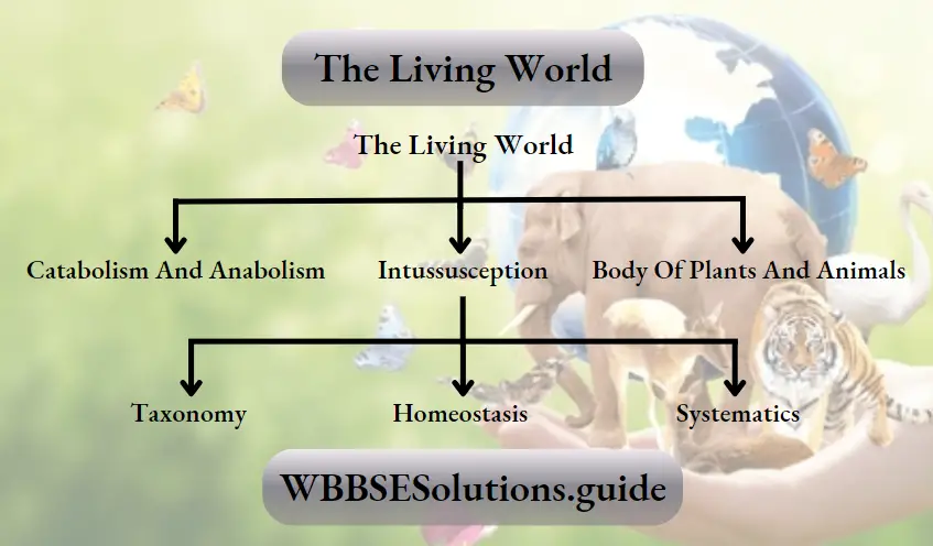
Question 2. What is intussusception?
Answer:
Intussusception
Overall growth of a body due to an increase in the dry weight of the protoplasm, is called intussusception.
Question 3. Which are the coordinators within the body of plants and animals?
Answer:
In case of animals, hormones act as chemical coordinators, while nerves act as physical coordinators. In case of plants, only hormones are present, which act as coordinators. Due to their presence, different cells, tissues, organs and organ systems function in a coordinated fashion.
Read and Learn More WBCHSE Solutions For Class 11 Biology
Question 4. What is homeostasis?
Answer:
Homeostasis
Homeostasis is the ability of maintaining a stable internal environment of the body irrespective of the changes in the external environment. For example, normal body temperature is 98.6°F. When the body temperature rises above this, hypothalamus sends signals all over the body, through the motor neurons. As a result, sweat is released which lowers the temperature to normal.

Question 5. What is taxonomy?
Answer:
Taxonomy
The branch of biology, that deals with the classification of organisms and provide required information for classification such as nomenclature; rules of nomenclature, identification, is known as taxonomy.
Living world important questions and answers for NEET PDF
Question 6. What is systematics?
Answer:
Systematics
The branch of biology, that reveals relationship j among different organisms with the help of identification, nomenclature, description, and classification is known as systematics.
Class 11 Biology WBCHSE
Question 7. What is the difference between taxonomy and systematics?
Answer:
The difference between taxonomy and systematics
The science which deals with identification, classification and nomenclature of organisms is known as taxonomy. On the other hand systematics is the science, that studies diversification of organisms — both past and present and the relationship among them, over the course of time.
Question 8. What is binomial nomenclature?
Answer:
Binomial nomenclature
The system of naming, that is done by using only genus and species name is known as binomial | nomenclature. For example, Pisum sativum.
Question 9. What are taxonomic category and taxonomic hierarchy?
Answer:
Taxonomic category
The different levels of classification are known as categories or taxonomic categories.
Taxonomic hierarchy
When these categories of classification are arranged in a definite order, then it is known as hierarchy or taxonomic hierarchy.
Class 11 biology living world chapter questions and answers
Question 10. What are monotypic and polytypic genus?
Answer:
Monotypic Genes
The genus which includes only one species is known as monotypic genus. For example, sapiens is the only species present in the genus Homo. Hence, Homo is a monotypic genus.
Polytypic Genus
The genus which includes more than one species is known as polytypic genus. For example, tigris and leo, both these species come under the genus Panthera. Hence, Panthera is a polytypic genus.
Class 11 Biology WBCHSE The Living World Multiple Choice Question and Answers
Question 1. Joint Forest Management Concept was introduced in India during —
- 1970s
- 1980s
- 1990s
- 1960s
Answer: 2. 1980s
Question 2. Which is the National Aquatic animal of India?
- River Dolphin
- Blue whale
- Sea-horse
- Gangetic shark
Answer: 1. River Dolphin
Question 3. Red List contains data or information on—
- All economically important plants
- Plants whose products are in international trade
- Threatened species
- Marine vertebrates only
Answer: 3. Threatened species
Short answer questions on the living world with solutions
Question 4. Nomenclature is governed by certain universal rules. Which one of the following is contrary to the rules of nomenclature?
- The first word in a biological name represents the genus name and the second is a specific epithet
- The names are written in Latin and are italicised
- When written by hand, the names are to be underlined
- Biological names can be written in any language
Answer: 4. Biological names can be written in any language
Question 5. Following are the two statements regarding the origin of life —
The earliest organisms that appeared on the earth were non-green and presumably anaerobes
The first autotrophic organisms were the chemoautotrophs that never released oxygen Of the above statements which one of the following options is correct?
- 2 is correct but i is false
- Both 1 & 2 are correct
- Both 1 & 2 are false
- 1 is correct but 2 is false
Answer: 2. Both 1 & 2 are correct
Question 6. Taxonomic categories showing correct hierarchical arrangement in ascending order is—
- Kingdom→ Order → Division→ Class → Genus → Species
- Species→ Genus→ Division → Class → Order → Kingdom
- Kingdom → Division → Class → Order → Family → Genus → Species
- Species→ Genus → Family → Order → Class → Division → Kingdom
Answer: 4. Species→ Genus → Family → Order → Class → Division → Kingdom
Question 7. Which one of the following shows the hierarchical arrangement of taxonomic categories of plants in descending order?
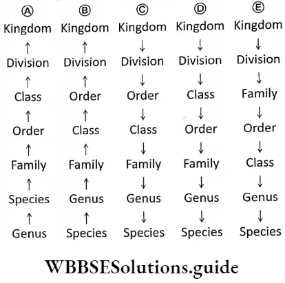
Answer: 4. Kingdom→Division→Class→Order→Family→Genus→Species
MCQs on living world with answers for NEET and competitive exams
Question 8. A taxonomic group of any rank is –
- Taxon
- Tribe
- Race
- Variety
Answer: 1. Taxon
Question 9. Which of the following is correctly sequenced?
- Phylum, class, order, family
- Phylum, order, class, genus
- Phylum, class, family, order
- Phylum, order, family, class
Answer: 1. Phylum, class, order, family
Question 10. Family is placed between—
- Genus and species
- Order and class
- Class and genus
- Order and genus
Answer: 4. Order and genus
Question 11. ICBN stands for
- Indian Congress of Biological Names
- International Code of Botanical Nomenclature
- International Congress of Biological Names
- Indian Code of Botanical Nomenclature
Answer: 2. International Code of Botanical Nomenclature
Difference between living and non-living things with questions and answers
Class 11 Biology WBCHSE The Living World Very Short Question And Answers
Question 1. Name the bird whose beak structure and feeding habit were studied by Darwin, while studying evolution.
Answer: Finch
Question 2. Name the book written by Charles Darwin.
Answer: Origin of Species
Question 3. What is taxon?
Answer:
Taxon
According to international rule of classification, each unit of classification is known as taxon.
Question 4. In which journal did Linnaeus publish his modified classification system?
Answer: Systema Naturae.
Question 5. What is hierarchy?
Answer:
Hierarchy
The arrangement of taxonomic categories one above the other in a definite logical way is known as hierarchy.
Question 6. Expand the abbreviation ICNCP?
Answer:
ICNCP
ICNCP stands for International Code of Nomenclature for cultivated Plants.
Question 7. What is genus?
Answer:
Genus:
The genus is a unit of classification which is formed by collection of species with similar | characteristics.
Question 8. What is meant by trinomial nomenclature?
Answer:
Trinomial nomenclature
The nomenclature that includes genus name, species name, and subspecies name is called: trinomial nomenclature.
Question 9. What is species?
Answer:
Species
Species is a group of living organisms hearing similar characteristic features and they are. capable of interbreeding.
Question 10. What is subspecies?
Answer:
Subspecies
There are several differences among organisms belonging to same species due to differences in geographical location. These species are thus divided into smaller groups known as subspecies.
Question 11. What do you mean by polytypic genus?
Answer:
Polytypic genus
The genus which contains more than one species is known as a polytypic genus. Example- Plasmodium is a polytypic genus. It has GO j species, some of which are, Plasmodium vivax, P. falciperum, P. malariae, etc.
Question 12. What is meant by couplets in identification key?
Answer:
Couplets in identification key
The pair of opposite characteristic features written in taxonomic key is known as couplet.
Question 13. What is identification key?
Answer:
Identification key
The identification key is a set of alternate characters arranged in such a fashion that by selection and elimination of them one can quickly identify the organism. It is an important tool for studying taxonomy It may be printed or computerised.
NEET previous year questions on the living world with solutions
Question 14. What is lead?
Answer:
Lead
The different characteristics which are written as a couplet are known as lead.
Question 15. Name the popular key of taxonomy.
Answer: Indented key is the popular key of taxonomy.
Question 16. Given below is the scientific name of mango.
Identify the correctly written name.
- Mangifera Indica
- Mangifera indica
Answer: 2. Mangifera indica
Question 17. Linnaeus is considered as father of taxonomy. Name two other botanists known for their contribution to the field of taxonomy.
Answer: George Bentham and Sir J.D. Hooker.
Question 18. What does ICZN stand for?
Answer:
ICZN
ICZN stands for International Code of Zoological Nomenclature.
Question 19. What is a museum?
Answer:
Museum
A museum is an institution that preserves objects of scientific, artistic, cultural or historical importance.
Question 20. Can you identify the correct sequence of Itaxonomical categories?
- Species —> Order —> Phylum —> Kingdom
- Genus —> Species —> Order —> Kingdom
- Species→ Genus→ Order → Phylum
Answer: 3. Species→ Genus→ Order → Phylum
Question 21. Taxonomic key is one of the taxonomic tools in the identification and classification of plants and | animals. It is used in the preparation of—
- Monographs
- Flora
- Both 1 and 2
- None of these
Answer: 3. Both 1 and 2
Question 22. The term systematics refers to—
- Identification and classification of plants and j animals
- Nomenclature and identification of plants and ; animals
- Diversity of kinds of organisms and their ; relationships
- Different kinds of organism and their classification
Answer: Both 1 And 2
Question 23. Which is the largest botanical garden in the world? Name a few well known botanical gardens in India.
Ans. Royal Botanical garden, ICew (London). Some well known botanical gardens in India are
- Acharya Jagadish Chandra Bose Indian Botanic Garden, Shibpur, Howrah;
- Lloyd Botanical Garden, Lucknow.
Question 24. Genus represents—
- An individual plant or animal
- A collection of plants or animals
- A group of closely related species of plants or animals
- None of these
Answer: 3. A group of closely related species of plants or animals
Class 11 Biology WBCHSE
Question 25. How is key helpful in identification of living organisms?
Answer: Key helps to identify various taxa such as genus, species, etc. It is a systemic representation of different identifying characters belonging to different taxa.
Question 26. How can you relate metabolism with growth?
Answer: Metabolism involves both catabolism and anabolism. Growth occurs when the rate of anabolism is greater than the rate of catabolism.
Question 27. How can you distinguish between human beings and other living organisms?
Answer: Human beings have ability to think about higher concepts that other animals cannot. This feature distinguishes human beings from other animals.
Question 28. Amoeba multiplies by mitotic cell division. Is this phenomenon growth or reproduction? Explain.
Answer: In unicellular organisms like Amoeba, cell division is the means of multiplication. So, it helps in reproduction. But it also helps in the growth of their colony. Thus, in unicellular organisms, mitosis helps in both growth and reproduction.