Longitudinal Tracheal Trunk
Longitudinal Tracheal Trunk Definition: The three pairs of air-filled longitudinal tubes present on each side of the abdominal cavity of a cockroach are called longitudinal tracheal trunks.
Longitudinal Tracheal Trunk Number and types: Three pairs of large longitudinal tracheal trunks are present on each side of the body- one pair dorsal, one pair ventral, and one pair lateral in position.
Therefore, a total of six longitudinal tracheal trunks are present in the body of a cockroach.
Longitudinal Tracheal Trunk Position: The dorsal and ventral pairs of longitudinal tracheal trunks are found in the median region of the abdomen. The lateral pair is present along the lateral part of the abdomen.
Number of longitudinal tracheal trunks in cockroach
Longitudinal Tracheal Trunk Structure: The paired tracheal trunks are interconnected by many transverse commissures or commissural tracheae or transverse tracheal connective (a commissure is a bundle of nerve fibers that cross the midline at the point of their entry or origin). Each longitudinal trunk is divided into many branches called tracheoles.
Read and Learn More: WBCHSE Notes for Class 11 Biology
Longitudinal Tracheal Trunk Function: The longitudinal tracheal trunk transports air (that has entered through the spiracles) throughout the body.
| Class 11 Biology | Class 11 Chemistry |
| Class 11 Chemistry | Class 11 Physics |
| Class 11 Biology MCQs | Class 11 Physics MCQs |
| Class 11 Biology | Class 11 Physics Notes |
Tracheae Definition: The air-filled, elastic, closed, membranous network of branching tubules that opens through the spiracles to the atmosphere, is called tracheae.
Tracheae Number: From the mesothoracic spiracles, 6 tracheae originate. The rest of the spiracles lead to 3 tracheae at each end, leading to 6 tracheae per segment.
Tracheae Structure:
- The trachea are hollow tubes that are made up of epithelial tissue.
- These tracheae arise from the longitudinal tracheal trunk and their transverse connectives.
- They undergo repeated branching and form a diffused network of finer tracheae.
- A type of cell known as tracheal end cell is found at the terminal of each trachea.
- Several finer tubes arise from these cells. These are known as tracheoles.
- The cytoplasmic appendages remain suspended in the cell sap.
- Along the length of the trachea, some swollen regions known as air sacs can be seen.
- These air-sacs act as the reservoirs of air.
- Each tracheal tube is lined by a thin strip of cuticle that is arranged in a spiral form.
- This chitinous spiral layer of cuticle is called taenidia or intima. It prevents the collapse of the tracheal walls when empty.
- It also provides an elastic nature to the tracheal walls. Smaller tracheae lack taenidia.
Tracheae Function: The network of tracheae helps in the transport of gases to the different cells.
Tracheoles Definition: Tracheoles are finer branches of tracheae that transport air directly to the body cells.
Number, structure, position: Tracheoles are very large in number. They have very thin walls (diameter lp) and are devoid of taenidia. Their inner wall consists of the protein, trachein.
This thin wall enables the tracheoles to come in direct contact with the cells.
Cockroach respiratory system longitudinal tracheal trunks
Tracheole Function: The opening of each tracheole within the tissues is immersed in the tissue fluid which supplies O2 to the cells and removes CO2 from the cells.
Tracheolar fluid is present inside the tracheoles. The level of the tracheolar fluid varies with the metabolic activity of the insect. It is more when the insect is inactive (or at rest).
On the other hand, this fluid gets completely reabsorbed into the tissues, and the space gets filled with air when the insect is more active.
” cockroaches breathe through “
Mechanism of breathing: In cockroaches, the mechanism of breathing includes two phases— inspiration and expiration.
The spiracles mediate inspiration and expiration as they facilitate the exchange of gases. Tergo-sternal muscle is a muscle located between the abdominal tergum and sternum of a cockroach. Contraction and relaxation of this muscle induces breathing.
The steps of inspiration and expiration are as follows—
Inspiration:
- Intake of atmospheric air or more specifically O2 into the tracheal system is called inspiration. It is a passive process (does not require expenditure of energy).
- The 1st and 3rd pairs of spiracles remain open all the time but, the remaining 8 pairs open only during inspiration.
- Relaxation of the tergo-sternal muscles expands the abdominal cavity. As a
result, the pressure in the abdominal cavity decreases. - This causes atmospheric air to enter through the spiracles. This air then passes through the trachea and finally reaches the tracheoles which contain tracheolar fluid.
- O2 diffuses into tissues through the tracheolar fluid and reaches the cells or tissues.
Expiration:
- The process of elimination of CO2 produced during metabolism is known as expiration. Expiration is an active process (requires expenditure of energy).
- Contraction of the tergo-sternal muscles decreases the volume of the abdominal cavity. As a result, the pressure inside the cavity increases.
- This causes air from the tracheoles and tracheae to be released through six abdominal spiracles.
- Opening and closing of spiracles are influenced by CO2 tension in hemolymph and O2 tension in the tracheae.
Structure of tracheal trunks in cockroach
Discontinuous gas exchange cycles
- Cockroaches exhibit the phenomenon of discontinuous ventilation or discontinuous gas exchange cycles (DGC).
- In this process, the exchange of gases is interrupted for certain periods during which spiracles remain closed.
- This discontinuous gas exchange cycle has three phases—closed phase (spiracles close), flutter phase (spiracles open slightly but close rapidly), and open phase (spiracles open completely).
- In cockroaches, most of the CO2 is released through the cuticle by diffusion through the cuticle.
- However, only a small amount of CO2 is eliminated through the trachea and spiracles.
Control of breathing: Different factors controlling breathing mechanism in cockroaches are—
5 Role of the nervous system: Breathing in cockroaches is coordinated and regulated by nerve centers in the thoracic ganglia. The coordinating centers in the thoracic ganglia are stimulated by variations in O2 and CO2 content.
Role of metabolic rate: When the cockroach is active, i.e., metabolic rate is high, the osmotic pressure of tracheal fluid increases. Most of the fluid gets absorbed by the cells.
This results in a better supply of O2 into tissues. However, when the cockroach is at rest or during low metabolic activity, the osmotic pressure of tracheal fluid reduces and the fluid fills up the terminal part of the tracheoles. A smaller amount of tissue fluid is absorbed by the cells.
This results in a slow rate of diffusion of O2.
Longitudinal tracheal trunks in insects explained
Thermal control: An increase in temperature increases the diameter of spiracles. This, in turn, allows more gaseous exchange.
A fall in the temperature of the brain to 8°C changes the breathing pattern of cockroaches from a continuous to a discontinuous type.
Chemical control: An increase in C07 concentration I increases the rate of breathing.
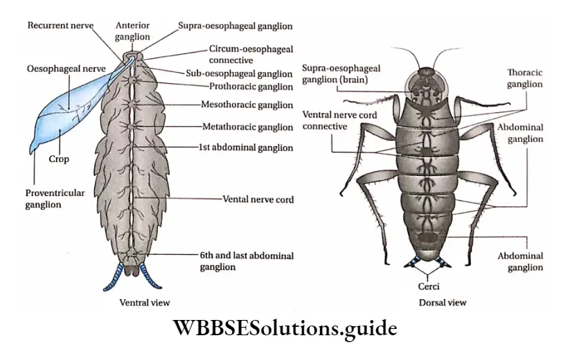
Nervous system Definition: The system of nerves and ganglia that coordinates and regulates various functions of the body in response to the environment, is called the nervous system.
“trachea of cockroach “
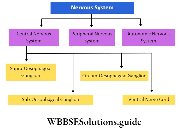
The nervous system of cockroaches has three components namely, the central nervous system, the peripheral nervous system, and the autonomic nervous system.
Central nervous system: The part of the nervous system, mainly comprising the brain, that coordinates the functioning of the body is called the central nervous system.
The components of the central nervous system are the supra-oesophageal ganglion, sub-oesophageal ganglion, circum-oesophageai ganglion, and ventral nerve cord.
Supra-oesophageal ganglia: It is a bilobed structure that acts as the brain.
Function of longitudinal tracheal trunks in cockroach
Position: It lies above the esophagus, almost in between the bases of the antennae, within the head.
Structure: Supra-oesophageal ganglia are formed by the fusion of three pairs of ganglia known as proto-, auto-, and tritocebrum. it is concerned mainly with sensory function.
Sub-oesophageal ganglia: It consists of a pair of ganglia that innervate the mouthparts.
Sub-oesophageal ganglia Position: It is located below the esophagus.
Sub-oesophageal ganglia Structure: It is formed by the fusion of the remaining three pairs of cephalic ganglia.
It is the main motor center, that is concerned with the motor functions. It innervates the muscles of mandibles, maxillae and labium and hypopharynx.
“Cockroach tracheal system anatomy”