
Biological Classification Introduction
Biological Classification
- Every day we come across different kinds of organisms. We see different kinds of plants in the fields, gardens, etc. But why are the plants all so different from each other? Or why are the animals we see around us so different from each other? This is because different organisms have different characteristic features.Φ
- Depending on these characteristics, they have been classified under different classes or groups. This is known as biological classification. In this chapter, we shall learn about the different types of classification of the organisms, that have been followed since ancient times to modern days.
Biological classification Definition: Biological classification is the scientific classification of living organisms based on their morphology, habitat, evolution similarities and differences with other organisms.
Read and Learn More: WBCHSE Notes for Class 11 Biology
- Classification of organisms has been carried out since the dawn of human civilisation. Aristotle (384-322 BC) had classified organisms into two large groups—plant kingdom and animal kingdom.
- But this system failed to classify several organisms, such as unicellular green algae, fungi, etc. These organisms could not be placed in any of the above-mentioned two groups.
- It even failed to distinguish between prokaryotes and eukaryotes. Later on, several other modified classification systems have been developed.
Importance of biological classification:
Biological classification is important in following ways—
- Identification of organisms: Using a proper classification system, organisms can be identified easily.
- Attaining knowledge about the living world: A classification system helps to categorise organisms on the basis of their morphological features, habitat, etc.
- Correlating different groups of organisms: Classification helps us to correlate different groups of organisms, through their similarities and dissimilarities.
- Discovery of new species: The system of classification makes it easier to classify newly discovered species. Since the dawn of civilisation, there have been many attempts to classify living organisms. But it was done without using scientific criteria.

Cyanobacteria or Blue-Green algae (BGA)
Cyanobacteria Definition: The gram negative, unicellular prokaryotes, containing photosynthetic pigments that can carry out photosynthesis, are called cyanobacteria or cyanophyta.
- Cyanobacteria contain photosynthetic pigments like chlorophyll and various accessory pigments. They are also called oxygenic photoautotrophs.
- Cyanobacteria are considered to be first organisms on earth that released oxygen. With evolution, several aerobic organisms developed from the cyanobacteria.
- They were known as | blue-green algae, myxophyceae or cyanophyceae. In 1978, ICNB (International Code of Nomenclature of | Bacteria) named them as cyanobacteria.
| Class 11 Biology | Class 11 Chemistry |
| Class 11 Chemistry | Class 11 Physics |
| Class 11 Biology MCQs | Class 11 Physics MCQs |
| Class 11 Biology | Class 11 Physics Notes |
Biological classification important notes for NEET PDF
Distribution: Cyanobacteria are usually free-living marshy regions. They are also found in sea, under the ice, desert region, lakes, etc.
General Features
General features Structure:
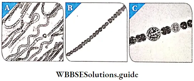
- Shape and size: Cyanobacteria are unicellular, They may exist as unit (Spirulina sp.), colony (Nostoc sp.) or filamentous (Oscillatoria sp.). The cells may be large, spherical or oval shaped, with size ranging from l//m to 50 pm.
- Protoplasm: The protoplasm is generally divided into two parts. The pigment-containing part, called chromoplasm or chromatoplasm, is present towards the periphery. While the inner region contains a colourless part called centroplasm. Thylakoids are present in the chromoplasm.
- Cell wall: They possess a cell wall with an inner thick peptidoglycan layer, similar to gram positive eubacteria. The outer membrane contains proteins, lipids and carotenoids. There is a mucilage layer outside the cells.
- Cell membrane: The lipoprotein-containing cell membrane is present just below the cell wall.
- Lamellasome: Similar to mesosome in bacteria, cyanobacteria have a circular extracellular appendage, that helps in respiration. These are known as lamellasomes.
- Thylakoids: All cyanobacteria contain photosynthetic pigments like chlorophyll a. They also contain blue phycobilin pigments, phycocyanin and allophycocyanin. Many members also contain another pigment, phycoerythrin, making the cells appear red, or sometimes black.
- These phycobilins are present in some special structures, on the thylakoid membranes. These structures are known as phycobilisomes. These are highly efficient channels that transfer captured solar energy (excitation energy) to the reaction centres of photosystems.
- Nucleoid: Cyanobacteria lack true nuclei and other organelles like mitochondria and chloroplastids. The naked, circular DNA (DNA without histone proteins) is arranged closely within the nucleoid. Plasmids may also be present.
- Ribosomes: 70S ribosomes may exist and form polyribosomes.
- Gas vacuoles: Cyanobacterial cells contain numerous gas vacuoles.Gas vacuoles provide buoyancy to the cells, allowing the cyanobacteria to float on the surface. This allows more exposure to sunlight for photosynthesis.
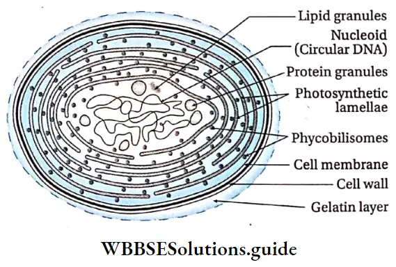
Stored materials: Cyanobacteria also contain various types of reserve food. They include—
- α-granules which are cyanophycean starch and look similar to glycogen (polyglucose) granules, which store carbon
- β- granules(lipid granules),
- Protein granules, polyphosphate or volutin granules, etc., which allow cells to accumulate energy and nutrients.
Locomotion: Cyanobacteria move by gliding along the surfaces of the substratum.
Reproduction: It reproduces by vegetative and asexual modes of reproduction. Sexual reproduction is totally absent.
- Vegetative reproduction: It occurs by the following : methods—
- Fission: Unicellular cyanobacterial cells usually divide and reproduce by fission. The cell divides into two daughter cells,
- Fragmentation: The filamentous members reproduce vegetatively through fragmentation. Each fragmented piece of the filament, germinates into a new colony.
- Hormogonia: At the end of the growing season, filaments or trichomes break into multicellular pieces. These pieces secrete thick wall around the mass of cells, forming structures called hormogonia: These hormogonia can germinate into new filaments at the onset of favourable conditions.
- Asexual reproduction: Many non-motile cyanobacteria reproduce asexually by spore formation. The different types of spores formed are—
- Endospore: These are formed within the cell, Here the cells increase in size and their protoplasm divides to form endospores, e.g., Dermocarpa.
- Exospore: These are produced outside the cell.
- By the dissolution of the apical region of the cell surface, the protoplast gets exposed. From this exposed region of the protoplasm, round spores are formed in basipetal succession. E.g., Chaemosiphon.
- Akinetes: These are formed close to the heterocysts. The akinete mother cells increase in size and then develop a wall around it. Under favourable conditions, they give rise to new filaments. E.g., Cylindrospermum.
- Heterocyst: Some filamentous cyanobacteria form special types of thick walled cells, known as the heterocysts. The outer layer of the cell wall is made up of pectin or cellulose and the inner layer is of cellulose. They are usually yellow in colour due to the presence of carotene. They have rudimentary reproductive structures, as they can germinate into new filaments, under favourable conditions. E.g., Anobaena sp., Nostoc sp., Spirulina sp., Scytonema sp.
Mycoplasma
Mycoplasma Definition: Mycoplasmas are simple, small, gram-negative, prokaryotic organisms that lack a cell wall and can survive without oxygen.
Nocard and Roux discovered mycoplasma in 1898. Mycoplasma is also known as PPLO (Pleuro Pneumonia Like Organism) or MLO (Mycoplasma Like Organism). They are the smallest living cells known. Many mycoplasmas are pathogenic to both animals and plants.
Example: Mycoplasma gallisepticum, Mycoplasma genitalium, Mycoplasma pneumoniae, etc.
General Features
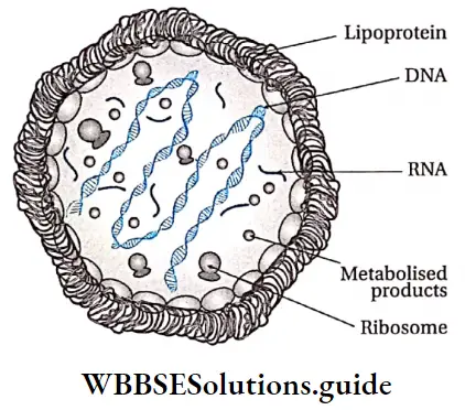
Cell type and structure: They are tiny, prokaryotic microorganisms (0.1-0.15/ym). The shape of mycoplasma varies from being spherical to filamentous with no cell walls. Due to absence of cell wall, they can change their structure.
This phenomenon is known as polymorphism. They contain acetylglucosamine in the cell membrane. Mesosome is absent. Ribosomes are present. Their genome consists of a double-stranded linear but coiled DNA molecule.
- Nutrition: They can be parasitic or saprophytic.
- Respiration: They are obligate anaerobes, usually without any electron transport system (ETS). Even if present, ETS is inactive. ‘
- Reproduction: Mycoplasma reproduce by binary fission. Sometimes, only the nucleoid shows binary fission, without cytokinesis.
- Effect on animal world: Several species are pathogenic in humans. These include M. pneumoniae, which causes pneumonia and other respiratory disorders. Another is M. genitalium, which causes pelvic inflammatory diseases.
- Distribution: Actinomycetes are primarily soil dwellers. But they are also widely distributed in a diverse range of aquatic ecosystem, including sediments | obtained from seabed.
General Features
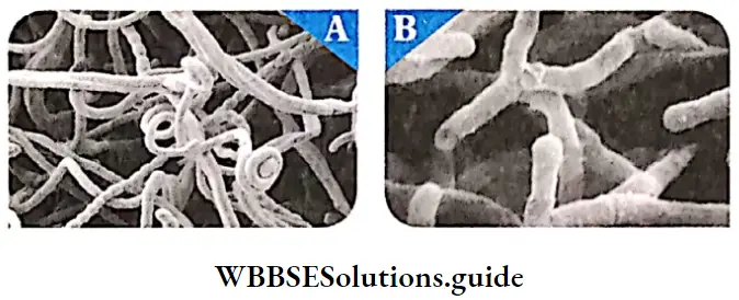
General Features Structure: Actinomycetes are gram positive, prokaryotic microorganisms that have branched hyphae. The hyphae are usually aseptate but septa may be | formed under special conditions in some forms. The hyphae form mycelium like structure. The cell wall is made up of peptidoglycan, teichoic acid, teichuronic acid and polysaccharides. The chemical composition of their cell wall is similar to that of gram-positive bacteria.
The rigid cell wall maintains the cell shape. There are tiny chromatin granules present within the cell.
- Nutrition: They are heterotrophic in nature. Some are saprophytic aerobes growing in soils and natural habitats.
- Respiration: They are facultatively anaerobic.
- Temperature sensitivity: They are generally mesophilic in nature. They are active at a temperature of 35°C.
- Effects on animal world: Some of the actinomycetes species are used to prepare antibiotics. Some actinomycetes also cause disease (Actinomycosis) in humans.
Spirochaetes
Spirochaetes Definition: The heterotrophic prokaryotes, that are j present in water and muddy regions are called j spirochaetes.
General Features
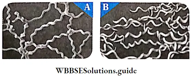
General Features Structure: Spirochaetes have long, helically coiled cells. The cells usually have diameter around 0.1 – 0.6pm and have cell wall. They are gram-negative bacteria. They are motile organisms. Spirochaetes are chemoheterotrophic in nature. They are sensitive to desiccation.
Effects on animal world: The majority of spirochaetes cause disease in man.
Examples: Leptospira sp., Borrelia sp., Treponema pallidum, etc.
Rickettsiae
Rickettsiae Definition: Rickettsiae intracellular, gram negative coccobacillary forms that ‘ multiply within eukaryotic cells.
Rickettsiae General Features
Rickettsiae Cellular structure: Cells are 0.3 – 0.5 x 0.8 – 2.0pm in size. The cell wall is chemically similar to that of gram negative bacteria. It contains diaminopimelic acid and lacks teichoic acid.
- Their outer membrane is composed mostly of lipopolysaccharides.
- They lack a distinct nucleus and membrane bound organelles. Flagella, pili, mucilage capsule are absent.
- They contain both RNA and DNA as nucleic acids. Mesosomes and ribosomes are also present in the cells.
- Nutrition: They are parasitic in nature (obtain nutrition from host cell).
- Growth and Reproduction: Rickettsia normally multiplies by transverse binary fission.
- Effects on animal world: In their arthropod vectors (insects that can carry the organism), the rickettsia multiply within the epithelium of the intestinal tract. They are transmitted to man, via the arthropod saliva, through a bite. In their mammalian host (i.e., man), they are found mainly in the endothelium of the small blood vessels, particularly in those of the brain, skin and heart.
- Rocky Mountain spotted fever is transmitted by the bite of a Dermacentor tick, which carries the rickettsia. This disease is common in warm-blooded animals, including humans.
Rickettsiae Example: Rickettsia typhi
Class 11 biology biological classification notes with diagrams
Kingdom Protista
- This is the kingdom comprising unicellular, eukaryotic organisms that have features similar to those of fungi, plants or animals. The concept of kingdom Protista was given by taxonomist E. Haeckel in 1866.
- All single-celled eukaryotes, other than green algae and red algae, have been placed under Protista. This kingdom forms a link between Kingdom Monera and the Kingdoms Fungi, Plantae and Animalia.
- Distribution: Most of the members of this kingdom live in water or aquatic habitats. Majority of them are found as planktons within marine water, fresh water, etc.
Kingdom Protista General Features
Kingdom Protista General Features Cell type: The members of this kingdom are tiny, unicellular eukaryotes. Some of them may exist as colony or filaments.
- Cell covering: Cell wall is usually absent; if present it may contain silica (e.g., diatoms). Cell membrane is covered by a pellicle, cuticle or shell. This forms a double-envelope system around the cells.
- Protoplasm: The protoplasm contains a well defined nucleus and other membrane bound organelles. The cells contain 80S (60S + 40S) ribosome in the cytoplasm. On the other hand, 70S ribosome is present in mitochondria and plastids.
- Chloroplast: The members may be photosynthetic or non-photosynthetic. Thylakoids, – containing photosynthetic pigments, are present within the j chloroplast of the photosynthetic members.
- Locomotion: Most of them can show locomotion. The cells may contain cilia, flagella or pseudopodia. The cytoplasm, along with organelles, show a flowing j movement. This movement is called cytoplasmic j streaming or cyclosis.
- Nutrition: Some are photoautotrophs, some heterotrophs, and some are mixotrophs (combining photosynthesis and heterotrophic nutrition). Some are holophytic, holozoic, parasitic, saphrophytic or symbiotic in nature. Some autotrophs are symbiotic in nature.
- Respiration: Most protists are aerobic, but parasitic and ones living deep in the ocean, are anaerobic.
- Reproduction: Protists reproduce asexually as well j as sexually.Asexual reproduction occurs by binary fission, j multiple fission, sporulation, cyst formation, etc. Sexual reproduction, on the other hand, occurs by syngamy and j conjugation. j
- Cell division: Both mitosis and meiosis occur.
- Life cycle patterns: The three basic life cycle patterns found in eukaryotes are represented by pr.otists. They are—Zygotic or haplontic, gametic or diplontic, sporic or haplodiplobiontic. At some point in the life cycle of many protists cysts (resistant cells) are formed.
Classification of Kingdom Protista
The members of Protista fall under the following three categories as shown in the chart below. These have been discussed under separate heads.
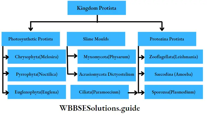
Photosynthetic protista
These are unicellular, eukaryotic organisms, containing photosynthetic pigments. They are mainly found in aquatic ecosystems. They are mainly of three types—
Chrysophyta, Dinoflagellata and Euglenophyta.
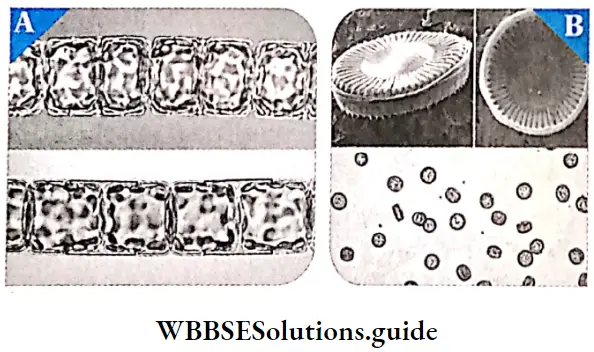
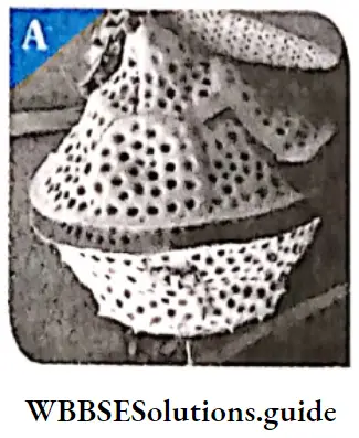
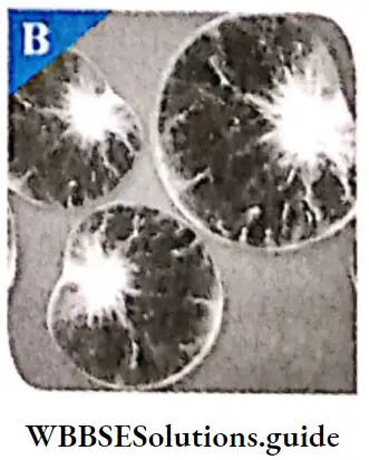
Chrysophyta: This group includes diatoms and golden algae. They are fresh water algae as well as marine dwellers. They are pollution indicators, i.e., they cannot grow in polluted regions, hence indicate the level of pollution.
Chrysophyta General Features
- Cellular structure: They are unicellular, microscopic and planktonic i.e., they float passively in water currents.The cells are enclosed within a shell made up of silica, called frustule.ln diatoms, the frustule divides to form two thin halves, which fit together as the lids in a soap-case.The cell wall is made up of silica along with cellulose, that makes it hard and porous. Chromatophores are present in the cells.
- These contain pigments like chlorophyll, /2-carotene and a special type of xanthophyll (diatomin). Fucoxanthin, which is a characteristic pigment of brown algae, is present. Due to the presence of /3-carotene and diatomin, diatoms appear golden.
- Locomotion: Diatoms do not have flagella. Mucilage and oil globules help them to float on water.
- Symmetry: Both bilateral and radial symmetry is observed among the members.
- Nutrition: Most diatoms are photosynthetic (autotrophic) but some have evolved to become heterotrophic.
- Reproduction: Diatoms reproduce by vegetative, asexual and sexual means. Vegetative reproduction takes place by cell division.Asexual reproduction occurs by binary fission. Sexual reproduction takes place by the formation of auxospores.
Chrysophyta Examples: Melosira sp., Cyclotella sp., Striatella sp., Hydrosira sp., etc.
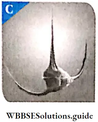
Dinoflagellata or Pyrrophyta: These are biflagellate, unicellular, marine algae.
Dinoflagellata General Features
Dinoflagellata Cellular structure: These organisms are mostly ; marine and photosynthetic. They appear yellow, green, brown, blue or red depending on the pigments present. Dinoflagellates may be covered by an outer covering outside their cells.
- Depending on their; presence, they are of two types—unarmoured (without outer covering) and armoured (with outer covering).
- Unarmoured dinoflagellates have a smooth pellicle or periplast. The cell wall has hard, cellulosic plates on the outer surface in case of armoured. These are known as the theca. The nucleus is large.
- It has distinct nucleolus and nuclear envelope. Chromatin is present in chromosome form. Histone proteins are absent. Such a nucleus is known as mesokaryon.
Flagella: Most of them have two flagella. One lies longitudinally and the other transversely, in a furrow between the wall plates.
- The flagella that is present in the longitudinal groove functions as a radar. The other flagella fits transversely in the furrow. It is used to create a rotational motion within the water.
- Nutrition: Most of these organisms are autotrophic. They have chlorophyll a, chlorophyll c, fucoxanthin, etc., as photosynthetic pigments.
- Stored food: Food is stored in the form of starch (polysaccharides) and oils (lipids).
- Reproduction: Dinoflagellates show two types of reproduction— asexual and sexual. During normal condition, they reproduce asexually, by cell division.
- But under certain stressful conditions, they undergo a different process of reproduction. Two dinoflagellates undergo fusion and form a diploid zygote.
- This diploid zygote state remains throughout the period of stress. Once conditions become favourable, the diploid zygote breaks off into small pieces called cysts, that grow into new organisms.
Examples: Gonyaulax sp., Gymnodium sp., etc.
Euglenophyta: They are unicellular, photosynthetic, fresh water protists that have features similar to those of plants and animals.
Euglenophyta General Features
- General Features Distribution: Majority of them are fresh water organisms, living in stagnant water.
- Structure: The cells of most euglenoids are spindle-shaped. There is no cell wall or other rigid structure covering the plasma membrane.
- Cell covering: Instead of a cell wall, they have a protein rich layer, called pellicle, which makes their body flexible.
- Flagella: They have two flagella, a short and a long one.The flagella are tinsel shaped and have hairs on them.Both the flagella arise from their respective basal granules.Blepharoplasts or basal bodies are present at the basal end of the flagella.
- Eye-spot: Eye-spot is present at the end of the flagella. It contains a red-coloured astaxanthin pigment, that acts as photoreceptor (sensitive to light). The eyespot appears to be connected to the flagellum by special strands of cytoplasm that transmit signals from one organelle to the other.
- Nutrition: Euglenoids are mostly autotrophic in nature. Photosynthetic euglenoids contain chloroplast just like green algae. Chlorophyll a, chlorophyll b and xanthophyll pigments are present in the chloroplast. Non-photosynthetic euglenoids are saprophytic in nature. Some of the non-photosynthetic euglenoids are holotrophic while some are mixotrophic in nature.
- Stored food: Euglenoids have a carbohydrate food reserve, called paramylon. It is generally present in the form of small pyrenoid granules.
- Locomotion: They generally swim in water, using their flagella, but may also move over wet land by creeping.
- Reproduction: Reproduction in euglenoids takes place by asexual means, i.e., by mitotic cell division. Example: Euglena viridis, etc

Euglenoids
The euglenoids were first defined by Otto Biitschli in 1884 as the flagellate order, Euglenida. They possess flexible cell coverings, that enable them to move about freely. They also ingest their food through a structure called gullet. So earlier they were considered as animals. However, they had certain characteristics similar to plants. Botanists subsequently created the algal division Euglenophyta. Presently they are classified as both animals and plants.
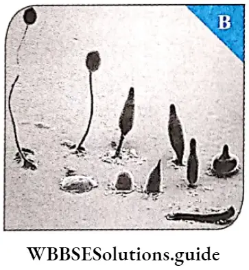
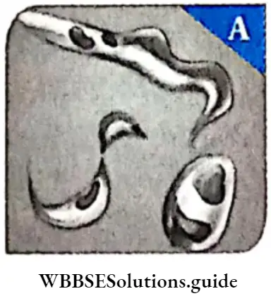
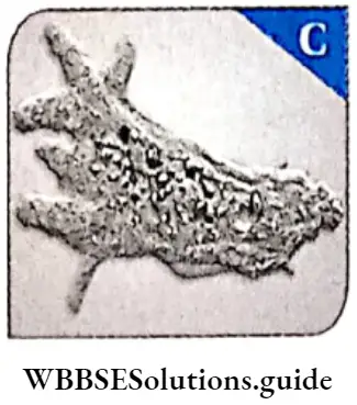
Slime moulds
Slime moulds Definition: Slime moulds are saprophytic protists that do not have a specific structure (amoeboid) and can grow in humid environment.
Slime moulds Distribution: Slime moulds were formerly classified as fungi. They are found mainly in wet, marshy regions. Some of the species of slime moulds are aquatic in nature.
Slime moulds General Features
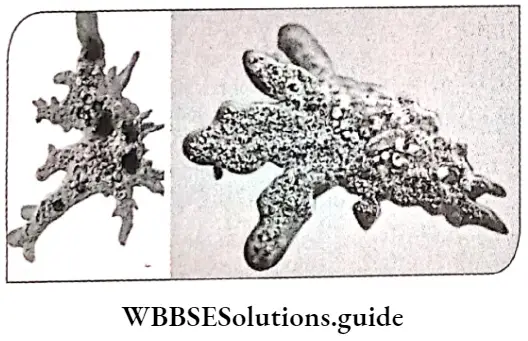
General Features Structure: During favourable conditions, they live as independent, amoeba-like cells. These structures have single nucleus and are called myxamoeba. They feed on fungi and bacteria. However, during scarcity of food and unfavourable temperature, they form an aggregated structure. This structure is called plasmodium.These structures may grow and spread over a large distance. The outer part of the body is covered by mucilage, hence the name slime mould.
- Locomotion: They move very slowly using pesudopodia. Such a motion is called amoeboid movement.
- Nutrition: They are saprophytic and phagotrophic in nature. Some of them are parasites.
- Reproduction: Both asexual and sexual types of reproduction are found. Asexual reproduction takes place by binary fission, plasmotomy (type of asexual reproduction in which a multinucleate protozoan cell divides into two or more multinucleate daughter cells, without mitosis), cysts or sclerotium formation, etc.
- Life cycle: Zygotic and gametic meiosis take place within the life cycle.
Slime moulds General Features Types: There are two main groups of slime moulds in the Kingdom Protista. They are—Plasmodial slime moulds and Cellular slime moulds.
Plasmodial slime moulds or Myxomycota or true not have a specific structure. They are large single-celled mass, without the cell wall but contain thousands of nuclei (structure called plasmodium). A slimy secretion is released by the slime mould.
Examples: Physarum sp., Fuligo sp.
Cellular slime moulds or Acrasiomycota: They spend most of their lives as haploid, single nucleus containing, amoeboid protists. They show amoeboid motion, hence called myxamoeba. Upon the release of a chemical signal, the myxamoeba aggregate into a clustered structure, known as a pseudoplasmodia. This pseudoplasmodia eventually form multicellular slugs. They contain a layer of cellulose, outside the spores.
Example: Physarum sp., Stemonitis sp., Fuligo sp., Dictyostelium sp.
Protozoa Protista
Protozoa are unicellular, heterotrophic, eukaryotic organisms. They live as predators or parasites. They are considered to be the primitive relatives of animals.
Protozoa Protista General Features
- General Features Symmetry: The body may be asymmetrical, or radially, bilaterally or spherically symmetrical.
- Shape: They are of different shapes—spherical or oval shaped. Some of them do not have a definite shape or structure, such as Amoeba sp.
- Body structure: They are eukaryotic, unicellular organisms. The cell may contain one or more nucleus, They have very thin cell membrane. Sometimes the cell membrane may be covered by pellicle (Paramoecium sp.) or calcareous or siliciferous shells (Elphidium sp.).
- Locomotion: Generally, members of protozoa such as Plasmodium, show contractile motion by pseudopodia, flagella or cilia. On the other hand, members like Monocystis, do not have organs for locomotion. Such organisms move about by cellular contractions, using contractile fibrils called myoneme threads.
- Osmoregulation: Protozoa living in fresh water are subjected to a hypotonic environment. Protozoa have contractile vacuoles, water vacuoles, etc., that collect and expel excess water. Some waste products and carbon dioxide are also excreted by this vacuole, along with the water. This is how protozoa perform osmoregulation.
- Nutrition: The mode of nutrition in most of the free-living protozoa is holozoic. Other modes of nutrition found in many protozoa are saprophytic, symbiotic or parasitic.
- Respiration: Aerobic respiration is seen in free-living organisms, while anaerobic respiration is seen in ectoparasites (parasites living on the body surface of the host).
- Excretion: Waste products of protozoans are nitrogenous wastes like ammonia, C02, excess water, etc. These are expelled from the body through the contractile vacuole.
- Reproduction: Both asexual and sexual reproduction are observed in these organisms. Asexual reproduction takes place by binary fission. Sometimes, multiple fission may also occur. Sexual reproduction may occur by syngamy and conjugation. Under unfavourable conditions, they form cysts, which become active during favourable conditions.
- Reserve food: Reserve food is generally glycogen (example,. Entamoeba sp.).
Protozoa Protista General Features Types: There are four major groups of protozoans according to the organ of locomotion. —
Biological classification chapter summary with key points
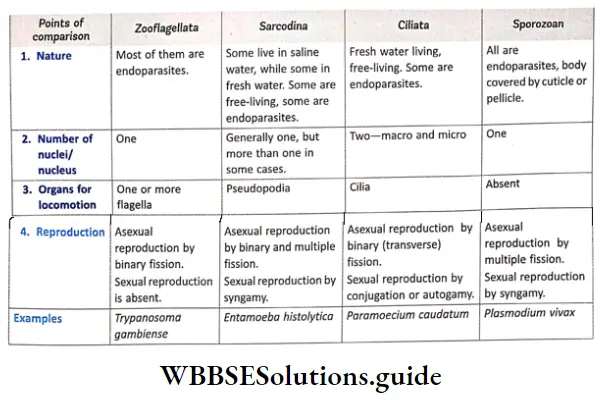
Zooflagellata, Ciliata, Sarcodina, Sporozoans
Zooflagellata (Flagellated protozoans):
Zooflagellata General Features
- The members of this group are either free-living or parasitic.
- They have one or more flagella. Some of them (parasitic forms) can cause diseases, e.g., Trypanosoma sp. causes sleeping sickness.
- The flagellated protozoans range from a simple oval cell with one or more flagella to collared flagellates (organisms with funnel-shaped collar of interconnected microvilli, at the base of the flagella).
- Most of the members have lost the ability to photosynthesise. They generally show parasitic nutrition, some may show saprophytic nutrition.
- Only asexual reproduction, by longitudinal binary fission, is observed. They may exist either as single cells (e.g., Paraphysomonas sp.) or as colonies (e.g., Codosiga sp.).

Sarcocfina (Amoeboid protozoans)
Sarcocfina General features
- These organisms live in fresh water, sea water or moist soil.
- The cells may contain more than one nucleus.
- The organisms move and capture their prey by stretching out their pseudopodia (false feet) as in Amoeba sp.
- The mode of nutrition is heterotrophic and holozoic. Some of them such as Entamoeba sp. are parasites.
- Marine forms have silica shells.
- Reproduction occurs both by asexual and sexual means. Asexual reproduction occurs by binary or multiple fission. Sexual reproduction occurs by syngamy.
Sarcocfina Example: Amoeba, etc.
Comparison among the four classes of protozoa:
General Features
- These are aquatic, actively motile organism.
- Most of them are free-living,however, some may form colonies. Some may also be endoparasitic in nature.
- The cilia are the main organs of locomotion.
- Most of these are heterotrophic and holozoic. They have modified structures for absorbing their food, such as cytostome, cytopyge or cytoproct. Digestion takes place within the food vacuoles.
- They have a cavity (gullet) that opens to the outside of the cell surface. The coordinated movement of rows of cilia makes any food in the water to enter the gullet.
- There are two nuclei in the cell, the larger one called macronucleus and smaller one called micronucleus.
- Both asexual and sexual reproduction are seen among the members. Asexual reproduction takes place by transverse binary fission. Sexual reproduction takes place by conjugation.
Example: Paramoecium sp.
Sporozoans:
Sporozoans General features:
- This group includes organisms that have an infectious spore-like stage in their life cycle.
- Generally, they are unicellular in nature. © They remain as endoparasites (parasites living within the body of the host) within vertebrates and invertebrates.
- During most of their life cycle, they are unable to move by themselves, as they lack organs for locomotion.
- The body is covered by a permeable cuticle or pellicle layer.
- The mode of nutrition is heterotrophic and saprozoic.
- Both asexual and sexual types of reproduction are observed in the organisms. Asexual reproduction occurs by multiple fission or schizogony. Sexual reproductiontakes place by syngamy.
- Life cycle is haplontic.
Sporozoans Examples: Pneumocystis carinii, Plasmodium vivax, etc.
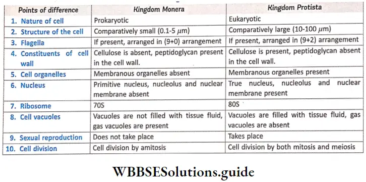
Differences between kingdom Monera and kingdom Protista:
Kingdom Fungi
This kingdom of the biological world comprises of eukaryotic, multicellular, non-chlorophyllous, spore-forming organisms are called fungi.
Kingdom Fungi Distribution: Fungi are found in air, water, soil and, on animals and plants. They prefer to grow in warm j and humid places. Most of the fungi grow in the soil, Some of the fungi may also be aquatic. Some fungi remain as parasites, within host organisms, both intracellularly and intercellularly.
Kingdom Fungi General features
Kingdom Fungi Body Structure: Most of the fungi are simple, unicellular or multicellular and filamentous in nature, filamentous fungi consist of long, slender thread-like structures called hyphae. Due to extensive growth, the hyphae form a cottony network, known as mycelium. Some hyphae are long, slender, thread-like in nature, called hyaline hyphae. Some hyphae are filamentous in
Nature, filled with multinucleated cytoplasm. These are called coenocytic hyphae. Some fungi have septa or cross walls in their hyphae and are called septate. Coenocytic hyphae do not have septa (aseptate), but septa develop within them during reproduction. There may or may not be pores across these septae, that allow transport of nutrients to other parts of the mycelium.
- Cellular structure: The cells are eukaryotic. The cell wall is composed of chitin and polysaccharides. Except chloroplast, all other organelles are present in the cell.
- Nutrition: Most fungi are heterotrophic. They absorb soluble organic matters from dead and decayed plant or animals. Hence, they are called saprophytes. They digest the extracellular substances and derive nutrition by absorbing them. Thus, fungi show an absorptive mode of nutrition. Some fungi can grow on other living organisms and draw nutrition from them (hosts). They are called parasites. The fungi can also live as symbionts—in association with algae as lichens and with roots of higher plants as mycorrhiza.
- Reserve food: The reserve food materials are- glycogen, starch, sucrose, maltose oil, etc.
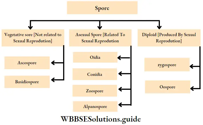
- Reproduction: Reproduction in fungi can be performed vegetatively through fragmentation, fission and budding. Asexual reproduction is performed by the formation of spores called conidia, sporangiospores, oidia, sclerotia, chlamydospores, aplanospores or zoospores.
- Sexual reproduction occurs, through isogamy, anisogamy and oogamy. The gametes unite to form different kinds of spores like oospores, ascospores and basidiospores. The various spores are produced in distinct structures called fruiting bodies or fructifications. Fertilisation involves the following three steps—
- Protoplasmic fusion of two motile or non-motile gametes, called plasmogamy.
- Subsequent fusion of two nuclei within the hyphae called, karyogamy.
- Meiosis within the zygote, resulting in the formation of haploid spores.
Sexual reproduction in fungi
- During sexual reproduction, two haploid hyphae of compatible mating types come together and fuse. In certain fungi, the two nuclei of opposite nature fuse immediately resulting in the formation of diploid zygotes (2n).
- However, in others (Ascomycetes and Basidiomycetes), an intervening dikaryotic stage (n + n, i.e., two nuclei per cell) occurs; such a condition is called a dikaryon and the phase is called dikaryophase of fungus. After dikaryophase ends, the parental nuclei fuse and the cells become diploid.
Classification Of Kingdom Fungi
Different scientists have classified fungi differently. Among them, the most widely accepted classification was propunded by Gwynne-Vaughan and Barnes (1927, 1937). This classification has been discussed below.
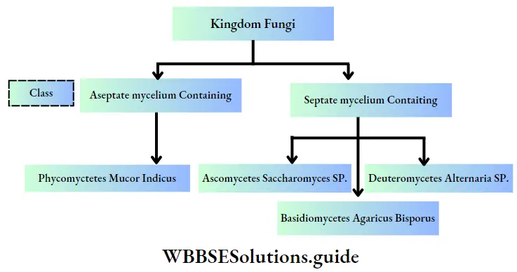
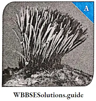
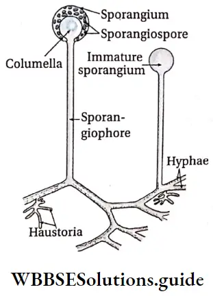
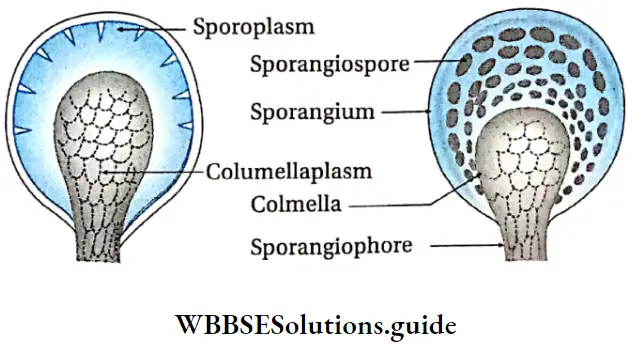
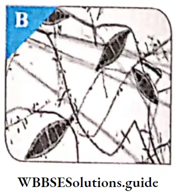
Phycomycetes
This is the class of fungi having aseptate mycelium.
Phycomycetes Distribution: Members of phycomycetes are found mostly in aquatic habitats. They may also grow as saprophytes on dead and decaying organic matters in moist and damp places. They also grow as obligate parasites on plants.
Phycomycetes Body structure:
- The mycelium is aseptate and coenocytic.
- The cell wall is made up of chitin and other polysaccharides (such as glucan, cellulose).
Phycomycetes Reproduction:
- Asexual reproduction takes place by zoospores (motile) or by aplanospores (non-motile). Zoospores are produced endogenously within the zoosporangium.
- Sexual reproduction takes place by isogamy and oogamy. The gametes are non-motile. Oospores are produced by the union of two dissimilar gametes. Zoospores are produced by the union of two similar gametangia through zygospore formation.
Phycomycetes Examples: Mucor sp., Rhizopus sp. (the bread mould) and Albugo sp. (the parasitic fungi on mustard).
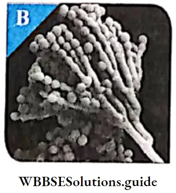
Ascomycetes
This is one of the three classes of fungi having septate; mycelium.
Ascomycetes Distribution: Members of ascomycetes are generally terrestrial in nature. They may also exist as saprophytes or parasites (on plants).
Ascomycetes Body structure:
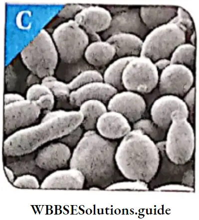
- Vegetative body is unicellular (yeasts) or multicellular.
- They commonly have a J r well-developed, branched septate mycelium.
- The cell wall is generally composed of chitin and glucans.
Ascomycetes Reproduction:
- Vegetative reproduction takes place by fragmentation (in filamentous form), fission and ; budding (in unicellular form).
- Asexual reproduction takes place by non-motile spores, such as conida, oidia and ; chlamydospores.
- The sexually reproducing units are called the ascospores. Ascospores are grown inside a small if specialised sac-like structure, called ascus (plural asci).
- The fruiting bodies (inside which asci are developed); are called the ascocarps.
- There are 8 ascospores within each ascus.
- The ascocarps may be cleistothecium (Penicillium sp.), apothecium (Ascobolus sp.), perithecium (Daldinio sp.) or ascostroma (Elsinoe veneta).
Ascomycetes Examples: Saccharomyces cerevisiae, Penicillium notatum E Aspergillus, Claviceps, Neurospora, etc.
Types of ascocarp :
In cleistothecium, the ascocarp is small or ovoid body that is closed from all sides. In apothecium, the ascocarp is cup-shaped or disc-shaped, while perithecium, it is flask-shaped with definite apical pore. In ascostroma (also known as pseudothecium), ascii are produced in cavities containing stroma.

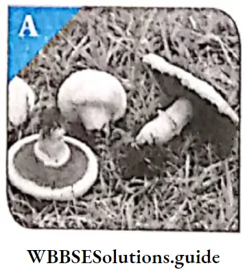
Basidiomycetes
This is another class of fungi having septate mycelium.
Basidiomycetes Body structure:
- Presence of well-developed, branched and septate mycelium.
- The mycelium is of two types—primary and secondary.
- The mycelial cells may contain one nucleus, called monokaryotic (or uninucleate, common in primary mycelium) or two nuclei, called dikaryotic (or binucleate, common in secondary mycelium).
- The secondary mycelia may organise and form fruit body.© The cell wall is mainly composed of chitin and glucans.
Basidiomycetes Nutrition: They are mostly saprophytic and parasitic.
Basidiomycetes Reproduction:
- Primary mycelium may reproduce asexually by oidia, conidia, chlamydospores, etc.
- Vegetative reproduction takes place by budding and fragmentation.
- Sex organs are absent. During sexual reproduction, the dikaryotic cell is formed by somatogamy, plasmogamy (dikaryotization of the cell by somatogamy of monokaryotic and dikaryotic mycelium).
- The dikaryotic phase persists for long period of time. Karyogamy occurs in club-shaped structures, called basidia or basidium.
- 4-haploid basidiospores are formed by meiosis. They are oval, cylindrical or spherical in shape. They may be unicellular and uninucleate.
- Basidiospores are developed exogenously on the horn-shaped or tubular outgrowths of the basidium. These outgrowths are called sterigmata.
Basidiomycetes Examples: Agaricus campestris (mushroom), Puccinia graminis (rust fungus), Ustilago (Smut) etc.
Different types of spores produced by the fungi have been depicted in the following flowchart.
Deuteromycetes
This is another class of fungi having septate mycelium.
Deuteromycetes Distribution: They are called the fungi imperfecti because only the asexual or vegetative phases of these j fungi are known. They are mostly terrestrial, with only a few being aquatic. They are generally decomposers and parasitic in nature, found as saprophytes in the soil.
Deuteromycetes Body structure: The vegetative body is mycelial and composed of profusely branched and septate hyphae. The sually multinucleated. In parasitic species, the hyphae grow intra or intercellularly.
Deuteromycetes Nutrition: Most of the fungi are saprophytic in nature. They act as decomposers of litter and help in mineral cycling. Some are parasites.
Deuteromycetes Reproduction:
- They reproduce mainly by asexual methods.
- The asexual reproduction takes place commonly by conidia, blastospores, chlamydospores and arthrospores.
Deuteromycetes Examples: Alternoria spv Colletotrichum sp. and Trichodermo sp.
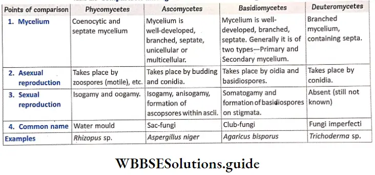
Compare among different classes of kingdom fungi:
Importance Of Fungi:
Fungi play a very important role in our daily life, Some fungi are useful. These are used as food, natural sources of medicine, etc. Some fungi are j directly or indirectly harmful to mankind. They spoil food and other substances and also cause diseases in; both plants and animals, Some common fungal diseases of plants are early blight of potatoes, late blight of potato, etc.
Poisonous fungi :
Some fungi like Amanita phalloides produce toxins, like a-amanitin which causes lesions of stomach cells, It also produces phalloidin which affects the liver, Fungi, like Aspergillus flavus, A. fumigatus and Penicillium islandicum can infect groundnut and mustard cakes. They produce a poisonous substance, called aflatoxin, that binds with DNA and inhibits transcription in the host plant cell. This results in the inhibition of protein synthesis, ultimately leading to cell death.
Handwritten notes on biological classification PDF download
kingdom Plante
Plants are multicellular organisms composed of eukaryotic cells. The cells are organised into tissues. The cells have cell wall that contains cellulose. The cells also contain plastids and large vacuoles in the cytoplasm. They obtain nutrition by photosynthesis. They are generally autotrophic and are called producers. Stored food is starch. Growth is not distinct. They continue to grow throughout their lifetime.
Examples: mosses, ferns, conifers, and flowering plants. We shall learn about this kingdom later.
kingdom Animalia
Animals are multicellular organisms composed of eukaryotic cells. The cells are organised into tissues. The cells lack cell walls. They respond to stimuli very fast. Their growth is distinct. They grow only upto a certain period of their lifetime. They do not carry out photosynthesis and obtain nutrients from other organisms. Thus they are heterotrophic and are decomposers in nature.
Examples: sponges, worms, insects, and vertebrates. We j shall learn about this kingdom in details.
Virus Viroids And Lichens
These are also considered to be members of the living world.
Virus
Virus Definition: Viruses are microscopic, acellular organisms, that form an intermediate group between living and non-living organisms. The term ‘virus’ is derived from the same word in Latin, meaning venom or poisonous fluid. It was coined by M.W. Beijerinck in 1898. Viruses and viroids have not been placed in classification system as they do not have a cell structure and are not considered living.
Discovery of virus:
- D.J. Ivanowsky (1892) recognised certain microbes as causal organism of tobacco mosaic disease. These organisms were, found to be smaller than bacteria because they could pass through bacteria-proof filters.
- M.W. Beijerinck (1898) demonstrated that the extract of the virus infected tobacco plants could cause infection even in healthy plants. He named the fluid as contagium vivum fluidum (meaning ‘contagious living fluid’).
- W.M. Stanley (1935) showed that viruses could be crystallised.
However, once they infect a cell, they behave like living organisms. They take over the replication machinery of the host cell to replicate themselves, thereby killing the host cell.
Therefore, the question still remains whether the viruses are living or non-living.
Virus General features: The viruses are members of the j intermediate group of organisms, between the living and the non-living. They are inert outside their specific host cell.
As soon as they enter the host cell, they become living and start reproducing. Viruses are cannot complete their life cycle without the specific host.
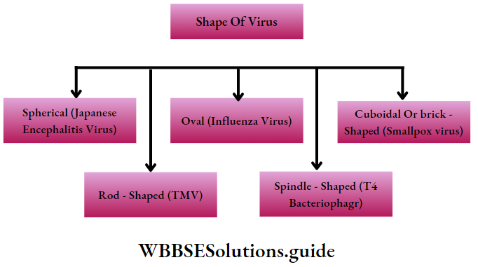
Virus Shape and size:
- Viruses are very minute (microscopic) organisms, that have an average diameter of 20-30nm.
- They may be spherical (e.g., Poliovirus, Influenza virus), cylindrical (e.g., TMV), brick-shaped (e.g., Smallpox virus), or spindle-shaped (Phage virus).
Virus Structure: Virus does not contain protoplasm or cell membrane. Virus consists of two parts—
- Nucleic acid (centrally placed) and
- Protein coat, sometimes with an additional envelope, made of lipoprotein. The protein coat, called capsid, is made up of small subunits, called capsomeres. These capsomeres are arranged in helical or polyhedral shapes. The capsid protects nucleic acid.
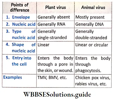
- Viruses contain either RNA or DNA as nucleic acid. A virus is a nucleoprotein (containing nucleic acid and protein) particle and the genetic material is infectious. In general, viruses that infect plants have single-stranded RNA.
- On the other hand, viruses that infect animals have either single or double-stranded RNA or double-stranded DNA. Bacteriophages (viruses that invade the bacteria) are usually double-stranded DNA viruses. Structural details is discussed under separate heads below.
Virus Living features: Presence of—
- Nucleic acid,
- Reproduction and
- Mutability.
Virus Non-living features:
- Lack of protoplast. The cell structure is not complete.
- Absence of respiration,
- High specific gravity like non-living things.
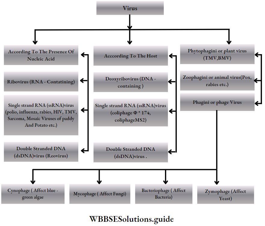
Virus General features Types: According to the presence of the nucleic acids and the host cell, the virus are classified into the following groups—
Structure of a virus
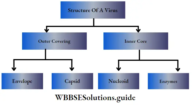
Outer covering
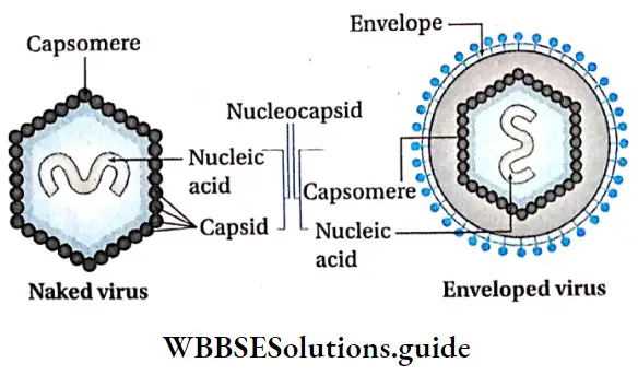
Outer covering Capsid: The protein coat surrounding the genome in a virus is called capsid. The capsid together, along with the enclosed nucleic acid, is called nucleocapsid. The capsid is made up of a number of protein subunits, called capsomeres.
Outer covering Envelope: Many mammalian viruses have a protein, carbohydrate or lipid bilayer surrounding the j nucleocapsid. This loose covering is called envelope, The envelope is a part of the host cell membrane. It: is made up of several units called peplomers. The envelope may possess several outgrowths called spikes.
- The virus which have lipid envelope outside the capsid are called lipoviruses. The viruses which do not have envelope are called naked virus (Example—TMV).
Internal or inner core
Nucleoid: The nucleic acids enclosed within the capsid region make up the nucleoid. Viruses contain only one type of nucleic acid, either DNA or RNA. DNA containing viruses are called deoxyviruses, whereas viruses having RNA are called riboviruses.
- They vary in the structure of their nucleic acid. The nucleoid forms viral genome. The nucleoid consists of about 1000-250,000 nucleotide pairs. The nucleoid remains inactive as long as it does not come in contact with the host cell, The amount of nucleic acid of a virus usually depends on its size.
Genetic material of plant virus:
Most of the plant viruses have RNA either single (TMV) or double stranded (Rice ragged stunt viruses), except a few have DNA, either single (Gemini viruses) or double stranded (Dahlia mosaic virus). Animal viruses have mostly double stranded DNA or either single (Polio virus) or double stranded RNA (Reo virus) and bacteriophages contain mostly double stranded DNA, but they also have single stranded RNA or single stranded DNA.
Enzymes: Hemagglutinin esterase, integrase, reverse transcriptase, viral neuraminidase, polymerase, lysozyme, etc., are some of the enzymes found in viruses.
Differences between plant and animal virus:
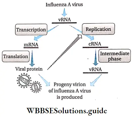
Viral Reproduction: Reproduction in virus takes place when its nucleic acid replicates within the host cell. The process of reproduction is slightly different in bacteriophage, from that of the other virus.
Reproduction in virus, other than bacteriophage: Principal events involved in viral replication, other than that in bacteriophage, are given below—
- Infection phase: It consists of two stages—
- Attachment: The first step in viral infection of a cell is attachment of the virus to the cell surface. Attachment is via temperature-independent ionic interactions. There are specific receptor molecules (generally protein, carbohydrate or lipid in nature), on the cell surface. Virus have a protein called viral attachment protein. This protein recognises the specific receptor on the cell and attaches the virus. Cells without the specific receptors are not susceptible to the virus,
- Penetration: After attachment, the virus has to enter or penetrate the host cell. The virus enters the cell in a variety of ways according to the nature of the virus. Plant viruses enter the host cell either via vectors (aphid, mites, etc.) or through damaged cell membrane. Animal viruses enter the host cell through processes like phagocytosis, endocytosis, etc.
- Eclipse phase: It occurs in two stages—
- Uncoating: The nucleic acid must be uncoated so that viral replication can begin. As soon as the nucleic acid becomes uncoated, the eclipse phase begins. This phase continues until new infectious virions are made.
- Synthesis of viral nucleic acid and protein: In this phase, the viral nucleic acid is replicated. Some 0 Assembly/maturation: New virus particles are assembled using the synthesised nucleic acids and coat proteins. Thus, the virus particles attain a maturation phase that follows the initial assembly process.
- Lysis (Release): Virus may be released due to cell lysis, or, if enveloped, may form bud on the cell.
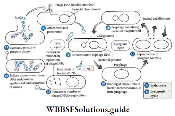
Reproduction in bacteriophage: Reproduction in j bacteriophage takes place by two cycles
- Lytic cycle
- Lysogenic cycle. The former type is seen in virulent | and the latter is found in avirulent or temperate phage. These cycles have been discussed under separate heads.
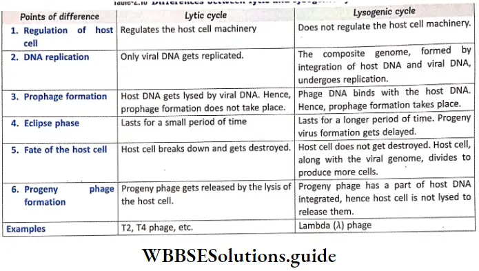
Lytic Cycle:
The cyclic reproduction mechanism, by which virulent | bacteriophage produces progeny virus by replication is called lytic cycle. It is common in T-even phages (T2, T4, etc.) which attack Escherichia coli. It takes place for about 20-30 minutes.
Lysogenic Cycle:
The cyclic reproduction mechanism by which phage virus introduces its nucleic acid into the host cell, divides to form progeny viruses is called lysogenic cycle. Lwoff (1953) discovered this cycle in Lambda (A) phages attacking £ coli. The phage involved in this cycle is called temperate phage, while the bacterium is of lysogenic strain.
Differences between lytic and lysogenic cycle:
Characteristics of some important viruses
Characteristics of some of the viruses have been discussed below in details—
Bacteriophage
Bacteriophage Definition: Bacteriophages are viruses that infect bacteria, reproduce within them and later on lyse them to release the progeny phage.
Bacteriophage Example: T2 phage, lambda phage, phage MS2, ΦX 174 phage, T4 phage.
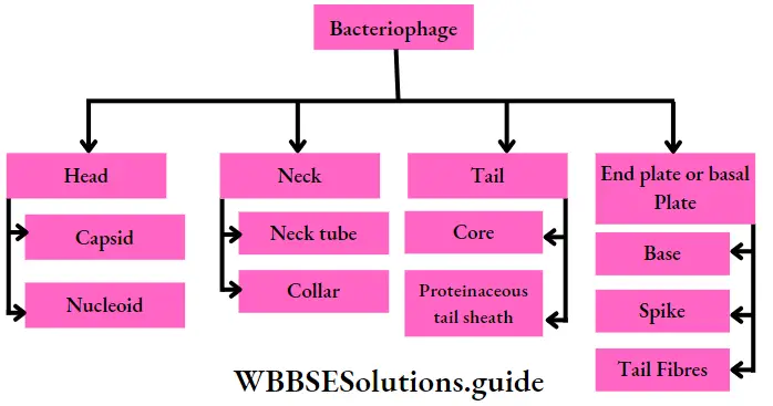
Bacteriophage Structure: T4 is one of the largest bacteriophages; it j is approximately ranging from 25nm-200nm long and 80-100nm wide. They are made up of four important parts—head, neck, tail and end plate or basal plate.
- Head: All phages contain a structure called head, which varies in size and shape.
- Some are icosahedral (with 20 sides) and others are filamentous. The head is covered by a bilayered protein membrane called capsid.
- Capsid is made up of several units called capsomeres. It encloses nucleic acid and acts as the protective covering.
- The nucleic acid is about 0.05nm long double stranded DNA, containing hydroxymethylated cytosine.
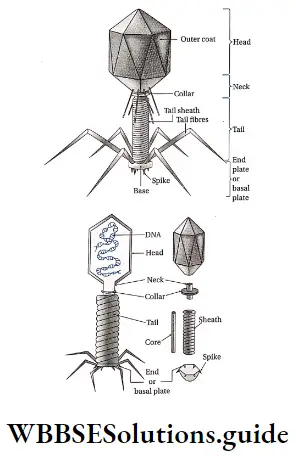
- Neck: It is a small part that extends just after the head. It is made up of neck tube and collar.
- Tail: Some phages have another structure called tail attached to the phage head. The tail has a hollow tube, called core. The nucleic acid is passed to the host cell during infection.
- T4 tail is surrounded by a Y contractile sheath, called tail sheath, which contracts during infection of the bacterium. At the end of tail, phages like T4 have another structure called base plate. One or more tail fibres remain attached to it.
- End plate or base plate: The tail has a plate-like structure at its end. This structure is called end plate or base plate. Six spikes are arranged, along with a long tail fibre, on this plate. The base plate and tail fibres are involved in the binding of the phage to the bacterial cell. All phages do not have base plates and tail fibres.
Tobacco mosaic virus
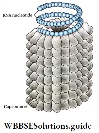
Tobacco mosaic virus Definition: The rod-shaped plant virus, that contains coiled RNA and causes mosaic diseases in tobacco plants are called tobacco mosaic virus (TMV).
Shape and size: TMV has a cylindrical hollow tube-like structure, with a length of 300nm and diameter 15-18 nm.
Tobacco mosaic virus Structure:
- The virus is composed of single strand of RNA, wrapped inside a capsid.
- The virus particle that the virus can attain the cylindrical structure.
- The capsid is composed of about 2130 capsomeres (70 j angstrom x 20 angstrom).
- The capsomeres are arranged like bricks in a cylindrical chimney-like structure. This gives the capsid a shape of spiral staircase.
- Each capsomere contains 158 amino acids.
- The single-stranded RNA is arranged spirally (as a spring), having 6400-7300 ; nucleotide units.
- The RNA strand encodes four : proteins. These include two proteins that replicate the ; viral RNA, a protein that transports the RNA from cell to cell, spreading the infection, and the capsid protein.
HIV, AIDS and AIDS wasting syndrome
HIV or Human Immunodeficiency Virus is a type of j retrovirus, that causes the disease AIDS (Acquired j Immunodeficiency Syndrome) in humans. AIDS wasting syndrome is a condition in which a person suffering; from AIDS loses at least 10% of his body weight, especially that of the muscles.
Tobacco mosaic virus Importance of virus
Viruses can be both useful and harmful and hence, have both advantages as well as disadvantages. Some of these are as follows—
Tobacco mosaic virus Importance of virus Advantages:
- Use as bactericide: Bacteriophages are sometimes j used in ‘polluted water as bactericides. They act as scavengers that eradicate the bacterial population j present in the water. Similarly, they can also kill the disease-causing bacteria.
- Use in scientific experiments: In space research, lysogenic phage cultures are used as radiation detector, Avirulent or temperate phages are useful in studying genetic recombination (transduction) and are used ; widely in biotechnological research.
- Treatment and prevention of diseases: Sometimes bacteriophages are used in therapy and prophylaxis of some bacterial diseases. Viruses are utilised in the production of vaccines used to develop immunity against viral infection.
Short notes on biological classification for quick revision
Importance of virus Disadvantages:
Causal agents of various diseases: Viruses are responsible for causing various diseases of both plants and animals. Some of the viral diseases in plants are tobacco mosaic, yellow vein mosaic of lady’s finger, leaf roll of potato, leaf curl of papaya, etc. The plant viruses cause damage to different parts like root, leaf, fruit, seed, etc. These incur economic losses by hampering the quality and quantity of the plant products. Some viral diseases in animals include small pox, meningitis, pneumonia, mumps, bronchitis, etc. In case of animals, the diseases may even prove fatal.
Oncovirus: Some of the viruses are responsible for causing cancer. These viruses are called oncoviruses.
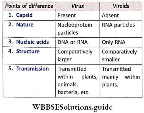
Viroids
Viroids Definition: The tiny, non-coiled, infectious, ssRNA containing particles that can cause diseases in plants are called viroids.
Viroids General features
Viroids Structure: The viroids do not have capsid (i.e., protein coat) around the RNA molecule.Their genome contains a circular RNA strand extensively base paired within itself. So, they can resist RNase attack.
Viroids Genome:
- Viroids have small, low-molecular weight, circular, single stranded RNA molecule. The ssRNA is made up of about 250-390 bases. This number of bases is insufficient to code even for one enzyme, required for replication. Some of the nucleotides even lack AUG(initiation codon).
- The molecular weight is about 75,000 to 1,20,000 Da.
- There are five domains in the viroid genome—TL (Terminal Left), P domain (Pathogenicity Domain), CCC
- Viruses can be both useful and harmful and hence, have both advantages as well as disadvantages. Some of these are as follows— leaf curl of papaya, etc. The plant viruses (Central Conserved Region), TR (Terminal Rigid), V Domain.
Viroids Nature:
- The viroids are smaller and simpler than virus particles.
- The viroids lack any protein coat and remain as free RNA.
- The size of RNA is 25-370 bases in viroids as compared to 3.2-20 kb in viruses.
- The ability to mutate is more in case of viroids, than in viruses.
- They generally reproduce under high temperature. They reproduce after attaching to the host cell DNA.
- Its RNA cannot be degraded by RNase enzyme.
Viroids that cause diseases in plants: Nearly twenty plant diseases are known to have caused by viroids. Some of such viroids are —Potato Spindle Tuber Viroid (PSTVd), Coconut Cadang-Cadang Viroid (CCVd), Avocado Sunblotch Viroid, etc.
Only two diseases such as Scrapie disease of sheep and Kuru disease of human are confirmed to be caused by viroids.
Lichens
The term ‘Lichen’ was given by Theophrastus. Its symbiotic characteristic was however, stated by, Schwander in 1867.
Lichens Definition: Lichens are symbiotic associations of fungi and algae.
Lichens Distribution: Lichens can grow over a range of habitats, such as stones, bark of trees, soil, even in water.
Lichens General features
- General features Structure: Lichens are composite thalloid, generally leaf-like or cylindrical in shape. The thalloid may be of different colours—grey, green, yellow, pink or brown. There are three layers in the structure of the thalloid— upper and lower layers are made up of fungal hyphae, with the middle cortex layer made up of algae. Algal layer is also known as gonidial layer.
- Nutrition: They depend on atmospheric source of nutrition. The lichens that grow on soil or rock, absorb nutrients from their substrate.
- Reproduction: Lichen reproduces by all the three means—vegetative, asexual, and sexual.Vegetative reproduction takes place by fragmentation, decaying of older parts, by soredia and isidia. Asexual reproduction takes place by the formation of oidia. Sexual reproduction takes place by the formation of ascospores or basidiospores. Only fungal component is involved in sexual reproduction.
- Components: The algal partner (known as photobiont or phycobiont) may be cyanobacteria (Nostoc sp.) or green algae (Trebouxia sp.).
- Generally the fungal partner (known as mycobiont) occupies most of the thallus and produces its own reproductive structures. It absorbs water and mineral salts and transports them to the algae.
- In about 98% lichens, fungal component is a member of ascomycetes, while the rest are those from basidiomycetes and deuteromycetes.
- The algal component, on the other hand, manufactures the food ; through photosynthesis. This food probably diffuses out from the algal body and is absorbed by the fungal component.
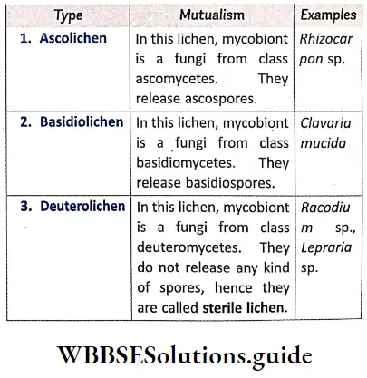
- Classification of lichens: Lichens are classified on the basis of mutualism and morphological structure.
Types of lichen according to mutualism:
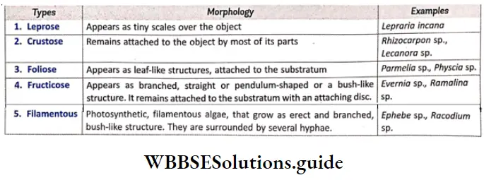
Types of lichen according to morphology:
Importance of lichens: The lichens are beneficial as well as harmful to mankind. They are used as food and fodder, and medicines. They also have various kinds of industrial uses.
- As food: Some species of Parmelia sp. are used as curry powder in India, while various others are used as food in other countries. Lichens like Lecanora saxicola and Aspicilia calcarea, etc., are used as food by several other organisms.
- As fodder: Ramalina fraxinea, R. fastigiato, Evernia prunastri, etc., are used as fodder for animals.
- As medicine: Lichens are medicinally important due to the presence of lichenin and some bitter alkaloids or astringent substances. They are used in the treatment of diseases like jaundice, diarrhoea, epilepsy, etc.
- Industrial uses: Lichens are also used in different kinds of industries—
- Tanning industry: Some lichens like Lobaria pulmonaria and Cetraria islandica are used in tanning leather.
- Brewery and distillation: Lichens like Lobaria \ pulmonaria are used in brewing of beer. In Russia and Sweden, Usnea florida, Cladonia rangiferina and Ramalina fraxinea are used in production of alcohol due to their rich lichenin content.
- Preparation of dye: Dyes obtained from some lichens have been used since ancient times for colouring fabrics, etc. Litmus, an acid-base indicator dye (substance that shows colour change in the presence of an acid or base), is extracted from Roccella tinctoria, R. montagnei and also from Lasallia pustulata.
- Cosmetics and perfumery: Certain aromatic compounds (organic compounds with characteristic odour) are available in lichen and are extracted.
These are used in the preparation of cosmetics and perfumes. Essential oils (oils present in cosmetics) extracted from Ramalina sp. and Evernia sp. are used in the manufacture of cosmetic soaps. - Harmful activities:
- Lichens like Amphiloma sp. and Cladonia sp. grow on mosses and cause total destruction of moss colonies.
- Different crustose lichens cause severe damage to window glasses and marble stones of old buildings,
- Lichens like Letharia vulpina (wolf moss) are highly poisonous due to the presence of vulpinic acid.
Ecological importance of lichens
- Pioneer of rock vegetation: Due to their ability to grow using minimum nutrients and water, crustose lichens can colonise fast.
- Accumulation of radioactive substances: Lichens can absorb different radioactive substances from the environment through bioremediation.
- Sensitivity to air pollutants: Lichens are very sensitive to air pollutants like S02, CO, C02, etc. Thus lichens are important bioindicators of these gaseous pollutants.
Notes
- Amitosis: A type of cell division, which involves simple
- Bioremediation: Using ‘ plants or microorganisms cleavage of the nucleus, followed by division of the j (naturally growing or artificially introduced) to consume cytoplasm. j or break down pollutants in the environment.
- Biota: Total collection of organisms of a particular region, at a specific time period.
- Chemosynthetic: Organisms which obtain energy by oxidising inorganic molecules into organic ones.
- Equatorial plane: Plane present between the poles of a cell, that remains perpendicular to the spindle apparatus in the dividing cell.
- Febrile: Feverish
- Heterotrophic: Organisms which cannot produce their own food.
- Histone: A type of protein that is wrapped around by DNA molecule to form chromatin fibre.
- Holozoic: A type of nutrition involving ingestion, digestion, absorption and assimilation of the food. Osmolability: Unstable towards changes in osmotic pressure.
- Peptidoglycan: A polymer made of sugar and amino ! acids, that forms the basic component of the cell wall.
- Pseudopodia: A type of transient organ of locomotion j produced by protoplasmic extension, present in some organisms.
- Ruminant animals: Animals that acquire nutrients from ! plant-based food, by fermenting it in a special stomach before digestion. This fermented ingesta is regurgitated I and chewed by these animals.
- Syngamy: Fusion of two cells, or their nuclei, during ; reproduction.
Points To Remember:
- Carolus Linnaeus (1753) classified living organisms into two kingdoms—Plant kingdom and Animal kingdom.
- R. H. Whittaker (1969) classified living organisms into five kingdoms. These kingdoms are Monera, Protista, Fungi, Plantae and Animalia.
- Kingdom Monera consists of only prokaryotes. Kingdom Protista, . Fungi, Plantae and Animalia consist of eukaryotes.
- Bacteria under the kingdom Monera was first discovered by Leeuwenhoek (1676).
- Cell wall of bacteria is composed of peptidoglycan or \ murein.
- The coiled structure formed by the invagination of j plasma membrane of Gram positive and Gram \ negative bacteria is called mesosome.
- The circular DNA molecule present in bacterial cell as I a substitute of nucleus is called nucleoid or genophore.
- Some bacteria (like Chlorobium) possess the j photosynthetic pigment bacteriochlorophyll. These bacteria are capable of photosynthesis.
- The protist which has characteristics of both animal j and fungi is called slime mould (Myxomycetes).
- Ascomycetes and basidiomycetes of kingdom Fungi i are called sac fungi and club fungi respectively.
- Edible mushroom is Agaricus bisporus.
- Each basidiocarp (mushrooms) has two parts—stalk like structure is called stipe and umbrella like structure is called pileus.
- The causative fungus of Black stem rust disease of wheat is Puccinia graminis tritici.
- Late blight of potato is caused by the fungus Phytopthora infestans.
- Lichen is a symbiotic organism formed by the association of algae and fungi. Its fungal part is mycobiont and algal part is phycobiont.
- Mycoplasma are prokaryotes that lack cell wall.
- Silica can be found in the cell wall of diatoms.
- The protozoa Plasmodium has four species. Each of them is a carrier of malaria. Such as
- Plasmodium vivax— Benign malaria
- Plasmodium falciparum—Malignant malaria
- Plasmodium malariae—Quartan malaria
- Plasmodium ovale—Mild tertian malaria
- The term virus was coined by the Dutch microbiologist Martinus Beijerinck. In the early 20th century. Frederick Twort discovered that bacteria could be attacked by viruses.
- Viruses are intermediate between living and non-living organisms.
- The viruses which invade the body of harmful bacteria and kill them are called bacteriophages.