Synthesis and secretion: The glands, formed by these glandular cells, synthesize and secrete mucus, hormones, enzymes, etc.
Storage: Some chemical substances are stored in these glandular cells, such as—protein in the stomach, lipids in the adrenal and sebaceous glands, and sugar and protein in the salivary gland.
Excretion: When present in sweat glands and sebaceous glands, glandular epithelium helps to excrete sweat and sebum, respectively.
Classification of glands: On the basis of the number of cells present in the epithelium, glands are divided into two types—
- Unicellular glands and
- Multicellular glands.
Unicellular gland: Unicellular gland may be defined as an isolated single secretory cell that itself functions as a gland.
Read and Learn More: WBCHSE Notes for Class 11 Biology
Unicellular gland Position: It is found in the mucous membrane of the stomach, intestine, rectum, and trachea.
Structure They are elongated and flask-shaped. The cell consists of a nucleus, surrounded by cytoplasm containing numerous Golgi bodies.
| Class 11 Biology | Class 11 Chemistry |
| Class 11 Chemistry | Class 11 Physics |
| Class 11 Biology MCQs | Class 11 Physics MCQs |
| Class 11 Biology | Class 11 Physics Notes |
The apical part of the cell contains granules which contain the glycoprotein mucin. The distal part of the cell becomes swollen and bursts. This causes secretion of mucin.
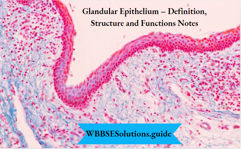
Glandular epithelium definition, structure, and functions notes PDF
Unicellular gland Functions: Some of them (goblet cells) secrete mucus which forms a protective layer on the intestinal mucous membrane,
- They neutralize acids and alkalis, thereby maintaining pH balance.
- They provide protection by engulfing bacteria and foreign particles that enter the trachea or alimentary canal.
- They lubricate the alimentary canal and trachea.
Example: Goblet cells, calciform cells.
what is glandular epithelium
Multicellular gland: Multicellular glands may be defined as glands comprising aggregates of secretory cells.
On the basis of types of secretion, there are three types of multicellular glands—
Endocrine glands are ductless glands. They pour their secretions (hormones) directly into the blood. Example pituitary, thyroid or adrenal glands etc.
Exocrine glands, these glands are provided with ducts. They pour their secretions (enzymes) through ducts into the target organs. Example salivary, gastric-OPlntestjnal glands,
Mixed glands, glands comprise both endocrine and exocrine parts. Example pancreas— whose exocrine secretory part is called ‘acinus’ and endocrine secretory part is made up of cells called ‘islets of Langerhans’.
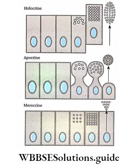
On the basis of the mode of secretion, glands are of three types. They are—
Holocrine gland: Secretion is discharged along with the entire cytoplasm of the cell. This leads to the disintegration of the cell. Example sebaceous gland.
Apocrine gland: The apical surface of the secretory cell is discharged along with the accumulated secretions. Examples mammary glands, and apocrine sweat glands.
Merocrine gland: The secretion is released to the external environment through the cell membrane slowly by diffusion. The whole cell remains intact. Examples salivary gland, and pancreatic gland.
Types of multicellular glands on the basis of the nature of ducts
Simple gland: Simple glands are multicellular glands with unbranched ducts. These are two Simple tubular glands, which are unbranched, tube-like simple glands. Example intestinal gland, sweat gland,
Simple saccular or alveolar glands, which are unbranched, sac-like simple glands. Example mucous glands in frog’s skin. It is absent in mammals.
Glandular epithelium structure and function with diagram
Compound gland: Compound glands are multicellular glands with branched ducts. These are of two types—
Compound tubular gland, the distal end of these glands is branched and tube-like. Example Brunner’s gland of duodenum.
Compound saccular or racemose gland, a distal end of these glands is branched and sac-like.
Examples exocrine pancreas, salivary gland, and sebaceous gland.
Tubulo-saccular/alveolar gland, distal ends of these glands comprise both tubule and sac-like structures. Examples are parts of the salivary gland, mammary gland, and glands of respiratory passage.
Functions of glands
Hormone secretion: The endocrine glands are involved in the secretion of hormones. For example, thyroxine is secreted from the thyroid gland, insulin is secreted from the endocrine part of the pancreas, etc.
Enzyme secretion: The exocrine glands secrete various enzymes. For example, ptyalin is secreted from the salivary gland, pepsin from the gastric gland, etc.
Compound or stratified epithelial tissue
Functions of glands
Hormone secretion: The endocrine glands are involved in the secretion of hormones. For example, thyroxine is secreted from the thyroid gland, insulin is secreted from the endocrine part of the pancreas, etc.
Enzyme secretion: The exocrine glands secrete various enzymes. For example, ptyalin is secreted from the salivary gland, pepsin from the gastric gland, etc. Compound orstratified epithelial tissue.
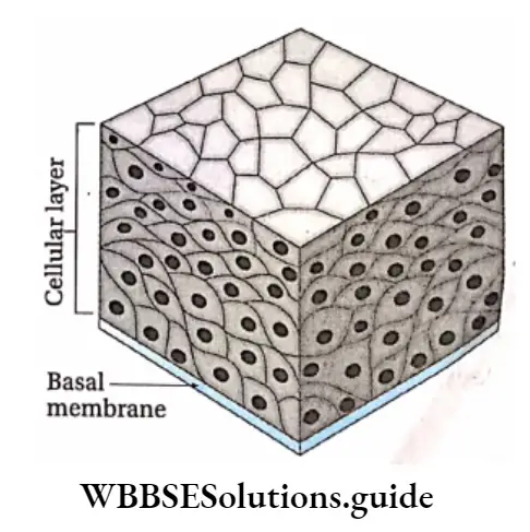
Transitional epithelial tissue Definition: The epithelial tissue that has multiple (3-4) layers of cells and shows properties of both simple and compound epithelium, is called transitional epithelial tissue.
Transitional epithelial tissue Position: It is found in the renal pelvis, broader parts of the ureter, upper part of the urinary bladder, urethra, and urinary duct.
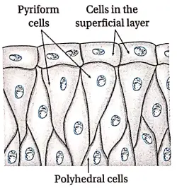
Transitional epithelial tissue Structure:
- The cells of the superficial layer are large, flat, irregular-shaped (generally quadrilateral), and may be binucleate.
- The next layer contains pyriform cells (flame-shaped cells). One end of these cells is round and the other end is narrow.
- Cells in the third layer are polyhedral. These cells are located in between the narrow ends of pyriform cells.
- The cells of the basal (bottom) layer are round or cuboidal in shape. © The nuclei are oval or spherical in shape and the cytoplasm is granular.
- This epithelium forms walls of the organs that undergo continuous expansion and contraction.
Transitional epithelial tissue Function:
- It prevents the reabsorption of ions during excretion.
- It regulates the distension of organs like the urinary bladder.
Stratified squamous epithelial tissue
Stratified squamous epithelial tissue Definition: Epithelial tissue in which the uppermost layer has squamous cells, that may or may not be keratinized, is called stratified squamous epithelial tissue.
Stratified squamous epithelial tissue Position: It is located in the epidermis of the skin, oral cavity, pharynx, esophagus, vag*na, cervix, cornea, etc.
Stratified squamous epithelial tissue Types: It is of two types—stratified keratinized squamous epithelium and non-keratinized stratified squamous epithelium. These are discussed under separate heads.
Stratified keratinized squamous epithelial tissue Definition: The epithelial tissue, consisting of multiple cellular layers, whose uppermost cellular layer contains keratin, is called stratified keratinized squamous epithelial tissue.
Stratified squamous epithelial tissue Position: It is found in the epidermis of the skin (nails, horns, the enamel of teeth, etc).
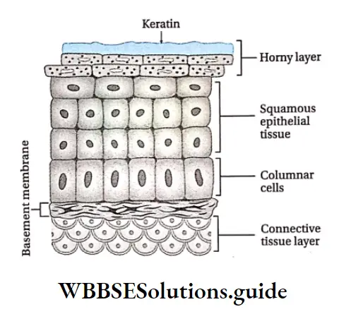
Stratified squamous epithelial tissue Structure:
- Cells of the superficial layer are dead. However, these cells store keratin and are thus cornified.
- Hence, this layer is called stratum corneum (Latin term, meaning ‘horny layer’).
- Cells of the middle layer are polygonal and squamous in nature.
- The lower layer has columnar cells which are attached to the basement membrane.
- The cells of the superficial layer are constantly sloughed off and replaced by new cells from the inner layers.
Function: Its major role is to protect the underlying soft tissues against mechanical injury and friction. It is water-resistant and hence, prevents water loss from the body.
Non-keratinized stratified squamous epithelial tissue Definition: The epithelial tissue, consisting of multiple cellular layers, in which the uppermost layer does not contain keratin is called non-keratinized stratified squamous epithelial tissue.
Types of glandular epithelium and their functions
Non-keratinized stratified squamous epithelial tissue Position: It is found in the lining of oral and nasal cavities, urethra, cervix, cornea, vag*na, anal canal, etc.
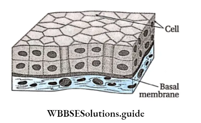
Non-keratinized stratified squamous epithelial tissue Structure:
- This tissue has multiple layers of cells. These cells lack keratin.
- The outer layer consists of living cells and the middle layer contains polyhedral cells.
- Cells attached to the basement membrane are columnar.
glandular
Non-keratinized stratified squamous epithelial tissue Function: Its major role is to protect the underlying soft tissues against mechanical injury and friction. It is water-resistant and hence, prevents water loss from the body.
Prickle cell: The cells at the basal surface of stratified keratinized squamous epithelial tissue are connected with each other by intercellular fibers and protoplasmic appendages.
These fibers and appendages are called prickles and the associated cells are called prickle cells due to their thorny appearance. These cells can withstand pressure.
Stratified Cubodial Epithelial Tissue
Stratified Cubodial Epithelial Tissue Definition: The epithelial tissue, consisting of multiple cellular layers, in which the uppermost layer does not contain keratin is called non-keratinised stratified squamous epithelial tissue.
Stratified Cubodial Epithelial Tissue Position: It is found in the larger duct of sweat glands, salivary glands, female urethra, and pancreas.
Structure:
The tissue is usually multi-layered.
Cuboidal cells are present in the uppermost layer and polyhedral cells are found in the middle layer.
Function: It provides strength to the walls of the lumen and also helps in the secretion of sweat, saliva, etc.
Class 11 biology glandular epithelium notes with examples
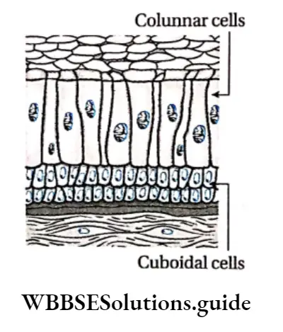
Stratified Columnar Epithelial Tissue Definition: The epithelial tissue, made up of multiple layers of cells, whose uppermost cellular layer has columnar cells is called stratified columnar epithelial tissue.
Stratified Columnar Epithelial Tissue Position: It is found in the pharynx, epiglottis, a mucous layer of the anus, and urethra.
Stratified Columnar Epithelial Tissue Structure:
The uppermost layer has columnar cells with an oval nucleus.
The lowermost layer has cuboidal cells.
Function: Its main function is to give protection to the underlying organ.