Muscular Tissue
Muscular tissue Definition: The tissue, derived from embryonic mesoderm, and involved in movement and locomotion by virtue of its property of contraction and relaxation is known as muscular tissue.
Muscular tissue Position: Some muscular tissues or muscles are associated with bones and so known as skeletal muscles, Some are associated with visceral organs and are called smooth muscles. Some muscles are associated with the heart, hence known as cardiac muscles.
Muscular tissue Origin: Muscular tissues have originated from mesoderm.
Muscular tissue Components: Muscular tissue is made up of muscle cells or fibers. The main components of the cell include 70% water, 20% proteins like actin, myosin, tropomyosin A, tropomyosin B, myogen, myoalbumin, myoglobulin, etc. 10% glycogen, 0.2% lipids like phospholipid and cholesterol, 1.5% inorganic salts like potassium phosphate, salts of calcium, sodium, magnesium, etc.
Read and Learn More: WBCHSE Notes for Class 11 Biology
Muscular tissue Structure
Muscular tissue Shape: Muscule Tissue Is Made of Bundles Of Narrow, Elongated Contractile Muscle Cell, called my myocytes. Due to their elongated nature, they are also known as muscle fibers.
muscles and muscle tissue
Muscular tissue Arrangement: They are contractile in nature. The contractile unit is made up of proteins such as mainly myosin, actin, troponin, tropomyosin, etc. The muscle fibers are arranged like a mesh.

Muscular tissue Matrix: Intercellular matrix is absent but muscle fibers are surrounded by connective tissue.
Muscular tissue Covering: Myofibril or muscle fiber is covered with a plasma membrane known as sarcolemma. They also have mitochondria, known as sarcosomes, and endoplasmic reticulum, known as sarcoplasmic reticulum.
Muscular tissue Innervation: Sensory nerves are present in muscles.
| Class 11 Biology | Class 11 Chemistry |
| Class 11 Chemistry | Class 11 Physics |
| Class 11 Biology MCQs | Class 11 Physics MCQs |
| Class 11 Biology | Class 11 Physics Notes |
Muscular tissue Circulation of blood: Blood vessels are present in the muscular tissue. So, blood circulation takes place within muscle fibers.
Some Special Contractile Cells
Besides muscle cells, some other cells also have the capacity to contract and expand. Some of these cells are described below.
Muscular tissue Myoepithelial cells: The contractile cells which originate from the ectoderm are called myoepithelial cells. Their contraction is slow and involuntary. They are found in the sweat glands, mammary glands, lacrimal glands, and salivary glands.
Muscular tissue Pericytes: These are contractile cells that wrap around the endothelial cells of capillaries and venules throughout the body. They are also known as mural cells.
Myofibroblasts: The contractile cells that contain actin and myosin are called myofibroblasts. They help in wound repair.
Myofibroblasts Functions
Coordination of organs: Muscles (especially involuntary muscles) control coordination between various organs, movement and locomotion, body posture, speech, facial expression, etc.
” types of muscle tissue”
Coordination of visceral organs: Involuntary muscles present within the visceral organs, help in the movement of the organs.
Some examples of these movements are—the contraction of the muscles of the GL tract during peristalsis, uterine wall contraction during childbirth, urinary bladder contraction during micturition, contraction, and relaxation of the walls of the blood vessels for regulating blood pressure, etc.
Circulation of blood: The circulation of blood throughout the body as well as the beating of the heart, both are coordinated by the contraction and relaxation of cardiac muscle.
Response to stimuli: The contraction of muscles helps us to respond to external stimuli.
Coordination of respiration: Respiration is regulated by the contraction and relaxation of respiratory muscles (skeletal muscles). It is also regulated by the smooth muscles lining the trachea and airways of the lungs.
Generation of heat: The movement of muscles during vigorous physical activities such as exercise etc., helps to generate heat in the body.
Types: There are several types of muscular tissue or muscles based on position, structure, mode of action, etc.
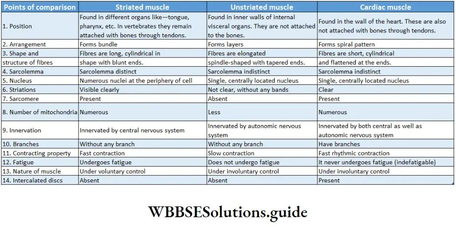
On the basis of structure, location, and function, the types of muscles are—striated or skeletal or voluntary muscles, non-striated or smooth visceral or involuntary muscles, and cardiac muscles. The structure and function of each of these muscular tissues are described below
Skeletal Muscle
Skeletal Muscle Definition: The muscles with alternate light and dark bands, which remain associated with the skeleton and can contract on will, are called striated skeletal or voluntary muscles.
Skeletal Muscle Position: These muscles remain attached to the bones or the skeleton and are controlled by the somatic nerve.
Characteristic features: Skeletal muscle is called striated because of its appearance.
It contains alternate light and dark bands, that are visible through a phase contrast microscope.
Components:
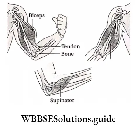
- The skeletal muscles (e.g. biceps, triceps) are spindle-shaped, i.e., swollen in the middle region and tapering at the ends.
- The muscles are attached to the bones by means of a hard, collagenous connective tissue, called tendon. One end of the tendon is bound to the perichondrium of bone and the other end is bound to the sarcolemma of the muscle fiber.
- Each muscle fiber is enclosed by a connective tissue membrane, known as an endomysium.
- A bundle of muscle fibers is known as a fasciculus (pi. fasciculi). This is enclosed by another connective tissue layer called the perimysium.
- Bundles of fasciculi are surrounded by another connective tissue layer called the epimysium.
- The main structural component of striated muscles or skeletal muscles is muscle cells or muscle fiber.
- The structural features of muscle fibre as seen under an electron microscope are described below.
Shape: Each muscle cell is elongated, cylindrical, unbranched fiber, with blunt ends. Each muscle fiber has a diameter of 10-100 pm and is 1-40 mm long.
“muscle tissue structure “
Sarcolemma: The muscle fiber is enclosed within a thin transparent membrane called sarcolemma. This membrane is semi-permeable in nature.
Sarcoplasm: The cytoplasm of muscle cells is known as sarcoplasm. The striated muscle fiber is multi-nucleated. During the formation of striated muscle fiber, its nucleus divides a number of times by mitosis.
This division is not followed by the division of cytoplasm. Due to this reason, muscle fibers are multi-nucleated.
Organelles: It contains sarcoplasmic reticulum (ER), sarcosomes (mitochondria), glycogen, and lipid droplets.
The most important organelle among them is
A-band sarcoplasmic reticulum. The terminal end of the sarcoplasmic reticulum is swollen and is called terminal cisternae. This part stores a huge amount of calcium ions.
These Ca2+ ions are used for muscle contraction.
T-tubule: The sarcolemma of muscle fibers invaginates and extends towards the cytoplasm, at some points.
These extended tube-like structures are called T-tubules or transverse tubules. Each T-tubule remains associated with two terminal cisternae of the sarcoplasmic reticulum.
This structure formed by the T-tubule and the two associated terminal cisternae is called a triad.
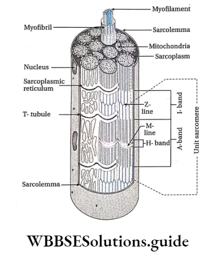
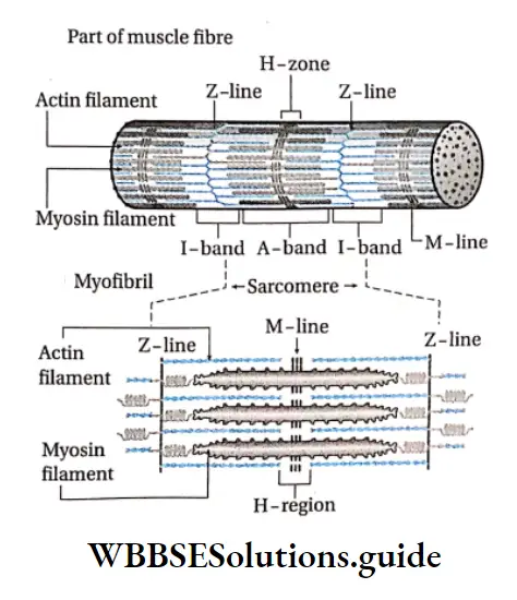
Myofibril: The sarcoplasm of skeletal muscle fiber consists of numerous longitudinal bundles of protein fibrils.
These fibrils are called myofibrils. Using an electron microscope, it can be seen that the myofibrils are made up of two types of thin, thread-like contractile protein filaments called myofilaments. The two myofilaments are thicker myosin filaments (100A in diameter) and thinner actin filaments (50A in diameter).
Each myofibril contains approximately 1500 myosin and 3000 actin filaments.
“function of muscle tissue “
The actin filament comprises actin, tropomyosin, and troponin proteins while the myosin filament is made up of myosin protein.
Alternate light and dark bands, called striations, appear throughout a myofibril. The structural details of these striations are as follows-
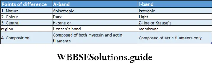
A Band And I Band: The Light Bands Of Myofilaments Which Are Composed Of Strands Of Only Actin known as l-bands or isotropic bands.
These bands are called ‘isotropic’ as they have the same refractive index in all planes.
The dark bands of myofilaments contain strands of myosin protein along with a few strands of actin protein scattered within. These are known as the A-bands or anisotropic bands.
These bands are called ‘anisotropic1 as they refract light differently in different planes.
Z-line or Z-disc: The thick line present at the center of the l-band is called the Z-line or Krause’s membrane. It is also known as Dobie’s line.
[The letter Z in ‘Z-line’ has been derived from the German word ‘Zwischenschiebe’ which means, Zwischen = between and Scheibe = disc.]
Sarcomere: The segment of the myofibril between two adjacent Z lines is known as sarcomere.
- It is the contractile unit of the myofibril and hence, that of the muscle.
- The ends of actin strands in adjacent sarcomeres are connected by the Z-line.
- Sarcomere = Half of l-band + One complete A-band + Another half of the next l-band
H-zone: Along the midline of each A-band is a lighter zone. This is known as the H-zone or Hensen’s zone.
M-line: A thick line is present vertically in the middle of the H-zone. This is known as the M-line.
[The letter ‘M’ in the M-line has been taken from the German word ‘Mittlescheibe’ which means ‘central disc’]
Function: The movement of the skeleton is under the conscious control of the body, hence controlled by striated muscles.
These include movement of limbs, fingers, toes, neck, etc. Changes during facial expressions Example ability to smile or to frown, are also controlled by voluntary muscles.
Smooth Muscle Definition: The muscles that are non-striated, involuntary in nature, and found in the visceral organs are known as visceral muscles or non-striated or involuntary muscles.
3 types of muscle tissue
Smooth Muscle Position: These muscles are found in the wall of the alimentary canal, urinary tract, blood vessels, uterus, fallopian tube, the dermis of the skin, iris, and ciliary bodies of the eyes.
Characteristic features:
- These muscles do not have any cross-striation and so these are known as smooth muscles.
- Due to their presence in the visceral organs, these are also known as visceral muscles.
- These muscles are controlled by the autonomic nervous system and hence, are involuntary in nature.
- The cells are capable of mitosis.
- These muscles can remain contracted for a long time. These muscles never get fatigued.
Components
Muscle fibers: Each muscle fiber is spindle-shaped. These are generally unbranched but, in some fibers, terminal branches can be seen.
Length and diameter: The average length of a muscle fiber is 20 pm with a maximum width of about 6 pm.
Outer covering: The outer covering of muscle fiber is called sarcolemma. It is indistinct in nature.
Cavioli and sarcoplasmic reticulum: Certain structures formed by sarcolemma within the smooth muscle cells are called cavioli. The inner portion of the ravioli contains the sarcoplasmic reticulum.
Nucleus: Cells are uni-nucleated with a centrally placed oval or elongated nucleus.
Mitochondria: Cells contain fewer mitochondria.
“muscular tissue function “
Golgi bodies: Cells contain smaller Golgi bodies.
Contractile protein: The sarcoplasm contains numerous fine contractile myofibrils. Unlike skeletal muscles, the myofibrils are not arranged in a distinct pattern in a smooth muscle cell.
The myofilaments, actin, and myosin, present within the myofibrils, are irregularly arranged.
They are not organized to form sarcomeres (structures formed by two adjacent l-bands with an A-band between), hence striations are not formed
Cellular connections: Adjacent smooth muscle cells are separated by a basal lamina.
However, this basal lamina is not continuous. It is absent in the regions where adjacent muscle cells are connected.
These regions that appear like band-shaped tight junctions are called fascia occludes or nexus.
Cellular connections Types
Single-unit smooth muscle: The muscles formed by the muscle fibers that contract together at the same time are called single-unit smooth muscles.
In such cases, the muscle cells are connected by tight junctions and hence, these muscles form a covering.
These tight junctions are known as cytoplasmic bridges. Due to this property of forming tight junctions, single-unit smooth muscle bundles form a syncytium.
This structure contracts in a coordinated manner, leading to muscle contraction.
Examples are the muscles of the uterus, gastrointestinal tract, and urinary bladder.
Multi-unit smooth muscle: The muscles formed by the muscle fibers that contract separately, each as a single unit are called multi-unit smooth muscles.
Examples are the muscles of walls of blood vessels, ciliary bodies, iris, esophagus, dermis, hair root, etc.
Cellular connections Functions
Peristalsis: The action of smooth muscles helps food to be carried along the gastrointestinal tract.
Regulation of opening and closing of orifice: Smooth muscle forms the structures called sphincters, which are present at the beginning of the orifices.
The sphincters control the opening and closing of a number of orifices in the body. This in turn regulates several physiological processes like the movement of food from the stomach into the intestine or evacuation of the urinary bladder and rectum.
“structure of skeletal muscle “
Controlling vasodilation: Vasodilation is controlled by the contraction of smooth muscles, thereby regulating blood pressure.
Contraction of the urinary bladder and uterus: Contraction of the urinary bladder during micturition and forceful contraction of the uterus during childbirth occurs with the help of smooth muscles.
Other functions: The smooth muscles of the stomach also help in the churning of food.
Cardiac Muscle Definition: The striated involuntary muscle present in the myocardium (wall) of the heart is known as cardiac muscle.
Cellular connections Position: These muscles are found in the wall of the heart.
Characteristic features:
- Cardiac muscles are associated with the heart.
- They are capable of involuntary and rhythmic movements.
- Cross striations are observed, hence they are a type of striated muscles.
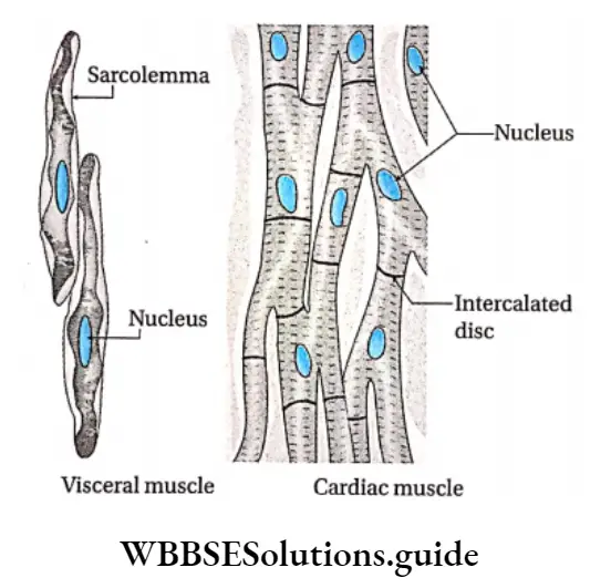
Components Muscle fibers: Cardiac muscle fibers are striated, and branched. The cells that form the cardiac muscle are called cardiomyocytes or myocardiocytes. They have a single, large, oval, central nucleus.
Sarcolemma: The outer covering of cardiac muscle (sarcolemma) is indistinct.
Syncytium: Cardiomyocytes are connected with one another forming a syncytium.
Intercalated disc: Sarcolemma of the cardiac muscle fiber is thickened at intervals to form plate or disc-like structures called intercalated discs.
They separate the cardiac muscle cells from one another. At each intercalated disc, the cell membranes of two adjacent cells fuse with one another, forming permeable communicating junctions (gap junctions). These junctions help in the transmission of nerve impulses.
Other types of cell junctions formed through intercalated discs include fascia adherens (binding site for actin, connect sarcomeres) and desmosomes (binding site for intermediate filaments, join the cells).
Sarcoplasm: Sarcoplasm is granular in nature. It has several mitochondria, scattered throughout. A small Golgi body, with few lipid droplets and sarcoplasmic reticulum (endoplasmic reticulum), are also present.
Myofilament: Numerous distinct myofibrils with alternate dark and light cross bands, forming prominent sarcomeres, are found within the sarcoplasm.
As stated earlier, myofibrils are made up of myofilaments, that are composed of actin and myosin protein strands. They are present as bundles.
T-tubules: Sarcolemma penetrates inside muscle fibers at specific regions. These structures formed by the sarcolemma are called transverse tubules or T-tubules. Together these tubules are known as T-system.
This network stores calcium ions, required for muscle contraction.
Functions: Pumping of blood through the heart takes place by alternate contraction and relaxation of cardiac muscles.
Besides the above-mentioned types, there are several types of muscular tissue, such as—
Types of muscles on the basis of function and pigments: Details regarding the classification (Locomotion and movement).
Types of muscles based on the nature of their activity: Muscles are classified on the basis of the nature of their activity into two groups.
White muscle or fast muscle: The skeletal or voluntary muscles which appear pale due to less blood vessels and the absence of myoglobin are called white muscles.
Such muscles show prominent striations. They react fast to impulses but cannot hold the contraction for a long time. Example muscles of the lip, tongue, etc.
Red muscle or stow muscle: Skeletal muscles that appear red due to more blood vessels and myoglobin are called red muscles.
Such muscles show fewer striations. They react slowly to impulses but can hold the contraction for a long time. Example muscles controlling body posture.
Few Concepts About Muscular Tissue
Refractory period: The brief period during which the muscle cannot get excited to a second stimulus after receiving the first stimulation is called the refractory period.
Heart muscles have a long refractory period. Hence, they do not fatigue easily.
Tetanus: In terms of physiology, if a series of I stimuli from the external environment are applied successively during a single contraction, then they fuse together leading to sustained contraction.
This phenomenon is called tetanus, Summation: A weak stimulation may fail to produce a contraction in the muscles.
But when this weak stimulation is applied repeatedly, it may produce considerable contraction in the muscles. This phenomenon is called summation.
Rigor mortis: The rigidity of muscles after death is called rigor mortis.