
Biology MCQ For NEET With Answers Pedigree Analysis And Genetic Disorders
Question 1. What is pedigree analysis?
- Record of inheritance pattern
- Linkage map
- Quantitative genetic
- Polygene analysis
Answer: 1. Record of inheritance pattern
Pedigree is a record of inheritance of certain genetic traits for two or more than two generations, represented in the form of a diagram.
Read And Learn More: NEET Biology Multiple Choice Question And Answers
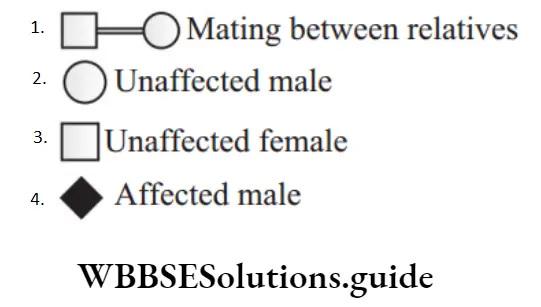
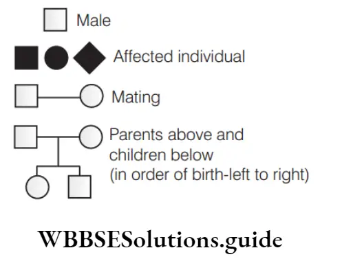
Question 2. Which one of the following symbols and its representation, used in human pedigree analysis is correct?
Answer:
1. Option (1) is correct. Rest other options are incorrect and can be corrected as
- Option (2) is unaffected female,
- Option (3) is unaffected male and
- Option (4) is affected individual with sex unspecified.
“pedigree analysis questions “
Question 3. The individual from which a pedigree analysis is initiated is called
- Proposita
- Propositus
- Both 1 and 2
- origin
Answer: 3. Both 1 and 2
If pedigree is initiated from male, it is called propositus. If pedigree is initiated from female, it is called proposita. So, an individual from which a pedigree is initiated could be proposita or propositus. Thus, option (3) is correct.
Biology MCQ For NEET With Answers
Question 4. Match the symbols with statement.
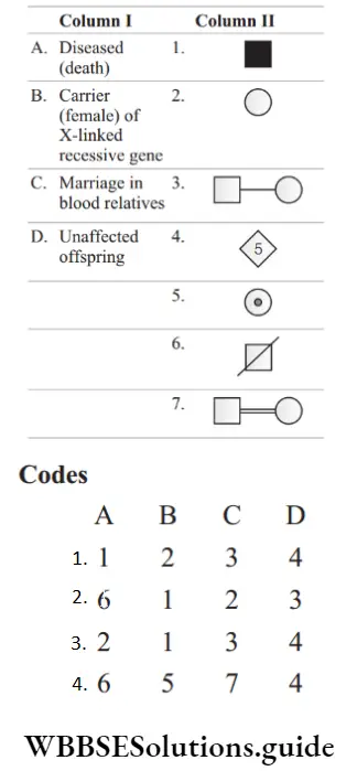
Answer: 4. A–6, B–5, C–7, D–4
NEET Biology Pedigree Analysis MCQs with answers
Question 5. Name the principle on which pedigree analysis work?
- Regression
- Probability
- Random mating
- ANOVA
Answer: 2. Probability
“principles of inheritance and variation pyq neet “
Pedigree analysis is used to study the transmission genetics where controlled process is not possible and it works on the principle of probability.
Question 6. The given symbol indicates.

- Carrier female
- Unaffected female
- Death of female
- Normal female
Answer: 1. Carrier female
Generally, the carriers are females therefore, they are rounded structure. Carrier is represented as dot or small circle within the gender symbol.
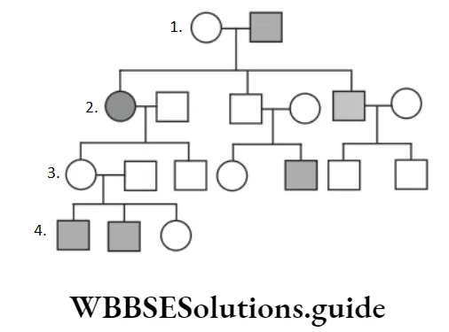
Question 7. Study the pedigree chart of a certain family given in figure and select the correct conclusion which can be drawn for the character.
“inheritance and variation mcq “
- The female parent is heterozygous
- The parents could not have had a normal daughter for this character
- The trait under study could not be colour blindness
- The male parent is homozygous dominant
Answer: 1. The female parent is heterozygous
The given pedigree chart shows that both the daughters received the gene from the parents, while son may be normal or affected. It shows that the female parent is heterozygous.
Biology MCQ For NEET With Answers
Question 8. Pedigree analysis represents
- Still birth
- Still death
- Still carrier
- Still mating
Answer: 1. Still birth
Symbol in pedigree chart represents still birth. It refers to foetal death at or after 20-28 weeks of pregnancy.
Question 9. Identify the symbols given below and the correct option with respect of A, B and C.
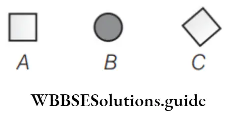
- A-Male, B-Affected female, C-Sex unspecified
- A-Affected male, B-Female, C-Sterile male
- A-Male, B-Female, C-Fertile
- A- Affected female, B- Sex unspecified, C- Male
Answer: 1. A-Male, B-Affected female, C-Sex unspecified
A–Male, B–Affected female, C–Sex unspecified
Important MCQs on Pedigree Analysis for NEET
Question 10. The given diagram A and B indicates.
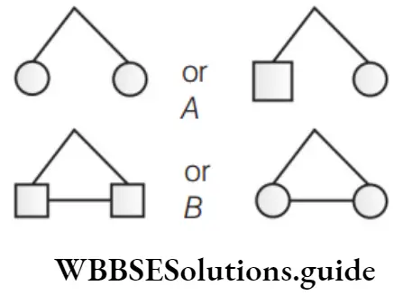
- A-Zygotic twins, B-Dizygotic twins
- A-Dizygotic twins, B-Identical twins
- A-Zygotic twins, B-Identical twins
- A-bizygotic twins, B-Dizygotic twins
Answer: 2. A-Dizygotic twins, B-Identical twins
“pedigree questions “
- A–Dizygotic twins are twins, which result from the fusion of two sperms with two ova. It is very rare in case of human beings.
- B–Monozygotic twins (identical twins) are twins, which result from the fusion of one sperm with one ovum leading to zygote formation.
- This zygote on division, give rise to two or more zygotes. In this, cells of all progenies have the identical genome. Thus, twins are identical.
Question 11. Following pedigree chart show.
- The trait is carried by Y-chromosome
- The trait is sex-linked recessive
- The trait is sex-linked dominant
- The trait is recessive autosomal
Answer: 1. The trait is carried by Y-chromosome
In the given pedigree chart, only males are affected. So, it can be easily inferred that the given trait is connected to Y-chromosome. The genes, which are present on the Y-chromosome are called holoandric genes and express in males only.
Biology MCQ For NEET With Answers
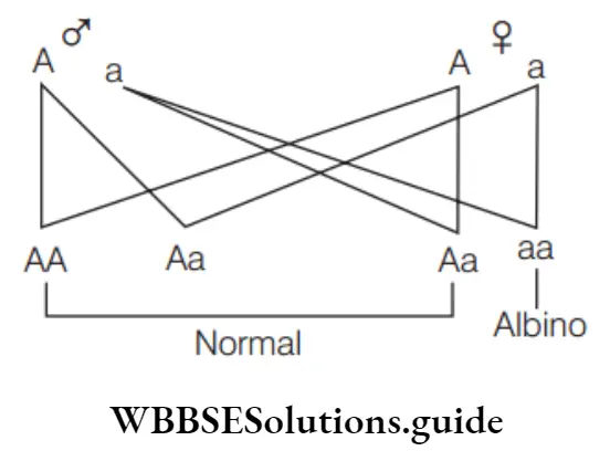
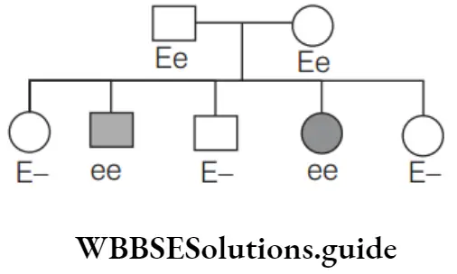
Question 12. Consider the following chart.
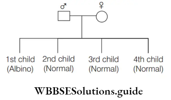
The first child of a couple is albino. Whereas, their second, third and fourth children are normal. Given that, A= normal and a = Albino, find the genotype of the mother and father.
Father – Mother
- Aa – AA
- AA – Aa
- AA – AA
- Aa – Aa
Answer: 4. Albinism is the recessive trait which occurs only when a homozygous condition is present. In the given cross, the progenies are both albino and normal. This is possible only when their parents are heterozygous for normal colour (Aa).
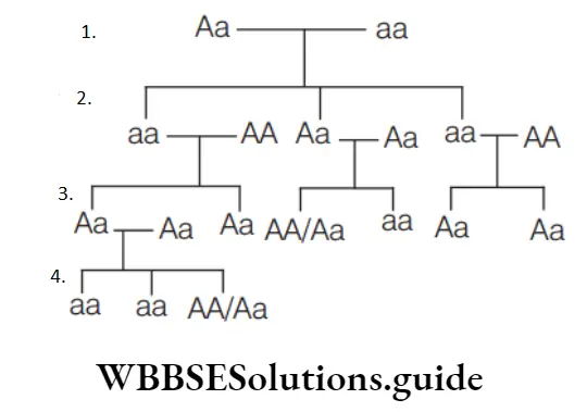
Question 13. Find out the genotype of father and mother in the given pedigree chart.
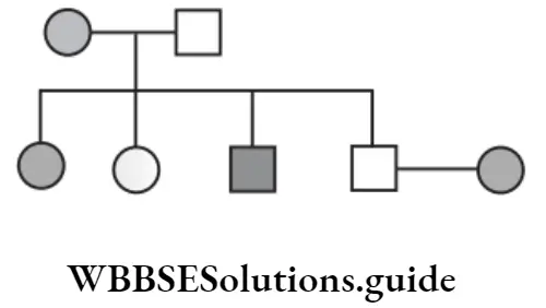
Mother Father
- AA AA
- Aa Aa
- AA aa
- Aa Aa
Answer: 4. aa Aa
The expected genotype of father and mother is Aa and aa, respectively. Since 50% male and female progenies are affected, mother would be homozygous recessive (aa) and father is heterozygous (Aa).
“monohybrid test cross ratio “
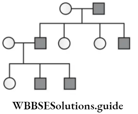
Question 14. Following pedigree chart show.
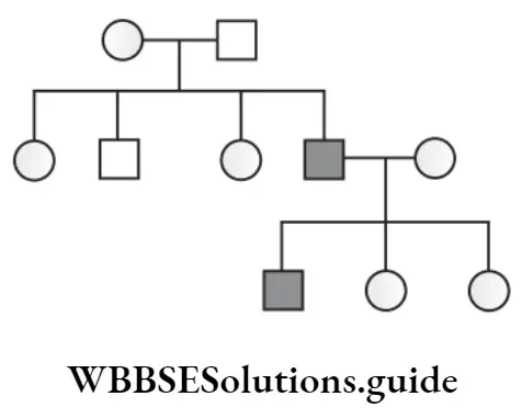
- Recessive and autosomal
- Recessive and sex-linked
- Dominant and sex-linked
- Dominant and autosomal
Answer: 2. Recessive and sex-linked
Given pedigree analysis indicates the transmission of recessive sex-linked trait from parents to their offspring because only 25% male progenies are affected.
Biology MCQ For NEET With Answers
Question 15. Identify the type of inheritance in the given diagram.
- Dominant X-linked
- Recessive X-linked
- Dominant Y-linked
- Cytoplasmic or mitochondrial inheritance
- Both 2 and 3
Answer: 4. Cytoplasmic or mitochondrial inheritance
- Cytoplasmic or mitochondrial inheritance is the inheritance in which the trait pass only from mother to all of their offspring.
- The genes of that inheritance present in the cytoplasm of ova that is way these genes go to all of their offspring. As sperm have very less cytoplasm, so this inheritance is not applicable for males.
NEET quiz on Pedigree Analysis and Genetic Disorders with solutions
Question 16. Hyperdactyly (the possession of more than 12 finger) is determined by the dominant allele (H) and normal condition by recessive allele (h). Observe the image given below. It represents family tree in which some members of the family are hyperdactylus. Identify A, B and C
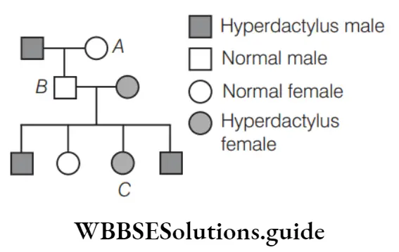
Find out the genotype of A, B and C.
- A-Hh, B-Hh, C-hh
- A-HH, B-Hh, C-hh
- A-Hh, B-HH, C-hh
- A-HH, B-HH, C-HH
Answer: 1. A-Hh, B-Hh, C-hh
- Dominant allele shows its effect in homozygous or heterozygous condition and recessive allele shows its effect only in homozygous condition.
- Given pedigree chart is possible only when the male parent in heterozygous (Hh) for hyperdactyle. If it is homozygous for hyperdactyle then the son would also be the hyperdactyle as well. Thus, option (1) is correct.
Biology MCQs with answers for NEET
Question 17. In a family, the father carried a trait which the mother did not. All their offspring displayed this trait. Though their daughters married normal males, a few grand-daughters carried this trait. Choose the correct pedigree chart for this condition.
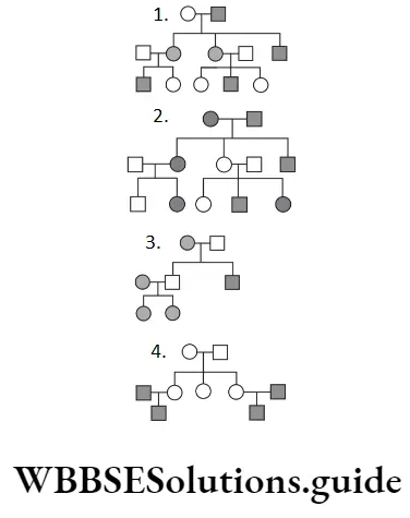
Answer: 1. Criss-cross inheritance is a type of sex-linked inheritance where a parent passes the traits to the grand child of the same sex through offspring of the opposite sex.
It appears in opposite sex progeny in subsequent generations. Pedigree chart in option (a) depict this inheritance pattern correctly.
Question 18. Study the pedigree chart given below. What does it show?
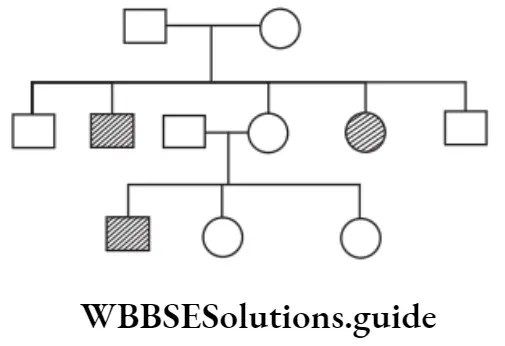
- Inheritance of a condition like phenylketonuria as an autosomal recessive trait
- The pedigree chart is wrong as this is not possible
- Inheritance of a recessive sex-linked disease like haemophilia
- Inheritance of a sex-linked inborn error of metabolism like phenylketonuria
Answer: 1. Inheritance of a condition like phenylketonuria as an autosomal recessive trait
The given chart shows inheritance of an autosomal recessive trait like phenylketonuria.An autosomal recessive trait may skip a generation but it affect both males and females equally. It appears in case of marriage between two heterozygous individuals.
“monohybrid cross ratio “
Question 19. Given below is a pedigree chart of a family with five children. It shows the inheritance of attached ear-lobes as opposed to the free ones. The squares represent the male individuals and circles represents the female individuals.
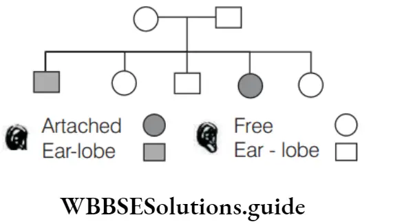
Which one of the following conclusions drawn is correct?
- The parents are homozygous recessive
- The trait is Y-linked
- The parents are homozygous dominant
- The parents are heterozygous
Answer: 4. The parents are heterozygous
The parents are heterozygous. It is an autosomal inheritance trait. It is controlled by a rare dominant allele say ‘E’ (free ear lobe). Here, the recessive homozygotes which do not have a dominant ‘E’ have attached ear lobes.
Biology MCQs with answers for NEET
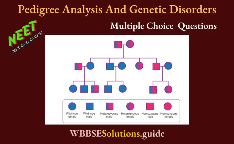
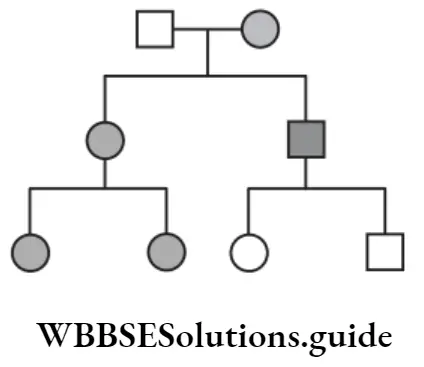
Question 20. Given below is a pedigree chart showing the inheritance of a certain sex-linked trait in humans.
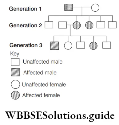
The trait traced in the above pedigree chart is
- Dominant X-linked
- Recessive X-linked
- Dominant Y-linked
- Recessive Y-linked
Answer: 1. Dominant X-linked
- The trait traced in the given pedigree is dominant X-linked. As seen in the figure, in first generation, male is affected, but female is not affected, i.e. female carries normal chromosomes in heterozygous condition.
- In second generation, both females are affected, but none of the males are affected. This shows that in spite of carrying single X-linked trait, they are affected, i.e. the trait is dominant.
- Likewise in the third generation when X-linked traits are again crossed with normal male, then, both male and female offspring carrying single Xlinked trait are affected. Thus, we can say that the trait is dominant X-linked.
Question 21. Assertion (A) In a pedigree analysis, represents five unaffected offspring. Reason (R) In pedigree analysis, the offspring are numbered with Arabic numerals (1, 2, 3 …..) and a generation is numbered with roman numerals (I, II, III ….).
- Both A and R are true and R is the correct explanation of A
- Both A and R are true, but R is not the correct explanation of A
- A is true, but R false
- Both A and R are false
Answer: 2. Both A and R are true, but R is not the correct explanation of A
Biology MCQs with answers for NEET
Both A and R are true, but R is not the correct explanation of A. In pedigree analysis, unaffected offspring are represented as non-shaded circle, square or diamond, representing the gender of progeny. The number of progenies are written in Arabic numerals within the gender symbol.
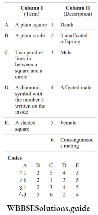
Question 22. Match the following columns.
“monohybrid ratio “
Answer: 4. A–3, B–5, C–6, D–2, E–4
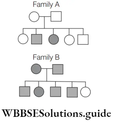
Question 23. Which of the following is true for a recessive disease in family A and B?
- In family A, both the parents are homozygous recessive
- In family B, both the parents are homozygous dominant
- In family B, both the parents are heterozygous recessive
- In family A, both the parents are heterozygous recessive
Answer: 4. In family A, both the parents are heterozygous recessive
- Option (4) is true whereas option (1), (2) and (3) are incorrect. Incorrect options can be corrected as In family A, if both the parents are homozygous recessive, then both should be diseased and should have 100% diseased progeny.
- In family B, if both parents are homozygous dominant, they would not have got the recessive disease in first place. If both are heterozygous recessive, then also they would not have got the disease, neither 80% of progeny would be diseased.
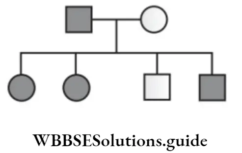
Question 24. In the following human pedigree, the filled symbols represent the affected individuals. Identify the type of given pedigree.
- Autosomal recessive
- Autosomal dominant
- X-linked dominant
- X-linked recessive
Answer: 1. Autosomal recessive
Autosomal recessive traits are the traits which are caused by recessive autosomal genes when present in homozygous condition. The given pedigree can be explained as follows.
As the trait appears only in homozygous recessive individuals (aa), therefore it is an autosomal recessive trait.
Question 25. Which one is the incorrect statement with regard to the importance of pedigree analysis?
- It helps to trace the inheritance of a specific trait
- It confirms that DNA is the carrier of genetic information
- It helps to understand whether the trait in question is dominant or recessive
- It confirms that the trait is linked to one of the autosome
Answer: 2. It confirms that DNA is the carrier of genetic information
- Statement in option (2) is incorrect and can be corrected as Pedigree is a chart showing the record of inheritance of certain genetic traits for two or more ancestral generations of an individual, abnormality or disease.
- It does not confirm that DNA is the carrier of genetic information. Rest statements are correct with regard to the importance of pedigree analysis.
Biology MCQs with answers for NEET
Question 26. The process of mating between closely related individuals is
- Self breeding
- Inbreeding
- Hybridisation
- Heterosis
Answer: 2. Inbreeding
Inbreeding is the process of mating between closely related individuals. It leads to the expression of recessive characters, most of which are harmful.
Question 27. The square, blackened shape and horizontal lines in pedigree analysis represents
- Female, healthy individual, parents
- Female, affected individual, parents
- Male, affected individual, parents
- Male, affected individual, progeny
Answer: 3. Male, affected individual, parents
In pedigree, square represents male, blackened square or circle represents affected individual. Horizontal line represents parents.
Question 28. Double lines in pedigree analysis show
- Unaffected offspring
- Sex unspecified
- Normal mating
- Consanguineous marriage
Answer: 4. Consanguineous marriage
Double lines in pedigree analysis represent mating between relatives, i.e. consanguineous mating. Normal mating is shown by single line. Unaffected offspring are depicted by open and clear symbols (i.e. for male and O for female).
Question 29. Eugenics is defined as
- The study of diseases resulting from environmental cause
- The study of congenital defects in man
- The study of human genetics from the point of view of the improvement of the genetic composition of the human stock during gestation or birth
- The diseases caused by consanguinity
Answer: 3. The study of human genetics from the point of view of the improvement of the genetic composition of the human stock during gestation or birth
Eugenics is the study of human genetics from the point of view of the improvement of the genetic composition of the human stock during gestation or birth.
Biology MCQs with answers for NEET
Question 30. The term ‘eugenics’ was given by Francis Galton and inborn errors of metabolism were studied by
- Galton
- Garrod
- Barr
- Both 1 and 2
Answer: 2. Garrod
The term inborn errors of metabolism were coined by a British physician, Archibald Garrod (1857– 1936), in 1908. He is known for work that prefigured the ‘one gene-one enzyme’ hypothesis, based on his studies on the nature and inheritance of alkaptonuria.
NEET expected MCQs on Pedigree Analysis 2025
Question 31. A heterozygous individual carrying recessive sex-linked gene is called
- Carrier
- Crossing over
- Transmitter
- Albino
Answer: 1. Carrier
An individual with one abnormal gene (but no symptoms) is called a carrier. A heterozygous individual carrying recessive sex-linked genes is called carrier.
Question 32. A hereditary disease which is never passed on from father to son is
- Autosomal linked disease
- X-chromosomal linked disease
- Y-chromosomal linked disease
- None of the above
Answer: 2. X-chromosomal linked disease
X-chromosome linked diseases can never be passed on from father to son because the X-chromosome of the father can only be transferred to daughter and Y-chromosome is only passed on to the son.
Question 33. Y-linked recessive gene is transferred from
- Father to son
- Mother to son
- Father to daughter
- Mother to daughter
Answer: 1. Father to son
Because only males have a Y-chromosome, in Y-linked inheritance, a mutation can only be passed from father to son.
Question 34. Mendelian disorder may be of
- Recessive
- Dominant
- Both 1 and 2
- Cannot be determined
Answer: 3. Both 1 and 2
Mendelian disorder may be dominant or recessive type, e.g. haemophilia, colour blindness, sickle cell anaemia, cystic fibrosis, phenylketonuria, thalassaemia. Thus, option (4) is correct.
Question 35. Sex linked disorder is
- Colour blindness
- Tuberculosis
- Diphtheria
- Leprosy
Answer: 1. Colour blindness
Colour blindness is a recessive sex-linked trait in which the eye fails to distinguish red and green colours. The gene for the normal vision is dominant.
Question 36. Which are sex-linked traits?
- Osteomalacia
- Dental disorders
- Pellagra
- Haemophilia and hypertrichosis
Answer: 4. Haemophilia and hypertrichosis
NEET Biology Mcq
Sex-linked traits are the traits whose determining genes are found on the sex chromosomes. All the sex-linked traits present on a sex chromosome are inherited together. For example, haemophilia and hypertrichosis are sex-linked traits.
Question 37. Identify a Mendelian disorder from the following.
- Down’s syndrome
- Turner’s syndrome
- Phenylketonuria
- Klinefelter’s syndrome
Answer: 3. Phenylketonuria
The pattern of inheritance of Mendelian disorders can be traced in a family by the pedigree analysis. Some common Mendelian or gene related human disorders are phenylketonuria, alkaptonuria, albinism, sickle cell anaemia and cystic fibrosis, etc.
Question 38. Which of these is not a Mendelian disorder?
- Down’s syndrome
- Sickle-cell anaemia
- Colour blindness
- Haemophilia
Answer: 1. Down’s syndrome
- Mendelian disorders are determined by mutations in single genes. They are transmitted to the offspring as per Mendelian principles.
- The pattern of inheritance of such Mendelian disorders can be traced in a family by the pedigree analysis. Down’s syndrome is chromosomal not Mendelian disorder.
Question 39. Identify a non-hereditary disease from the following.
- Thalassemia
- Haemophilia
- Cystic fibrosis
- Cretinism
Answer: 4. Cretinism
Cretinism is a congenital deficiency of thyroid hormone. It is a non-hereditary disease.
Question 40. A man and a woman, who do not show any apparent signs of a certain inherited disease, have seven children (2 daughters and 5 sons). Three of the sons suffer from the given disease but none of the daughters affected. Which of the following mode of inheritance do you suggest for this disease?
- Sex-linked dominant
- Sex-linked recessive
- Sex-limited recessive
- Autosomal dominant
Answer: 2. Sex-linked recessive
Traits governed by sex-linked recessive genes produce disorders in males more often than in females or express themselves in males even when represented by a single allele because Y-chromosome does not carry any corresponding alleles.
NEET Biology Mcq
Question 41. Huntington’s chorea is
- Recessive mutation associated with chromosome 14
- Dominant mutation associated with chromosome 4
- Recessive mutation associated with chromosome X
- Sex-linked disease
Answer: 2. Dominant mutation associated with chromosome 4
Huntington’s chorea is a progressive brain disorder caused by a single defective gene on chromosome-4. This defect is dominant as anyone who inherit it from a parent will develop the disease.
Its symptoms include difficulty in concentrating, memory lapses, depression, mood swings, etc.
Question 42. The traits which are expressed in only a particular sex though their genes occurs in the opposite sex too are known as
- Sex-linked trait
- Sex influenced trait
- Sex-limited traits
- None of the above
Answer: 3. Sex-limited traits
- Sex-limited traits are genes that are present in both sexes of sexually reproducing species but are expressed in only one sex and remain ‘turned off’ in the other.
- In other words, sex limited genes cause the two sexes to show different traits or phenotypes, despite having the same genotype.
Question 43. The recessive genes located on X chromosome in humans are always
- Lethal
- Expressed in males
- Sublethal
- Expressed in females
Answer: 2. Expressed in males
X-linked recessive genes are expressed in females only if there are two copies of the gene (one on each X-chromosome). However, for males, there needs to be only one copy of an X-linked recessive gene in order for the trait or disorder to be expressed. Thus, these are always expressed in males.
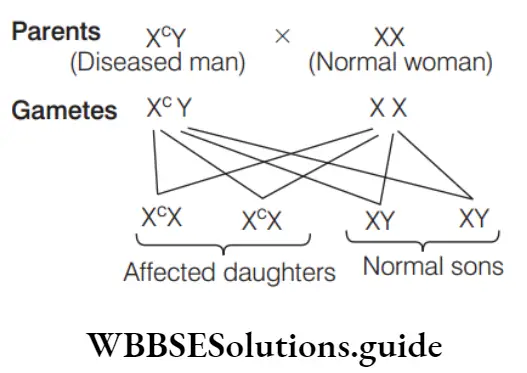
Question 44. A diseased man marries a normal woman. They give birth to three daughters and five sons. All the daughters were diseased and sons were normal. The gene of this disease is’
- Sex-linked dominant
- Sex-linked recessive
- Sex-limited character
- Autosomal dominant
Answer: 1. Sex-linked dominant
Since only daughters are affected, the disease is X-linked. Daughters get one copy of affected X chromosome from father and other normal copy from mother. The trait is expressed in daughter in heterozygous condition, therefore it is a dominant trait. Thus, the gene of this disease is sex-linked dominant.
NEET Biology Mcq
Question 45. Abnormal gene is replaced by normal gene by
- Gene therapy
- Mutation
- Cloning
- None of these
Answer: 1. Gene therapy
- If a child or an embryo (foetus) is diagnosed to carry a defective gene leading to disability, this defect can be corrected by replacement of defective gene with a normal gene.
- It is done by correcting the defective gene through gene targeting or by gene augmentation either through increasing the number of copies of the gene or through a higher level of expression of the introduced gene. Such a correction of genetic defect is described as gene therapy.
Question 46. Which one of the following techniques is employed in human genetic counselling?
- Serological technique
- Polyploidy
- Pedigree analysis
- Genetic engineering
- Amniocentesis
Answer: 3. Pedigree analysis
Pedigree analysis is the techniques which is employed in human genetic counselling because it shows the phenotypes and family relationships of the individuals through charts of family histories.
Question 47. Which of the following is an inherited disorder?
- Leprosy
- Goitre
- AIDS
- Albinism
- Parkinson’s disease
Answer: 4. Albinism
Out of the given options, only albinism is an inherited disorder. Albinism is the recessive trait which occurs only when a homozygous condition is present.
Question 48. Carrier of genes of colour blindness are present in
- Father
- Mother
- Father and mother
- None of the above
Answer: 2. Mother
Colourblindness is a X-linked recessive trait. A man possesses only one gene for colour vision, whereas woman possesses two. Therefore,
only mother will be a carrier when she has gene for colour blindness in one X-chromosome.
NEET Biology Mcq Chapter Wise
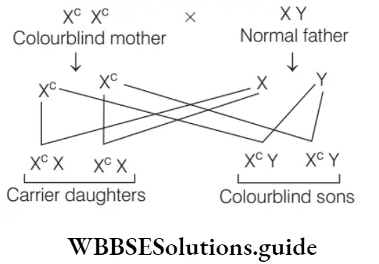
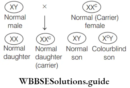
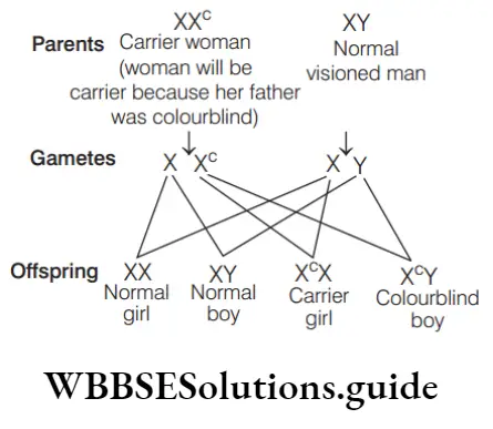
Question 49. A marriage between normal visioned man and colourblind woman will produce
- Colourblind sons and 50% carrier daughters
- 50% colourblind sons and 50% carrier daughters
- Normal sons and carrier daughters
- Colourblind sons and carrier daughters
Answer: 4. Colourblind sons and carrier daughters
- Colour blindness is the inability of certain human beings to distinguish red from green colour. It is produced by a recessive gene which lies on X chromosome.
- A marriage between a normal visioned man (XY) and colourblind woman (Xc cX ) results in
colourblind sons (XcY) and carrier daughters (XXc).
Question 50. Which is true for colour blindness?
- It is a sex-linked disorder
- A person is not able to recognise red and green
- The person lacks red and green pigments differentiating cells in eyes
- All of the above
Answer: 4. All of the above
All the given statements are true for colour blindness. Thus, option (d) is correct.
Question 51. A person whose father is colourblind marries a lady whose mother is a daughter of a colourblind man. Their children will be
- All sons colourblind
- Some sons normal and some colourblind
- All colourblind
- All daughters normal
Answer: 4. All daughters normal
To solve the given quesiton, genotype of different individuals is needed to be predicted in steps. To find out the genotype of person, whose father is colurblind.
XCY × XX
↓
Offspinings XCX, XCX, XY, XY
As all sons will be normal, therefore the genotype of the person will be XY. ….
To find out the genotype of lady whose mother is a daughter of colourblind man
XCY × XX
↓
XCX, XCX, XY, XY
So, the mother of lady would be carrier, having genotype XCX … Performing cross between to find out lady’s genotype.
XX × XCY
↓
XCX, XX, XCY, XY
As 50% daughters are carrier and 50% daughters are normal. So, the lady can be normal or carrier having genotype XX, XCX, respectively. …
Now considering both the genotypes of the lady and the genotype of the person, the result would be as follows
XY × XCX
↓
XCX, XY, XY, XCY, XY × XX
↓
XX,XX, XY , XY
Above cases show that if, mother (lady) is carrier then option (1) and (3) are not true. Option (2) is true and option (4) stating all daughters normal (though phenotypically) is also true. If mother is normal then options (1), (2) and (3) are not true and option (4) is true. So, from both the cases, it is concluded that option (4) is true.
NEET Biology Mcq Chapter Wise
Question 52. More men suffer from colour blindness than women because
- Women are more resistant
- Male sex hormone, testosterone causes the disease
- The colourblind gene is carried by Y-chromosome
- Men are hemizygous and one defective gene is enough to cause colour blindness
Answer: 4. Men are hemizygous and one defective gene is enough to cause colour blindness
- More men suffer from colour blindness than women because men are hemizygous and one defective gene is enough to cause colour blindness.
- Also colourblindness is a sex-linked recessive disease. The recessive genes located on X-chromosome of humans are always expressed in males.
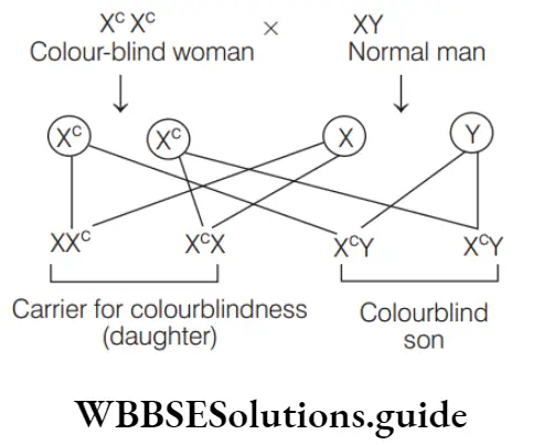
Question 53. If a colourblind woman marries a normal visioned man, their sons will
- All be colourblind
- All be normal visioned
- Be one-half colourblind and one-half normal
- Be three-fourths colourblind and one-fourth normal
Answer: 1. All be colourblind
A marriage between normal visioned man (XY) and colourblind woman (XCXC), results in colour blind sons (XcY) and carrier daughters (XXc). Thus, all sons will be colourblind.
Question 54. A woman with normal vision, but whose father was colourblind, marries a colourblind man. Suppose that the fourth child of this couple was a boy. This boy
- May be colourblind or may be of normal vision
- Must be colourblind
- Must have normal colour vision
- Will be partially colourblind since he is heterozygous for the colourblind mutant allele
Answer: 1. May be colourblind or may be of normal vision
Colour blindness is a recessive sex-linked trait. Since the woman’s father was colourblind. She should be carrier of the colourblind gene (X cX). When she marries to colourblind man their progeny could be as given below
Thus, the boy progeny of this couple may be colourblind or normal visioned.
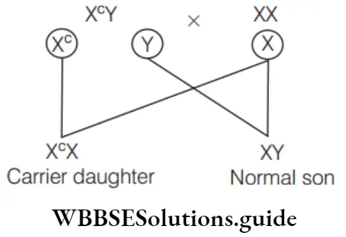
Question 55. A normal woman, whose father was colourblind is married a normal man. The sons would be
- 75% colourblind
- 50% colourblind
- All normal
- All colourblind
Answer: 2. 50% colourblind
Woman whose father is colourblind would have genotype XcX. So, woman is carrier. She marries a normal man, then progeny are
Thus, 50% colourblind sons would born.
Question 56. If all the sons of a couple are colourblind then
- Mother is homozygous colourblind
- Mother is homozygous and father is normal
- Mother is homozygous and father is colourblind
- Mother is normal and father is colourblind
Answer: 1. Mother is homozygous colourblind
Whenever mother is homozygous colourblind, all her sons will be colourblind irrespective of their father’s genotype.
So, option (1) is correct.
NEET Biology Mcq Chapter Wise
Question 57. A child’s father is colourblind and mother has also a gene for the same. The probability of the girls to be colourblind is
- 100%
- 50%
- 25%
- 75%
Answer: 2. 50%
If the mother carries the gene but is not colourblind (X XC ) and father is colourblind (X YC ) then, there is a 50% chance that their sons and daughter will be colourblind.
Question 58. Both husband and wife have normal vision though their fathers were colourblind. The probability of their daughter becoming colourblind is
- 0%
- 25%
- 50%
- 75%
Answer: 1. 0%
The probability of the daughter becoming colourblind arises only when the father is also colourblind. Husband-XY, Wife – XXC (wife is carrier because her father is colourblind and husband is normal).
So, probability of their daughter becoming colourblind is 0%.
Question 59. I. Haemophilia
- Cystic fibrosis
- Sickle-cell anaemia
- Colour blindness
- Cancer
- Plague
- Phenylketonuria
- Down’s syndrome
Choose the correct options for Mendelian disorders.
- 1, 2, 3, 4, 6, 8
- 1, 2, 3, 4, 7
- 1, 2, 3, 4, 5, 6
- 1, 2, 3, 4, 5, 8
Answer: 2. 1, 2, 3, 4, 7
- Option (2) is correct. Genetic disorder mainly caused due to alternation and mutation in a single gene.
- These genes are transmitted to offspring by the principle of inheritance. Mendelian disorders can be dominant or recessive, e.g. haemophilia, colour blindness, sickle-cell anaemia, cystic fibrosis, phenylketonuria, thalassaemia. Rest other diseases do not represent Mendelian disorder.
Question 60. A colourblind man, whose parents had normal vision and whose paternal and maternal grandparents had normal vision, probably inherited the gene for colour blindness from his
- Maternal or paternal grandmother
- Maternal or paternal father
- Father
- Mother
Answer: 4. Mother
A male always carries X- chromosome from his mother. Thus, if a male is colourblind, this gene is inherited from his mother only.
NEET Biology Mcq Chapter Wise
Question 61. The cure for night blindness is administration of vitamin …p… medicines but colour blindness is not curable because it is a …q… disease. Identify p and q.
- p-A, q-genetic
- p-B, q-autosomal
- p-C, q-non-genetic
- p-D, q-genetic
Answer: 1. p-A, q-genetic
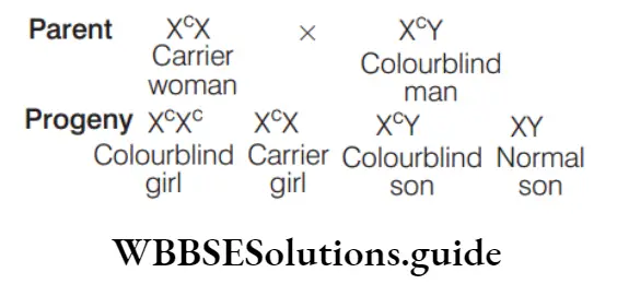
Question 62. A colourblind man marries the daughter of another colourblind man whose wife has a normal genotype for colour vision. In their progeny
- All the children would be colourblind
- All their sons are colourblind
- None of the daughters would be colourblind
- Half of their sons and half of their daughters would be colourblind
Answer: 4. Half of their sons and half of their daughters would be colourblind
Colour blindness is a X-linked recessive disorder. It shows criss-cross inheritance. A colourblind man has a genotype XcY. He married daughter of a colourblind man (XCY) and a normal woman (XX), thus his wife should have XCX genotype. The genotypes of their children can be worked out as follows
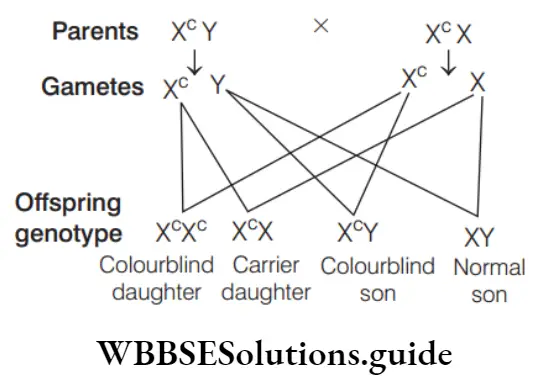
Thus, 50% of their sons and 50% of their daughters would be colourblind.
Question 63. If daughter of colourblind marries another colourblind then possibility of their first daughter to be colourblind is
- 50%
- 25%
- 7%
- None of these
Answer: 1. 50%
- Daughter of a colourblind person will be a carrier of the disease and only one of the two X-chromosomes will bear the recessive gene for the disease (X Xc ).
- If she marries another colourblind man (X Y)c then the possibility of first daughter to be colourblind is 50%.
Question 64. A colourblind man marries a woman with normal sight who has no history of colour blindness in her family. What is the probability of their grandson being colourblind?
- Nil
- 0.25
- 0.5
- 1
Answer: 3. 0.5
When a colourblind man (XCY) marries a normal woman (XX), all of their daughters are carriers and all of their sons are normal, as shown in following cross.
When the carrier daughter (XXC) is married to a normal man, the probability of their son being colourblind is 0.25, as shown in following cross.
From above crosses, it is clear that the probability of occurrence of colour blindness in the grandson of a colourblind man and a normal woman is 0.5.
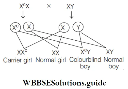
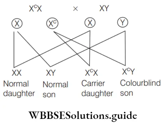
Question 65. Proportion of colourblind children when normal man marries to carrier woman is
- 25%
- 50%
- 75%
- 100%
Answer: 1. 25%
When a normal man (XY) marries a carrier woman (XCX) for colour blindness, then 25% of progeny would be colourblind.
Question 66. A normal-visioned man whose father was colourblind, marries a woman whose father was also colourblind. They have their first child as a daughter. What are the chances that this child would be colourblind?
- 100%
- 0 %
- 25%
- 50 %
Answer: 2. 0 %
If a normal-visioned man marries a woman whose father was also colourblind, then his wife would be carrier of this disease (if her mother was normal).
This trait is passed to children but daughters produced by this couple are carrier not the colourblind.
Thus, 0% daughters are colourblind.
Question 67. Red-green colour blindness in humans is governed by a sex-linked recessive gene. A normal woman whose father was colourblind marries a colourblind man. What proportion of their daughters is expected to be colourblind?
- 3/4
- 1/2
- 1/4
- All
Answer: 2. 1/2
Normal woman whose father was colourblind would be a carrier (XCX). She married a colourblind man (XCY) and thus, half (1/2) of their daughters are expected to be colourblind.
Question 68. In colour blindness, a person fails to differentiate between
- Red and blue
- Red and green
- Red and black
- Red and white
Answer: 2. Red and green
Red and green colour blindness results in defect in either red or/and green cone cells of eye, resulting in failure to discriminate between red and green colour.
Question 69. Of both normal parents, the chance of a male child becoming colourblind are
- Nil
- Possible only when all the four grandparents had normal vision
- Possible only when father’s mother was colourblind
- Possible only when mother’s father was colourblind
Answer: 4. Possible only when mother’s father was colourblind
On crossing carrier colourblind woman with a normal man, the sons become colourblind. This is an example of criss-cross inheritance. The genes for colour blindness is inherited from mother’s father.
Question 70. A colourblind son will be born when
- Mother is carrier and father is normal
- Mother is colourblind and father is normal
- Mother is carrier and father is colourblind
- All the cases are correct
Answer: 4. All the cases are correct
A colourblind son will be born to a carrier mother and a colourblind mother irrespective of father being colourblind or not. A carrier mother is a normal female who carries recessive allele for colour blindness in heterozygous state. Thus, option (d) is correct.
Question 71. Anish is having colourblindness and married to Sheela, who is not colourblind. What is the chance that their son will have the disease?
- 100%
- 50%
- 25%
- 0%
Answer: 2 & 4 50% & 0%
If Sheela (mother) is a carrier then chances of having colourblind son are 50%. If Sheela is normal then chances of their son being colourblind are 0%. It can be represented as follows
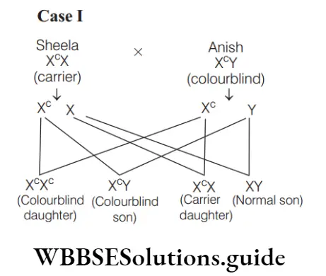
Thus, there are 50% chances of their son being colourblind.
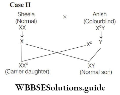
Thus, there are 0% chances of their son being colourblind. Thus, both options (b) and (d) are correct.
Question 72. Colour blindness, in which all the colours are perceived as grey, is termed as
- Monochromasia
- Chromasia
- Dichromasia
- All of these
Answer: 1. Monochromasia
- Colour blindness is the inability of certain human beings to distinguish between colours. It is produced by an X-linked recessive gene by causing lack of one of the primary cone pigments of the retina.
- Colour blindness, in which all colours are perceived as grey, is termed monochromasia. The person suffering from this disease is completely colourblind.
Question 73. If both parents of a male child are normal, what are the chances of the child being colourblind?
- It is impossible
- It is possible only if father’s mother was colourblind
- It is possible only if mother’s father was colourblind
- It is possible even when all the four grandparents had normal vision
Answer: 3. It is possible only if mother’s father was colourblind
- In this problem, an X-linked recessive allele is passed from an affected male, to a daughter who is a heterozygous carrier and subsequently to an affected grandson.
- Thus in given case, maternal father is likely to be colourblind. Such criss-cross trait is called as diagynic trait.
Question 74. If a colourblind man marries a woman who is homozygous for normal colour vision, the probability of their son being colourblind is
- 0
- 0.5
- 0.75
- 1
Answer: 1. 0
If a colourblind man marries a woman who is homozygous for normal colour vision, the probalility of their son being colourblind is zero.
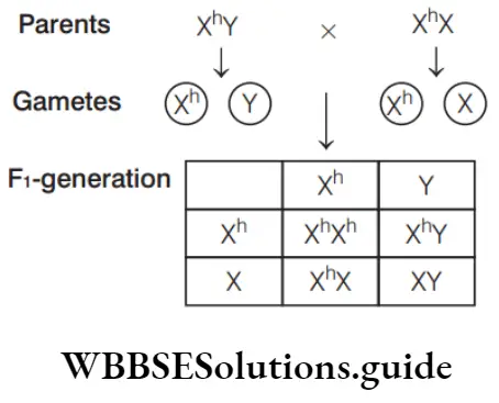
Question 75. X h is the chromosome with gene for haemophilia and X is the chromosome with normal gene. Which of the following individuals will act as carrier for haemophilia?
- Xh Y
- XY
- Xh Xh
- Xh X
Answer: 4. Xh X
Males are never the carrier of sex-linked recessive traits, e.g. haemophilia Heterozygous female for this condition (e.g. X X)h acts as carrier. XY is normal male. X Xh h and X h Y are haemophilic.
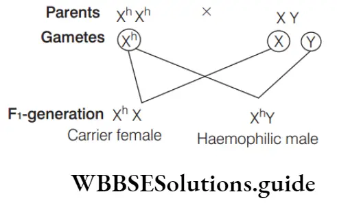
Question 76. Haemophilic man marries a normal woman. Their offsprings will be
- All haemophilic
- All boys haemophilic
- All girls haemophilic
- All normal
Answer: 4. All normal
- Haemophilia is a defect of blood which prevents its clotting. It is caused by a recessive gene located in the X-chromosome.
- When a haemophilic man (X h Y) marries a normal woman (XX), they would produce carrier girls (XX h ) and normal boys (XY), i.e. all their offspring will be normal.

Thus, option (4) is correct.
Question 77. The most common type of haemophilia results from the congenital absence of
- Factor 2
- Factor 5
- Factor 8
- Factor 11
Answer: 3. Factor 8
- Haemophilia is a group of inherited blood disorders in which the blood does not clot properly.
- It is caused by a fault in one of the genes that determine how the body makes blood clotting factor 8 or 9. These genes are located on the X-chromosome 10.
Question 78. A man known to be a victim of haemophilia marries a normal woman whose father was known to be a bleeder. Then it is expected that
- All their children will be bleeders
- Half of their children will be bleeders
- One fourth of their children will be bleeders
- None of their children will be bleeder
Answer: 2. Half of their children will be bleeders
Man is haemophilic with genotype XhY. The normal woman whose father was bleeder or diseased, would be a carrier of disease (XhX).
Thus, half of their children (50% girls and 50% boys) would be bleeders.
Question 79. Haemophilic female marries a normal male, the theoretical ratio of their offsprings regarding haemophilia will be
- All offspring are haemophilic
- All girls are haemophilic
- All sons are haemophilic
- Half daughters and half sons are haemophilic
Answer: 3. all sons are haemophilic
When a haemophilic female (XhXh) marries a normal male (XY), all sons would be haemophilic .
Question 80. If a boy’s father has haemophilia and his mother has one gene for haemophilia, what is the chance that the boy will inherit the disease?
- 25%
- 50%
- 75%
- 100%
Answer: 2. 50%
The boy’s father is haemophilic and mother is carrier.
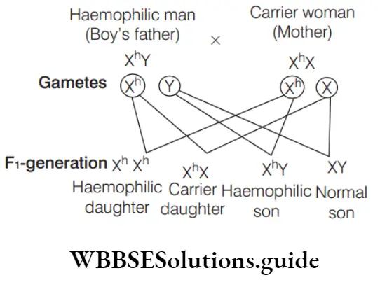
Thus, there are 50% chances that boy will inherit the disease.
Question 81. A woman carrier with two genes for haemophilia on one X-chromosome and for colour blindness on other X-chromosome marries a normal man. How will the progeny be?
- 50% haemophilic colourblind sons and 50% normal sons
- 50% haemophilic daughters (carrier) and 50% colourblind daughters (carrier)
- All sons haemophilic and colourblind
- Haemophilic and colourblind daughters
Answer: 3. All sons haemophilic and colourblind
- Haemophilia is a defect of blood which prevents its clotting. Colour blindness is the inability of certain human beings to distinguish red from green colour.
- Both these diseases are produced by a recessive gene which lies on the X-chromosomes. A woman having one gene for haemophilia on one X-chromosome and other gene for colour blindness on another X-chromosome will have genotype X Xh c.
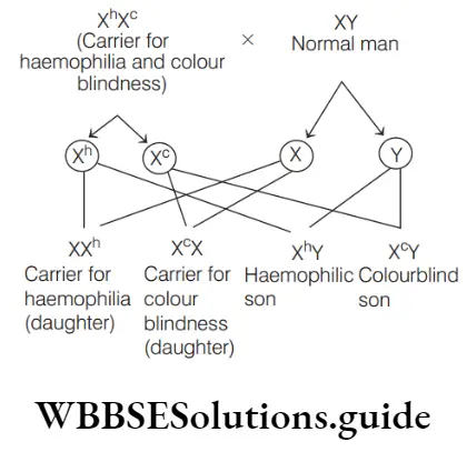
Thus, sons would either be colourblind or haemophilic and daughters will be the carriers.
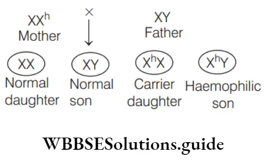
Question 82. Of a normal couple, half the sons are haemophilic, while half the daughters are carriers. The gene is located on
- X-chromosome of father
- Y-chromosome of father
- One X-chromosome of mother
- Both the X-chromosomes of mother
Answer: 3. One X-chromosome of mother
Of a normal couple, half the sons are haemophilic, while half the daughters are carriers. The gene is located on one X-chromosome of mother. Cross between a haemophilic carrier female X h X and normal male would yield 50% of the sons being haemophilic and 50% of the carrier daughters.
Question 83. Assertion (A) Haemophilia is a genetically linked disease. Reason (R) It is predominantly found in females.
- Both A and R are true and R is the correct explanation of A
- Both A and R are true, but R is not the correct explanation of A
- A is true, but R is false
- Both A and R are false
Answer: 3. A is true, but R is false
- A is true, but R is false. Reason can be corrected as Haemophilia predominantly occurs in males, since the gene can be passed from mother to son.
- A single copy of defected gene can cause disease in males. In contrast, woman is affected in the presence of two defected alleles which is a rare phenomena.
Question 84. Which is genetically not possible?
- Haemophilic father transfers the haemophilic gene to his son
- Haemophilic father transfers the haemophilic gene to his daughter
- Carrier mother transfers the haemophilic gene to her son
- Carrier mother transfers the haemophilic gene to her daughter
Answer: 1. Haemophilic father transfers the haemophilic gene to his son
- Statement in option (1) is not possible and can be corrected as Haemophilia is a sex-linked disease. It follows criss-cross inheritance in which father does not pass the sex-linked allele of a trait to his son.
- The same is passed to the daughter, from where it reaches the grandson, i.e. diagynic. It is because the males have only one X-chromosome which is transferred to the female offspring.
Question 85. Which of the following diseases is also called Christmas disease?
- Sickle-cell anaemia
- Haemoglobinuria
- Myocardial infarction
- Haemophilia-B
Answer: 4. Haemophilia-B
Haemophilia-B occurs due to deficiency of factor IX (Christmas factor). The patient may experience prolonged bleeding following any injury or wound and in severe cases there is spontaneous bleeding into muscles and joints.
Question 86. Which of the following most appropriately describes haemophilia?
- Recessive gene disorder
- X-linked recessive gene disorder
- Chromosomal disorder
- Dominant gene disorder
Answer: 2. X-linked recessive gene disorder
- Genes related with haemophilia are always present on X-chromosome and it is a recessive gene disorder as it expresses itself in females when it comes a homozygous condition.
- It causes a defect in the clotting factor formation, thus a simple cut can bleed continuously leading to even death. Thus, it is also known as ‘Bleeders’ disease or ‘Royal Disease’ as Queen Victoria is a carrier for this disease.
Question 87. One child is haemophilic (sex-linked trait), while its fraternal twin brother is normal. Which one of the following information is most appropriate?
- The mother must have been heterozygous
- The child is a monozygotic twin
- The other child is a female and the father is haemophilic
- The haemophilic child is a male
Answer: 1. The mother must have been heterozygous
The mother must have been a heterozygous haemophilic carrier (X h X). One of the twins would have inherited the normal X-chromosome and the other would have received the X-chromosome carrying the gene for haemophilia.
Question 88. Choose the incorrect statement with regard to haemophilia.
- It is a recessive disease
- It is a dominant disease
- A single protein involved in the clotting of blood is affected
- It is a sex-linked disease
Answer: 2. It is a dominant disease
Statement in option (2) is not correct. Haemophilia is sex-linked recessive disease. Rest statements are correct with regard to haemophilia.
Question 89. G-6-P dehydrogenase deficiency is associated with haemolysis of
- Leucocytes
- Lymphocytes
- Platelets
- RBCs
Answer: 4. RBCs
- Glucose-6-P dehydrogenase is the first enzyme of glucose oxidation during pentose phosphate pathway.
- RBC contains haemoglobin which combines with oxygen to form oxyhaemoglobin which gives its oxygen for oxidation of food. In haemolysis, there is destruction of RBCs with release of haemoglobin into plasma resulting in jaundice.
- Thus, RBCs cannot provide oxygen for oxidation of food thereby, causing deficiency of G-6-P dehydrogenase.
Question 90. A female becomes haemophilic definitely if
- Mother is carrier
- Father is carrier
- Father is affected
- Both mother and father affected
Answer: 4. Both mother and father affected
Males and female are definitely haemophilic, if their father and mother, both are haemophilic.
Question 91. A male human is heterozygous for autosomal genes A and B and is also hemizygous for haemophilic gene h. What proportion of his sperms will be abh?
- 1/8
- 1/32
- 1/16
- 1/4
Answer: 1. 1/8
Chance of getting ‘a’ = 1/2
Chance of getting ‘b’ = 1/2
Chance of getting ‘h’ = 1/2
Chance of getting sperms with abh
1/2 × 1/2 × 1/2 = 1/8
Question 92. Marriages between close relatives should be avoided because it induces more
- Blood group abnormalities
- Mutations
- Recessive alleles to come together
- Multiple births
Answer: 3. Recessive alleles to come together
The risk of a child inheriting two copies of a dangerous recessive allele is elevated in marriages between close relatives as compared to non-related marriages since they have a greater chance of inheriting the same recessive allele from their common ancestors.
Question 93. Haemophilia is a genetic disorder, in which
- Blood fails to coagulate after an injury
- There is delayed coagulation of blood
- Blood clots in blood vessels
- Blood cell count falls
Answer: 1. Blood fails to coagulate after an injury
Haemophilia is a defect of blood which prevents its clotting or coagulation after an injury. It is produced by a recessive gene which lies on the X-chromosomes.
Question 94. In which disease, the RBC of a person becomes half moon-shaped?
- Haemophilia
- Sickle-cell anaemia
- Thalassemia
- Leukemia
Answer: 2. Sickle-cell anaemia
Sickle-cell anaemia is a biochemical disorder in which shape of RBCs become sickle-shaped or half moon-shaped due to the defective haemoglobin. Haemoglobin becomes useless for oxygen transport.
Question 95. Haemophilia is related to which of the following?
- Colour blindness
- Polio
- Cataract
- Tumour
Answer: 1. Colour blindness
Haemophilia and colour blindness are sex-linked (X-linked recessive) disease. Option (a) is correct.
Question 96. Which one is ineffective against antibiotics?
- Bacterial infected wound
- Bacterial infected throat
- Haemophilia
- Bacterial infected gonorrhoea
Answer: 3. Haemophilia
Haemophilia is a genetic disease. Therefore, it is not affected by any antibiotics. These are used to treat bacterial diseases.
NEET Biology Genetic Disorders MCQs with explanations
Question 97. The α and β-globin chain is coded by genes located on chromosome number
- 16 and 11
- 10 and 12
- 9 and 8
- 15 and 16
Answer: 1. 16 and 11
The genes that encode the alpha (α) globin chains are on chromosome 16. Those that encode the non-alpha or β-globin chains are on chromosome 11.
Question 98. Persons suffering from sickle-cell anaemia normally do not suffer from
- Cholera
- Malaria
- High blood pressure
- Hepatitis
Answer: 2. Makaria
- Sickle-cell anaemia is caused by the formation of an abnormal haemoglobin called haemoglobin-S which differs from normal haemoglobin-A in only one amino acid, i.e. 6th amino acid ofβ-chain.
- Despite having harmful effect, the allele for sickle-cell anaemia continues to persist in human population because it has survival value in malaria infested areas like tropical Africa.
- Malarial parasite is unable to survive in abnormal shaped erythrocyte. Thus, persons suffering from sickle-cell anaemia normally do not suffer from malaria.
Question 99. Sickle-cell anaemia, a hereditary disease was first described by
- James B Herrick
- William Harvey
- Carl Landsteiner
- J Priestley
Answer: 1. James B Herrick
James Bryan Herrick (1861-1954), an American physician was credited with the description of sickle-cell anaemia (an autosomal hereditary disorder). He was one of the first physicians to describe the symptoms of myocardial infarction.
Question 100. The gene of sickle-cell anaemia is inherited by
- Blood cells
- Bone cells
- Sex chromosomes
- Autosomes
Answer: 4. Autosomes
Sickle-cell anaemia is inherited in an autosomal recessive pattern, which means that both copies of the gene in each cell have mutations.
Question 101. Assertion (A) Sickel-cell anaemia is a genetically determined disorder affecting many newborn babies. Reason (R) It is caused by heterozygosity for allele HbS producing a single amino acid substitution in the α-chain of the normal haemoglobin molecule determined by allele Hb A.
- Both A and R are true and R is the correct explanation of A
- Both A and R are true, but R is not the correct explanation of A
- A is true, but R false
- Both A and R are false
Answer: 3. A is true, but R false
A is true, but R is false. Reason can be corrected as Sickle-cell anaemia is caused when Hb S allele is present in homozygous condition. It is the resultant of single amino acid substitution in β-chain of haemoglobin.
Question 102. Sickle-cell anaemia results due to mutation caused by
- Substitution
- Insertion
- Deletion
- Duplication
Answer: 1. Substitution
- Sickle-cell anaemia is caused due to inheritance of a defective allele coding for β-globin. It results in the transformation of Hb A into Hb S in which glutamic acid is replaced by valine at sixth position in each of two β -chains of haemoglobin.
- The substitution of amino acid in the globin protein results due to the single base substitution at the sixth codon of the beta globin gene from GAG (Glu) to GUG (Val). Sickle-cell anaemia is a disease where the red blood cells become sickle-shaped instead of biconcave disc-shape.
Question 103. Genetic or chromosomal symbol used for the person who is having sickle-cell anaemia is
- Hb HbS S
- Hb Hba a
- Hb Hbg g
- Hb Hbb b
Answer: 1. Hb HbS S
Genetic or chromosomal symbol used for person who is having sickle-cell anaemia is Hb S Hb S.
Question 104. Both sickle-cell anaemia and Huntington’s chorea are
- Congenital disorders
- Pollutant-induced disorders
- Virus-related diseases
- Bacteria-related diseases
Answer: 1. Congenital disorders
- Sickle-cell anaemia is a biochemical disorder in which shape of RBCs become sickle-shaped due to the defective haemoglobin.
- Huntington Chorea in a disease in which atrophy of brain occurs resulting to respiratory irregulations, articulation of speech and irregular limb movements take place. They both are genetic disease present in any person since birth hence, called congenital diseases.
Question 105. Sickle-cell anaemia is a case of
- Single mutation in a gene on chromosome 10
- Pleiotropism
- Both 1 and 2
- None of the above
Answer: 2. Pleiotropism
Sickle-cell anaemia is a classic example of the mixed benefit given by the staying power of pleiotropic genes, as the mutation to Hb-S provides the fitness benefit of malaria resistance to heterozygotes, while homozygotes have significantly lowered life expectancy.
Question 106. In sickle-cell anaemia, the sequence of amino acids from the first to the seventh position of the β -chain of haemoglobin-S(HbS ) is
- His, Leu, Thr, Pro, Glu, Val, Val
- Val, His, Leu, Thr, Pro, Glu, Glu
- Thr, His, Pro, Val, Pro, Val, Glu
- Glu, His, Leu, Pro, Val, Glu, Glu
- Val, His, Leu, Thr, Pro, Val, Glu
Answer: 5. Val, His, Leu, Thr, Pro, Val, Glu
The amino acid sequence in person with sickle-cell anaemia is Val, His, Leu, Thr, Pro, Val, Glu.
Question 107. Sickle-cell anaemia is caused due to the substitution of
- Valine at the 6th position of beta globin chain by glutamine
- Valine at the 6th position of alpha globin chain by glutamic acid
- Glycine at the 6th position of alpha globin chain by glutamic acid
- Glutamic acid at the 6th position of beta globin chain by valine
- None of the above
Answer: 4. Glutamic acid at the 6th position of beta globin chain by valine
Sickle-cell anaemia is the example of point mutation in which the glutamic acid (Glu) is replaced by valine (Val) at the sixth position of β-globin chain of haemoglobin molecule.
Question 108. A marriage between two carriers of sickle-cell anaemic gene will result into
- 1 normal and 2 carriers
- 1 sickle-cell anaemic
- 2 normal and 2 sickle-cell anaemic
- Both 1 and 2
Answer: 4. Both 1 and 2
Sickle-cell anaemia occurs when a person inherits two sickle-cell genes, one from each parent. If both partner carry sickle-cell gene, there will be 1 normal, 2 carriers and 1 sickle-cell anaemic progeny.
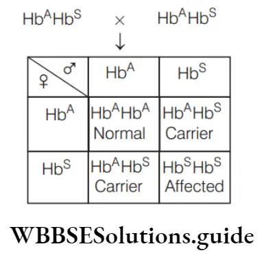
Question 109. Marriage between two sickle-cell carriers results into normal and sickle-cell carrier progenies in the ratio of
- 2: 1
- 3: 1
- I: 2 : 1
- 1: 2
Answer: 4. 1: 2
Question 110. A couple, both carriers for the gene sickle-cell anaemia planning to get married, wants to know the chances of having anaemic progeny?
- 100%
- 75%
- 50%
- 25%
Answer: 2. 25%
A marriage between two carriers of sickle-cell anaemia will produce normal, carrier and anaemic with progeny in 1: 2: 1 ratio. Out of this 25% are diseased or anaemic, 50% are carrier and 25% are normal.
Question 111. Choose a false statement with reference to sickle-cell anaemia.
- Have genotype Hb / HbS S
- Have genotype Hb / HbA A
- Substitution of glutamic acid to valine
- Have sickle shape RBC
Answer: 2. Have genotype Hb / HbA A
Statement in option (2) is false and can be corrected as Genotype- Hb A /Hb A represents a normal individual. Whereas sickel-cell anaemia is represented by genotype Hb S/Hb S Rest options are correct.
Question 112. Sickle-cell anaemia has not been eliminated from the African population because
- It is not a fatal disease
- It provides immunity against malaria
- It is controlled by dominant genes
- It is controlled by recessive genes
Answer: 2. It provides immunity against malaria
Question 113. Sickle-cell anaemia occurs as a result of …A… mutation in …B… of haemoglobin. Fill the correct option for A and B.
- A–point, B– β-chain
- A–chromosomal, B– β-chain
- A–allele, B– β-chain
- A–non-allele, B– α-chain
Answer: 1. A–Point, B–β-chain.
Question 114. In sickle-cell anaemia, GAG is replaced by
- GGA
- GUG
- UUG
- GGG
Answer: 2. GUG
GAG that code for glutamic acid in haemoglobin mRNA is replaced by GUG code which code for valine in haemophilic haemoglobin mRNA. So, in sickle-cell anaemia GAG is replaced by GUG.
Question 115. The genotype of a carrier carrying a gene for sickle-cell anaemia is
- HbS
- HbA/HbS
- HbA
- HbO
Answer: 2. HbA/HbS
Sickle haemoglobin is often shortened to S or Hb S. If a person have only one copy of the sickle haemoglobin along with one copy of the more usual haemoglobin (A or Hb A ) he is said to have sickle-cell trait. This is not an illness but means that he ‘carry’ the gene and can pass it on to children.
Question 116. Which type of thalassemia is more dangerous?
- Thalassemia minor
- Alpha thalassemia
- Thalassemia junior
- None of the above
Answer: 2. Alpha thalassemia
Thalassemia minor is a less serious form of the disorder. There are two main forms of thalassemia that are more serious. In alpha thalassemia, at least one of the alpha globin genes has a mutation or abnormality. In beta thalassemia, the beta globin genes are affected.
Question 117. Which of the following disease is called Cooley’s anaemia?
- Down’s syndrome
- Thalassemia
- Haemophilia
- Turner’s syndrome
Answer: 2. Thalassemia
Thalassemia major or Cooley’s anaemia is the most severe form of beta thalassemia in which the complete lack of beta protein in the haemoglobin causes a life-threatening anaemia that requires regular blood transfusions and extensive ongoing medical care.
Question 118. Thalassemia and sickle-cell anaemia are caused due to a problem in globin molecule synthesis. Select the correct statement.
- Both are due to a qualitative defect in globin chain synthesis
- Both are due to a quantitative defect in globin chain synthesis
- Thalassemia is due to less synthesis of globin molecules
- Sickle-cell anaemia is due to a quantitative problem of globin molecules
Answer: 3. Thalassemia is due to less synthesis of globin molecules
- Statement in option (3) is correct. Other statements are incorrect and can be corrected as
- Thalassemia is a quantitative problem whereas sickle-cell anaemia is a qualitative problem. Thalassemia occurs due to either mutation or deletion resulting in reduced rate of synthesis of one of globin chains of haemoglobin.
- In sickle-cell anaemia due to point mutation, glutamic acid (Glu) is replaced by valine (Val) at the sixth position ofβ-globin chain of haemoglobin molecule.
Question 119. The disease caused due to the quantitative abnormality of polypeptide chain of globin chain synthesis is
- Down’s syndrome
- Thalassemia
- Turner’s syndrome
- Klinefelter’s syndrome
Answer: 2. Thalassemia
In thalassemia, the reduced production of one of the globin chains upsets the balance of alpha to beta chains and causes abnormal haemoglobin to form or causes an increase of minor haemoglobin components, such as HbA2. Thus, it is caused due to quantitative abnormality of globin chains.
Question 120. Thalassemia is
- An autosomal recessive disease
- An autosomal dominant disease
- A sex-linked dominant disease
- A sex-linked recessive disease
Answer: 1. An autosomal recessive disease
Thalassemiais is an autosome-linked recessive disease. It occurs due to either mutation or
deletion resulting in reduced rate of synthesis of one of globin chains of haemoglobin.
Question 121. Which of the following statement about Huntington’s disease is true?
- Genetic test to detect the presence of the allele responsible for Huntington’s disease do not exist at this time
- The onset of Huntington’s disease is typically between birth and three years of age
- There is currently no effective treatment of this disease
- It is caused by the expression of recessive allele
Answer: 3. There is currently no effective treatment of this disease
- Statement in option (3) is true. Other statements are false and can be corrected as Genetic test to detect Huntington’s disease counts the number of CAG repeats in HD gene, using DNA from blood sample.
- The presence of 36 or more repeats confirms the disease. Huntington’s disease develops between 30 and 50 years of age. It is not caused by the expression of recessive allele.
Question 122. Which of the following disease is characterised by excessive trinucleotide repeats (CAG)?
- Cystic fibrosis
- PTC testing
- Dwarfism
- Huntington’s disease
Answer: 4. Huntington’s disease
Question 123. In α -thalassemia, the ……………… chromosome is affected.
- 16th
- 17th
- 18th
- 22nd
Answer: 1. 16th
In α-thalassemia, production of α-globin chain is affected. It is controlled by the closely linked genes HBA1 and HBA2 on chromosome 16. It occurs due to the mutation or deletion of one or more of the four genes.
Question 124. If parents are carriers for thalassemia, which is an autosomal recessive disorder, what are the chances of pregnancy resulting in an affected child?
- 50%
- 25%
- 100%
- No chance
Answer: 2. 25%
- In the given case, both the partners are unaffected carriers for the gene, i.e. have heterozygous genotype Tt. People homozygous for the autosomal recessive gene of thalassemia suffer from severe haemolytic anaemia.
- Heterozygous people are also not normal, but show the defect in a less severe form (Thalassemia minor). Genotype of carrier parents is Aa (male parent) and Aa (female parent).
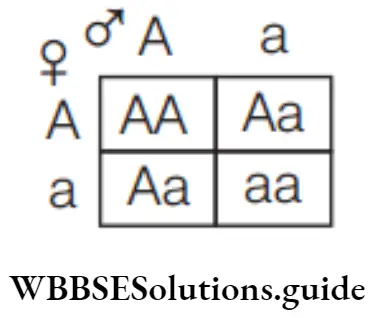
AA → Normal child (25%)
Aa → Carrier child (50%)
aa → Affected child (25%)
Question 125. Phenylketonuria disease is a
- Autosomal dominant
- Autosomal recessive
- Sex linked recessive
- Sex linked dominant
Answer: 2. Autosomal recessive
Phenylketonuria is an autosomal recessive disease that is being carried on chromosome 12.
Question 126. With respect to phenylketonuria identify which statement is not correct.
- It is a case of aneuploidy
- It is an example of pleiotropy
- Caused due to autosomal recessive trait
- It is an error in metabolism
Answer: 1. It is a case of aneuploidy
Statement in option (1) is incorrect and can be corrected as Phenylketonuria occurs due to defective gene of chromosome 12, not due to aneuploidy.
Question 127. In phenylketonuria, the phenylalanine gets converted to
- Acetic acid
- Phenyl acetic acid
- Phenyl pyruvic acid
- Pyruvic acid
Answer: 3. Phenyl pyruvic acid
Due to the absence of phenylalanine hydroxylase, the phenylalanine changes into phenyl pyruvic acid in phenylketonuria. Lack of this enzyme is due to autosomal recessive defective gene on chromosome number 12.
Question 128. In order to lessen the suffering of phenylketonurics, their diet should have
- No phenylalanine and no tyrosine
- Low phenylalanine and normal requirement of tyrosine
- Normal recommended amount of phenylalanine
- Normal recommended amount of both phenylalanine and tyrosine
Answer: 2. Low phenylalanine and normal requirement of tyrosine
- Phenylketonuria is an inborn error of metabolism in which an individual lacks an enzyme (phenylalanine hydroxylase PAR) that converts amino acid phenylalanine into tyrosine.
- In order to lessen the suffering of phenylketonurics, their diet should have low phenylalanine and normal requirement of tyrosine.
Mock test on Pedigree Analysis for NEET preparation
Question 129. Assertion (A) Phenylketonuria is an inborn error of metabolism. Reason (R) Phenylalanine is not converted into alanine in individuals suffering from this disease.
- Both A and R are true and R is the correct explanation of A
- Both A and R are true, but R is not the correct explanation of A
- A is true, but R false
- Both A and R are false
Answer: 3. A is true, but R false
- A is true, but R is false. Phenylketonuria is an inborn, autosomal, recessive metabolic disorder in which the homozygous recessive individual lacks the enzyme phenylalanine hydroxylase needed to change phenylalanine (amino acid) to tyrosine (amino acid) in liver.
- It results in hyperphenylalaninemia, which is characterised by accumulation and excretion of phenylalanine, phenylpyruvic acid and related compounds.
- Question 130. A person affected with phenylketonuria, lacks an enzyme that converts the amino acid phenylalanine intoValine
- Proline
- Histidine
- Tyrosine
- Methionine
Answer: 4. Tyrosine
Question 131. The disorders such as alkaptonuria and phenylketonuria are referred to as
- Acquired disease
- Congenital disease
- Infectious disease
- All of the above
Answer: 2. Congenital disease
Alkaptonuria and phenylketonuria are inborn metabolic disorders (congenital).
Question 132. Albinism and phenylketonuria are disorders due to
- Recessive autosomal genes
- Dominant autosomal genes
- Recessive sex genes
- Dominant sex genes
Answer: 1. Recessive autosomal genes
Albinism and phenylketonuria are recessive autosomal trait which occurs only when a homozygous condition is present.
Question 133. Condition of sex chromosomes in male child with Patau’s syndrome is
- XX
- XY
- XO
- XXY
Answer: 2. XY
Patau’s syndrome is a serious rare genetic disorder caused by having an additional copy of chromosome 13 in some or all of the body’s cells. It’s also called trisomy 13.Thus, the genetic makeup of male child would be normal, XY.
Question 134. Progressive degeneration of skeletal muscle, mostly due to genetic disorder occurs in
- Myasthenia gravis
- Muscular dystrophy
- Tetany
- Osteoporosis
- Arthritis
Answer: 2. Muscular dystrophy
- Muscular dystrophy is a group of muscle diseases, marked by weakness and wasting of selected muscles, in which there is a recognisable pattern of inheritance.
- The affected muscle fibres degenerate and are replaced by fatty tissue. The muscular dystrophies are classified according to the patient’s age at onset, distribution of the weakness, the progression of the disease and the mode of inheritance. Isolated cases may occur as a result of gene mutation.
Question 135. Muscular dystrophy is
- Dominant
- Sex-linked dominant
- Sex-linked recessive
- All of the above
Answer: 4. All of the above
Muscular dystrophy is inherited as X-linked, autosomal dominant or autosomal recessive trait.
Question 136. Match the type of syndromes listed under Column 1 with the causes given under Column 2.
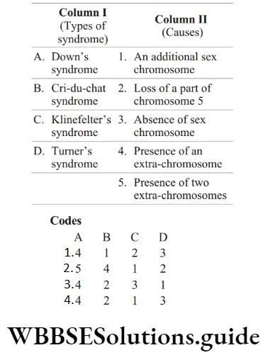
Answer: 4. A–4, B–2, C–1, D–3
Question 137. The hereditary disease in which the urine of a person turns black on exposure to air due to the presence of homogentisic acid is known as
- Ketonuria
- Phenylketonuria
- Haematuria
- Alkaptonuria
Answer: 4. Alkaptonuria
Alkaptonuria is an autosomal recessive disorder and inborn metabolic disorders (congenital). Alkaptonuria in man is caused due to accumulation of homogentisic acid. On exposure to air, urine containing this acid turns black.
Question 138. Edward’s syndrome is an abnormality leading to mental deficiency caused by trisomy of chromosome number
- 5
- 9
- 15
- 18
Answer: 4. 18
Edward’s syndrome occurs due to trisomy of 18th chromosome.
Question 139. Which of the following disorder is not hereditary?
- Haemophilia
- Cataract
- Sickle-cell anaemia
- Colour blindness
Answer: 2. Cataract
Cataract is not hereditary. Some inherited genetic disorders that cause other health problems can increase the risk of cataracts.
Question 140. Which of the following is not a X-linked recessive disease?
- Haemophilia
- Colour blindness
- Thalassemia
- Glucose-6-phosphatedehydrogenase deficiency
Answer: 3. Thalassemia
Thalassemia is an autosomal recessive disease.
Question 141. Usually, the recessive character is expressed only when present in double recessive condition. However, single recessive gene can express itself in human beings when the gene present on
- Any autosomes
- The X-chromosome of the female
- The X-chromosome of the male
- Either can autosome or X-chromosome
Answer: 3. The X-chromosome of the male
- Females have two X-chromosomes, whereas males have one X and one Y-chromosome. Genes on the X-chromosome can be recessive or dominant. Their expression in females and males is not the same.
- Genes on the Ychromosome do not exactly pair up with the genes on the X chromosome. X-linked recessive genes are expressed in females only if there are two copies of the gene (one on each X-chromosome).
- However, for males, there needs to be only one copy of an X-linked recessive gene in order for the trait or disorder to be expressed.
Question 142. Select the incorrect statement from the following.
- Galactosemia is an inborn error of metabolism
- Small population size results in random genetic drift in a population
- Baldness is a sex -limited trait
- Linkage is an exception to the principle of independent assortment in heredity
Answer: 3. Baldness is a sex -limited trait
Statement in option (3) is incorrect and can be corrected as Baldness is a sex influenced trait. The dominance of alleles may differ in heterozygotes of the two sexes. Rest other statements are correct.
Question 143. Consider the following statements.
- Myotonic dystrophy is an autosomal dominant trait.
- Sickle-cell anaemia is an autosomal recessive trait.
- Hypertrichosis is a holandric inheritance.
- Failure of segregation of alleles results in chromosomal gain.
- Cystic fibrosis is a Mendelian disorder.
Choose the correct option.
- 1, 2, 3 and 4
- 1, 3, 4 and 5
- 1, 2, 4 and 5
- 1, 2, 3, 4 and 5
Answer: 4. 1, 2, 3, 4 and 5
All the given statements are correct.
Question 144. Phenylketonuria, Huntington’s disease and sickle- cell anaemia are caused due to the disorder associated with
- Chromosome-7, chromosome-11, chromosome-12
- Chromosome-11, chromosome-4, chromosome-12
- Chromosome-7, chromosome-22, chromosome-46
- Chromosome-12, chromosome -4, chromosome-11
Answer: 4. Chromosome-12, chromosome -4, chromosome-11
Phenylketonuria–Chromosome 12 Huntington Diseases–Chromosome 4 Sickle-cell anaemia – Chromosome 11
Question 145. Mental retardation in man associated with sex chromosomal abnormality is usually due to
- Increase in X complement
- Moderate increase in Y complement
- Large increase in Y complement
- Reduction in X complement
Answer: 1. Increase in X complement
Mental retardation is usually found when there are one or more extra X-chromosomes in males.
Question 146. The interphase nuclei of a person are showing two sex chromatins/nucleus. The possible genotype of the person is
- 44 + XXY
- 44 + XYY
- 44 + XXX
- 44 + XXYY
Answer: 3. 44 + XXX
- Soon after the start of cell division during embryonic development, one of the two X-chromosomes of the somatic cells in normal females becomes functionally inactive.
- This inactive X-chromosome condense (heterochromatic) and can be seen as Barr body. Individual with two sex chromatins, show 44+ XXX genotype, in which only one remains active, while the others two are sex chromatins.
Question 147. In man, which of the following genotypes and phenotypes may be the correct result of aneuploidy in sex chromosomes?
- 22 pairs + XXY males
- 22 pairs + XX females
- 22 pairs + XXXY females
- 22 pairs + Y females
Answer: 1. 22 pairs + XXY males
Aneuploidy is the condition of cells having more or less than integral multiple of typical haploid chromosome number. Thus, 22 pairs + XXY is an extra sex chromosome due to non-disjunction of a pair of sex chromosome either in an ovum or sperm.
Question 148. The diagrammatic representation of the chromosomes of an individual is called
- Idiogram
- Karyotype
- Phenotype
- Diploidy
Answer: 1. Idiogram
The diagrammatic representation of the chromosomes of an individual is called idiogram whereas karyotype is the morphological representation of somatic chromosomes of an individual in descending order.
NEET practice test on Pedigree Analysis and Genetic Disorders
Question 149. The basic set of chromosomes in an organism is known as
- Karyotype
- Idiogram
- GTenome
- Plasmosome
Answer: 1. Karyotype
The basic set of chromosomes in an organism is known as karyotype.
Question 150. Given below is a highly simplified representation of the human sex chromosomes from a karyotype. The genes a and b could be of
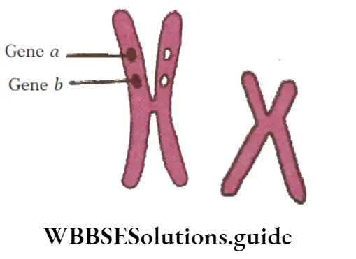
- Colour blindness and body height
- Attached ear lobe and Rhesus blood group
- Haemophilia and red-green colour blindness
- Phenylketonuria and haemophilia
Answer: 3. Haemophilia and red-green colour blindness
The figure shows, human sex chromosome with genes ‘a’ and ‘b’. So, ‘a’ and ‘b’ are sex linked genes, that are inherited through sex chromosomes of X type (X is larger than Y). The most important character of sex linked inheritance is colour blindness and haemophilia.
Question 151. Which of the following statement about Barr bodies is wrong?
- It was first discovered in female cat by Murray Barr
- It is also present in males
- It is present in all females
- It lies against the nuclear membrane and appears like rounded disc
Answer: 2. It is also present in males
Statement in option (2) is wrong and can be corrected as Barr body is a condensed heterochromatic copy of the X chromosome, visible by staining the interphase nucleus of somatic cells of the homogametic sex, e.g. the human female. It is not present in normal
males.
Question 152. Number of Barr body present in each somatic cell of a female is
- 1
- 2
- 3
- 4
Answer: 1. 1
One Barr body is present in each somatic cell of female (XX). The number of Barr bodies is one less than the number of X-chromosomes in the cell.
Question 153. Barr body is missing in the female suffering from
- Huntington’s disease
- Tay-sach’s disease
- Klinefelter’s syndrome
- Turner’s syndrome
Answer: 4. Turner’s syndrome
Turner’s syndrome (44 + XO) is a genetic disorder that affects only females. In this condition, cells of the affected female possess only one X-chromosome, instead of two. Due to the presence of only one X-chromosome, no Barr body is present in females suffering from Turner’s syndrome.
Question 154. Barr body is found in the cytoplasm during
- Interphase in cell of female mammal
- Interphase in cell of male mammal
- Prophase in cell of female mammal
- Prophase in cell of male mammal
Answer: 1. Interphase in cell of female mammal
Barr body is a mass of condensed sex chromatin in the interphase nuclei of normal female somatic cells due to inactive X-chromosome. Thus, Barr body represents the inactivated X-chromosome.
Question 155. Barr body is observed in
- Basophils of male
- Neutrophils of female
- Basophils of female
- Eosinophils
- Neutrophils of male
Answer: 2. Neutrophils of female
Barr body is found attached to nuclear envelope in oral mucosa, anywhere in the nucleus in nerve cells and as drumstick or small rod at one side of nucleus in neutrophil or polymorphonuclear leucocytes (Davidson and Smith).
Question 156. Which of the following genotypes of man shows presence of one Barr body?
- XY
- XXXY
- XXY
- All of these
Answer: 3. XXY
XXY type of genotypes of man shows presence of one Barr body. The total number of Barr bodies is always one less than the total number of X-chromosome present in the cell of the organism. Thus, XXY would contain one Barr body.
Question 157. Studies related to human sex-linked traits depicts that mostly
- Male are affected
- Female are carrier
- Both 1 and 2
- Neither 1 or 2
Answer: 3. Both 1 and 2
Males are affected and females are carrier in most cases regarding human sex-linked traits. The recessive genes located on X-chromosome of humans are always expressed in males, e.g. colour blindness is a recessive sex-linked trait in which the eye fails to distinguish red and green colours. The gene for the normal vision is dominant.
Question 158. Barr body is produced due to partial inactivation of one X-chromosome in female. This is called
- Dosage compensation
- Facultative heterochromatisation
- Both 1 and 2
- None of the above
Answer: 3. Both 1 and 2
- Barr body is produced due to partial inactivation of one X-chromosome and development of facultative heterochromatin in it. Any of the two X-chromosomes can become heterochromatic.
- It begins in the late blastocyst stage (roughly 16 day of embryonic life). Partial inactivation of one X-chromosomes in females is called dosage compensation.
Question 159. If the genetic complement of a person is XXXX, how many Barr bodies can you locate?
- One
- Two
- Three
- Four
Answer: 3. Three
The total number of Barr bodies is always one less than the total number of X-chromosomes present in the cell or the organism. Therefore, it should be 3 in case of XXXX.
Question 160. A medical technician while observing a human blood smear under the microscope notes the presence of a Barr body close to the nuclear membrane in the WBC. This indicates that the person under investigation is a
- Colourblind
- Haemophilic
- Normal female
- Normal male
Answer: 3. Normal female
The total number of Barr bodies is always one less than the total number of X-chromosomes present in the cell or the organism. Thus, presence of one Barr body indicates that person under investigation is normal female.
Question 161. Three copies of chromosome – 21 in a child with Down’s syndrome have been analysed using molecular biology technology to detect any possible DNA polymorphism with reference to different alleles located on chromosome – 21. Results showed that out of 3 copies, 2 of the chromosomes of the child contain the same alleles as one of the mother’s alleles. Based on this when did the non-disjunction event most likely occur?
- Paternal meiosis-1
- Maternal meiosis-1
- Paternal meiosis-2
- Maternal meiosis-2
Answer: 2. Maternal meiosis-1
- Down’s syndrome is an autosomal aneuploidy, caused by the presence of an extrachromosome number 21. Both the chromosomes of pair 21 pass into a single egg due to non-disjunction during oogenesis.
- The non-disjunction is more common in females. The frequency of non-disjunction of chromosome pair 21 increases with mother’s age. Usually non-disjunction of chromosomes takes place in maternal meiosis-I.
Question 162. G-6-P-O deficiency inheritance is an example of ………….. inheritance.
- X-linked recessive
- Autosomal dominan
- Autosomal recessive
- X-linked dominant
Answer: 1. X-linked recessive
Glucose-6- phosphatedehydrogenase deficiency is most common human enzyme defect. It (also known as favism) is an X-linked recessive genetic condition that predisposes to haemolysis (spontaneous destruction of RBCs) and results in jaundice in response to a number of triggers, such as certain food, illness or medication.
Question 163. Syndrome stands for
- A group of symptoms
- Viral disease
- Diseased condition
- Dwarf organism
Answer: 1. A group of symptoms
Syndrome stands for the group of symptoms, which indicates to a particular disease.
Question 164. Match the syndrome and chromosomal abnormalities given in columns 1 and 2, respectively.
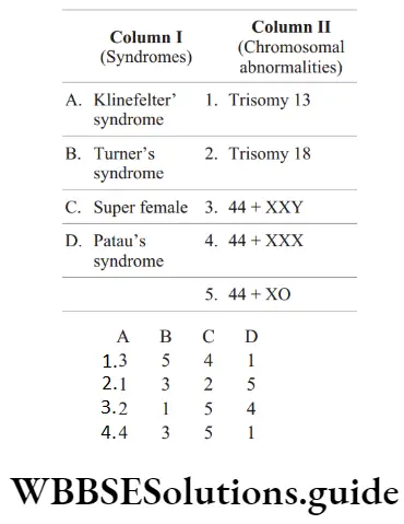
Answer: 1. A–3, B–5, C–4, D–1
Question 165. Trisomy 18 is
- Edward’s syndrome
- Patau’s syndrome
- Turner’s syndrome
- Klinefelter’s syndrome
Answer: 1. Edward’s syndrome
- Edward’s syndrome is due to an extra chromosome number 18. Thus, the total number of chromosome is 47 instead of normal 46. Patau’s syndrome is due to trisomy of chromosome 13.
- Turner’s syndrome is due to monosomy (2n –1), the individual has 45 chromosomes (44 + XO). Klinefelter’s syndrome is due to trisomy of sex (X) chromosome. The individual has 47 chromosomes (44 + XXY).
Question 166. Which of the following chromosomal constitution refers to Jacob’s syndrome in human?
- 44 + XO
- 44 + XXY
- 44 + XYY
- 45 + XYY
Answer: 3. 44 + XYY
Jacob’s syndrome is a rare genetic disorder that occurs due to aneuploidy. It is characterised by the presence of an extra Y-chromosome making the chromosome complement 44 + XYY. Diseased persons show higher growth rate. Any other symptoms are not prominent.
Question 167. The syndrome in which individual somatic cell contains three sex chromosomes XXX is called
- Klinefelter’s syndrome
- Turner syndrome
- Down’s syndrome
- Super female
Answer: 1. Klinefelter’s syndrome
Klinefelter’s syndrome is formed by the union of an XX egg and a normal Y sperm or normal X egg and abnormal XY sperm. The individual has 47 chromosomes (44 + XXY) or (47+XXX).
Question 168. Low pitched voice, bearding and moustaches are a type of
- Sex limited traits
- Sex linked trait
- Nullisomic traits
- Sex influenced traits
Answer: 4. Sex influenced traits
Low pitched voice, bearding and moustaches are a type of sex influenced traits.
Question 169. One of the following in man is controlled by dominant genes
- Colour blindness
- Brachydactyly
- Nyctalopia
- Juvenile glaucoma
Answer: 2. Brachydactyly
Brachydactyly is a shortening of the fingers and toes due to unusually short bones. This is an inherited condition, and in most cases does not present any problems for the person who has it. It is an autosomal dominant condition, which means you only need one parent with the gene to inherit the condition.
Question 170. Name the syndrome which is caused when there is trisomy of the sex chromosomes.
- Turner’s syndrome
- Down’s syndrome
- Klinefelter’s syndrome
- Patau’s syndrome
Answer: 3. Klinefelter’s syndrome
- Klinefelter’s syndrome is formed by the union of an abnormal XX egg and a normal Y sperm or normal X egg and abnormal XY sperm. The individual has 47 chromosomes (44 + XXY) due to the trisomy of sex chromosomes.
- Such persons are sterile males with undeveloped testes, mental retardation, sparse body hair and long limbs and with some femalecharacteristics such as enlarged breasts.
- It is considered that the more the X-chromosomes, the greater is the mental defect. As the syndrome has two X-chromosomes, one Barr body is seen in this case.
Question 171. Which one of the following conditions in humans is correctly matched with its chromosomal abnormality/linkage?
- Klinefelter’s syndrome-44 autosomes + XXY
- Colour blindness – Y-linked
- Erythroblastosis foetalis – X-linked
- Down’s syndrome – 44 autosomes + XO
Answer: 1. Klinefelter’s syndrome-44 autosomes + XXY
- Option (1) is correct as Klinefelter’s syndrome is a genetic disorder affecting men in which an individual gains an extra X chromosome, so that the usual karyotype of XY is replaced by one of XXY.
- Other options are not correct. Colour blindness is X-linked, Down’s syndrome is 21 trisomy and erythroblastosis foetalis is related to Rh incompatibility.
Question 172. What is the genetic disorder in which an individual has an overall masculine development gynaecomastia and is sterile?
- Turner’s syndrome
- Klinefelter’s syndrome
- Edward’s syndrome
- Down’s syndrome
Answer: 2. Klinefelter’s syndrome
Individuals with Klinefelter’s syndrome have trisomy of sex chromosome as 44 + XXY (47). They show overall masculine development, gynaecomastia and are sterile.
Question 173. Spermatogenesis is absent in person suffering from
- Klinefelter’s syndrome
- Down’s syndrome
- Turner’s syndrome
- Thalassemia
Answer: 1. Klinefelter’s syndrome
Klinefelter Syndrome (KS) was first described by Harry F. Klinefelter in 1942 as a clinical entity characterised by gynecomastia, small testes, absent spermatogenesis, normal to moderately reduced Leydig cell function and increased secretion of FSH.
Question 174. In Klinefelter’s syndrome, the sex chromosome compliment is
- 22 A+ XXY
- 22A + XY
- 22A+XO
- 22 A + XX
Answer: 1. 22 A+ XXY
Klinefelter’s syndrome is formed by the union of an XX egg and a normal Y sperm or normal X egg and abnormal XY sperm. The individual has 47 chromosomes (22A + XXY).
Question 175. A man having Klinefelter’s syndrome is
- Intersex with secondary sexual characters on the side of female
- Male with secondary sexual characters of female
- Female with secondary sexual characters of male
- Normal fertile male
Answer: 2. Male with secondary sexual characters of female
Phenotypically Klinefelter individuals are males, but they can show some female secondary sexual characteristics and are usually sterile.
Question 176. Which of the following set of syndromes show 47 chromosomes in their genetic makeup?
- Down’s syndrome, Patau’s syndrome, Edward’s syndrome
- Turner’s syndrome, Edward’s syndrome, Klinefelter’s syndrome
- Klinefelter’s syndrome, Turner’s syndrome, Edward’s syndrome
- All of the above
Answer: 1. Down’s syndrome, Patau’s syndrome, Edward’s syndrome
- Syndromes which show 47 chromosomes (instead of normal 2n = 46 chromosomes) in the genetic
makeup of affected individual include Down’s syndrome, Patau’s syndrome, Edward’s syndrome and Klinefelter’s syndrome. - Individuals suffering from Down’s syndrome contain 45+XY chromosomes in males and 45 + XX chromosomes in females. Patau’s syndrome (13-trisomy) is due to an extra chromosome number 13 and Edward’s syndrome (18-trisomy) is due to an extra chromosome number 18.
- Klinefelter’s syndrome occurs due to trisomy of sex (X) chromosome and the affected individual possesses 47 chromosomes (44+XXY). Turner’s syndrome occurs due to monosomy of sex chromosome and the affected individual possesses 2n = 45 chromosomes (44+XO) instead of 46.
Question 177. Edward, Patau, Down’s syndromes are
- Change in autosomes
- Change in sex chromosomes
- Mutation due to malnutrition
- Both change in sex chromosome and autosomes
Answer: 1. Change in autosomes
Edward’s, Patau’s and Down’s syndromes are disorders which occur due to autosomal chromosomal changes. Edward’s syndrome occurs due to trisomy of 18th chromosome, Patau’s syndrome arises due to trisomy of 13th chromosomes and Down’s syndrome occurs due to trisomy of 21st chromosomes.
Question 178. Number of sex chromosomes is normal in
- Super female
- Turner’s syndrome
- Klinefelter’s syndrome
- Down’s syndrome
Answer: 4. Down’s syndrome
Down’s syndrome is an autosomal aneuploidy. The 21st pair of chromosomes has three chromosomes instead of two. The sex chromosomes remain unchanged.
Question 179. Who reported that Down’s syndrome is due to extra 21st chromosome?
- JL Down (1866)
- Lejeune (1959)
- Klinefelter (1942)
- Huntington (1872)
Answer: 1. JL Down (1866)
- For centuries, people with Down’s syndrome have been alluded in art, literature and science. It was not until the late 19th century, however, that John Langdon Down, an English physician, published an accurate description of a person with Down syndrome.
- It was this scholarly work, published in 1866, that earned Down the recognition as the ‘father’ of the syndrome.
Question 180. In Down’s syndrome, karyotyping has shown that the disorder is associated with trisomy of chromosome number 21 usually due to
- Non-disjunction during egg cell formation
- Non-disjunction during sperm cell formation
- Non-disjunction during formation of egg cells and sperm cells
- Addition of extra chromosome during mitosis of the zygote
Answer: 3. Non-disjunction during formation of egg cells and sperm cells
Down’s syndrome caused by trisomy 21, arises due to an occasional non-disjunction during meiosis when a gamete comes to possess an extra chromosome (n+ 1).
Fusion of this chromosome with a normal gamete containing normal chromosome (n) result in [n + (n + 1)] trisomy.
Question 181. Freqeuency of Down’s syndrome increases when the maternal age is
- Above 35 years
- Below 35 years
- During 1st pregnancy
- If she is a mother of at least 3 children
Answer: 1. Above 35 years
The chance of a woman having a baby with Down’s syndrome increases as she gets older. Only 1 in 2000 births from teenage mothers are affected by Down’s syndrome, but this goes up to 1 in 10 in 48-years old women. The risk starts above 35 years of age
Question 182. Consider the following statements.
- Short statured with small round head.
- Tongue is furrowed and mouth partially opened.
- Also called Mongolian Idiocy.
- Physical, psycomotor and mental development is slow.
Choose the correct option.
- Down’s syndrome
- Turner’s syndrome
- Klinefelter’s syndrome
- Patau’s syndrome
Answer: 1. Down’s syndrome
All the listed symptoms belong to Down’s syndrome 21 trisomy.
Question 183. Down’s syndrome is caused by an extra copy of chromosome number 21. What percentage of offspring produced by an affected mother and a normal father would be affected by this disorder?
- 100 %
- 75 %
- 50 %
- 25 %
Answer: 3. 50 %
Down’s syndrome is the example of autosomal aneuploidy. Here, an extra copy of chromosome 21 occurs. As it is an autosomal disease, the offspring produced from affected mother and normal father should be 50%.
Question 184. Which of the following occurs due to monosomy of sex chromosome?
- Down’s syndrome
- Turner’s syndrome
- Haemophilia
- Sickle-cell anaemia
Answer: 2. Turner’s syndrome
- Turner’s syndrome is caused due to the absence or monosomy of one of the X-chromosome, i.e. 45 chromosomes total with sex chromosomes as XO.
- Such females are sterile as ovaries are rudimentary. Besides this, other features include lack of other secondary sexual characters, etc.
Question 185. Which is correct for Turner’s syndrome?
- It is a case of monosomy
- It causes sterility in females
- Absence of Barr body
- All of the above
Answer: 4. All of the above
- All given statements are correct regarding Turner’s syndrome. It is a genetic disorder of women caused by the absence of the second sex chromosome (such women are XO, rather than the normal XX).
- It is characterised by a lack of ovaries and menstrual cycle. Affected women are sterile and lack secondary sexual characteristics, In the case of Turner’s syndrome, it is 1 – 1 = 0, i.e. Barr bodies are absent. Thus, option (4) is correct.
Question 186. A sterile female having reduced mental ability is said to be suffering from
- Turner’s syndrome
- Klinefelter’s syndrome
- Down’s syndrome
- Edward’s syndrome
Answer: 1. Turner’s syndrome
- Unlike some other genetic conditions, Turner’s syndrome does not cause mental retardation or decreased intelligence.
- However, some girls with Turner’s syndrome have learning disabilities, especially in areas involving spatial skills, such as math. It can affect their ability to connect with others.
Question 187. The person with Turner’s syndrome has
- 45 autosomes and X sex chromosome
- 44 autosomes and XYY sex chromosomes
- 45 autosomes and XYY sex chromosomes
- 44 autosomes and X sex chromosome
Answer: 1. 45 autosomes and X sex chromosome
Turner’s syndrome is caused due to absence of one of the X-chromosomes, i.e. 45 with XO.
Question 188. Turner’s syndrome in humans is caused by
- Autosomal aneuploidy
- Sex chromosome aneuploidy
- Polyploidy
- Point mutation
Answer: 2. Sex chromosome aneuploidy
Turner’s syndrome is 23 monosomic (sex chromosome aneuploidy) conditions, in which only one X-chromosome is present.
Question 189. Turner’s syndrome was first discovered by
- JL Down
- Huntington
- HH Turner
- Lejeune
Answer: 3. HH Turner
Turner syndrome was first discovered in 1938 by Dr. Henry Turner while studying a group of 7 girls who all had the same unusual developmental and physical features.
Question 190. A human female with Turner’s syndrome
- Has 45 chromosomes with XO
- Has one additional X chromosome
- Exhibits male characters
- Is able to produce children with normal husband
Answer: 3. Exhibits male characters
- Turner’s syndrome is due to monosomy (2n – 1). It is caused by the union of an allosome free egg (22 + 0) and a normal X sperm or a normal egg and an allosome free sperm (22 + 0).
- The individual has 2n = 45 chromosomes (44 + XO) instead of 46. Such people are sterile
females who have rudimentary ovaries filled with connective tissues (therefore, also called gonadal dysgenesis), undeveloped breasts, small uterus, puffy fingers, short stature, male like characters like beared, masculinity, etc.