
Epithelial Tissue
Types of tissues: The bodies of all vertebrates and most of the invertebrates are made of a variety of tissues.
However, all the tissues may be grouped into four main types. These are epithelial tissue, connective tissue, muscular tissue, and nervous tissue.
Epithelial Tissue Location: Epithelial tissue is present mainly in two regions—the external covering of the body (skin) and the walls (both inner and outer) of all internal organs.
Epithelial Tissue Components: Epithelial cells are compactly arranged with minimum intercellular spaces.
The tissue is made up of three major components. They are—the non-cellular basement membrane, epithelial cells, and intercellular cementing material. This cementing material is a mucoprotein complex containing hyaluronic acid and calcium salt.
Read and Learn More: WBCHSE Notes for Class 11 Biology
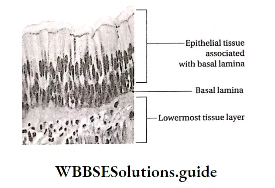
Epithelial Tissue Structure: An epithelium consists of one or more layers of cells. The outer layer of cells, when present in the skin, is exposed to the external environment outside the body.
“types and functions of epithelial tissues notes for class 11”
But, this layer is also present on the outer surface of internal organs. Here, it is exposed to a lumen or cavity within the body. The deep inner layer of cells is bound by a basement membrane.
| Class 11 Biology | Class 11 Chemistry |
| Class 11 Chemistry | Class 11 Physics |
| Class 11 Biology MCQs | Class 11 Physics MCQs |
| Class 11 Biology | Class 11 Physics Notes |
simple squamous epithelial tissue diagram
Basement membrane: Basement membrane is not actually a membrane. Rather, it is an extracellular matrix present between the epithelial tissue and the loose connective tissue below it.
It is composed of a network of collagenous fibers which are formed from secretions of the underlying cells of the connective tissue. It consists of two layers—the outer thin basal lamina and the inner thick, fibrous, reticular lamina (also known as lamina reticularis).
“short notes on epithelial tissues for NEET and exams”
The basal lamina is further made up of two layers—lamina lucida and lamina densa. The lamina lucida is closer to the epitheium while the lamina densa is closer to the connective tissue.
The lamina reticularis is attached to the basal lamina with collagen fibrils. This layer is also attached together by microfibrils made of fibrillin protein.

Functions of the basement membrane of epithelium
- Anchorage: It binds the epithelium to the loose connective tissue underneath and holds the layers -c6
- Growth: Controls the growth and differentiation of cells within the epithelial tissue.
- Exchange of molecules: Acts as a filter during an exchange of large molecules between epithelium and the underlying connective tissue.
- Signaling: Helps in cell-to-cell signaling.
Lamina propria: It is a layer of loose vascular connective tissue, present at the base of the basement membrane. It provides mechanical support to the epithelium.
It also contains several blood vessels and nerves. These blood vessels and nerves provide nutrition and innervation to the epithelium.
“detailed notes on epithelial tissues with diagrams”
Papilla: The hair-like appendages that lie at the junction of epithelium and the underlying connective; tissue are called papillae (sing, papilla). They hold the epithelium in position.
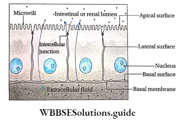
Surface layers of cells: The epithelial cells have three surfaces. These are as follows—
Basal surface: It is the lower surface of the cells, adjacent to the basement membrane.
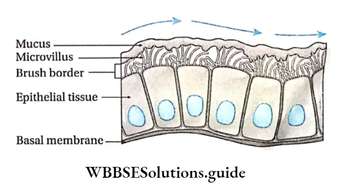
Apical surface: It is the upper surface of the cells, that remains free. It increases the surface area for absorption.
This property is achieved by modification of the surface into structures like microvilli, stereocilia, etc.
Lateral surfaces: These are surfaces of the cells, on both sides, facing adjacent epithelial cells.
“classification of epithelial tissues and their functions”
Intercellular junctions: These are specialized regions between the plasma membrane of adjacent cells. These are also known as cell junctions. They provide mechanical support and help in cell-to-cell communication.
However, sometimes they may act as impermeable barriers, preventing the transport of certain molecules between the cells.
“difference between simple and stratified epithelial tissues”
Intercellular junctions are of the following types—
Tight junctions: These are specialized regions between adjacent epithelial cells where the cell membranes are fused together by means of sealing strands.
They bind the epithelial cells together and check the passage of molecules and ions between them.
Gap junctions: These are specialized junctions directly connecting the cytoplasm of cells. They allow the passage of ions and small molecules from one cell to the adjacent one.
“epithelial tissue characteristics and examples notes”
Adherens junctions: Junctions present in heart muscles and skin epithelium. They join the actin filaments present in the muscles to form a continuous belt.
Desmosomes: Intercellular junctions of epithelia and cardiac muscle. They are of two types—Belt and Spot desmosomes. The belt desmosomes are belt-like and the spot desmosomes are spot-like or circular in appearance.
“squamous epithelial tissue “
Cilia and Flagella: These are microtubular structures that arise from the plasma membrane of the epithelial cells. Cilia are found in animal cells whereas flagella are primarily found in prokaryotes and unicellular eukaryotes.
They help in locomotion as well as to propel away harmful, unwanted particles.
Cilia and Flagella Functions: The functions of epithelial tissue are as follows—
Cilia and Flagella Protection: Epithelial tissue forms the skin of animals. It also forms the wall of internal organs, thus protecting them from injuries.
Terrestrial vertebrates have keratin in their skin cells. This makes them resistant to water loss from their skin. Ciliated epithelium, lining the respiratory tract, sweeps away impurities with the help of cilia.
“types and functions of epithelial tissues PDF notes download”
Cilia and Flagella Absorption: The gut is lined with epithelial tissue that absorbs nutrients from food. The lungs are also lined with epithelial tissue which helps them to absorb oxygen.
Cilia and Flagella Secretion: Glandular epithelium forms the exocrine and endocrine glands. Endocrine glands secrete hormones into the circulation. Exocrine glands secrete mucus, saliva, wax, milk, etc., through ducts.
“importance of epithelial tissue in protection and absorption”
Contractile property: Myoepithelium is the contractile epithelium, present in sweat glands, mammary glands etc. It helps in the flow of secreted fluids from the glands.
Reproduction: Gametes (such as sperm and ova) are produced from the germinal epithelium lining the testis and ovary.
“functions of epithelial tissue in different organs”
Sensation: It is perceived with the help of sensory epithelium present in the skin, taste buds in the tongue, nasal epithelium, etc.
Excretion: Ultrafiltration by Bowman’s capsule and tubular reabsorption during urine formation is carried out by renal epithelium in the kidney.
Transport: The beating of cilia in ciliated epithelium present in food pipe, respiratory tract, reproductive tract, etc., helps to transport food, mucus, gametes, etc.
Respiration: The epithelium of alveoli in the lungs helps in gaseous exchange.
Classification: Based on the shape of the cells that lie above the basement membrane and on the number of layers of cells, the different types of epithelial tissues are shown in the given flowchart.
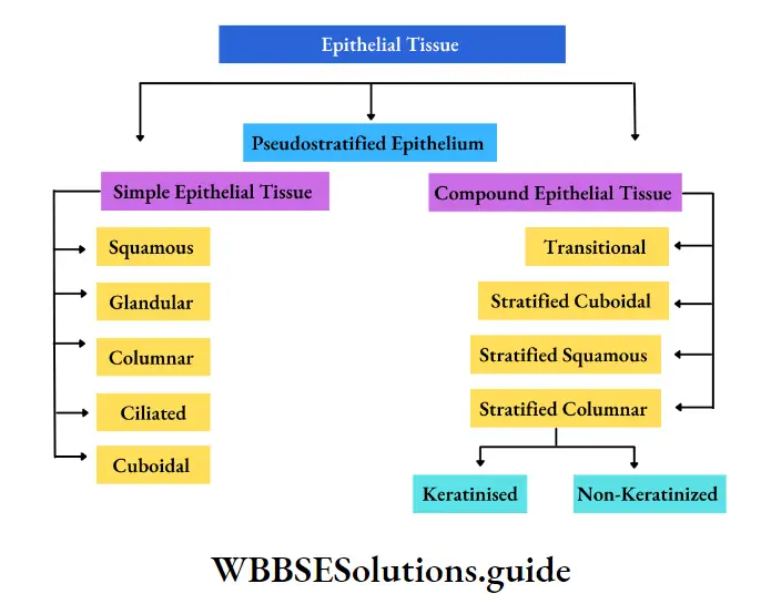
Simple epithelial tissue Definition: The epithelium that is made up of a single layer of cells, is called simple epithelial tissue or epithelium.
Based on the structure of the cells, the simple epithelium is classified as—squamous epithelium, cuboidal epithelium, columnar epithelium, ciliated epithelium, and glandular epithelium.
“structure and types of epithelial tissues explained”
Squamous Epithelial Tissue Definition: The epithelial tissue that consists of a single layer of large, flattened, polygonal cells is called squamous epithelial tissue.
Squamous Epithelial Tissue Position: It is found in the alveoli of lungs, endothelium of capillaries, Bowman’s capsule and Henle’s loop of the nephron, pericardium and the inner lining of the heart, and the peritoneal lining of the coelom.
Squamous Epithelial Tissue Structure:
- Cells of this tissue are flat, polygonal in shape with irregular margins. Their shapes resemble the scales of fish. It is also known as pavement epithelium as it forms a pavement-like structure.
- Cells are compactly arranged without any intercellular spaces.
- The nuclei are oval or disc-shaped and centrally located.
- Cytoplasm may be clear or granular.