Human Circulatory System
The human circulatory system consists of the muscular heart, the network of blood vessels and blood. Blood is the circulating fluid which flows through the blood vessels after being pumped by the heart.
Types: Two types of circulatory systems are found in human beings— blood and lymphatic systems.
Heart
The heart is the main organ of the circulatory system. In vertebrates, it is located on the ventral side of the oesophagus. The human heart is more complex and the Function of lymph nodes advanced as compared to other organisms.
Heart Definition: A multi-chambered muscular organ, that pumps blood throughout the body in a rhythmic process, is known as the heart.
” circulatory system questions”
Location: The heart is situated in between the lungs in the middle of the thoracic cavity, slightly tilted to the left. The heart rests on the superior surface of the diaphragm, posterior to the sternum.
Read and Learn More Class 11 Biology Notes
Shape and size: The size of the heart is closely related to the size of the organism’s body.
The base of the heart is broad and flat, about 9 cm in breadth. The length of the heart of an adult is about 10-12 cm. Externally, the heart appears as a cone-shaped structure.

Shape and size: The size of the heart is closely related to the size of the organism’s body.
The base of the heart is broad and flat, about 9 cm in breadth. The length of the heart of an adult is about 10-12 cm. Externally, the heart appears as a cone-shaped structure
| Class 11 Biology | Class 11 Chemistry |
| Class 11 Chemistry | Class 11 Physics |
| Class 11 Biology MCQs | Class 11 Physics MCQs |
| Class 11 Biology | Class 11 Physics Notes |
Wall of the heart
The wall of the heart is composed of the following layers—
Pericardium: The heart is enclosed in a double-layered sac, called pericardium. It is the fibrous outer wall made of dense connective tissue.
The pericardium is further divided into two layers —
Fibrous pericardium: It is the outer fibrous transparent sac that covers the heart.
Serous pericardium: The inner side of the pericardium is formed of a serous layer and is known as serous pericardium.
This serous pericardium is again divided into two layers, the inner layer—visceral pericardium and the outer layer—the parietal pericardium.
“WBCHSE class 11 biology notes for human circulatory system”
Visceral Pericardium: This layer is also called the epicardium. It is the innermost layer of the pericardium. It is composed of a single sheet of squamous epithelial cells overlying delicate connective tissue. This layer adheres to the heart.
Parietal Pericardium: This layer lies between the visceral pericardium and the fibrous pericardium.

The pericardial cavity lies between the visceral pericardium and the parietal pericardium. This cavity is filled with pericardial fluid which serves as a shock absorber and reduces friction between the pericardial membranes.
” questions on circulatory system”
The primary function of the pericardium is to—
- Protect The Heart
- Anchor The Heart To The Surrounding Structures, Such As The Diaphragm And The Great Vessels; And
- Prevent The Heart From Overfilling.
Myocardium: The myocardium is a thick contractile layer present just below the visceral pericardium.
It is composed of muscle cells, known as myocardiocytes. This layer is more thick in the ventricles than in the atria. This layer of the left ventricle is thicker than the right ventricle.
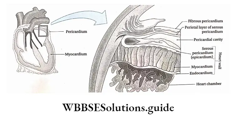
Endocardium: The endocardium is the innermost layer of the heart wall and lines the heart’s chambers.
It is made of squamous epithelium that rests on a thin connective tissue layer. It is continuous with the endothelium of the great blood vessels—the inferior and superior vena cava.
Chambers Of The Heart
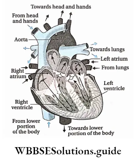
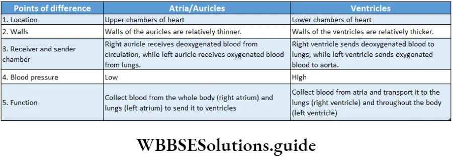
The heart is a hollow, four-chambered, muscular organ that consists of the upper right and left atria (singular: atrium) and the lower right and left ventricles.
The atria are separated by a thin muscular wall called the interatrial septum and the ventricles are separated by a thick muscular wall called the interventricular septum.
There is a translucent, depression present in the septum between the right and left atrium, known as fossa ovalis. In the embryonic stage, there is an opening in place of the fossa ovalis.
It is known as foramen ovale. The heart actually functions as two separate pumps.
“detailed notes on human circulatory system for class 11 WBCHSE”
The right atrium and ventricle act as one pump to propel deoxygenated blood to the lungs. At the same time, the left atrium and ventricle act as another pump to propel oxygenated blood collected from the lungs towards the heart throughout the systemic circulation.
” body fluids and circulation bank of biology”
The right atrium and right ventricle: The inner surfaces of both atria are mostly smooth except for a low network of muscular ridges called musculi pectinati. The right atrium receives deoxygenated blood through the openings of the superior vena cava, inferior vena cava & coronary sinus.
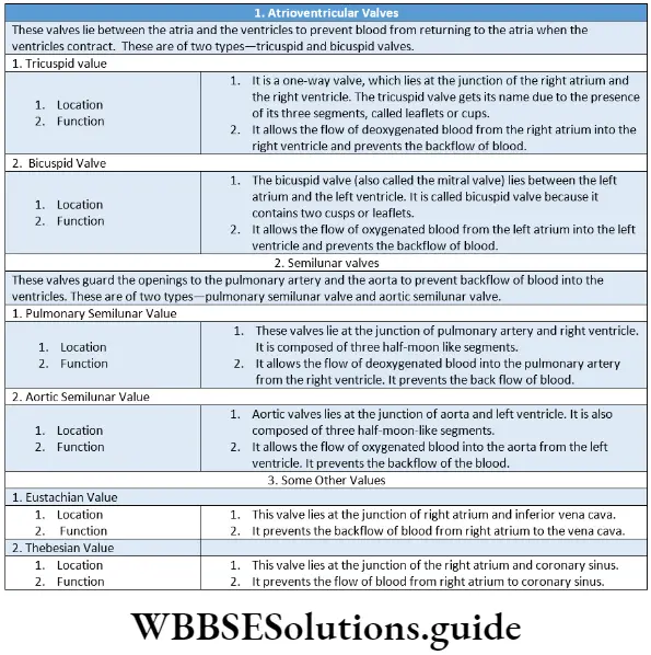
The right atrium is connected to the right ventricle through an opening, known as the right atrioventricular orifice or aperture.
The right atrioventricular orifice is guarded by the right atrioventricular valve or tricuspid (formed of three flaps or cusps) valve.
This valve regulates unidirectional blood flow from the right atrium to the right ventricle and prevents the backflow of blood.
There are numerous muscular ridges on the inner wall of the right ventricle, known as columnae carneae.
They contain some finger-like muscular projections, known as papillary muscles. The cusps or leaflets of the tricuspid valve are attached to the wall of the right ventricle by strong, elastic threads, known as chordae tendineae. The opening and closing of the tricuspid valve are controlled by the papillary muscles through the chordae tendineae.
The pulmonary artery originates from the right ventricle. There is a seminular valve present at the juncture of the pulmonary artery and right ventricle.
Left atrium and left ventricle: The left atrium and left ventricle are also connected to each other through an opening known as the left atrioventricular orifice or aperture.
“structure and function of human circulatory system notes”
The left atrioventricular orifice is guarded by the bicuspid (formed of two cusps) or mitral valve.
The left atrium receives four pulmonary veins, two from the left and two from the right lung.
These pulmonary veins bring oxygenated blood from the lungs. They do not have any valve to regulate the blood flow. Blood flows obliquely from the atrium to the ventricle which is regulated by the mitral valve.
The left ventricle which is larger than the right ventricle, has much thicker walls. Like the right ventricle, it also possesses columnae carneae and papillary muscles. Chordae tendineae are also seen.
The lower part of the left ventricle receives blood from the left atrium. The upper part, which has mainly fibrous walls, is termed the aortic vestibule, and it ends in the aorta, the largest artery of the body.
The junction of the vestibule and aorta contains dense fibrous tissue that encircles the aortic valve and is continuous with the cardiac skeleton.
mcq on body fluids and circulation pdf
Cardiac grooves
- All the different chambers of the heart are separated from each other by grooves. These grooves are known as cardiac grooves.
- The left and right atria are separated from each other by an interatrial groove. Atria and ventricles are separated from each other by the coronary sulcus or atrioventricular groove.
- The grooves by which two ventricles are separated from each other are known as anterior interventricular sulcus and posterior interventricular sulcus.
Junctional Tissues Of Heart
Junctional Tissues Of Heart Definition: The tissues of the cardiac muscles, responsible for the origin and conduction of cardiac impulses (waves of electric potential generated by stimulation of cardiac muscles by specific stimulus), are known as junctional tissues of the heart.
Junctional Tissues Of Heart Types:
- Sino-atrial (SA) Node,
- Atrioventricular (AV) node,
- Bundle of His,
- Purkinje fibres,
- Internodal tract.
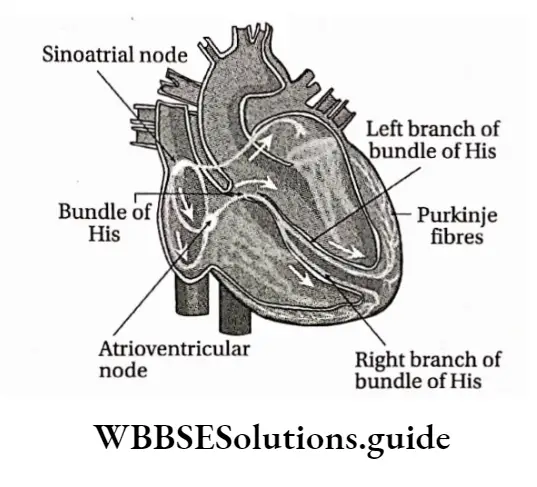
- Sinoatrial (SA) node
Location: Situated on the right wall of the right atrium, near the entrance of the superior vena cava.
Characteristics: The SA node is a small mass of muscle tissue, about 3.0 mm in width.
“blood circulation process explained for WBCHSE class 11”
Function: This is known as the “Natural Pacemaker” or heartbeat regulator. It initiates impulses automatically at a more rapid rate than any other
part of the conduction system.
Its rate of discharge of impulses determines the rate of heartbeat. The electrical signal spreads out over the walls of the atria, causing them to contract.
Atrioventricuiar (AV) node
Location: It is located in the right posterior portion of the interatrial septum.
Characteristics: Muscles of AV nodes are cardiac muscle fibres. It contains a very small amount of myofibrils.
AV muscle fibres are comparatively smaller than the SA node. The AV node and the conduction tissue surrounding it are known as the AV junctional tissue.
The AV node is also called the ‘reserve pacemaker of heart’ because during functional inactivity of the SA node, it can initiate heartbeats, but at a slower rate.
Function: The action potential spreading through the right atrium causes depolarization of the AV node.
When the impulses reach the AV node, there is a slight delay that allows the atria to finish their contraction before the ventricles begin their contraction.
The AV node then conducts impulses to the muscles of the ventricles through a bundle of His and Purkinje fibres.
Bundle of His and its branches
Location: It is a band of nerve fibres that originates from the posterior part of the AV node, and then passes along the interventricular septum to the ventricles.
Characteristics: A Swiss physician, Wilhelm His, first described this tissue. It divides into right and left branches which proceed along the right and left sides of the interventricular septum to the tips of the two ventricles. It is also called an AV bundle.
Function: These specialised fibres conduct the impulses at a very rapid velocity (about 2 m/sec). The bundle of His constitute the only electrical link between the atria and the ventricles.
Purkinje fibres
Location: The Purkinje fibres, named after their discoverer Johannes von Purkinje, are the tree-like terminal branchings of right and left bundles of His.
Characteristics: Cells of these fibres consist of high amount of glycogen-contining sarcoplasm. There are thousands of myofibrils present around the cells.
“heart structure and working class 11 biology WBCHSE notes”
Function: Purkinje fibres conduct the impulses at high velocity (about 4 m/sec) throughout the ventricles. During the ventricular contraction, they carry the impulses from both the left and right branches of the bundle of His to the myocardium of the ventricle.
Internodal tract
Location: These are located between the SA node and the AV node.
Characteristics: There are three internodal tracts connecting the SA node and AV node—the anterior tract of Bachman, a middle tract of Wenckebach and the posterior tract of Thorel.
Function: Their main function is to carry impulses from the SA node through the right atrium to the AV node.
As the impulses pass along the internodal tracts, the right atrium contracts, pushing the blood it contains, through the tricuspid valve into the right ventricle.
Myocardial tissue has four main characteristics that integrate the heart’s electrical and mechanical activity
“short notes on human circulatory system for WBCHSE board exams”
Automaticity: It is the ability to initiate an impulse or stimulus. The pacemaker cells (SA node) of the cardiac conduction system spontaneously depolarize in the absence of external stimulation. The AV-junctional tissue and the His-Purkinje network also possess the property of automaticity.
Excitability: It is the ability to respond to an impulse or stimulus. The cells are electrically irritable because of an ionic imbalance across the cell membranes.
Cells thus respond to external stimuli from chemical, mechanical or electrical sources. Atrial and ventricular myocardial fibres respond to the impulse generated by the pacemaker cells of the cardiac conduction system by depolarization and repolarization.
Conductivity: It is the ability to transmit impulses to other areas of the heart. Cells of the conduction system and the myocardial muscle fibres, both have this property.
“difference between systemic and pulmonary circulation notes”
Contractility: It is the ability of cardiac muscles to shorten, responding to stimuli with mechanical action. Myocardial fibres respond mechanically to electrical stimulation by contracting.
The course of circulation of blood through the human heart The circulation of blood through the human heart occurs through periodic systole and diastole.
They are discussed below—
Atrial diastole: This is the relaxation time of both the atria. During this time deoxygenated blood enters into the right atrium through superior and inferior vena cava and oxygenated blood from the lungs enters into the left atrium through pulmonary veins.
The mixing of the blood is prevented due to the presence of an interatrial septum between the atria.
Auricular or atrial systole: This is contraction time for both atria. The deoxygenated and oxygenated blood move from the right and left atria respectively to the corresponding ventricles.
“composition of blood and its function in circulation notes”
The tricuspid and bicuspid valves present at the atrioventricular grooves open to guide the flow of blood.
Ventricular diastole: This is the relaxation time of the ventricles. During this time, the left ventricle receives oxygenated blood from the left atrium and the right ventricle receives deoxygenated blood from the right atrium.
Ventricular systole: It is the time for contraction of both the ventricles. The deoxygenated blood in the right ventricle flows to the lungs through the pulmonary artery.
“important points on blood vessels and circulation for WBCHSE”
The oxygenated blood from the left ventricle flows to different parts of the body through the aorta.
The bicuspid and tricuspid valves close during this time and prevent the backflow of the blood to the atria. At the same time the semilunar valves open and guide the blood flow to the respective blood vessels and prevent the backflow of the blood to the ventricles.