
- WBBSE Class 10th Results Will Be Released on May 19, 2023
- WBBSE Madhyamik Model Question Paper 2023 Geography And Environment Set 1
- WBBSE Madhyamik Model Question Paper 2023 Geography And Environment Set 2
- WBBSE Madhyamik Model Question Paper 2023 Geography And Environment Set 4
- WBBSE Madhyamik Model Question Paper 2023 History And Environment
- WBBSE Model Question Paper 2023 History And Environment Set 1
- WBBSE Model Question Paper 2023 History And Environment Set 2
 Vasantha
WBBSE Solutions Class 10 Life Science Chapter 3 Heredity And Common Genetic Diseases Long Answer Questions
Chapter 3 Heredity And Common Genetic Diseases Long Answer Questions
Question 1. What do you understand by the term heredity? Who is the father of genetics? On which living material did he do his experiment? State two
conclusions derived from his monohybrid cross experiment.
Answer:
(1) Heredity:- The transmission of parental characters from one generation . to the successive generation of organisms is called heredity.
(2) Gregor Johann Mendel is the father of genetics.
(3) He conducted his experiment on a garden pea plant (Pisum sativum).
(4) Conclusions derived from his monohybrid experiment:・
(1) Characters are controlled by genes. Genes are found in pairs. One gene controls only one character. There are two factors for a single character. Such as tall and dwarf are two factors for length which is a single character. In these two factors, one is dominant while the other one is recessive. The factor which expresses itself in a generation is called the dominant factor and the next which is unable to express itself in a generation is called the recessive factor.
(2) Both factors of a character are present in a hybrid. But these factors are never mixed up with each other. They are separated during gamete formation. Hence a gamete contains only one factor for a character. Thus we can say that a gamete is pure.
“WBBSE Class 10 Life Science Chapter 3 solutions, Heredity and Common Genetic Diseases, long answer questions”
Question 2. Explain what would be the ratio of the pure red and pure white flower-bearing pea plants in the F, generation when a cross is made between two pea plants: one pure red flower (dominant) and the other bearing pure white flower (recessive).
Answer:
(1) Monohybrid cross between pure red and pure white pea plant flower:-
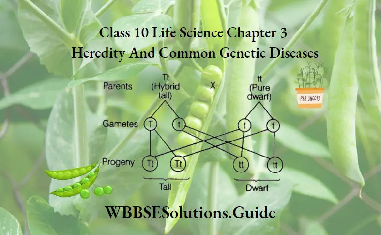
Two pea plants, one bearing pure red flower and another bearing pure white flower, are selected. Flowers appear on both plants at the same time. These plants are considered as peas plants is bisexual plants. So, to check the self-pollination the anther of the flower of one plant is cut and the stigma of the other plant is covered with a paper. Now artificial cross-pollination is done. As a result of this, the seeds for F, are obtained. When these seeds are sown all the plants bear red flowers. These are the plants of the F, generation. When the plants of F, bear flower, self-pollination is allowed. As a result of this action seeds for F, are obtained when these seeds germinate. Now plants are produced. These plants are called the F, generation plants. The colour of flowers in all the plants of the F, generation is not red. The genotypic ratio of F, generation is 1: 2: 1. So, the ratio of pure red and pure white flowers in F, generation is 1: 1.
Question 3. How did Mendel explain the occurrence of tallness of all pea plants in the F, generation in his experiment on monohybrid cross? Discuss in brief how he also explained the occurrence of growing tall and dwarf pea plants in the ratio of 3: 1 in the F, generation in the same experiment.
Answer:
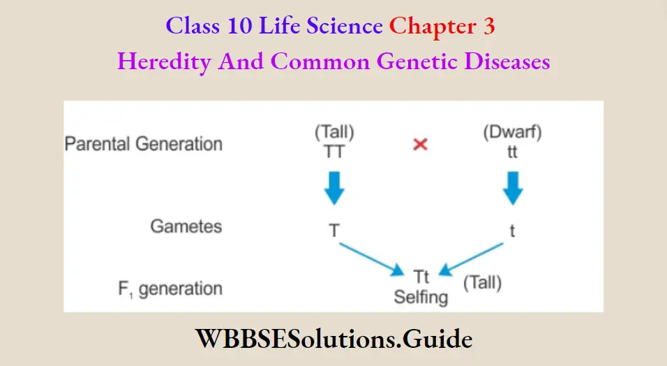
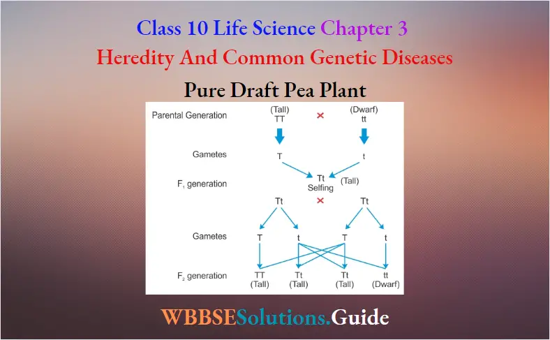
(1) Explanation of occurrence of all tall plants in F, generation of Mendel’s monohybrid experiment:- Mendel selected two sets of pea plants. One set consisted of tall plants (50cm – 200 cm in height) and the other of short plants (up to 50 cm in height). He first made it certain that seeds from tall plants invariably produced tall plants and the dwarfs produced only the dwarfs in successive generations, that is, they are true breeding. He then artificially cross-fertilized tall plants with dwarf plants of the parental generation. To achieve this, he removed the anther of tall plant and covered the stigma of the dwarf plant Then he dusted the pollen grains of a dwarf plant to the stigma of the tall plant. He also made reciprocal crosses. So that each one had a chance to set as male and female parent. He collected all seeds upon ripening and raised the hybrid from these seeds. He referred to this generation as F, All plants of this generation were tall.
Mendel explained the reasons behind the occurrence of all tall plants in F, He suggested that each parent contributed a factor to the offspring. This factor was later termed a gene. The factor contributed by the tall parent was dominant over the factor of dwarfness. He suggested that though both factors were present in the F, the dwarf factor remained unexpressed as it was masked by the factor for tallness.
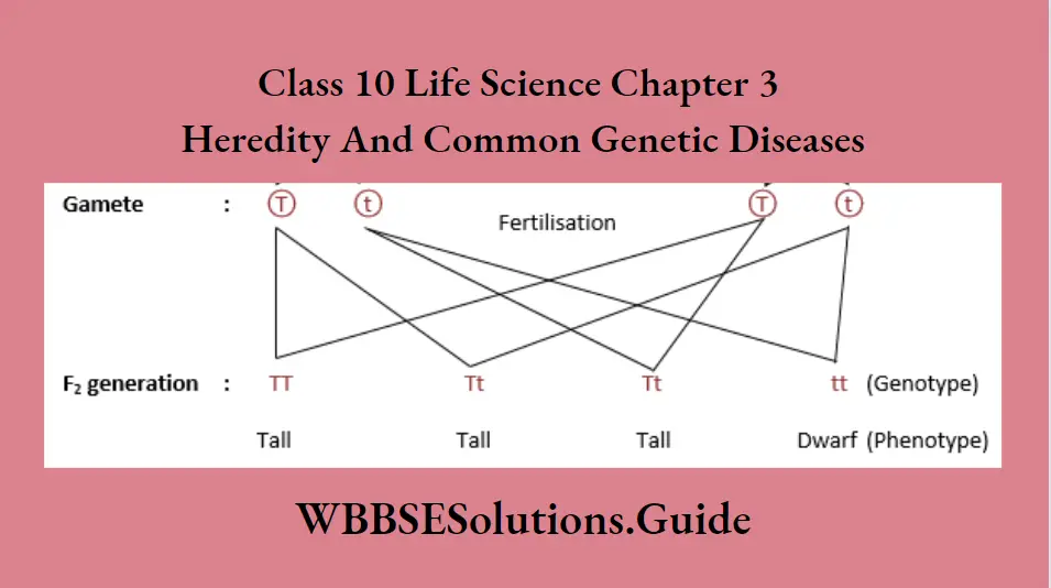
(2) Explanation of the occurrence of tall and dwarf plants in the ratio of 3: 1 in F, generation in the same experiment:・He allowed individuals of F, for self-pollination and subsequently self-fertilization occurred. These self-fertilized seeds are sown in the field for germination. He observed that all the plants were not tall, rather some of the plants became dwarf. So, the tall and dwarf plants were designated as F, generation. Mendel observed that 75% of plants became tall and 25% of plants became dwarf in the F, generation. The ratio of tall and dwarf plants is 3: 1. It was found that the dwarf of F, the generation when self-pollinated was true-breeding, while of the tall plants, only 25% were true breeding. The remaining 50% of the tall plants in the
next generation, produce tall and dwarf plants in the ratio of 3: 1.
In the F, generation, however, the characters segregated and produced tall and dwarf plants. Based on these observations, Mendel postulated the law of segregation, which stated that pairs of contrasting characters of a hybrid separate from each other in subsequent generations.
Question 4. Write the first and second laws of Mendel related to heredity in the form of a definition.
Answer:
First Law of Mendel (Law of Segregation):・Whatever factor may come together in the zygote, they never lose their identity and the factors or genes making up the pair are separated at meiosis during the formation of gametes to produce the next generation. Thus, each gamete contains one factor or gene for a character but the paired form is restored after fertilization.
Second Law of Independent Assortment:- Two or more pairs of factors coming together in the F, separate at meiosis and combine in various ways forming the plants in F and succeeding generations and each pair of factors is inherited independently of the other.
“West Bengal Board Class 10 Life Science Chapter 3, long answer questions, with solutions”
Question 5. Explain the terms dominant and recessive characters, phenotype and genotype with the help of Mendel’s monohybrid cross experiment.
Answer:
Mendel’s monohybrid cross experiment:- Mendel selected two sets of garden pea plants. One set consisted of pure tall plants and the other of pure dwarf plants. He then cross-pollinated tall plants with dwarf plants artificially. He also made reciprocal crosses. He collected all the seeds. He sowed them in the field for germination. He observed that all the plants were tall. He called them the F, generation.
The tall plants of F, generation were allowed for self-pollination and subsequently for self-fertilization. Again, seeds are collected and sown in the garden field for germination. He observed that all the plants were not tall. Some of the plants became dwarfs. He called them as F, generation. Mendel observed that in the F, generation 75% of plants became tall and 25% became dwarf. The ratio of tall and dwarf plants is 3: 1. It was found that the dwarf of F, the generation when self-pollinated was true-breeding, while of the tall plants, only 25% were true breeding.
The remaining 50% of the tall plants in the next generation produce tall and dwarf plants in the ratio of 3: 1.
Based on the observations of the monohybrid cross, we can clarify some points:-
(1) F, generation obtains one factor from the pure tall plant and another factor from the pure dwarf plant. It has both factors for a single character’s tallness. Hence it is a hybrid. Here only the pure tall factor expresses itself. So, this plant is a hybrid tall. The pure dwarf factor is unable to express itself.
(2) The factors of a hybrid are segregated during the formation of gametes to produce the next generation. A gamete contains only one factor for a character. Thus, it is clear that a gamete is always pure.
Keeping the above facts in view, we can define the following terms in this way:-
(1) Dominant:・The factor present in a hybrid expresses itself in a generation is called dominant. Such as tall (TT) factor is a dominant factor of tallness character.
(2) Recessive:・The factor present in a hybrid unable to express itself in a generation is called recessive. Such as dwarf (tt) factor is a recessive factor of tallness character.
(3) Phenotype:- The characteristic of an organism which is externally visible is called phenotype. Example – In F, generation the phenotype of three plants is tall and one plant is dwarf.
(4) Genotype:- The characteristic of an organism which is given based on its entire genetic constitution is called genotype. Example ・In F, generation one plant is pure tall (TT), two plants are hybrid tall (Tt) and the last plant is pure dwarf (tt).
“Class 10 WBBSE Life Science Chapter 3 long answers, for board exam preparation”
Question 6. Mention Mendel’s dihybrid cross experiment with the help of a checkerboard and also mention the phenotypic ratio of the F, generation based on the experimental results.
Answer:
A cross between two sets of plants involving two pairs of contrasting characters is called a dihybrid cross.
Experiment:- For this cross, Mendel selected two plants. One plant possesses round and yellow seeds while another plant possesses wrinkled and green seeds. He arranged artificial cross-pollination. Seeds obtained from this cross were round and yellow. So these factors are dominant, while the wrinkled and green factors are recessive. He called them seeds of the first filial generation (F,).
Plants obtained from these seeds were allowed for self-pollination. They yielded four types of seeds : (1) Round and yellow, (2) Wrinkled and yellow, (3) Round and green, and (4) Wrinkled and green. }
These results are explained by a factor pair Y-y for yellow-green and another factor pair R-r for round-wrinkled. The round and yellow parent can be written as RRYY and the wrinkled and green parent as rryy, the first parent produces gametes which are Ry and the gametes of the second parent are ry. When these gametes unite, the resulting F is RrYy. As round (R) and yellow (Y) factors are dominant over wrinkled (r) and green (y) factors, the F is a hybrid round and yellow.
When the F, in turn, produces gametes, all combinations occur at the time of meiosis. Therefore, both among male and female gametes, four types of gametes RY, ry, Ry and ry are produced. The male and female gametes unite truly and produce a second filial generation (F,). The phenotypic ratio of this generation is 9:3:3: 1.
(Dihybrid cross between pea plants having round yellow seeds and wrinkled green seeds.)
Question 7. State the modern concept of heredity.
Answer:
(1) Characters of parents are transmitted to the progeny through sperms and ova.
(2) Chromosome numbers in each species are constant.
(3) Sperms and ova carry half the number of chromosomes of that species.
(4) Genes are present on chromosomes in a single linear order.
(5) Each gene is present in a particular position of a chromosome.
(6) Gene is composed of DNA.
(7) Gene has the capacity of self-duplication and also mutation.
(8) The fundamental material of heredity is the gene.
(1) Genes are the bearer of hereditary characters.
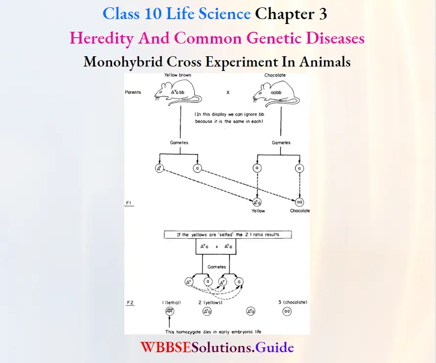
Question 8. Describe the monohybrid cross experiment in animals (Guineapig).
Answer:
To perform a monohybrid cross a black hair guineapig and a white hair guinea-pig are. selected. Now a cross is allowed. In the F, generation all the offspring are black. From this, it is concluded that the black colour is dominant over the white colour.
Now a cross is made between an adult male and a female of the F, generation. In the F, generation, black and white coloured guineapigs are produced in the ratio of 3: 1.
Question 9. Describe dihybrid cross in animals.
Answer:
Dihybrid cross in animals:
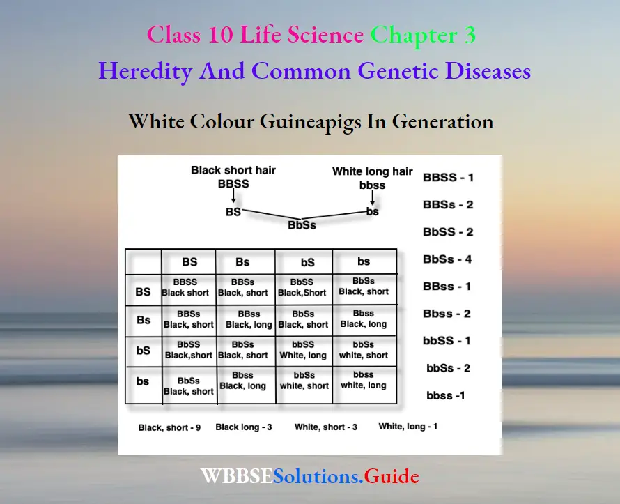
Two different-sex guinea pigs are selected. One has pure black and rough hair while another has white and smooth hair. Now, a cross is allowed. All the F, offspring are black and rough-haired.
Now, F, hybrids are interbred. They will produce 16 offspring in F, generation in the ratio 9:3:3:1
Dihybrid experiment on guineapig
Black and Rough = 9
Black and Smooth = 3
White and Rough = 3
White and Smooth = 1
Phenotypic ratio =9:3:3:1
“Class 10 WBBSE Life Science Chapter 3 long answer questions, Heredity and Genetic Diseases”
Question 10. Explain how the sex of a child is established.
Answer:
Sex of a child is established as follows:
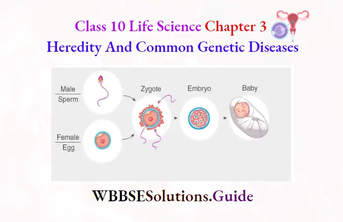
Sex is a characteristic which is transmitted from parents to offspring. Genes are grouped to form chromosomes. The human species has twenty-three pairs of chromosomes. With one exception, the twenty-two members of each pair of chromosomes are alike. The exception is the pair which determines sex always referred to as the sex chromosomes. The somatic cells of the female have a pair xx but the somatic cells of the male have a pair of sex chromosomes which are not alike, xy, y being much smaller than x. Each gamete gets only one sex chromosome; all the ova have x but half the sperms have x and the other half y.
Allowing for every possibility, in the fusion of the gametes the chances are equal that an ovum will be fertilized by x x-bearing sperm or a y-bearing sperm. But the resulting zygote has its sex determined at the moment of fertilization. If xx chromosomes are present the child will be a girl, if xy are Present the child will be a boy.
Question 11. Write four reasons for selecting pea plants by Mendel for his experiments on heredity. Explain with reasons which one between sperm and ovum
is responsible for the determination of sex in human beings.
Answer:
Reasons for selecting pea plants by Mendel for his experiments on heredity
(1) Mendel selected garden peas as his experimental material because
(1) Sweet pea plant is an annual plant. :
(2) It has a large number of pairs of contrasting characters.
(3) Its flowers are complete and bisexual. So it avoids the problems of pollination.
(4) Reproduction ability is excess. :
(2) Explanation to show between sperm and ovum which one is responsible for the determination of sex in human beings:- Sex is a character which is transmitted from parents to offspring. Genes are grouped to form chromosomes. The human species has twenty-three pairs of chromosomes. With one exception, the twenty-two members of each pair of chromosomes are alike. The exception is the pair which determines sex, always referred to as the sex chromosomes. The somatic cells of the female have a true pair xx but the somatic cells of the male have a pair of sex chromosomes which are not alike, xy, y being much smaller than x. Each gamete gets only one sex chromosome; all the ova have x but half the sperms have x and the other half y.
“Heredity and Common Genetic Diseases WBBSE Class 10, long answer questions, solved answers”
Allowing for every possibility, in the fusion of the gametes the chances are equal that an ovum will be fertilized by x x-bearing sperm or a y-bearing sperm. But the resulting zygote has its sex determined at the moment of fertilization. If xx chromosomes are present the child will be a girl, if xy are present the child will be a boy. So, sperm is responsible for the determination of sex.
Question 12. Write the full name of the Father of Genetics. What are phenotype and genotype? If a YYRR (pure dominant yellow and round seeded) pea plant is hybridised with a year (pure recessive green and wrinkle seeded) pea plant, what will be the genotype of the plants obtained in F, generation? Write the phenotypic ratio obtained in the F, generation in the said dihybrid cross.
Answer:
Father of Genetics
(1) Gregor Johann Mendel.
(2) Phenotype:- An organism’s externally visible characteristics are called phenotype. Example = In F, generation of monohybrid cross the phenotype of three plants is tall and one plant is dwarf.
(3) Genotype:- The characteristics of an organism which are given based on its entire genetic constitution are called genotype. Example ・In F, generation of a monohybrid cross one plant is pure tall (TT), two plants are hybrid tall (Tt) and the last plant is a pure dwarf (tt)
(4) The genotype of F, plants will be RrYy
(5) The phenotypic ratio of F, generation is 9:3:3: 1,
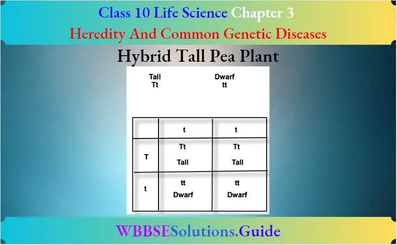
Question 13. What is a hybrid? If a hybrid black-haired Guineapig is crossed with a white-haired Guineapig, what will be the phenotypes of the Guineapigs of the First filial (F,) generation and why? Explain with reasons.
Answer:
Hybrid – An organism having two different genes (contrasting genes) affecting the same trait is called a hybrid.
50% Hybrid black-haired guinea pig and 50% white-haired guinea pig will be produced because Black hair is dominant over White hair.
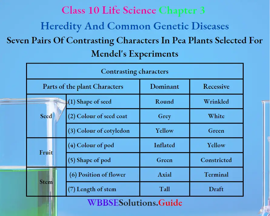
Question 14. Write seven pairs of contrasting characters in pea plants selected for Mendel’s experiments.
Answer:
Question 15. In which name is the first law known Explain with a checkerboard the experiment Mendel performed to reach that conclusion.
Answer:
(1) The first law is known as the law of segregation.
(2) Explanation:- Mendel selected two sets of pea plants. One set consisted of tall plants (50cm – 200 cm in height) and the other of short plants (up to cm in height). He first made it certain that seeds from tall plants invariably produced tall plants and the dwarfs produced only the dwarfs in successive generations, that is, they are true breeding. He then artificially crisscross-fertilized plants with dwarf plants of the parental generation. To retrieve this, he removed the anther of tall the plant and covered the stigma of the dwarf plant. Then he dusted the pollen grains of a dwarf plant to the stigma of the tall plant. He also made reciprocal crosses, so that each one had a chance to set as male and female parent. He collected all seeds upon ripening and raised the hybrids・from these seeds. He referred to this generation as F, A, plants of this generation were tall.
“WBBSE Class 10 Life Science Chapter 3 important long answer questions, exam-focused solutions”
Mendel explained the reasons behind the occurrence of all tall plants in F, He suggested that each parent contributed a factor to the offspring. This factor was later termed a gene. The factor contributed by the tall parent was dominant over the factor of awareness. He suggested that though both factors were present in the F, the dwarf factor remained unexpressed as the factor for tallness masked it.
He allowed individuals of F, for self-pollination and subsequently self-fertilization occurred. These selfself-fertilizedds are sown in the field for germination. He observed that all the plants were not tall, rather some of the plants became dwarf. So, the tall and dwarf plants were designated as F, generation. Mendel observed that 75% of lants became tall and 25% of plants became dwarf in the generation. The ratio of tall and dwarf plants is 3: 1. It was found that the dwarf of F, generation when self-pollinated were true breeding, while the tall plants only 25% were true breeding. The remaining 50% of the tall plants in the next generation, produce tall and dwarf plants in a ratio of 3: 1.
In the F, generation, however, the characters segregated and produced tall and dwarf plants. Based on these observations, Mendel postulated the law of segregation, which stated that pairs of contrasting characters of a hybrid separate from each other in subsequent generations.
Question 16. Explain Mendel’s second law of heredity with the help of the dihybrid cross experiment by Mendel. If a sperm containing the X-chromosome of a man fertilizes an ovum of a woman, what will be the sex of the child developing from the resulting zygote?
Answer:
(1) Experiment:- For this cross, Mendel selected two plants. One plant possesses round and yellow seeds while another plant possesses wrinkled and green seeds. He arranged artificial cross-pollination. Seeds obtained from this cross were round and yellow. So these factors are dominant, while the wrinkled and green factors are recessive. He called them seeds of the first filial generation (F,).
These results are explained by a factor pair Y-y, for yellow-green and another factor pair, R-r for round-wrinkled. The round and yellow parent can be written as RRYY and the wrinkled and green parent as rryy, the first parent produces gametes which are Ry and the gametes of the second parent are ry. When these gametes unite, the resulting F is RrYy. As round (R) and yellow (Y) factors are dominant over wrinkled (r) and green (y) factors, the F is a hybrid round and yellow.
When the F, in turn, produces gametes, all combinations occur at the time of meiosis. Therefore, both among male and female gametes, four types of gametes RY, ry, Ry and ry are produced. The male and female gametes unite truly and produce a second filial generation (F,). They yielded four types of seeds :
(1) Round and yellow,
(2) Wrinkled and yellow,
(3) Round and green,
(4) Wrinkled and green.
The phenotypic ratio of this generation is 9: 3:3: 1.
(2) Second Law or Law of Independent Assortment Two or more pairs of factors coming together in the F, separate to meiosis and combine in various ways forming the plants in F, and succeeding generations and each pair of factors inherited independently of the other.
(3) The child will be female.
“Best long answer questions for WBBSE Class 10 Life Science Chapter 3, solved Q&A”
Question 17. What do you understand by the terms homozygous・and Heterozygous・conditions? Explain the law of Independent Assortment.
Answer:
(1) Homozygous – The individual having a genotype, with two of the same alleles for a trait is called Homozygous.
Example – TT or tt
(2) Heterozygous – The individual having a genotype, with distinct alleles for the same trait is called Heterozygous.
Example – Tt.
(3) Law of Independent Assortment – When two or more pairs of factors come together in the F, separate at meiosis and combine in various ways forming the plants in the F, and succeeding generations and each pair of factors is inherited independently of the other.
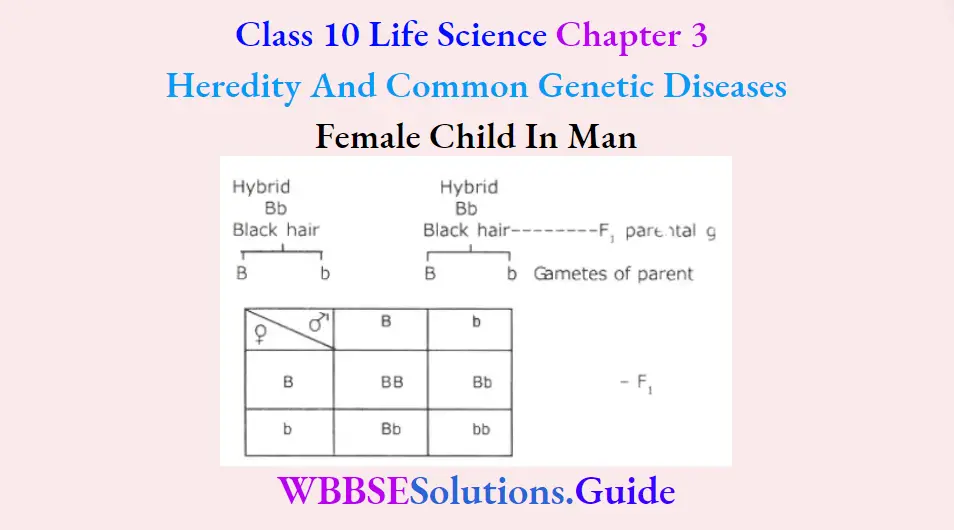
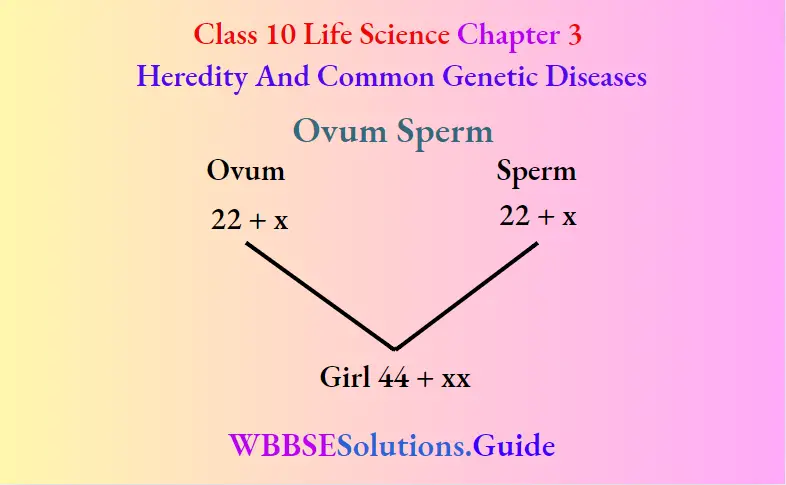
Question 18. Black hair is dominant and white hair is a recessive character of guineapig. Explain the result of a cross between two hybrid black guineapigs with the help of a checkerboard. What types of gametes unite to form a female child in man?
Answer:
(1) When hybrid black Guineapigs are interbred they will produce 75% black colour and 25% white colour Guineapigs in F, generation. The phenotypic ratio of this generation is 3: 1, while the phenotypic ratio is 1: 2: 1.
(2) Female gamete ovum contains (22x+x) chromosome. Male gamete sperm also contains (22x+x) chromosome. These two gametes unite to form a female child.
Question 19. Briefly explain the following behavioural adaptations :
(1)Problem-solving in chimpanzees,
(2) Communication in honeybees.
Answer:
(1) Problem-solving chimpanzees: Just like humans, chimpanzees create and use tools to make their lives easier. Termites are one of the himpanzees・favorite foods, but how to reach the creatures deep within their mounds presents quite a problem. Chimpanzees pick up a twig and stripe the leaves off of it Then they stick the twig into one of the holes in the termite mound, leave it there for a moment, and slowly pull it out as termites clung to the twigs they pick them off with their lips and scrunched them. They are using the stem as a tool to fish・for insects. Chimpanzees have also been seen using tools such as stone hammers to chop up and reduce food into smaller bite-sized portions. Chimpanzees like eating nuts. They hammer them open with stone or wood.
Non-human primates especially chimpanzees-self-medicate. Chimpanzees in the wild appear to practice herbal medicine. They consume numerous items with medicinal properties, such as anti-bacterial agents and deworming herbs.
“WBBSE Class 10 Life Science Heredity and Common Genetic Diseases, detailed long answer questions”
(2) Communication in honeybees Perhaps the most famous and fascinating language of the honey bee is communicated through a series of dances done by
foraging worker bees who return to the hive with news of nectar, pollen, or water. The details of the dance languages were worked out by Karl von Frisch (1967). Two common types of dances are the so-called round dance and the waggle dance. The dance in a figure-eight (8) patterns. It involves a shivering side-to-side motion of the abdomen. The bee first runs straight ahead for a precise distance wagging her
Question 20. Explain in brief the dominant and recessive characters from the result of Mendel’s monohybrid cross experiment in the pea plant. What is genotype?
Answer:
(1) Mendel’s monohybrid cross experiment ・Mendel selected two sets of garden pea plants One set consisted of pure tall plants and the other of Pure dwarf
plants He then cross-pollinated all plants with dwarf plants artificially. He also made reciprocal crosses. He collected all the seeds. He sowed them in the field for germination. He observed that all the plants were tall. He called them the F, generation. Based on observations of the Monohybrid cross we can Clarify some points:-
(1) F, generation obtains one factor from the pure tall plant and another factor from the pure dwarf plant. It has both the factors for single character tallness. Hence it is a hybrid. Here only the Pure Tall factor expresses itself. So this plant is a hybrid tall. The pure dwarf factor is unable to express itself. Keeping the above facts in view, we can define the following terms in this way:-
(1) Dominant:- The factor present in a hybrid expresses itself in a generation is called dominant. Such as the tall (TT) factor is a dominant factor of tallness character.
(2) Recessiv:- The factor Present in a hybrid unable to express itself in a generation is called recessive. Such as the dwarf (tt) factor is a recessive factor of tallness character.
Genotype The genetic constitution of an organism is called genotype. Example-Pure tall (TT), hybrid all- Tt, pure dwarf-tt.
Question 21. Why did Mendel select pea plants for his experiments? Mention any three reasons for it. What is hybridisation? Why is Mendel called the father of genetics? What do you mean by character
Answer:
Mendel selected garden peas as his experimental material because ・;
(1) These are annual self-fertilised plants with bisexual flowers.
(2) The plants could be grown easily and crossing could be done without any difficulties.
(3) The hybrid plant obtained from the cross between two varieties of pure plants ais completelyfertile.
The artificial process which is used to produce hybrid is called hybridization. He performed cross-breeding experiments on sweet pea plants analysed the records obtained from observations and formulated them in the form of laws.
Any inheritable feature is called a character.
WBBSE Class 10 Life Science Chapter 1 Control And Co-Ordination In Living Organisms Long Answer Questions
WBBSE Chapter 1 Control And Co-Ordination In Living Organisms Long Answer Questions
Question 1. Describe in brief the locomotion in Amoeba.
Answer.
Locomotion in Amoeba:
Amoeba locomotes with the formation of pseudopodium (singular). During locomotion, a blunt pseudopodium is formed in the direction of movement simultaneously the pseudopodia (pi.) on the opposite side are withdrawn.
“WBBSE Class 10 Life Science Chapter 1 long answer questions, Control and Co-ordination”
Locomotory organ: Pseudopodia.
Type of locomotion: Pseudopodial or amoeboid movement Process: Several theories have been put forward to explain the formation of pseudopodia, of these the sol-gel theory forwarded by Hyman (1917) and later supported by Mast (1925) is the most accepted.
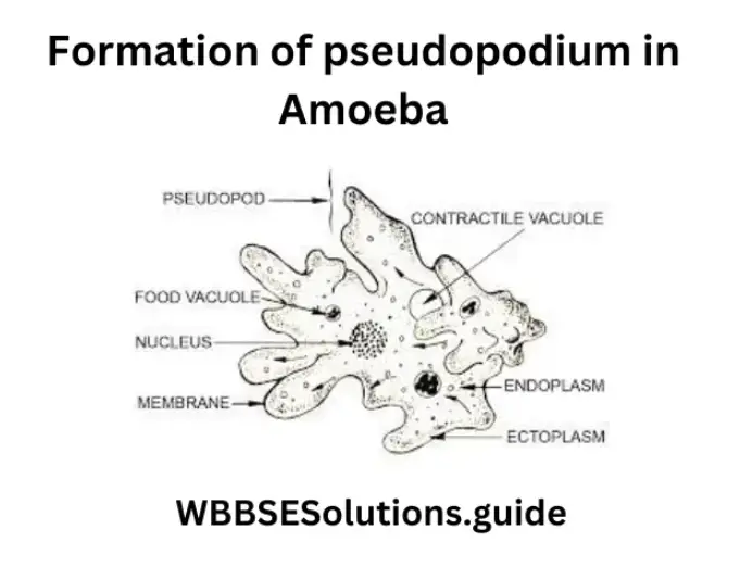
The body of Amoeba is differentiated into three parts. These are (1) a thin and elastic Plasmalemma, (2) an outer non-granular ectoplasm, and (3) an inner granular endoplasm. The endoplasm is further differentiated into an outer jelly-like plasma gel and an inner more fluidy plasmasol. In the formation of pseudopodium, four processes occur simultaneously
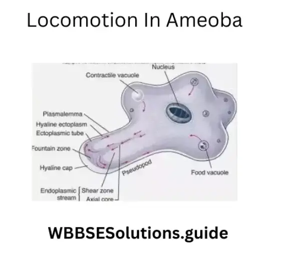
- At the advancing end of the body, the plasma gel partially changes to the solstice (solution) and thus becomes thinner and weaker than the rest of the plasma gel.
- The plasma gel of the opposite end contracts and causes hydraulic pressure on the plasmas.
- Due to the hydraulic pressure, the plasmas are pushed forward towards the softened plasma gel which, being weak, cannot withstand the pressure and develops an outward bulge to receive the plasmas.
- At the periphery of the bulge, the plasmas change into the gel state (gelation). Thus, a tube of plasma gel with a core of plasmas and a covering of ectoplasm is formed. This is the pseudopodium. Pseudopodium is formed in the direction of movement
- Simultaneously the pseudopodia on the opposite side are withdrawn and the amoeba moves forward in the direction of the newly formed pseudopodia.
Question 2. Describe in brief the locomotion in Earthworms.
Answer.
Locomotory organs in earthworms are :
1) Buccal Cavity 2) Setae 3) Body muscles. The buccal cavity acts as a sucker and the setae act as legs. Body muscles are of two types – circular and longitudinal. The alternate contraction and relaxation of these muscles make the body thin and thick respectively.
Type of Locomotion: Creeping locomotion.
Process: Locomotion in earthworms is described in the following six steps :
1. The locomotion starts with the contraction of the circular muscles, which extends to the anterior end. The anterior half of the earthworm becomes thin and elongated.
2. After extending fully the earthworm attaches its mouth (buccal cavity) to the soil which acting as a sucker firmly grasps the soil.
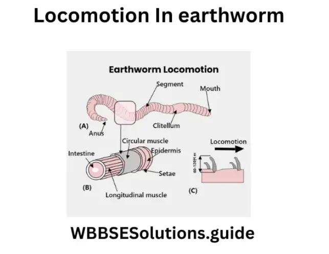
3. Then the setae of the thin region protrude out by the contraction of protractor muscles. The protruded setae firmly grip the soil.
4. At this time the posterior half of the body is free from the soil, and its longitudinal muscles are now ready to contract Due to their contraction the posterior half becomes short
5. After shortening the posterior half, the setae of this region protruded out to take a firm grip on the soil.
6. When the posterior half is completely anchored, the mouth and the setae of the anterior half are withdrawn and the earthworm begins to elongate moving forward. Thus the forward movement is by alternate waves of elongation passing over the whole body. During locomotion, the coelomic fluid serves as a kind of hydraulic skeleton. Sometimes the earthworm may move in a backward direction. It has been calculated that the earthworm can travel a distance of 25 cm in 1 minute.
Question 3. Describe in brief the locomotion of cockroaches.
Answer.
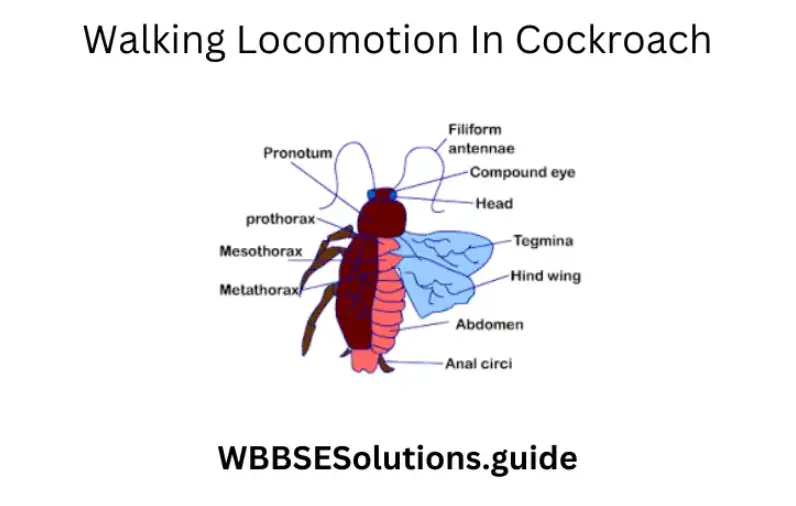
Locomotion of cockroaches
1) Locomotory organs: 1) Three pairs of thoracic legs.
- Pairs of Antagonistic (extensor and flexor) muscles act along the ball and socket joint which divides the leg into five parts.
- Two pairs of wings are present at the posterior side of the second and third segments of the thorax.
- The dorsoventral muscles and longitudinal muscles.
2. Process of Walking Locomotion: It takes place as follows:- The extensor and flexor muscles first contract which causes the straightening of joints in 1st and 3rd leg of one side and the 2nd
- leg of the other side.
- As a result, these legs get lifted and move forward.
- The other three legs provide support to the body and remain attached to the ground with the help of a claw and sticky pad arolium provided at the terminal portion of limbs.
- Next to it, the extensor and flexor muscles relax which causes the bending of the legs. As such the three legs come back on the ground.
- Similarly, the other three legs are moved forward whilst the remaining ones provide support and in this way cockroach moves forward.
2. Flying Locomotion: The male cockroach only flies during the breeding season and in times of danger. The female cockroach cannot fly.
- Process of locomotion:
The forewings (upper wings) of cockroaches are thick and leathery and are called mesothoracic wings. They are not used for flying. The hind wings (lower wings) are smaller and thin and are called meta-thoracic wings. They are used for flying. The movement of wings is brought about by dorsoventral muscles and longitudinal muscles. The contraction of dorsoventral muscles lowers the dorsal wall of the tergum and thus the wings move upward. The contraction of longitudinal muscles arches the wall upward and so the wings move down. This process is repeated and the movement of the hind wing in the manner of ‘8’ helps to support the body and moves the body forward.
Question 4. Describe in brief the locomotion in fish.
Answer.
Locomotion in fish
Swimming is brought about by the whole of the body.
Fins do not contribute at all to forward movement The main function of fins is to control the stability and direction of the fish.
Locomotory organs: Myotome muscles, tail & tail fins.
“West Bengal Board Class 10 Life Science Chapter 1, Control and Co-ordination, solved long answers”
Type of Locomotion: Swimming.
Type of fins: Two kinds of fins are present on the trunk and tail: paired fins and unpaired or median fins.
The paired fins are of two types :
1. Pectoral fins: Present close to the head and correspond to front legs of other vertebrates (two in number).
2. Pelvic fins: Correspond to the hind legs (two in number).
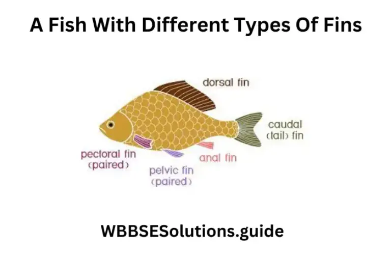
The unpaired or median fins are of three types :
1. Dorsal fin: Present along the top middle line of the trunk (1).
2. Caudal fin : Grows from the tail (1).
3. Anal or Ventral fin: Grows along the middle on the lower side behind the anus (1).
Role of fins in swimming :
1. The paired fins (pelvic and pectoral) control the pitching movement of the fish, causing it to swim downwards or upwards according to the angle at which they are held against water.
2. Pectoral fins help to maintain the fish at rest at any depth.
3. The paired fins are also how the fish slows down and stops locomotion. Thus they act as brakes.
4. The median fins (dorsal, caudal, and ventral) control the rolling and yawing movements of the fish by increasing the vertical surface area presented to the water. During rolling, the median fins prevent the fish from wobbling, i.e., tilting right and left on its axis. Yawing is the tendency to turn in different directions in the same horizontal plane.
Role of myotomes in locomotion (swimming) in Fish :
The fish actively swims by the side-to-side movements of the body particularly the tail. These movements start from the head and continue to the tail like a wave. During the wave of the side-to-side movements, the different regions of the body, from front to back, kick the water backward and sideward, pushing the fish forward. The wave-like movements of the body are a result of the contraction of myotome muscle blocks from the head to the tail in a serial manner and alternation on the right and the left sides.
![]()
The two successive equal and opposite sideways movements of the tail cancel each other out and the fish moves forward.
Swim bladder: Fishes can stay at a particular depth in the water by making their bodies weightless compared with the surrounding water. Most fish achieve this buoyancy by having in their body cavity an air-filled bladder running just beneath the spinal column. This air-filled bladder is known as the swim bladder (buoyancy organ). It acts like a float in providing buoyancy or upthrust in water.
Question 5. Describe in brief the tactic movement in plants.
Answer.
The tactic movement in plants:
Tactic or Taxis: This type of movement of locomotion occurs due to external stimuli like light, chemicals temperature, etc. The direction of the movement is
controlled by the direction of the stimulus. Tactic movement is of three types:
1) Phototactic movements: Many unicellular algae move towards diffuse light (positive phototactic movements) or away from the light of high intensity (negative phototactic movements), e.g. the movements of the chloroplast in the palisade cells which move and arrange and rearrange themselves in the cell in response to light stimulus and the movements of zoospores of certain algae which move in response to light
2) Thermotactic movements: Here, temperature regulates the change of place or position of a plant Some algae move towards the moderately warmer region of water (positively thermotatic), but away from a region of very high temperature (negatively thermotactic), e.g. the rapid rotational cytoplasmic movement in the leaf of Vallisnaria due to increase in temperature.
3) Chemotactic movements: These movements are induced by specific chemical substances acting as external stimuli. Such movements may be either positively chemotactic (e.g. spermatozoids of fern moving towards the ovum under the influence of malic acid secreted by the archegonium) or negatively chemotactic (e.g. movements of certain bacteria and algae away from acidic or alkaline medium).
Question 6. Briefly describe the different types of tropic movements in plants.
Answer.
Tropic movement in plants: It is a type of movement of curvature that comes under the category of induced or paratonic movement
Definition: When the direction of movement (response) of the plant organs bears a definite relation with either towards or away from the external stimulus, then the movement is called tropic movement
Tropic movements are of the following types, depending upon the nature of the stimuli:
1. Phototropism 2. Geotropism 3. Hydrotropism.


1. Phototropism or Heliotropism: The movement of plant organs in response to the source of light and they are said to be positively phototropic while the roots move away from the source of light and are called negatively phototropic, whereas the leaves and branches grow perpendicular to the sun rays and are said to be diaphototropic.
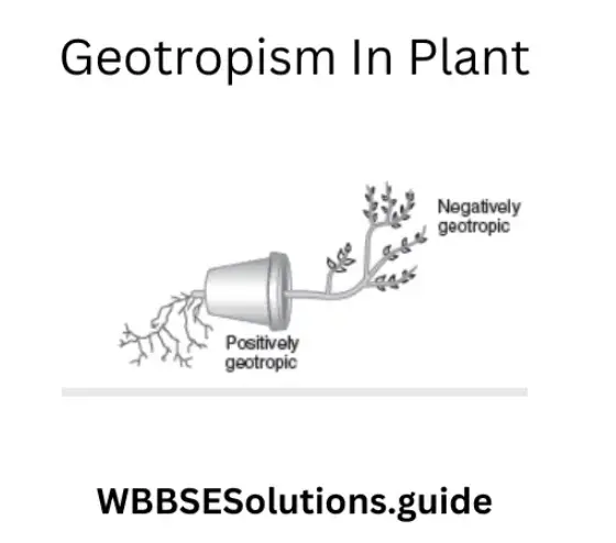
2. Geotropism: The movement of the plant organs induced by the force of gravity and soil is known as Geotropism or Geotropic movement. Generally, the primary roots move towards the force of gravity in the soil while the primary shoot moves away from the gravitational force of the earth. Thus they are positively and negatively geotropic respectively. The lateral roots and branches grow normally at right angles (perpendicular) to the force of gravity and the movement is called diageotropic. The breathing roots of the Rhizophora plant are an exception, showing negative geotropism.
3. Hydrotropism: The movement of plant organs in response to the source of water is called Hydrotropism or Hydrotropic movement The roots are said to be positive hydrotropic because they move towards the source of water while the shoot system moves away from the water and are said to be negative hydrotropic.
Question 7. Describe nastic movement in plants.
Answer.
Nastic movement in plants: When the curvature movements are induced by the influence of external stimuli light and temperature, they are termed as nastic (paratonic) movement. Nastic movements in which the response, i.e., the movement of plant organs, bears no relation to the direction of the stimulus. The stimulus acts on the protoplasm of cells from all sides.
According to the nature of the stimulus, nastic movements may be photonasty, thermonasty, chemonasty, nyctinasty, seismonasty, etc. where the stimuli are light; temperature, and chemical substances. In nastic movement, the direction of movement is not determined by the external stimuli. The response of the plant organ is always the same from whatever direction the stimulus may come.
“Class 10 WBBSE Life Science Chapter 1 long answer questions, important notes and solutions”
1) Photonasty — The movements are concerned with the opening and closing up of flowers, e.g. the flowers of Pentapetes phoenicea fully open at noon, whereas they close at night The opening and closing of leaves of many leguminous plants in day and night afford best examples of photonasty; in these, light from all direction acts as stimulus which causes opening and closing up of leaflets. Similarly, the leaves of Oxalis expand at day time and remain semiclosed and drop at night Light and darkness cause variation in the turgidity of the cell of pulvinar tissues, hence the movement.
2) Thermonasty — The example of the tmonasticcurvature is afforded by the opening of the flowers of Tulips, where an increase in temperature affects the opening of the flower; a consequent fall in temperature brings about the closing of flowers.

This is due to the change in the turgor condition of pulvinar tissue.
3) Chemonasty — It is the movement induced by chemical substances. This type of movement is seen in insectivorous plants. As soon as an insect sits in the center of a leaf of sundew (Drosera sp.), the tentacles move towards it
4) Nyctinasty — It is the most common of the Nastic movements, where changes in temperature and light during the day and night induce visible response which may be termed as nyctinasty. Thus the opening and closing of certain flowers are examples of nyctinasty. Nyctinastic movement of variation due to changes of turgor in the pulvini is also exhibited by the leaf or leaflets of Leguminosae (e.g. clover, Indian Telegraph
(Oesmodium grants), Acacia, Bauhinita, etc), Oxalidacease (e.g. Oxalis acetosella and also some species of fern and Marsilea). This movement is controlled by both light and temperature. The individual factors acting singly may induce photonastic and monastic movements. These movements are brought about by changes in the turgor of the cells of the pulvinus at the base of the petiole.5) Seismonasty — When a leaf of Mimosa pudica is subjected to sudden touch or shock of any kind, the leaf and its parts droop down very rapidly, and the leaflets close up, altogether, presenting a new appearance.
The movements are brought about by changes in the turgor pressure of the cells of the pulvinus.
Question 8. Give the hormonal explanation of geotropism
Answer.
Hormonal explanation of geotropism
Hormonal explanation of Geotropism: Due to the force of gravity accumulation of hormones takes place more on the undersurface of the root and stem. As root cells need less hormone concentration for their growth, the upper surface grows faster than that of the under surface and as a result root bends downward.
Question 9. Describe the flying locomotion in cockroaches.
Answer.
Flying: Cockroaches rarely fly. Generally, male cockroach flies. There are two pairs of wings held at right angles to the thorax during flight. The fore wings are tough and leathery but the hind wings are thin, membranous, and folded like a fan. The wings are without muscle. The movement of wings is controlled by two different sets of muscles in the thorax. These muscles are not connected to the wings. On muscle runs from the roof (tergum-dorsal surface) to the floor (sternum-ventral surface) of the thorax which is termed dorsoventral muscle or tergo-sternal muscle. The second muscle, the longitudinal, muscle runs anteroposteriorly
Contraction of the dorsoventral muscles (elevator muscle) lowers the dorsal wall of the roof (tergum) and thus forces the wing up. Contraction of the longitudinal muscle
(depressor muscle) arches the dorsal wall or roof (tergum) upwards and so forces the wings down. This process is rapidly repeated for flight In flight the tough leathery forewings
are held at right angles to the body and do not beat. Only the membranous hind wings beat in a complicated manner supporting the body as well as driving it forward.
Question 10. Describe the process of bipedal locomotion in man.
Answer.
The process of bipedal locomotion in man
Process of Bipedal Locomotion in Man: Walking in man is brought about by movements of the legs produced by sets of antagonistic muscles contracting and relaxing alternately. In the first step of movement, the heel bone is raised above the ground due to the contraction of calf muscles. Simultaneously extension of the pelvic joint and knee joint
takes place by contraction of the muscles in front of the thigh. In the next step, muscles contract thrusting the toe backward against the ground, so propelling the body forward, and then lifting the toe from the ground. In the next step, the leg is pushed forward.
When the leg moves forward the body also bends forward and due to the force of gravity and with the relaxation of the muscle the heel first comes down at the ground level and sets down in a new position. The toe then comes down to the ground level.
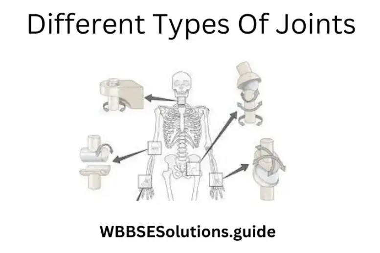
Question 11. What is a synovial joint? What are its different types?
Answer.
Synovial Joint: A movable joint containing synovial fluid, like a white portion of an egg, is called a synovial joint Synovial joint consists of a ligament, covering of the articular capsule le, synovial membrane surrounding the synovial cavity, synovial fluid inside and articular cartilage.
Following are the different synovial joints:
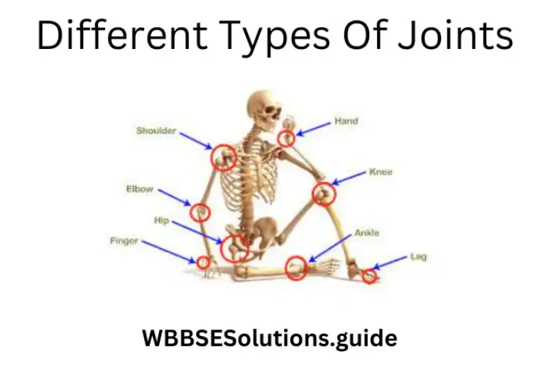
- Ball and socket joint: In this type of joint the ball-shaped head (end) of a bone articulates within the cup-shaped head (end) of the other bone; e.g. at the hip the femur or thigh bone joins the pelvis or hip girdle and at the shoulder where the humerus in the upper arm joins the scapula or shoulder blade. Ball and socket joints allow movement in three planes.
- Hinge joints: Hinge joints, like those in the elbow, finger, in knee, permit movement in one plane only.
- Pivot joint: In this type of joint, the concave end of a bone is attached to the ball-like end of another bone, where the first bone rotates around the head of the second bone. Example: atlas (1st vertebra) and axis (2nd vertebra) joint.
- Condyloid Joint: In this type of joint the convex end of a bone joins with the concave end of another bone. Here the movement takes place in one plane only.
For example, joints between the radius and carpal, metacarpals, and phalanges. The different movable joints of the skeleton are enclosed by a capsule of fibrous connective tissue, the synovial membrane. The ends of each bone are covered with a smooth, slippery layer of cartilage. In the cavity, the bursa, between the articular cartilage, a lubricating fluid, the synovial fluid, is found that is secreted by the membrane lining the joint cavity.
Question 12. Define and classify joints. Name the part of the plant which moves away from the soil. Name the part of the plant that moves towards sunlight. What types of movements are there?
Answer.
Joints
The point at which two separate bones meet is called a joint. There are several different kinds of joints in our body.
1. Immovable joint: In this type of joint no movement is possible between the two bones. The sutures between the bones of the brain box are examples of immovable joints.
2. Partially movable joint: Here only very little ‘ (partial) movement occurs between the two bones. For example, the joint between a rib and the breastbone or between the vertebrae.
3. Movable joint: In this type of joint varying degrees of movement are possible between the two bones forming the joint.
Question 13. Write two functions of the nervous system. What is a synapse? What is meninges?
Answer.
Functions of the nervous system:
Functions of the Nervous system (a) It controls the different activities of different organ systems of the body, (b) It coordinates functions of organ systems.
Synapse:
Synapse The physiological junction between the dendrites of one neuron and the terminal dendrites of another neuron is called a synapse.
Meninges:
Meninges The membranes that cover the brain are collectively called meninges.
Question 14. What is nerve? Write one characteristic of each of the afferent and efferent nerves. Mention the functions of the cerebrum.
Answer.
Nerve:
Nerve A collection of nerve fibers covered by myelin sheath is called a nerve.
Characteristics of afferent and efferent nerves
- Afferent nerve is made up of sensory neurons and it carries impulses from the sense organ to the central organ.
- Efferent nerve is made up of motor neurons and it carries impulses from the central organ to the motor organ.
Function of cerebrum
- It controls all types of voluntary movements of muscles.
- It regulates and co-ordinates voluntary movements.
- It controls the movement of the ball.
Question 15. Write two functions of the cerebral cortex. Describe the protective part of the eyes.
Answer.
Functions of the cerebral cortex
Functions of cerebral cortex (1) The Cerebral cortex establishes the conditioned reflex actions through the development of new connections with different subcortical centers.
It controls the activity of the autonomic nervous system.
The sclera is the outermost layer of the eye. It is made of very tough connective tissue and forms the white portion of the eye. This part is called the cornea. It is covered by a membrane called conjunctiva.
Question 16. Wis an isa neuron? Discuss the relation between a neuron and a nerve. What is ganglion?
Answer.
Neuron
- Neurone The structural and functional unit of the nervous system is called the neurone.
- Relation between the neuron and a nerve Neurone is the structural and functional unit of the nervous system. A bundle of nerve fibers covered with special connective tissue is called a nerve. Relation The nerve fiber of a neuron is the main material of a nerve. Hence, neurons take an active part in the production of a nerve.
- Ganglion The structure formed by the fusion of cell bodies of neurons located outside the Central Nervous System is called a ganglion.
Question 17. What do you understand nervous system? Mention the characteristics and features of afferent and efferent nerves. What is a synapse?
Answer.
Nervous system:
Nervous system The organ system is only present in higher groups of animals whose main function is to control and coordinate the functions of other organ systems of the body, called the nervous system.
Characteristic features of afferent nerve
- It is made by nefibersbres of sensory neurons.
- It carries impulses from the receptor of sense organs to central organs (brain and spinal cord).
Characteristic features of efferent nerve
- It is made by nerve fibers of motor neurons.
- It carries impulses from the central organ to the motor organ (Muscles and glands).
- Synapse The physiological junction between the terminal, dendrite neuronurone with the dendrite of another neuron is called a synapse.
Question 18. Describe in brief the three main parts of the human brain. Mention two functions of skin as a sense organ.
Answer.
Three main parts of the human brain
Structure of three main parts of the human brain
- Cerebrum,
- Cerebellum,
- Medulla oblongata.
Cereb It is the largest part of the brain. It is ® the part of the full brain. Its mass is 1050 gm. It has a fissure in its length due to which it seems it is made by two hemispheres. The shape of each part is semi-spherical. This part has sulci and gyrus.
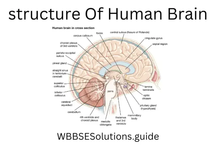
Cerebellum It is fully covered by the cerebrum. It is divided into two parts (left and right). The left part is related to left-organ scans and the right part is related to the right-side organs of the body.The
Medulla oblongata is the lowest part of the brain. It is located between the cerebellum and the Spinal Cord. Its outer and inner parts are made up of white matter and grey matter respectively.
Two functions of skin as a sense organ
- It receives external stimuli! (i.e., hot, cold, pain of Alpin)
- It realizes pressure and weight
Question 19. What is a nerve? What is its function? Mention the characteristics of two main types of nerves and cite examples.
Answer.
Nerve:
Nerve A collection of nerve fibers covered by myelin sheath is called a nerve.

Function of a nerve It carries impulses from receptor organs to the central organ and from the central organ to motor organs.
Characteristics of afferent and efferent nerves (1) Afferent nerve is made up of sensory neurons and it carries impulses from the receptor of the sense organ to the central organ.
Efferent nerve is made up of motor neurons and it carries impulses from the central organ to the motor organ.
Examples
- Olfactory nerve,
- Optic nerve.
Question 20. Write one main difference between sensory and motor nerves with one example each. Mention one function of each of gibberellin, cytokinine, and thyroxine.
Answer.
The main difference between sensory and motor nerves:
- Function of Gibberellins It promotes the development of fruits without seed.
- Function of Cytokinine It takes part in the cytokinesis of plant cells.
- Function of Thyroxine By increasing the blood sugar level, synthesizing protein (in low dose), and depressing protein synthesis (in high dose) it influences metabolism.
Question 21. Describe the structure of the human eye. Explain the mechanism of vision.
Answer.
The structure of the human eye
Each eye is spherical. It lies in a cavity in the skull called the eye socket and is attached to it by three pairs of muscles that move it in different directions. The eyeball is hollow.
Structure Eye is made up of three layers
- Sclera,
- Choroid,
- Retina.
- Sclera It is the outermost layer. It is made of very tough connective tissue and forms the white portion of the eye. This part is called the cornea. It is covered by a membrane called conjunctiva.
- Choroid It is the middle layer. It contains many capillaries. It supplies nutrients to the inner parts of the eye. The choroid forms the pigmented muscular curtain which is called the iris. The hole of the iris is called the pupil which is round-shaped
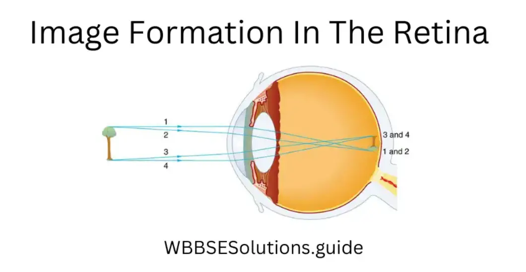
Retina The innermost layer which is a very delicate sheet of tissue, called the retina. This layer contains the rod and cone cells which are sensitive to light and color respectively. These cells are more closely packed at a spot point. This spot is called the yellow spot Optic nerve meets at a point of this layer.
This point is called the blind spot. Here sensory cells are absent. The place of the retina at the front of the eye is taken by the circular transparent biconvex lens which is held in place by suspensory ligament.
“Control and Co-ordination in Living Organisms WBBSE Class 10, long answer questions, with answers”
The suspensory ligament is attached to the choroid coat at its outer edge. The part of the eye in front of the lens is filled with watery aqueous humor. The part behind the lens is filled with telly-like vitreous humor.
Mechanism of vision All the transparent parts of the eye allow light to pass through them. The pigmented parts limit the amount of light entering the eye. The iris allows light to enter the eye only through the pupil. The cornea, aqueous humor, lens, and vitreous humor all act as refracting media.
They make the images falling on the retina clear and detailed. The images formed on the retina are inverted (real), but in the brain, this is corrected to give the impression of an erect object
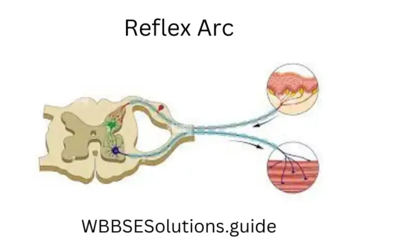
Question 22. How many types of reflex actions are there? Explain with examples.
Answer.
Types of reflex action There are two types of reflex action :
Inborn or unconditioned reflex action.
Conditioned reflex action.
Inborn or unconditioned reflex action A reflex action that is present in the body from the time of birth is called unconditional or unconditioned reflex action.
Example – Knee Jerk reflex action.
Acquired or conditioned reflex action The reflex action that is acquired as a result of repeated training after birth is called acquired or conditioned reflex action.
Example – Salivation in the absence of food but in the presence of a stimulus like a ringing bell.
Question23. What is Auxin hormone? Write three functions of this hormone.
Answer.
Auxin hormone:
(1) Auxin: A group of nitrogenous orgaadsadds containing an indole group produced within meristematic tissue of root and stem apex is called Auxin.
(2) Functions of auxin –
(1) Controlling weeds: By spraying 2,4-D (2,4 – Dichloro phenoxy acetic acid) unwanted dicotyledonous weed plants can be destroyed from the field mono-cotyledonousous crops like wheat.
(2) Rooting of cutting: Cutting is a method of artificial vegetative reproduction. One end of the branch is dipped into a solution of Auxin before placing it into the ground. It increases the rate of rooting.
(3) In parthenocarpy: It helps in the formation of seedless fruits.
Question 24. What is the unit of the nervous system? Mention the names of its three major parts. What is reflex action? Cite one example of reflex action.
Answer.
(1) Name of the unit of nervous system Neurone.
(2) Name of three major parts of neuron (1) Cell body, (2) Dendron, (3) Axon.
(3) Reflex action The involuntary automatic actions controlled by the Central Nervous System are called reflex action.
Example Uplift of leg when touched with a hot object
Question 25. Mention the sources of gibberellins and kinins. Discuss the applicative aspects of plant hormones in agriculture.
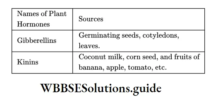
(2) Applicative aspects of plant hormones in agriculture
(1) In the destruction of weeds Dicotyledonous herbs like amaranthus are removed from the field of monocotyledonous crops like wheat by the spray of 2.4 -D and 2,4,5 Tricholorophenoxy acetic acid.
(2) Prevent sprouting The development of adventitious buds of tubers is controlled by the use of auxin. So, potatoes can be stored for a longer time.
(3) For early rooting During cutting IAA and IBA are used at the cut ends. It initiates early rooting at the cut ends.
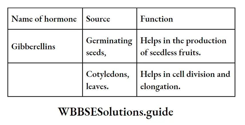
Question 26. Write two functions of gibberellines. How is glucose regulated in the blood? Which hormone decreases glucose levels in the blood?
Answer.
Functions of Gibberellins –
(1) It checks and minimizes the dormancy period of seeds.
(2) It takes part in sex determination.
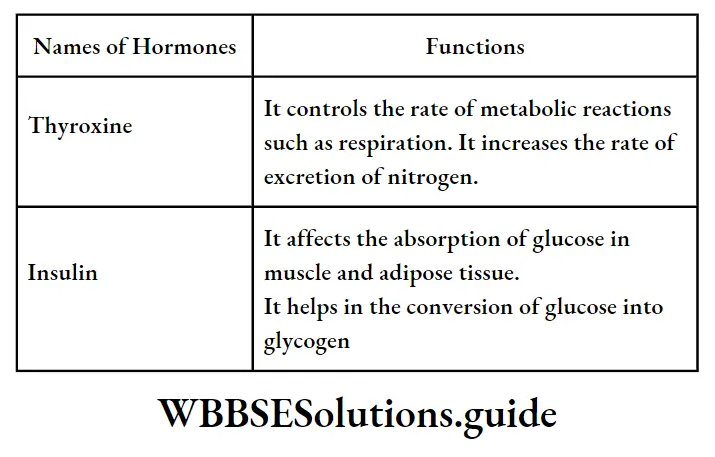
(2) Process of regulation of glucose in the blood:-
Regulation of glucose in the blood is done by insulin. It increases the rate of glucose absorption and stimulates the process of glycolysis. It also helps in the synthesis of glycogen in liver and muscle cells. It prevents the conversion of glycogen to glucose. It induces the synthesis of fat from glucose in adipose tissue and maintains the glucose level in the blood.
(3) Insulin.
Question 27. Why are hormones called chemical coordinators in the living body? Write the site of formation and any two functions of the Gibberelline hormone.
Answer.
(1) Hormones a£ chemical co-ordinator in the body of an organism:- Hormones are complex organic chemicals that are produced in one part of an organism and obtain information about their function from here and then transported to another part of the organism where they perform their function. Hormones control the various metabolic reactions and bring a co-ordination between different metabolic reactions.
Question 28. Which hormone is known as the ’emergency hormone’ and why? Write the sources of gibberellin and any two functions of the hormone.
Answer.
(1) Adrenalin hormone is known as emergency hormone. Reason:- It is secreted at the time of danger and helps to control sudden excitement by influencing the nervous system and increasing blood flow, thereby helping the person to face the abnormal condition successfully.
Question 29. Where is the thyroid gland located? Name the hormones secreted from it. What are the main functions of those hormones?
Answer.
(1) The thyroid gland is situated at the root of the throat, one on either side of the trachea. The two lobes are joined by an isthmus, which is situated in front of the 2nd, 3rd, and 4th tracheal rings.
(2) (1) Thyroxine (2) Calcitonin.
(3) Functions of Thyroxine
(1) Thyroxine increases the metabolic activities of almost all the tissues of the body.
(2) It has effects on growth.
(3) It causes vasodilation in most body tissue, thus increasing blood flow.
(4) It increases the rate of heartbeat
(5) It increases the rate of respiration.
(6) It increases the rapidity of secretion of the digestive juice.
Function of Calcitonin
(1) It promotes the deposition of calcium in the bones and decreases extracellular fluid calcium ion concentration.
(2) It maintains calcium balance in the blood.
Question 30. What is the source of estrogen and testosterone hormones in the human body and write two functions of each.
Answer.
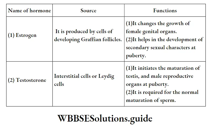
Question 31. Mention the names of two female sex hormones and one function of each of those.
Answer.
(1) Estrogen (2) Progesterone.
The function of Estrogen Estrogen controls the development of female sex organs and female features.
The function of Progesterone Progesterone controls the uterus changes in the menstrual cycle.
Question 32. Wherefrom are TSH and thyroxine hormones secreted? Mention any two functions of the thyroxine hormone. Name the diseases caused by to hyposecretion of this hormone among children and adults.
Answer.
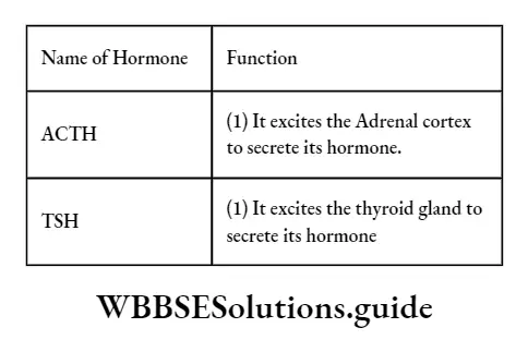
(1) TSH (Thyroid Stimulating Hormone) is secreted from the anterior lobe of the pituitary gland.
Thyroxine is secreted from the thyroid gland.
(2) Function of thyroxine hormone (1) It controls metabolic reaction.
(2) This hormone helps in the development of the nervous system.
(3) Children – Cretinism, Adult – Myxoedema.
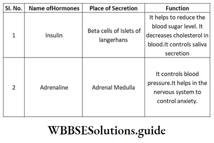
Question 33. Mention the source of Insulin. Mention the functions of this hormone. Which hormone is called emergency hormone and why?
Answer.
(1) Beta cells of islets of Langerhans (pancreas) are the source of Insulin.
(2) It reduces the blood sugar level by converting glucose into glycogen.
(3) Adrenalin is called emergency hormone. Because it gives courage to the
individual during an emergency period, i.e., anger, fear, etc.
Question34. Which hormone is known as the “Emergency hormone”? From which gland is it secreted? Discuss two functions of that hormone. Mention the place of secretion and the place of action of auxins.
Answer.
(1) Name of Emergency Hormone Adrenaline.
(2) Name of the gland Adrenal medulla.
(3) Function of Adrenaline (1) It increases the rate of blood circulation, (2) It increases the rate and depth of respiration.
(4) Place of secretion of auxins Apical meristematic tissue of root and stem.
(5) Place of action of auxins It acts at the place of cell division and cell elongation.
(3) Sites of secretion and functions of insulin and adrenaline hormones :
Question 35. Name three animal hormones that are not secreted from the pituitary gland mention their sources and write the functions of any two of those hormones.
Answer.

(2) Functions of thyroxine and insulin
Question 36. Which gland is considered the master gland and why? Mention the name band function of two trophic hormones secreted from this gland.
Ans. (1) (1) Pituitary gland is considered as the Master gland.
(2) Reason This gland is considered a Master gland because some hormones secreted from its anterior lobe control the functions of another endocrine gland.
(2) (1) TSH (Thyroid Stimulating Hormone) It initiates the thyroid gland to secret thyroxin.
(2) GTH (Gonado Trophic Hormone) influences the growth of the testis in males and ovaries in females.
Question37. Write two characteristics of hormones. Why is a hormone called the “chemical messenger”? Mention one functional difference between the nervous system and the endocrine system.
Answer:
Characteristics of hormones
- Their molecular weight is low.
- Their remaining quantity is destroyed and excreted at the end of the reaction.
- Because they have received orders from their place of production (such as endocrine glands) and act accordingly on the target organs.
- Nervous System
- It does not take part in metabolism.
- Endocrine System
- It takes part in metabolism.
Question 38. Name two hormones that are responsible for apical dominance and division of cytoplasm in plant cells respectively. Write the full name of two hormones produced in the anterior pituitary and one function of each.
Answer.
(1) Hormone responsible for apical dominance —Auxin
(2)Hormone responsible for the division of —– cytoplasm
(3) (1)TSHACTH —ThyroidAdrenoStimulatingCortico (2)TrophicHormoneHormone—Cytokinin.
Question 39. Describe the parts of the Nervous system.
Answer.
Parts of the Nervous system
(1) Central Nervous System (CNS): This part of the nervous system is the supreme controller of all body responses. CNS includes :
(1) Brain or Encephalon, which occupies the cranial cavity, and contains the higher governing centers.
(2) Peripheral Nervous System (PNS): All nerves originating from the CNS and constituting the PNS are subdivided into the following two components :
(1) Cerebrospinal nervous system: It is the somatic component of the PNS which includes 12 pairs of cranial nerves (originating from the brain) and 31 pairs of spinal nerves (originating from the spinal cord). It innervates (supplies) the somatic structures of the head and neck, limbs, and body wall.
(2) Autonomic nervous system (ANS): It is the visceral component of the PNS which includes the visceral or splanchnic nerves. It innervates the viscera, glands,
blood vessels and nonstriated muscles.
ANS consists of two divisions :
(1) Sympathetic and (2) para-sympathetic systems.
These two systems have antagonistic effects, i.e., while one system promotes the activity of the organs, the other system retards. Thus they control and co-ordinate the activities of internal or visceral organs. This coordination is involuntary
Question 40. What is the unit of the nervous system? Describe its structure and functions.
Answer.
Unit of the nervous system
(1) Neuron(e) is the unit of the nervous system.
(2) Structure of Neuron (e): It is the structural and functional unit of the nervous system and consists of a nerve cell body with all its processes.
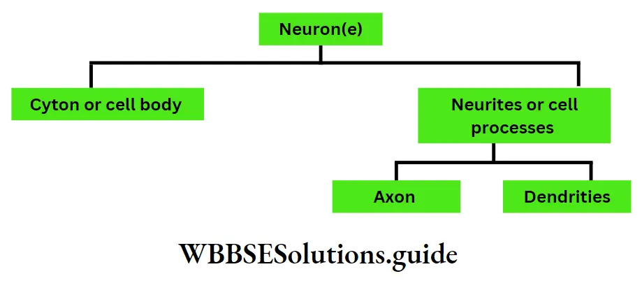
1. CYTON : (also called soma or perikaryon)
The soma is present In the following places of the nervous system.
(1) In the grey matter of CNS.
(2) small clusters called ganglia in the PNS.
It is found in different sizes and shapes (spherical, oval, spindle, etc.) It consists of the following :
1. Neuroplasm: It is a mass of cytoplasm surrounded by a cell membrane.
Various structures are suspended in it
2. Nucleus: It occupies the central part of the soma and contains, usually, one prominent nucleolus.
Function: Controls all the functions of the cyton.
3. Nissl bodies (also called tigroid substances): They are named after the discoveror Franz Nissl. These are granular structures (maybe rod-shaped) present all over the cyton. They may extend in the dendrites but not in the Axon.
Function: They synthesize the proteins of neurons.
4. Mitochondria: It is present both in cyton and axon.
Function: Performs respiration (production of ATP)
“WBBSE Class 10 Life Science Chapter 1 important long answer questions, exam-focused”
5. Neuro-fibrils: These are thread-like structures. They are present in both soma and neurites.
Function: They transmit impulses.
6. Centrioles: In the past, it has often been stated that centrioles are not present in neurons but studies with the electron microscope have shown that centrioles are present.
Function: They help in the production and maintenance of micro-tubules. Besides these, some other structures are also present
1. Golgi apparatus
4. Lyso-somes
2. Neurites or cell processes :
The processes arising from the cell body of a neuron are called neurites. These are of two kinds : (1) Dendrites and (2) Axon.
1. Dendrites or Dendrons: These are short; numerous, afferent processes arising from the different regions of the cyton. They are characterized by the following :
(1) They are tapering processes, i.e., thick at the origin but much thinner at the end.
(2) They branch repeatedly and irregularly.
(3) They bear numerous small spines called Gemmules to increase the surface area.
(4) Their cytoplasm consists of neurofibrils,missess granules, and mitochondria.
(5) They are rough in external appearance.
Functions :
1. They receive the sensory impulses and conduct them towards the cell body.
2. In some cases they modify to act as receptors, e.g. Dendrons of olfactory cells modify into olfactory rods and receive impulses or olfaction (smell) in the olfactory region of our nose.
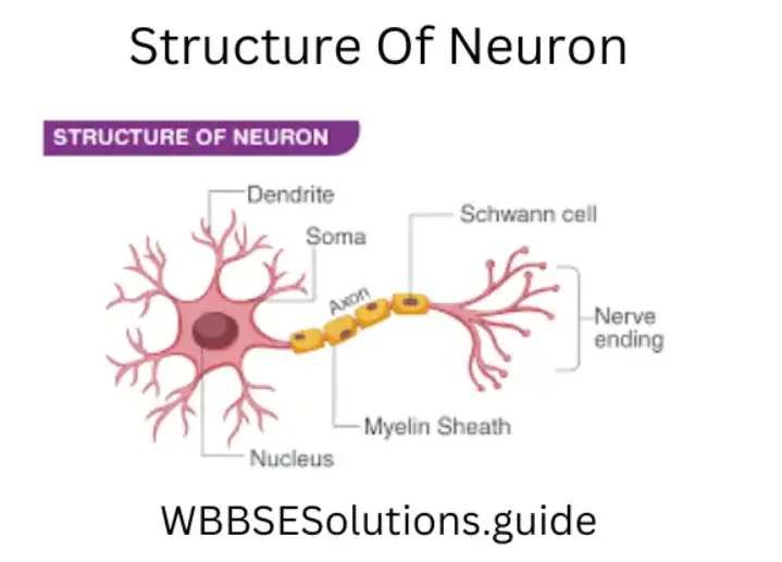
2. Axon or Axis Cylinder or Neuraxon: It is a single long efferent process arising from the axon hillock of the cyton and is of uniform thickness. It is better known as “nerve fiber”. Based on the presence of myelin sheath, it is of two types:
(1) Myelinated and (2) Non-Myelinated nerve fibers.
(1) Myelinated or Medullated Nerve Fibers: As the name suggests, they are surrounded by a layer of the myelin sheath. The nerves look white (due to the high refractive index of the myelin sheath). The great majority of nerve fibers in our body are myelinated nerves. They are found in
(1) The white matter of central nervous system.
(2) The peripheral nerves. It consists of the following parts from inside outwards
(1) Axis Cylinder: It constitutes:-
The Axoplasm: It is the central core of the axon and is pasty (semi-fluid) in nature. Within the axoplasm (1) mitochondria, (b) axoplasmic vesicles and
(2) neuro fibrils can be seen but its granules are absent (It also contains axis fibrils that run parallel to the axis of the fiber).
Axolemma: The axoplasm is ensheathed (covered) by a membrane called axolemma. It is a non-nucleated, semi-permeable membrane.
Functions: Many substances like proteins are not synthesized by the axon but are formed by the cyton. These substances are transported from the cyton to the whole of the axon by the axoplasm. This transportation is called axonal flow.
(2) Myelin sheath or Medullary sheath: Outside the axis cylinder lies the myelin sheath. Myelin has a whitish appearance. The sheath is made up of lipid materials and protein. It is formed by:-
(1) The Schwann cells (In peripheral nerves)
(2) Oligodendrocytes (In the nerves of CNS)
Functions : (1) Increases the speed of conduction of nerve impulse, (2) Acts as an insulator by reducing loss of electrical activity.
(3) Neurilemma (Neurolemma) (Also called Schwann cell sheath): This is the externalmost covering of the axon. It is unbroken and nucleated.
Functions: It helps in the regeneration of nerves.
(4) Nodes and Inter-nodes: The myelin sheath does not form a continuous sheath but is interrupted at regular intervals. The gaps so produced are seen as constrictions or nodes known as the Nodes of Ranvier. The segment of the axon between two successive nodes is called an “Inter-nodal segment”. The longer the segment, the faster the rate of conduction of nerve impulses.
(5) Schwann Cells: They lie between the neurilemma and the myelin sheath. In between two nodes of Ranvier, there is a single cell consisting of one (and only one) nucleus of Schwann.
Function: They are responsible for the formation of the myelin sheath.
(6) Endoneurium: It is a tube of connective tissue that envelopes each nerve
Fibre outside the neurilemma.
Wbbse Class 10 Life Science Question Answer
(7) Branches of axon: An axon may give off a variable number of branches. At its termination, the axon breaks up into several fine branches called telodendria. From the sides of the axon, several branches arise at right angles and are known as collaterals. The collaterals and telocentric often form small bulbous swellings at the synapse called terminal boutons (or bouton teminaux).
2) Amyelinated or Non-medullated nerve fibers (Also called fibers of the remark): These nerve fibers are called non-medullated because they have no myelin sheath. Thus, their diameter is very small. It consists of an axis cylinder covered by neurilemma with a single cell of Schwann. Nodes of Ranvier are absent They are found in :
(1) The grey matter of CNS. (2) The peripheral nerves. The function of Axon: They transmit the impulses away from the cell body to the other neurons via a synapse.
Question 41. How many types of neurons are there?
Answer.
Neurons are classified on the following basis:-
(1) based on the direction of transmission of impulses :
(1) Motor neurons: Transmit impulses from the CNS to the effectors.
(2) Sensory neurons: Transmit impulses from receptors to the CNS.
(3) Intermediate neurons: Confined to the CNS and connect the motor and the sensory neurons.
(2) based on the number of fibers arising from nerve cells :
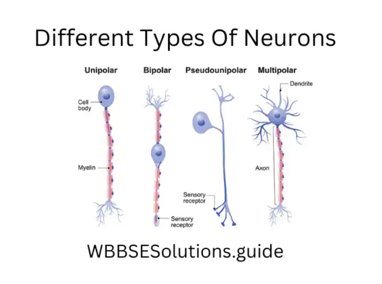
(1) Apolar: Having no axon or dendrites.
(2) Unipolar : A neuron with a single fibre.
(3) Bipolar: A neuron with two fibers, e.g. a neuron with one dendrite and one axon.
(4) Multipolar: A neuron with many fibers.
(3) based on the presence of myelin sheath :
(1) Myelinated: Those neurons whose axons have a covering- of the myelin sheath.
(2) Non-Myelinated: Those neurons whose axons do not have a covering of myelin sheath.
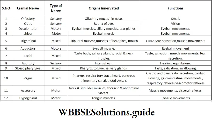
Question 42. Name the different cranial nerves. Mention the organs innervated and their functions.
Answer:
Question 43. What are the characteristic features of afferent and efferent nerves?
Answer.
Characteristic features of afferent nerve :
1. It is also called sensory nerve.
2. It arises from the dorsal horn and runs through the dorsal nerve root of the spinal cord.
3. It connects the sensory organs to the brain and spinal cord.
4. It carries the stimulusthe the brain and spinal cord the the rom sensorgansrgan.
5. They are generally unipolar or bipolar.
Example : 1st, 2nd & 8th cranial nerves.
Characteristic features of efferent nerve :
1. It is also called a motor nerve.
2. It arises from the right ventral horn and runs through the ugh ventral nerve root the the of spinal cord.
3. It connected the cts brain and spinal cord to the effector organs, that is, muscles and glands.
4. It carries impulses from the brain and spinal cord to the effector organs.
5. They are generally multipolar. Ex: 11th & 12th cranial nerves.
Wbbse Class 10 Life Science Question Answer
Question 44. What is the importance of the nervous system? Write the name of the structural and functional unit of the nervous system.
Answer.
Importance of the nervous system
(1) The importance of the nervous system is explained as follows :
(1) It controls, harmonizes, and regulates all voluntary muscular activities, involuntary activities such as breathing, heating of the heart, rates of secretion of some endocrine gland and ds, rapidly changing visceral ever to bring about coordination among the various organs of the body.
(2) It is responsible for memory and intelligence and provides higher mental processes, i.e., consciousness, ambition, and judgment It is also responsible for emotional activities. Thus it enables us to remember, to think, and to reason.
(3) The nervous system acts as a museum of past incidents and experiences.
(4) The stimuli in the form of nerve impulses travel along the incoming nerve from the receptors to the spinal cord and the brain. Here the impulses are analysed and order is passed into other outgoing nerves which lead the information from the brain to the effector organs which then makes a suitable response.
(2) The structural and functional unit of the nervous system is the neuron.
Question45. Mention the functions of any three of the arts of neurons.
Answer.
The function of the ions of three main neurons neurons are
(1) Cyton or cell body: Its functions are
1. It receives nerve impulses from its dendrite and transmits them to the axon.
2. It produces nerve processes.
3. It also provides space for the attachment of nerve processes.
4. The Nissl bodies contain RNA and act like ribosomes.
5. It has some degeneration generation and regeneration of neurons.
(2) Dendrites (Dendron) Its functions are
1. They impulses impulses from the axon of the other, neuron and send the impulses
to the cell body.
2. It establishes fun relations relation with other neurons.
3. In some neurons dendrites are modified into receptors.
3) Axon: Its functions are:-
1. Axons act as efferent processes that receive impulses from the cell body and transmit
them to the next neuron through synapses or to the muscles through
junctions.
2. It establishes junctional relations with other neurons.
3. It takes part in the formation of nerve fibers.
Question 46. What is a synapse? Mention one similarity and one dissimilarity between the functions of the nervous system and hormones in the animal body.
Ans.
Synapse – The specialized junctions of neurons with one another or with effector organs (muscle or gland) are termed synapses.
OR, The point of junctional communication or contact between the dendrites of one neuron and the axon or cell body of another neuron is called a synapse.
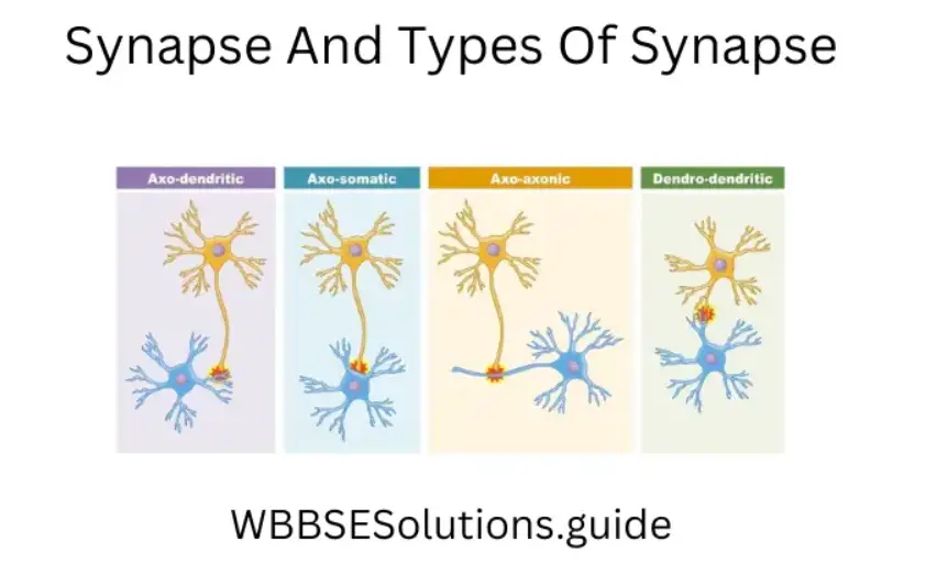
(2) Distribution – Synapses occur in the central nervous system and the ganglia of the autonomic nervous system.
(3) Types – 1 . Axoaxonic
2. Axosomatic 3. Axon dendritic.
(4) Function – Transmission of impulse between the neurons.
2. Basis Nervous system And Hormone
similarity coordination
Dissimilarity physicochemical chemical coordinator
Rapid in action slow in action
47. What is neurone ? Discuss the relation between a neuron and a nerve. What is ganglion?
Answer.
Neurone
(1) Neurone or nerve cell is the structural and functional unit of the nervous system. It consists of main three parts, viz. 1. Cell body or cyton., 2. Axon.,
3. Dendron. Its main function is the transmission of impulses.
(2) Neurone-nerve relation: A nerve is a bundle of nerve fibers ensheathed by connective tissue. Neurone is made up of an axon, having cyton or cell body and dendrites. Axon is covered by a sheath called the myelin sheath, together with neurilemma forming the axis cylinder. When two or more axis cylinders are covered by a connective tissue called endoneurium with blood vessels, then this bundle is called nerve fiber. Two or more nerve fibers may be joined by a perineurium, which may be covered by another epineurium to form a nerve.
(3) Ganglion: A mass of nervous tissue containing many cell bodies and synapses usually enclosed in a connective tissue sheath. In vertebrates, most ganglia occur outside the CNS (exceptions are basal ganglia in the brain).
In invertebrates, ganglia occur along the nerve cords and most anterior pairs are analogous to the vertebrate brain, invertebrate ganglia constitute a part of the CNS
Types : (1) Basal Ganglia: It receives impulses from the thalamus and influences motor impulses.
(2) Submandibular ganglia It relays impulse to the submandibular and sublingual salivary gland.
Question 48. What is the brain? A chart shows the different parts of the human brain.
Answer.
Brain
The brain is the master organ of the body. The human brain, a highly specialized organ, is present in a bony case called cranium which protects it from external injuries. The brain of an adult weighs about 1350 gms and is mainly composed of nervous tissue. In the brain, the grey matter is situated on the surface while the white matter forms the interior.
The brain can be differentiated into three main regions: forebrain, midbrain, and hindbrain.
Wbbse Class 10 Life Science Question Answer
Question 49. Describe the structure & functions of the forebrain.
Answer.
Cerebrum :
1. It is the largest part of the brain and is proportionally larger in man than in any other animal.
2. It consists of two cerebral hemispheres joined together by a broad curved thick band of nerve fibers called corpus callosum.
3. The cerebral hemispheres have an outer region of densely packed nerve cells called the cerebral cortex. It forms the grey matter of the brain.
4. Each cerebral hemisphere is divided into four lobes namely frontal, parietal, temporal, and occipital by three deep and wide fissures. The specific areas control different activities of the body.
5. The surface area of each hemisphere is increased by numerous infoldings or convolutions called gyri (singular gyrus). The gyri are separated by depressions called sulci.
6. The cerebral cortex is the region of various kinds of activities. These can be broadly classified into three areas.
Functions: 1. The sensory areas receive impulses from receptors and register impressions of what we see, hear, and feel.
2. The motor areas transmit impulses to various organs and control voluntary movements like the activities of the trunk, limbs, and face.
3. The association areas give the ability to register impressions and respond by interpreting past experiences. It is associated with memory, learning, reasoning, and intelligence
(2) Thalamus: It is a region present at the center of the forebrain.
All sensory information that reaches the cerebral hemispheres first passes through the thalamus.
Function: Its function is to interpret sensory signals and then channel them to the appropriate cerebral cortex region.
(3) Hypothalamus: It is present beneath the thalamus, hence the name hypothalamus (Hypo = below). It consists of grey matter scattered in the white matter.
Functions: 1. It is a control center for many internal control mechanisms or the autonomic nervous system and is associated with temperature regulation, water balance, hunger, blood pressure, etc.
2. It is also a reflex control center and is associated with sleeping, feeding, emotions, etc.
3. In association with the pituitary gland it secretes neurohormones and controls the release of hormones from the anterior pituitary.
4. It also controls the synthesis and release of posterior pituitary hormones.
Question 50. Name the cells from which the midbrain is made up. Mention its functions.
Answer.
Midbrain: It, consists of groups of nerve cells – grey matter scattered in the white matter. It connects the forebrain and the hindbrain. It has four corpora quadrig
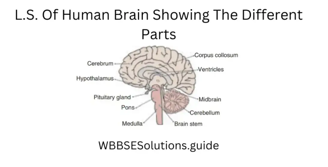
Functions: 1. It is the reflex center of every movement and auditory response.
2. The lower part of mid brain is associated with muscular reflexes and balancing reflexes. Mid brain can not be easily distinguished as it is enclosed by the forebrain.
(2) LH: In males, it is known as ICSH. (1) In Female: It controls ovulation.
(2) In Male: It helps the secretion of testosterone hormone.
Question 51. Mention the structure & functions of different parts of the hindbrain.
Answer.
Hindbrain: It consists of the cerebellum on the dorsal side and the brain stem (pons and medulla) on the ventral side.
Cerebellum: It is somewhat at the back of the head and consists of two cerebellar hemispheres just like the cerebrum. It also has grey matter on the surface and white matter in the central part It gives the appearance of two halves of a large walnut
Function:
(1) It is a large reflex center for the coordination of muscular body movements and
(2) maintenance of posture or equilibrium.
(3) The cerebellum is also associated with certain voluntary muscular movements originating in the forebrain.
Pons: It lies just above the medulla and acts as a bridge carrying ascending and descending tracts between the brain and spinal cord.
Function: It sends impulses to the cerebellum and controls breathing.
Medulla oblongata or medulla: It is the posterior-most part of the brain and continues into the spinal cord. Various ascending and descending tracts cross over from left to right and right to left in the medulla. Any damage to one of its sides causes paralysis on the opposite side of the body.
Function: 1. Since it carries the nerve tracts connecting the spinal cord to the brain, all communications between the brain and the spinal cord pass through it
2. It also contains the cardiac, respiratory, and vasomotor centers that control
complex activities like heart action, respiration, coughing, etc.
Question 52. What is a plant hormone? Name the different types of plant hormones.
Answer.
Plant hormone:
(1) Phytohormones: The organic compounds, synthesized in certain parts of the plants but exerting their influence in some other parts are called plant hormones
or phytohormones.
(2) In plants only five substances are officially classified as plant hormones. However, some other substances are being studied that may eventually be classified also as hormones.
The five main plant hormones are :
1. Auxin 2. Gibberellin 3. Cytokinin 4. Abscisic acid 5. Ethylene.
Wbbse Class 10 Life Science Question Answer
Question 53. What is auxin? How many types of auxin are there? Mention the physiological functions of auxin.
Answer.
Auxin:
(1) Auxin is a generic term applied to those hormones whose primary function is cell elongation.
The first auxin to be discovered is Indole 3-acetic acid (IAA). Other auxins, either natural or synthetic, resemble IAA in their physiological effects.
(2) The natural and synthetic auxins are as follows :
1. Natural Auxin: Auxins which are synthesized in plants are called natural auxins. Ex : IAA (Indole acetic acid), IPA (Indole propionic acid).
2. Synthetic Auxin: Auxins which can be synthesized in the laboratory are called synthetic auxins. Ex: NAA (Naphthalene acetic acid), IBA (Indole butyric acid); 2, 4-D (2, 4-Diclorophenoxy acetic acid), MCPA (2 Methyle 4-Chloro Phenoxy Acetic acid).
(3) The major physiological functions of auxins are as follows
1. Cell elongation
2. Apical dominance.
3. Phototropism
4. Geotropism
5. Root initiation
6. Parthenocarpy.
Delaying senescence of leaves by preventing the formation of abscissions layer.
Question 54. What are gibberellins? Who isolated them? Mention the functions of gibberellins.
Answer.
Gibberellins:
(1) Gibberellins are phytohormones first extracted from the fungus Gibberella fujikuroi (Fusarium moniliforme), the fungus that causes the banana disease, also called foolish seeding disease.
(2) The first Gibberellin was isolated in crystalline form by Yabuta and Sumiki in 1938. Chemically they are related to compounds called terpenoids.
(3) The major physiological functions of gibberellins are as follows:
1. Elongation of internodes.
2. Promotion of flowering.
3. Induction of parthenocarpy.
4. Breaking dormancy of the seeds and buds.
5. Induction of bolting, i.e., conversion of genetically dwarf plants into tall plants as well as their flowering.
Question 55. What are cytokinins? Who discovered it? Mention the important functions of cytokinins.
Answer.
Cytokinins:
(1) Cytokinins are phytohormones which induce cell division.
(2) The first cytokinin named kinetin was discovered by Miller and Skoog in 1954. Kinetin is a 6-furfury laminopurine – a degradation product of nucleic acid. Later in 1964 Letham and Miller isolated another cytokinin named Zeatin from corn milk, i.e., extract of immature corn (maize) caryopses. However, the most widely occurring cytokinin in plants is isopentenyl adenine (IPA).
(3) The major functions of cytokinins are as follows :
1. Induction of cell division.
2. Cell enlargement
3. Root and shoot initiation.
4. Breaking of seed and bud dormancy.
Question 56. Mention the source of the auxin hormone. Discuss the functions of that hormone in the plant body.
Answer.
(1) Source: Meristematic cells of growing regions like root tip, and shoot tip.
(2) Functions of auxin :
1. Cell division: Auxin hormone helps in nuclear division and thus promotes cell division. As a result, cells divide profusely which causes the growth of the meristematic region.
2. Cell elongation: Auxin softens the cell wall by removing calcium linkage present in the cell wall. It increases the uptake of water in the cell, thus the size of cells increases.
3. Callus formation: Auxin helps in callus formation for the healing of wounds. When applied to cuttings or wounds the permanent cells of the pith and the cortex region divide and redivide to form a callus – a wound tissue.
4. Cambial activity: Cambium cells become active due to the movement of IAA (Indole Acetic Acid) from the developing buds towards the region of reactivation of cambium during the growing season and take part in secondary growth.
5. Apical dominance: Auxin allows the growth of the apical or terminal bud and inhibits the growth of lateral buds.
6. Xylem formation: Auxin helps in xylem formation. The division of xylem cells takes place in the presence of auxin
7. Tropic movement: Auxin occurs at the root apex and stem apex. Auxin tends to move away from the light and towards gravity.
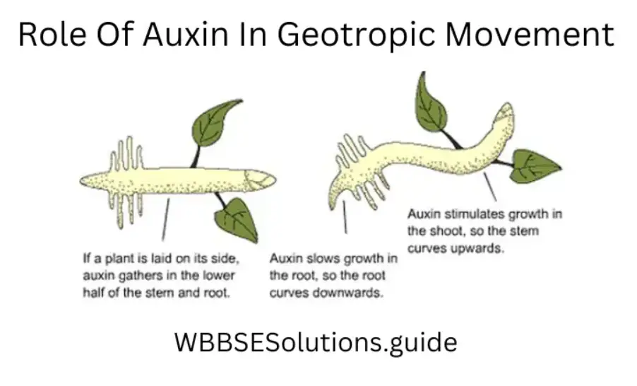
Auxin controls two types of movement:1) Phototropic movement – When light falls the auxin moves towards the darker side. The action of auxin is different in stem and root In stem more auxin causes more growth while in root more auxin causes less growth. So the stem moves towards the light showing positive phototropism and the root moves away from the light, that is towards the dark showing negative phototropism.
2) Geotropic movement – As stated earlier, the auxin moves towards gravity. More auxin causes more growth in the stem so it grows away from gravity showing negative geotropism and as more auxin causes less growth in the root it moves towards gravity showing positive geotropism.
“WBBSE Class 10 Life Science Control and Co-ordination, detailed long answer questions”
Question 57. Name the different hormones secreted from the anterior pituitary gland. What are their functions? Mention the hyper and hypo activities of the anterior pituitary gland.
Answer.
The different hormones secreted from the anterior pituitary are :
(1) ACTH, (2) GH (3) TSH, (4) GTH.
(1) Functions of ACTH :
1. Stimulates adrenal glands to produce cortisone.
2. It also affects profound changes in the adrenal structure.
3. It stimulates lipolysis in adipose tissue and mobilises fat from it, this is called adipokinefic active of ACTH.
(1) Hypersecretion leads to Cushing’s disease.
(2) Hyposecretion leads to Addison’s disease.
(2) Functions of STH :
1. Promotes overall growth of the body.
2. Increases metabolism.
3. Reduces nitrogen excretion.
4. Promotes protein synthesis.
5. Promotes blood sugar level.
6. Reduces fat
7. Stimulates lactation in women after childbirth.
(1) Hypersecretion causes Gigantism in infants and Acromegaly in adults.
(2) Hyposecretion causes Dwarfism in infants and Simmond’s disease in adults.
(3) Functions of TSH :
1. It stimulates the growth and secretion of the thyroid gland.
2. It increases the iodine uptake.
3. It affects general metabolism.
4. It stimulates glycolysis & TCA cycle.
5. It enhances the release of stored thyroid hormones.
(1) Hypersecretion leads to Grave’s disease.
(2) Hyposecretion results in the enlargement of the thyroid gland.
(4) Functions of GTH: It influences Gonads (ovary & testis). It is of two types –
(1) FSH & (2) LH or ICSH.
(2) FSH: 1) In Females: Oestrogen secretion: It is a gonadotrophic hormone. It is gametogenic in action and stimulates the development of the ovarian follicles and secretion of estrogen hormone.
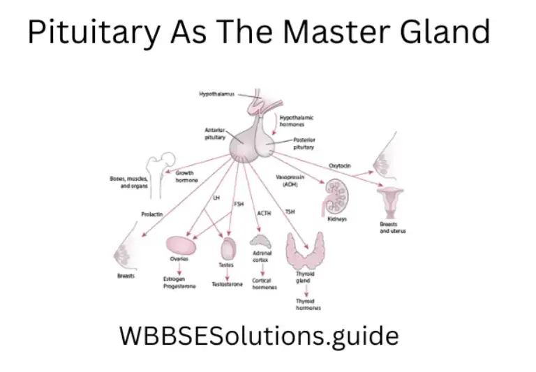
2) In Male: Spermatogenesis: It helps in cortex spermatogenesis (formation) of sperms.
(2) LH: In males, it is known as ICSH. (1) In Female: It controls ovulation.
(2) In Male: It helps the secretion of testosterone hormone.
Question 58. Mention the site of secretion and function of the Thyroxin hormone. What are the effects of its hypo and hypersecretion?
Answer.
(1) Site of Secretion – Thyroid gland.
Functions of Thyroxine (a calorigenic hormone) :
1. Regulates basic metabolism (BMR).
2. Controls absorption of glucose from the intestine.
3. Increases the blood sugar level.
4. Increases the heartbeat, cardiac output & blood pressure.
5. Controls body temperature by calorigenic effect
6. Causes normal development of the central nervous system.
7. Helps in the maturation of R.B.C.
(1) Hypersecretion causes Grave’s disease or Exophthalmic goiter.
(2) Hyposecretion causes Cretinism in children, Myxoedema in adults & Simple
Goitre (swelling of the neck, i.e., enlargement of the thyroid gland).
Question 59. Mention the site of secretion of Insulin. Discuss the functions of this hormone. Mention its hyper and hypo action.
Answer.
Site of Secretion: B cells of Islets of Langerhans of the pancreas.
(1) Functions of Insulin :
1. Glucose absorption: It influences glucose absorption in muscles and adipose tissues.
2. Controls glucose metabolism: Under its influence synthesis of glycogen from glucose in muscles and liver increases. It also reduces the breakdown of glycogen into glucose.
3. Antiketogentic: Under its influence lipids are inhibited from oxidation to produce ketone (acetone) bodies, therefore, insulin is termed as an anti-ketogenic hormone.
Hypersecretion of Insulin: Increased insulin secretion leads to hypoglycemia, i.e., decreased glucose level in the blood.
Hyposecretion of Insulin: Less insulin secretion leads to diabetes mellitus.
Question 60. Mention the site of secretion of adrenaline hormone. Discuss the function of this hormone. Mention its hyper and hypo activities.
Answer.
Site of secretion: Medulla of the adrenal gland.
(2) Functions of Adrenaline: Emergency hormone: It helps to control sudden excitement by influencing the nervous system and increasing blood flow and, therefore, is known as an emergency hormone. Controls blood pressure: It influences the blood vessels and controls blood pressure. Influences respiration: It controls the muscles of the respiratory organs and
thereby influences respiration. 3-F’s Hormone: Under conditions of fight, flight, or fright it causes changes that increase the efficiency of the animal.
For Eg.: (1) Increases heartbeat and breathing.
(2) Diversion of blood from the gut to muscles.
(3) Conversion of glycogen to glucose in the liver.
Hypersecretion: It causes rapid heartbeat, headache, and high.blood pressure, excess glucose in the blood. It also causes Cushing’s syndrome.
Hyposecretion: Addison’s disease: in the early stage of the disease, symptoms
are dark pigmentation of the skin (especially the hand, neck, and face).
Question 61. Name the hormones secreted from gonads. Mention their functions.
Answer.
1. Ovary and testis (singular) are known as gonads. The ovaries are paired oval bodies located in the pelvic cavity of a female. The important hormones secreted by the ovaries are estrogen, progesterone, and relaxin.
(1) Oestrogen: Site of secretion: Graffican follicle of the ovary, placenta.
Function : (1) Oestrogen stimulates the growth, development, and functional activities of primary and secondary sex organs.
(2) It regulates the menstrual cycle and stimulates breast development
(3) Oestrogen also has a cholesterol-lowering effect
Hypersecretion: It leads to disturbance in menses or cancer formation.
Hyposecretion: It causes failure of menses and ill-developed genital tract
(2) Progesterone: Site of Secretion: Corpus luteum of the ovary.
Functions : (1) Promotes secretory changes in uterine endometrium for pregnancy,
(2) implantation of zygote and
(3) formation of placenta,
(4) Also promotes the development of mammary glands.
Hyposecretion causes abortion and misconceptions.
2. Testes (plural) are located in the scrotal sac, hanging outside of the body at the junction of two legs in males. The main hormone secreted from the testis is Testosterone. Site of secretion: Interstitial cells of Leydig cells of the testis.
Functions :
(1) They are required for the normal maturation of sperm.
(2) Effect the growth of accessory sex organs, (3) development of secondary sexual characters in males, and (4) inhibits thymus.
Hyposecretion causes poor development of sexual characters leading to feminism.
Wbbse Class 10 Life Science Question Answer
Question 62. In the human body, locate the different endocrine glands.
Answer.
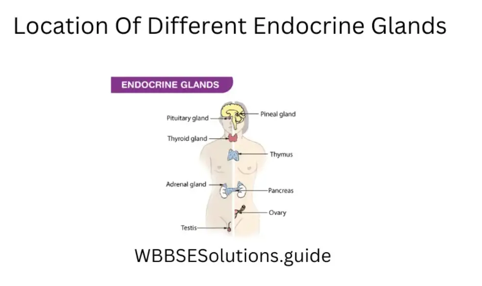
Question 63. Briefly describe the mechanism of sensing environmental changes and stimuli in plants.
Answer.
Mechanism of sensing environmental changes and stimuli in plants:
(1) Plant organs sensing light with photosensitive compounds: They are also called phototropins, cryptochromes, and phytochromes, each reacting very specifically to certain wavelengths of light These light sensors tell the plant whether it is day or night, how long the day is, how much light is available and from where the light comes.
Shoots grow towards light and roots usually grow away from light. These responses are called phototropism and skototropism respectively. This phenomenon is brought about by the plant hormone along with the pigments mentioned above.
Many plants exhibit certain phenomena at specific times of the day; for example, certain flowers open only in the mornings. Plants keep track of the time of the day with an internal molecular clock. This internal clock coupled with the ability to perceive light also allows plants to measure the time of the day and so find the season of the year.
The seeds of many plants sprout only after they are exposed to light This response is carried out by phytochrome signaling. Plants are also able to sense the quality of light and respond appropriately.
For example, in low light conditions, plants produce more photosynthetic pigments. If the light is very bright or if the levels of harmful UV increase, plants produce more of their protective pigments, that act as sunscreens.
(2) Production of signaling molecules: Wounded tomatoes are known to produce the volatile odor methyl-jasmonate as an alarm signal. Plants in the neighborhood can then detect the chemical and prepare for the attack by producing chemicals that defend against insects or attract predators.
(3) Hormonal signaling in plants: Plants systematically use hormonal signaling to coordinate their development and morphology. Morphogenesis in a developing plant is brought about by a delicate balance between auxin and cytokinin. Phytochrome, gibberellin, and the hypothetical hormone florigen bring about the modification of apical meristem to floral meristem.
(4) Trapping of prey by insectivorous plants: Stinging hairs of insectivorous plants like Sundew close the leaflets when they come in contact with the insect body. This is primarily due to the monastic movement
(5) Shock-based movement in plants: Any type of mechanical or electrical pressure releases the water molecule from the pulvinus in Mimosa pudica and the plant droops down. After some time with the discontinuity of the touch, the plant again regains its original configuration.
This is called the seismonastic movement This was denoted by Acharya J.C. Bose, who showed that a low electricity shock of 1.3 volts can result in drooping and retraction movement in Mimosa pudica.
Bose uses a crescograph to measure the rate of growth of a plant which is a device to magnify plant growth up to 10,000 times when they are stimulated by different stimuli like temperature, chemicals, gases & Electricity and thereby they not only proved that plants have life but also showed that they show varied responses to stimuli.
6) Movement due to osmotic stress: The leaflets of the Indian Telegraph plant Desmodium gyrans automatically go up and down with the loss and gain of turgidity of the basal leaflets. As the leaflets go down, they lose turgidity, become light, and go up.
(7) Growth movement due to touch: The tendril of a pea or vine grows like a spring-surrounding a solid rod-like support and there by the plant despite having a non-woody stem that can stand to erect This phenomenon occurs by the initial touch of the solid support causing the bending of the tendril (thigmotropism) followed by growth in the spring like manner (circumnutation).
Wbbse Class 10 Life Science Question Answer
Question 64. How are the physiological activities of plants regulated to increase agricultural yield?
Answer.
Regulation of physiological activities of plants leading to an increase in Agricultural Productivity :
(1) Apical growth of plant shoots and roots is promoted by auxin, so plant size decreases.
(2) Healing of wounds by the formation of callus tissue through auxin prevents infection or insect damage.
(3) Prevention of abscission of leaves by auxin causes pre-harvest loss of leaves.
(4) Parthenocarpic (seedless) fruit development with the help of auxin makes fruit like grapes economically more viable.
(5) Destruction of weed hosts with certain synthetic auxin compounds like 2,4-D reduces the competition from different weed plants.
(6) Initiation of intercalary growth by elongating the internode by gibberellin (GA3), induces growth for different stem vegetables.
(7) Transformation of apical bud to floral bud by GA3, induces more branching.
(8) Gibberellin induces early germination of seed.
(9) Morphogenesis by the induction of root or shoot through auxin-cytokinin balance is very useful for plant tissue culture.
(10) Cytokinin induces a bushy habit of a plant which is useful for plants like tea, where more apical buds and leaves are required.
Question 65. Briefly explain with examples the chemical coordination in the human body.
Answer.
There are several examples of coordination in our body —
(1) Goosebumps, result from the erection of the hairs in our skin. It is an involuntary, or uncontrollable response to cold temperatures or strong emotions. When we are in a cold environment, our thermoreceptors signal to our brain that our body temperature is decreasing.
This triggers the release of the hormone epinephrine (adrenaline), which targets the erector pili (smooth muscles surrounding hair follicles) and causes them to contract The contraction of these muscles causes our hairs to stand erect. These raised hairs cause the insulating air layer to expand, allowing your body to retain more heat.
(2) Most cells in the human body use the sugar called glucose as their major source of energy. To ensure a constant supply of glucose to cells, blood glucose levels must be maintained at relatively constant levels. In a healthy person, blood glucose levels are restored to normal levels primarily through the actions of two pancreatic hormones, namely insulin and glucagon.
(3) In the modern environment one is exposed to various stressful conditions. Stress can lead to changes in the serum level of many hormones. Some of these changes are necessary for the fight or flight response to protect oneself.
Adrenaline, along with nor-epinephrine is largely responsible for the immediate reactions we feel when stressed and combating stress. Along with the increase in heart rate, adrenaline also gives you a surge of energy that you might need to run away from a dangerous situation and also focuses your attention.
(4) Blood pressure is the pressure exerted by circulating blood upon the walls of vessels. Hormonal responses exist for both lowering and raising blood volume. The kidneys provide a hormonal mechanism (the renin-angiotensin-aldosterone system) for the regulation of blood pressure by managing blood volume.
Epinephrine and nor-epinephrine hormones, secreted by the adrenal medulla, raise blood pressure by increasing heart rate and the contractility of the heart muscles and by causing vasoconstriction of arteries and veins.
Antidiuretic hormone (ADH), a hormone produced by the hypothalamus and released by the posterior pituitary, raises blood pressure by stimulating the kidneys to retain water (raising blood pressure by increasing blood volume).
(5) Basal Metabolic Rate (BMR) is the rate of energy expended by animal (such as human) bodies when at rest. The thyroid hormones triiodothyronine (T3) and thyroxine (T4) are the main regulators of BMR. BMR increases when the quantity of these hormones in the blood increases. Some other hormones such as testosterone, insulin, and human growth hormone (HGH) can also increase the body’s metabolic rate.
(6) Sex Hormones: In most animal species the brain controls and regulates sexual behavior primarily using hormones. The sex hormones are estrogen and testosterone. Sexual behavior is influenced by the hypothalamus. It stimulates the pituitary gland to release the sex hormones.
In the female the target gland of the gonadotrophic hormone is the ovary, the ovary has two functions, the first one is to produce eggs, and the other is to secrete hormones. Estrogen and progesterone are the hormones released by the ovary?
Question 66. “Accommodation is the ability of the eye lens to adjust its focal length”. Explain the statement.
Answer.
Accommodation- viewing near and distant objects:
The accommodation is a reflex action of the eye, in response to focusing on a near object, then looking at a distant object (and vice versa), comprising coordinated changes in convergence, lens shape, and pupil size.
The lens is responsible for accommodation, the adjustment of the eye for observing near or far objects. It does this by changing its shape. This changes the focal length of the lens and permits it to focus the image formed on it by the cornea.
The lens is suspended from the muscular ciliary body by many delicate zonule fibers attached to its equator. In the normal relaxed condition (that is, with no tension placed on the fibers by contraction of the ciliary muscle) the lens’ shape is such as to refract rays from distant objects, and the eye is focused at infinity.
Accommodation for close vision requires tension to be exerted to deform the lens. Contraction of the muscles of the ciliary body tugs at the fibers and changes the shape of the lens. The eye adjusts the shape of the lens to keep objects in focus.
Looking at distant objects: Ciliary muscles relaxed -*• lens becomes thin -»• focal length of the lens increases. Looking at objects closer to the eye: Ciliary muscles contract -*ÿ increases the curvature of the eye lens -*• lens becomes thick -*ÿ focal length of the lens decreases.

An example of accommodation can be cited from daily life situations. Good driver vision is a crucial factor in road safety, both for the pedestrians and passengers of the car. This is required to avoid road accidents by taking note of obstacles, traffic signals, and road directions.
Distance acuity is probably the most important visual skill for driving. Distance acuity is the ability to focus and see clearly at far distances. Even the simplest reactions in driving take at least 0.4 seconds.
A driver has to change focus quickly and easily from the road to the dashboard and back again. Being able to see color is also pretty important in the car.
Question 67. Briefly explain different defects of vision and their corrective measures.
Answer.
Defects of visions and corrective measures:
A -person with normal eyes can, by accommodation, see all objects that are at a distance greater than about 25 cm from the eye. If due to certain abnormalities the eye is unable to accommodate itself to various distances, then the eye is said to be defective. Some common defects of the eye are –
(1) Myopia: Near-sightedness, also called myopia is a common name for impaired vision in which a person sees near objects clearly while distant objects appear blurred. In such a defective eye, the image of a distant object is formed in front of the retina and not at the retina itself. Consequently, a nearsighted person cannot focus clearly on an object farther away than the far point of the defective eye.
Causes: This defect arises because the power of the eye is too great due to the decrease in focal length of the crystalline lens. This may arise due to either-(1) excessive curvature of the cornea, or (2) elongation of the eyeball.
Correction: This defect can be corrected by using a concave (diverging) lens. A concave lens of appropriate power or focal length can bring the image of the object back to the retina itself.
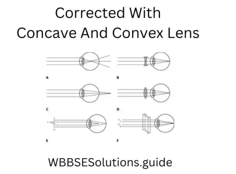
(2) Hyperopia or hypermetropia: Farsightedness, also called hyperopia or hypermetropia, common name for a defect in vision in which a person sees near objects with blurred vision,
while distant objects appear in sharp focus. In this case, the image is formed behind the retina.
Cause: This defect arises because either (1) the focal length of the eye lens is too great, or (2) the eyeball becomes too short, so that light rays from the nearby object, say at point
N, cannot be brought to focus on the retina to give a distinct image.
Correction: This defect can be corrected by using a convex (converging) lens of appropriate focal length. Eyeglasses with converging lenses supply the additional focusing power
required for forming the image on the retina.
“Best long answer questions for WBBSE Class 10 Life Science Chapter 1, solved answers”
(3) Presbyopia: Presbyopia is a progressive form of farsightedness that affects most people by their early 40s. The power of accommodation of the eye decreases with aging. Most people find that the near point gradually recedes.
Causes: It arises due to the gradual weakening of the ciliary muscles and diminishing flexibility of the crystalline lens.
Correction, Simple reading eyeglasses with convex lenses correct most cases of
presbyopia. Sometimes, a person may suffer from both myopia and hypermetropia.
Such people often require bi-focal lenses. In the bi-focal lens, the upper portion of the bi-focal lens is a concave lens, used for distant vision. The lower part of the bi-focal lens is a convex lens, used for reading purposes.
Question 68. What do you mean by bipedal locomotion? what are the characteristics of plant hormones?
Answer.
Bipedal locomotion
In the animal kingdom, only man has the effective capacity to walk and run on his two legs. Locomotion in man is la affected by the movement of legs, though the hands keep the balance of the body, Thistle moving.
The kind of locomo is called the bipedal locomotion.
1. They are mainly produced at the tips, root tips, etc.
2. Plant hormones are natural, artificial, and postulated.
3. Chemically plant hormones are acidic or alkaline in nature.
4. Plant hormones are secreted from meristematic cells.
5. These are transported from the site of secretion to the site of action by conducting tissues, mainly by phloem.
6. Plant hormones are mostly growth hormones.
Question 69. What are the characteristics of hormones? What are the characteristics of animal hormones? How is nerve impulse transmitted through synapse?
Answer.
Characteristics of hormones
1. These are present in the body in minute quantities and can act at very low concentrations.
2. They often act at a site remote from their site of production.
3. They are transported through the body fluids.
4. A hormone may act at more tissue tissue target tissues.
5. They are degraded after their actions are over.
6. They influence the activity of a process but cannot initiate a pro-quick-acting quickly acting (slow-acting or acting (estrogen).
2. These are secreted from the endocrine glands.
3. It is carried from the site of secretion to the target organ by the blood flow.
4. It acts in a very low concentration.
5. It is destroyed and excreted as soon as its function is over.
6. Chemically they steroids steroid site peptides steroid or peptides in nature.
7. Perform various activities including the growth of the organism.
8. They help the body cope with emergency demands such as infection, trauma,
emotional stress, dehydration, starvation, hemorrhage, and temperature extremes.
They are secreted in response to specific stimuli.
When an impulse arrives at the tip of the axon it releases neurotransmitters like adrenaliacetylcholineholine from the synaptic vesicles. The chemical diffuses across the synaptic cleft and reaches the dendrite of the next neuron causing depolarization of the membrane there. A new impulse then passes through the next neuron.
Question 70. Write the common characteristic features of the endocrine gland cataract cataracts cataract occur. How can it be rectified?
Answer.
1. Anatomical characters — The endocrine glands are devoid of excretory ducts and are highly vascularised.
2. Chemical characters — They synthesize some definite hormones and secrete them into the blood leaving the organ.
3. Functional — The materials secreted by such glands have some definite physiological actions. That means each gland has some definite functions in the body as evidenced by the fact that surgical removal of the gland leads to impairment of certain physiological functions that are corrected by administration of the gland extract to the animal.
The reason for this defect is that the lens loses its transparency and becomes opaque due to the deposition of protein materials and calcium minerals in the lens. This opaque the e condition of the lens does not allow the light rays from objects to pass through the lens. This defect can be rectified by surgically removing the lens and it has to be replaced by a highly convex lens.
Question 71. Which animal hormone was discovered first? Write its origin and function. What was the contribution of Acharya Jagadish Chandra Bose to sensitivity in plants?
Ans. (1) Secretin.
(2) Secretin is secreted from the small intestinal mucosa. It stimulates the secretion of bile from the liver as well as the secretion of bicarbonate and water in the pancreatic juice. Acharya Jagadish Chandra Bose made substantial discoveries in plant physiology. One of his inventions is the Crescograph, which he used to measure plant’s response to different stimuli. He demonstrated with experiments that plants too crescographph Crescograph was used to record the plant pulses when it was connected to a plant called Desmodium gyrans.
The plant root was hydrobromic hydrobromic acid till its set, m. Initially, the plant showed a steady deflection of the pointer in the crescograph and soon it became unsteady and then vibrated violently and finally stopped suddenly, indicating the death of the plant
Question 72. What is apical dominance? Name the hormone responsible for the formation of lateral buds. How can apical dominance be demonstrated sensitivity Sensitivity?
Answer. (1) Apical dominance is a physiological phenomenon in plants in which the growth of the apical bud inhibits the development of lateral buds.
(2) Auxin is the hormone responsible for the formation of lateral buds.
(3) Apical dominance can be easily demonstrated by cutting the API, and cal the bud because in the absence of the apical bud, active growth starts in the lateral buds. Sensitivity is the ability to detect the change in the environment and react to the stimuli. It is moreanimalssunced in animals the ability to show locomotion. Some of the lower plants can exhibit locomotion like in unicellular or colonial green algae and their response to stimuli is easily detectable which is in terms of positive or negative tactic movement But higher plants are fixed to the substratum with the help of roots and they cannot move from place to place, so they exhibit growth movement which is very slow and cannot be de CrtnCertain Though
certain plants are relatively more sensitive and they exhibit turgor movement in response to electrical or mechanical shock.
Question 73. (1) What is auxin? (2) What is its role in phototropism? Mention any two functions of GTH.
Answer. (1) Auxin is a class of plant growth hormone. It is produced at the tips of root & shoot.
(2) Phototropism is the movement of plant parts towards or away from light Auxin controls this movement by its differential presence in a particular area causing unequal rates of growth of the two sides of that plant part functions to functions of G.Follicle-stimulatingcle stimulating hormone increases, the growth of ovary stimulates to secrete estrogen from Grafian follicles in females and in males it stimulates seminiferous tubules of the testis and causes spermatogenesis.
(2) Luteinising hormone stimulates corpus luteum in females to seprogesteroneer one, and in males, it stimulates the interstitial theal cells of the testis to secrete testosterone.
Question 74. Classify animal hormones according to their chemical nature giving three examples of each class a ss. What is a hormone?
Answer. Animal hormones are chemically of three types
(1) Protein or peptide hormones: Ex – STFI, ACIH, insulin.
(2) Amines & modification acids: Ex – Adrenaline, thyroxine, melatonin.
(3) Steroid hormones : Ex – Estrogen, progesterone, testosterone.
(1) Hormones are organic substances produced by a group of cells in one part of the body that after being transported by body fluid influence growth and other physiological activities at a site remote from its place of production.
(2) Hchemical agents agent released from one group of cells which travel through the body fluid to affecgroupsgroupsferent group of cells. (Bayliss and Starling -1904).
Question 75. What are the functions (importance) of the nervous system? Define thermonasty with an example.
Answer.
Functions (importance) of the nervous system
(1) It controls all the voluntary and involuntary activities.
(2) It regulates and coordinates the activities of the bodies.
(3)It receives, transmits andinformationinformations, and then formulates a response for the effector organs.
(4) It helps in thinking and learning.
(5) It maintains the equilibrium of the body) It is the sum of the past incidents and experiences.
(ininIt helps in maintaining the integrity between an organism and the external environment.
(8) It is the seat of imagination, intelligence, judgment, ideas, etc.
(9) It also controls the emotional activities.
The movement of plant organs in variations to variation in temperature is known as thermonasFlowersower and s of tulips, crocus open when the temperature increases during daytime and clotemperatureee temperature falls at night.
Question 76. What is the the necessity of nervous system in the body? What i photo nasty? What is tropism?
Answer.
Necessity of nervous system in the body
The body of a man (or any other multicellular animal) is made of many organs specialized to perform different functions. In any society which includes people doing different sorts of work, an administrator is required to activities the activities of all the people.
Similarly in our body, a controller is required to coordinate and regulate all the functions (both voluntary and involuntary) andcoordinatorinator is no other than the nervous system. The movement of plant organs caused by the change in light intensity is called photonasty.
This type of movement occurs due to the o change in turgor pressure of certain cells involved in the movement of the organ. The tropic movements are the movements of the plant organs in which the direction of movement is determined by the direction of stimuli. If the movement is towards the source of the stimulus, it is called positive; if it is away from the source, it is called negative.
Question 77. What is the advantage of a seneuroneurone having numerous synaptic connections? What tactic movement?
Answer.
Across the synapse, the impulse is transmitted through chemicals, sucacetylcholineholine, and adrenaline. A sensory nerve has to carry the stimulus quickly to the brain or spinal cord across a synapse. This depends on moacetylcholineholine secretion which acts as a stimulus transmitter.
More synaptic connections increase the transmitting efficiency of the nervous stimulus. Tactic movement or Taxism: The movement of the entire plant, e.g. Chlamydomonas, Valvox, etpartspartsr plant parts like sperm antherozoids of moss or fern in response to external stimulus is called ‘tactic movement’ or ‘taxis’.
Question 78. Explain why nerve impulse flows more rapidly in myelinated nerve fibers than in non-myelinated fibers. What is the function of Corea in the human eye?
Answer.
In non-myelinated, nerve fiber depolarization changes are repeated all over the length of the fiber, so the action potential has to travel the entire length of the fiber. On the other hand, in myelinated fiber depolarization occurs only at nodes of Ranvier which do not contain the myelin sheath; so the action potential hops through nodes of Ranvier and is faster. This type of conduction is known as saltatory.
(1) It allows the light to pass into the eye.
(2) It acts as a refrective medium.
“Class 10 WBBSE Life Science Chapter 1 long answer Q&A, for board exam preparation”
Question 79. Why should edible salt be ed? Where is testosterone produced? State one advantage of stereoscopic vision.
Answer.
Iodized edible salt helps in the formation of thyroxin because iodine is an essential part of it. So, if edible salt is not iodized the body will not get sufficient iodine; tha a ere will be a deficiency of thyroxin. Testosterone is setestesrom testets. The stereoscopic vision is of great importance for estimating the distance and shape of the object
Question 80. What is the the source of adrenalin hormone? What is it called an emergency hormone? What is stereoscopic vision?
Answer. (
1) Source of adrenalin hormone – AdrThe adrenalinalind.
(2) Adrenalin hormone one is known as emergency hormone. Reason It is the secretomes time of danger and helps to control excitement by influencing the nervous system and increasing blood flow, thereby helping the person to face the abnormal condition successfully.
The visit here-dimensional three dimensional) the aspect of the object portrayed is called stereoscopic vision.is found in animals having binocular vision, e.g. in man.
Question 81. Which hormone is released from the testes? Mention its functions.
Answer.
(1) Testosterone is the the released from the testes.
(2) Functions of testosterone
(1) It promotes the growth of secondary semalesmalesgans in males.
(2) It facilitates sperm production, (3) It also promotes the growth of muscles and bones.
Quesa a tion 82. Why is a hormone called chemical messenger? Mention the secretion motion and function of estrogen. What is hypermetropia and how can it be corrected? What is meant by “binocular vision”?
Answer.
(1) Because they have received order from their place of production (such as endocrine glands) and act accordingly on the target organs.
(2) (1) Site of secretion Graffian follicles of ovary.
(2) Function It helps in the development of female sex organs and the growth of mammary glands.
Haarmetropia ithe the s a defect of the eye ithe n which the person cannot see nearby objects. clearly but the distant vision is clear. It can be corrected by using a convex lens. The vision in which both eyes participate in viewing an object is called binocular vision, e.g. in man.
Question 83. Mention the place of secretion and function of Auxin, ACTHand, and Adrenaline hormone. What is myopia and how can it be corrected?
Answer.
Place of secretion and function
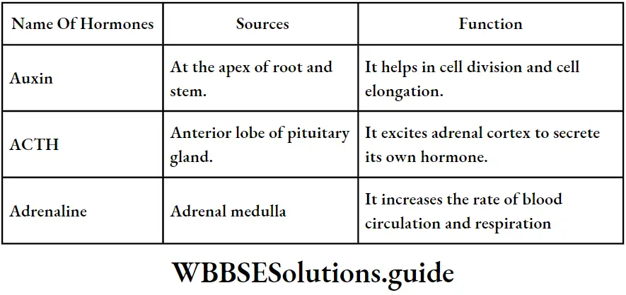
Myopia is the defect of the eye in which a person can see nearby things clearly but distant objects are blurred. It can be corrected by using a concave lens.
Question 84. Write two functions of each of ACTH, TSH and, STH in the human body. What type of lens is used to correct the following defects of the eyes: (1) Astigmatism (2) Myopia (3) Hypermetropia?
Answer.
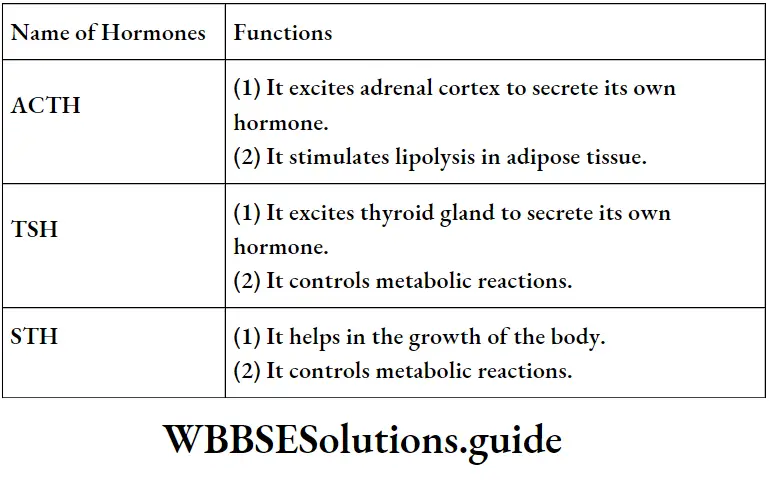
(1) Spherical lens (2) Concave lens (3) Convex lens.
Question 85. Name three animal hormones that are not secreted from the pituitary gland and mention their sources. Name some major eye defects of man.
Answer.
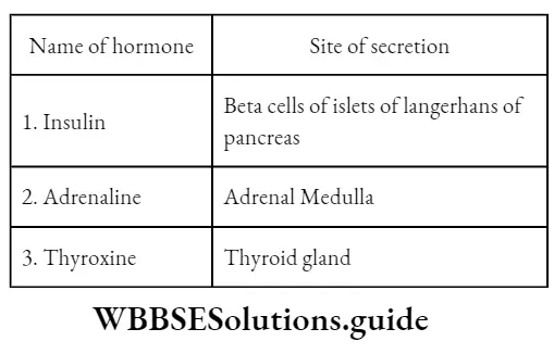
The major eye defects of man are – Myopia, Hypermetropia, Astigmatism, Glaucoma, CatarCataractsColour-blindness, etc.
Question 86. Write the full names of any three hormones secreted from the anterior pituitary gland. What types of visions are associated with rods and cones?
Answer.
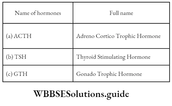
The rods are associated with night vision (dim light) and cones are associated with coloucoloron.
Question 87. Write any three characteristics of animal hoa a rmos. What is a blind spot?
Answer.
Three characteristics of animal hormone
(1) Their places of secretion (endocrine gland) and action are different
(2) They are concentrations concentration.
(3) The remaining quantity of hormone is destroyed and removed from the body by the process of excretion n.
The point on the retina having no rods and cones is called the blind spot It is insensitive to light so no image is formed here.
Question 88. What do you mean by endocrine and exocrine glands? Give examples. Mention the location of the nd function of the yellow spot.
Answer.
(1) Endocrine glands The duct the less glands of an animal body secrete the hormones directly into tbloodstreameam are known as endocrine glands. Ex.:-
Pituitary gland.
(2) Exocrine glands Glands that discharge their secretion through ducts are called exocrine glands. Ex. Salivary glands.
The yellow spot is located on the retina. It is responsible for the formation of the sharpest image.
Question 89. Mention the sites of secretion and functions of insulin and adrenaline hormones. What is ‘Fovea’ or ‘Yellow Spot’?
Answer.
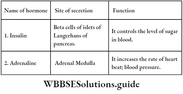
The fovea or yellow spot is the region on the retina just opposite the lens where cones are very densely packed.
“WBBSE Life Science Class 10 Chapter 1 long answers, Control and Co-ordination, study guide”
Question 90. Give names of one plant hormone and two animal hormones and mention their functions. What is the function of aqueohumour vitrehumormour?
Ans.
(1) Plant Hormone – Auxin. Function It helps in growth and controls tropic movement.
(2) Animal Hormone (1) Thyroxine. Function It controls metabolic reactions.
(2) Insulin. Function maintains blood sugar level. humorous urus and vitreous humor maintain the spherical shape of the eyeball.
Question 91. Mention the place of secretion and two functions of each of GTH, Insulin, Thyroxine, and Adrenaline the hormones in the human body. What are receptors?
Answer. Place of secretion and two functions
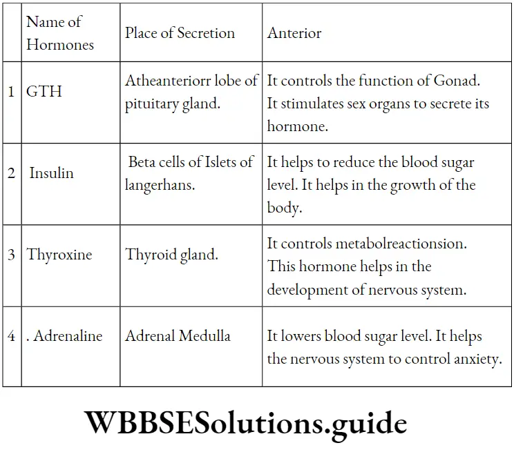
Structures at the end of sensory nerve fibers collect information from the external and internal environment of the body.
Question 92. Describe three important functions of gibberellins. Name the pigments present in rod and cone cells.
Answer:
The function of Gibberellin (1) It checks and inimizesises the dormancy period of seeds.
(2) It helps in parthenocarpy.
(3) It helps in the determination of sex.
Rod cells contain Rhodopsin and cone cells contain Iodopsin.
Question 93. What parthenocarpy? Mention two differences between Auxin and anHormone-producing more producing glands are called endocrine glands. Explain.
Answer.
Parthenocarpy
(1) Parthenocarpy The production of seedless fruit using artificial plant hormones but withfertilizingsoffthe the ovum is called parthenocarpy.
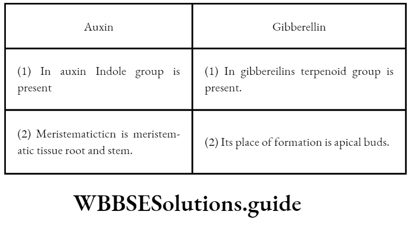
Because these ducts have no duct and in the absence of ducts they directly pour down their secretions (hormones) into the blood vessels.
Structural Organization In Animals Question And Answers
Biology Class 11 WBCHSE Structural Organisation In Animals Question And Answers
Question 1. What are animal tissues?
Answer: The cells in an animal body which are identical in structure, origin and function, organise to form animal tissues.
Question 2. What is histology?
Answer: The branch of science that deals with the structure, origin, function, variation etc., of tissues is called histology.
“structural organization in animals questions and answers for class 11”
Question 3. Name the four main tissues in an animal body. State the embryonic origin of these tissues.
Answer: The four main animal tissues are—epithelial tissue, connective tissue, muscular tissue and nervous tissue.
The embryonic origin of these tissues is—
- Epithelial tissue—ectoderm, endoderm, mesoderm
- Connective tissue—mesoderm
- Muscular tissue—mesoderm
- Nervous tissue—ectoderm.
| Class 11 Biology | Class 11 Chemistry |
| Class 11 Chemistry | Class 11 Physics |
| Class 11 Biology MCQs | Class 11 Physics MCQs |
| Class 11 Biology | Class 11 Physics Notes |
Question 4. Which tissue lacks blood vessels?
Answer: Epithelial tissue lacks blood vessels. The cells of epithelium obtain nutrition and other necessary substances through diffusion from the intercellular fluid.
Read and Learn More WBCHSE Solutions For Class 11 Biology
Question 5. What is a brush border?
Answer: In most cases, the free surface of epithelial tissue folds to form tiny finger-like structures which are known as microvilli.
“important questions on the structural organization in animals with answers”
Class 11 Biology Solutions
The epithelium that is lined by microvilli appears like a brush and so is called a brush border.
Question 6. Which chemical constituent is secreted by goblet cells?
Answer: Goblet Cell Mainly Sexreates Mucin.
Question 7. Compare the secretion process of holocrine, apocrine and merocrine glands.
Answer:
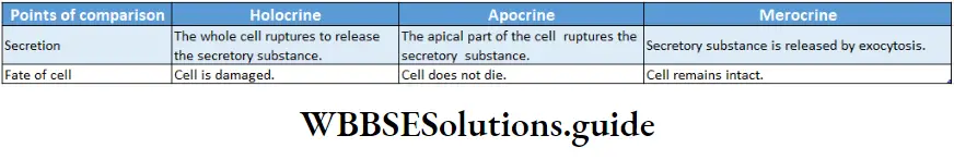
Question 8. What are the main parts of connective tissue?
Answer: The main components of connective tissue are- cells, matrix and fibres. Besides, blood vessels are associated with connective tissue. gill
Question 9. Name some immune cells of connective tissue.
Answer: Macrophages, mast cells etc., are some immune cells of connective tissue.
“structural organization in animals NEET questions and answers PDF”
Question 10. What fibres are present in areolar connective tissue?
Answer: Two types of fibres are seen in areolar connective tissue
White fibres: Collagen-containing, white, unbranched, thick fibres.
Yellow fibres: Elastin-containing, yellow, branched, thin fibres.
Question 11. Name three main cartilages.
Answer: Three main cartilages are—hyaline cartilage, fibrous cartilage and elastic cartilage.
Question 12. What are red and white muscles?
Answer: Mammals have two types of striated/skeletal muscles—red muscles and white muscles.
“MCQs on structural organization in animals with answers”
Myoglobin containing red-coloured, slow-contracting muscles are called red muscles. These muscles work slowly for a long time without getting fatigued. Example muscles of the back.
Myoglobin lacking pale-coloured, fast-contracting muscles are called white muscles. These muscles work fast for a short time. Example muscles of eye orbit.
Question 13. Why is skeletal muscle called striated?
Answer: Skeletal muscle fibres have alternate light and dark bands. So, skeletal muscle is called striated muscle.
“short answer questions on structural organization in animals”
Actually, the parts of the muscle fibres that have more actin protein appear dark. Again, the parts of the muscle fibres that have more myosin protein appear light.
Question 14. Which contractile protein fibres are present in striated muscles?
Answer: Striated muscle has two contractile protein fibres— thin fibres or actin fibres and thick fibres or myosin fibres.
Question 15. Name the neuroglial cells of the nervous system,
Answer: The neuroglial cells are—oligodendrocytes, astrocytes, microglia and ependymal cells.
Question 16. What are the divisions in each body segment of a cockroach?
Answer: Each body segment of a cockroach has four chitinous parts—tergum (dorsal), sternum (ventral), and pleurite (two lateral parts).
Question 17. What is elytra?
Answer: A pair of chitinous wings that originate from the j dorsal surface of the mesothorax is called elytra. It helps the cockroach to change direction.
Question 18. State the position of crop and gizzard.
Answer: In cockroaches, the crop is located at the posterior part of the alimentary canal and gizzard is located next to the crop.
Question 19. Name the parts of the mouth of a cockroach.
Answer: The parts of the mouth of a cockroach are- maxillae (1 pair), mandible (1 pair), labium, labrum, and hypopharynx.
Question 20. Why isn’t a respiratory pigment present in cockroaches?
Answer: Cockroaches lack any respiratory pigment. It is because atmospheric air enters the body through spiracles and is directly transported to tissues through the trachea and tracheoles. So, it does not require any pigment.
Question 21. What type of metamorphosis occurs in cockroaches? What is the name of the moulting hormone?
Answer: Metamorphosis in cockroaches is of hemimetabolous type and the moulting hormone is ecdysone.
“previous year structural organization in animals questions and answers”
Question 22. What are Ostia? Mention their function.
Answer: Ostia: Openings in the lateral surface of each cavity of the heart in cockroaches are called ostia. These are covered by valves.
Function: Valves allow the flow of haemolymph from the pericardium to the heart through the ostia but prevent its backflow.
Class 11 Biology Solutions
Question 23. What are alary muscles?
Answer: These are a kind of muscles, located on both sides of the heart of a cockroach. They help the heart to contract and relax. Cockroach possesses 12 pairs of alary muscles.
Question 24. What is the collateral gland?
Answer: The branched gland present within the fat bodies in the ovariole of female cockroaches is called the collateral gland. The secretions from this gland form ootheca.
Question 25. What is hepatic caecae?
Answer: The eight fine tubules present at the junction of the gizzard and mesenteron are called hepatic caecae.
The frontal end of these tubules is closed and the distal ends open into the alimentary canal. Digestive juices are secreted by these glands.
Biology Class 11 WBCHSE Structural Organisation In Animals Multiple Choice Questions
Question 1. Which type of tissue correctly matches its location?
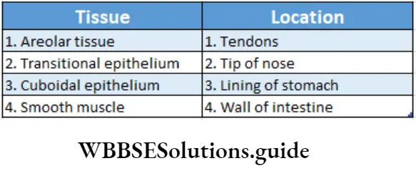
“chapter 7 class 11 biology notes “
Answer: 4.
Question 2. Which of the following features is not present in Periplaneta americana?
- Indeterminate and radial cleavage during embryonic development
- Metamerically segmented body
- Schizocoelom as body cavity
- Exoskeleton composed of N-acetylglucosamine
Answer: 1. Indeterminate and radial cleavage during embryonic development
Question 3. The body cells in cockroaches discharge their nitrogenous waste in the haemolymph mainly in the form of—
- Calcium carbonate
- Ammonia
- Potassium urate
- Urea
Answer: 3. Potassium urate
Question 4. Choose the correctly matched pair—
- The inner lining of salivary ducts—ciliated epithelium
- The moist surface of the buccal cavity—glandular
- Epithelium
- Tubular parts of nephrons—cuboidal epithelium
- The inner surface of bronchioles—squamous epithelium
Answer: 3. Tubular parts of nephrons—cuboidal epithelium
Class 11 Biology Solutions
Question 5. Choose the correctly matched pair—
- Tendon—Specialised connective tissue
- Adipose tissue—Dense connective tissue
- Areolar tissue—Loose connective tissue
- Cartilage—Loose connective tissue
Answer: 4. Cartilage—Loose connective tissue
Question 6. The inner surface of the bronchi, bronchioles and fallopian tubes are lined by—
- Cuboidal epithelium
- Columnar epithelium
- Squamous epithelium
- Ciliated epithelium
Answer: 4. Ciliated epithelium
“difference between epithelial, connective, muscular, and nervous tissue questions”
Question 7. What type of cartilaginous tissue is found in the intervertebral discs?
- Costal cartilage
- Hyaline cartilage
- White fibrous cartilage
- Yellow elastic cartilage
Answer: 3. White fibrous cartilage
Question 8. The lining of intestine and kidneys in humans is—
- Keratinised
- Brush border
- Ciliated
- None of these
Answer: 2. Brush border
Question 9. The vagina, oesophagus, and urethra contain which type of tissue?
- Stratified squamous epithelium
- Simple squamous epithelium
- Ciliated epithelium
- Columnar epithelium
Answer: 1. Stratified squamous epithelium
“earthworm, cockroach, and frog anatomy questions and answers”
Question 10. The inner lining of the ducts of sweat glands and pancreatic duct is formed by this epithelium—
- Stratified cuboidal
- Stratified non-keratinised squamous
- Transitional
- Pseudostratified
Answer: 1. Stratified cuboidal
Question 11. Dense regular connective tissue is present in—
- Ligament and tendons
- The joint capsule and Wharton’s jelly
- Periosteum and endosteum
- Pericardium and heart valves
Answer: 1. Ligament and tendons
Question 12. The structural unit of bone is—
- Chondrin
- Cyton
- Osteon
- Ossein
Answer: 3. Osteon
Question 13. Read the following statements and choose the correct answer—
- Gap junctions cement adjacent cells together.
- Areolar tissue contains fibroblasts, macrophages and mast cells.
- Tight junctions facilitate the cells to communicate with each other.
- Adhering junctions helps to stop substances from leaking across tissues.
- Cells of connective tissue except blood secrete fibres of structural proteins called elastin,
Choose the Correct Option
- 1, 2 and 3 only are incorrect
- 2, 3 and 4 only are incorrect
- 3 and 5 only are incorrect
- 1, 2 and 5 only are incorrect
- 2, 4 and 5 are incorrect
Answer: 1. 1, 2 and 3 only are incorrect
Class 11 Biology Solutions
Question 14. Which of the following is not a sensory structure in cockroaches?
- Antennae
- Eyes
- Anal cerci
- Maxillary palps
- Proventriculus
Answer: 5. Proventriculus
Question 15. Choose the wrongly matched pair regarding the position of reproductive structure in earthworms.
- Testes—10th and 11th segments
- Spermatheca—6th and 9th segments
- Male genital pore—9th segment
- Ovaries—Intersegmental septum of 12th and 13th segments
Answer: 3. Male genital pore—9th segment
Question 16. Female genital pore—14th segment In cockroaches, arthrodial membrane—
- Forms the hind wings
- Covers the compound eyes
- Forms the hypopharynx
- Forms the tegmina
- Joins the sclerites
Answer: 5. Joins the sclerites
Question 17. Which of the following animals has enucleated erythrocytes?
- Earthworm
- Sepia
- Frog
- Rat
Answer: 4. Rat
Question 18. The outer covering of cartilage is called—
- Peritoneum
- Periosteum
- Endosteum
- Perichondrium
Answer: 4. Perichondrium
Question 19. Bone marrow is absent in—
- Reptiles
- Amphibians
- Fishes
- Birds
Answer: 4. Birds
“structural organization in animals long answer questions for board exams”
Question 20. Debove’s membrane is a layer of—
- Muscular tissue
- Epithelial tissue
- Connective tissue
- All of these
Answer: 3. Connective tissue
Question 21. Cardiac muscle is found in—
- Pericardium
- Endocardium
- Myocardium
- Peritoneum
Answer: 3. Myocardium
Question 22. The H-zone in skeletal muscle fibre is due to—
- The absence of myofibrils in the central portion of a band
- The central gap between myosin filaments in the a-band
- The central gap between actin filaments extending through myosin filaments in the a-band
- Extension of myosin filaments in the central portion of the a-band
Answer: 3. The central gap between actin filaments extending through myosin filaments in the a-band
Class 11 Biology WBCHSE Structural Organisation In Animals Short Answer Type Questions
Question 1. What is a basement membrane?
Answer: The layer of collagenous fibres present between the upper epithelial layer and lower subepithelial loose connective tissue is called the basement membrane.
Question 2. What is epithelial tissue?
Answer: The animal tissue which forms the covering of the body and the inner and outer walls of some organs is called the epithelial tissue.
Question 3. What is germinal epithelium?
Answer: The epithelial tissue that helps in the formation of gametes and lines the testis and ovary is called germinal epithelium.
Question 4. What is endocardium?
Answer: The squamous epithelium in the inner wall of the heart is called endocardium.
Question 5. What are the cellular components of blood?
Answer: The cellular components of blood are red blood corpuscles (RBCs), white blood corpuscles (WBCs) and blood platelets.
Question 6. What type of tissue occurs in hairs, nails and horns?
Answer: Stratified cornified squamous epithelial tissue {occurs in hairs, nails and horns.
Question 7. Which is the hardest structure in our body?
Answer: Bone is the hardest structure in our body.
Question 8. State the function of Malphighian tubules,
Answer: Malpighian tubules are the main excretory organs of cockroaches. They collect excretory substances from body fluid and transport them to the alimentary canal.
Question 9. What is endothelium?
Answer: All tissue on the inner wall of blood vessels and lymphatic vessels is called endothelium.
Question 10. What is glandular tissue?
Answer: The epithelial tissue that takes part in secretory functions is called glandular tissue.
Question 11. What is a mixed gland? Give an example.
Answer: The glands which are constituted of both endocrine and exocrine components are called mixed glands. For example, the pancreas is a mixed gland.
Question 12. What do you mean by synapse?
Answer: The junction between the axon of one neuron and the dendron of another neuron where neurotransmitters are released is called a synapse.
Question 13. Mention the main function of epithelial tissue.
Answer: The main function of epithelial tissue is to provide protection from injuries.
Question 14. What is a bipolar neuron?
Answer: The neuron which has one dendron and one axon extending from the cyton is called a bipolar neuron.
Question 15. What is astrocyte?
Answer: Supporting cells of the peripheral nervous system that provide nutrition to a neuron are called astrocytes.
Question 16. What is myosin?
Answer: The contractile protein of myofilaments in muscles is called myosin.
Question 17. What is a sarcomere?
Answer: The contractile unit of muscles is known as sarcomere.
“structural organization in animals solved questions PDF download”
Question 18. Name one plasma protein.
Answer: y-globulin protein is a plasma protein.
Question 19. Mention the name of the contractile proteins of the muscles.
Answer: Actin and myosin are the contractile proteins in the muscles.
Question 20. Mention the function of adipose cells.
Answer: Adipose cell stores fat and provides insulation to the body, thereby preventing loss of heat.
Class 11 Biology WBCHSE
Question 21. What is a neurilemma?
Answer: The outermost covering made up of Schwann cells, that surrounds the axon is called neurilemma.
Question 22. What is an intercalated disc?
Answer: Sarcolemma of the cardiac muscle fibre is thickened at intervals to form plate or disc-like structures called intercalated discs.
They appear as dark lines that separate the individual cardiac muscle cells from one another.
Question 23. Mention the scientific name of the American cockroach.
Answer: The scientific name of the American cockroach is Periplaneta americana.
Question 24. Mention the phylum and class of cockroaches.
Answer: Cockroach belongs to the phylum Arthropoda and class Insecta.
Question 25. What role is played by the gizzard of cockroach?
Answer: Gizzard contains chitinous teeth which cut and chum the food into pieces.
Question 26. How many spiracles are found in a cockroach?
Answer: 10 pairs of spiracles are found in cockroaches.
Question 27. Name the different mouth parts of the cockroach.
Answer: The labrum, mandible, maxillae, labium and hypopharynx are the mouth parts of cockroaches.
Question 28. How many segments are found in the thorax of a cockroach?
Answer: Three segments are present in the thorax of the cockroach.
Question 29. How many segments are present in the abdomen of a cockroach?
Answer: Ten segments are present in the abdomen of cockroaches.
Question 30. Name the different digestive glands of cockroaches.
Answer: The digestive glands of cockroaches are salivary gland, glandular cells of midgut epithelium and hepatic caecae.
Question 31. What is the function of the mandible of a cockroach?
Answer: The mandible acts as the palate and helps in chewing, crushing and grinding the food.
Question 32. How many segments are there in the leg of cockroach?
Answer: The leg of the cockroach has five segments.
Question 33. What do you mean by haemocoel?
Answer: The haemolymph-filled body cavity of a cockroach is called a haemocoel.
Question 34. What is anal style?
Answer: The paired appendages articulated with a ninth abdominal segment of male cockroaches are called anal style.
Question 35. Where are the sclerites present in cockroaches?
Answer: Sclerites are present in the exoskeleton of cockroaches.
Question 36. Which mouth part of the cockroach is comparable to our tongue?
Answer: The Hypopharynx of cockroaches is comparable to the human tongue.
Question 37. What is the use of crops of cockroach?
Answer: Crop stores food for cockroaches.
Question 38. How many sinuses are found in cockroaches?
Answer: Three sinuses are present in cockroaches. They are dorsal, middle and ventral sinuses.
Question 39. A muscular fibre tapers at both ends and does not show striations. Name the muscle fibre.
Answer: Smooth or non-striated muscle fibres taper at both ends and do not show striations.
Question 40. How does a gap junction facilitate the intercellular communication?
Answer: Gap junction connects adjacent cells through the cytoplasm and thus allows cellular communication.
Question 41. Justify the name ‘pseudostratified epithelium’.
Answer: In the case of pseudostratified epithelium, all tire cells touch the basement membrane but may or may not reach the free surface.
The nuclei of these cells are arranged in different planes. As a result, they seem to form several layers, giving a pseudostratified appearance.
Structural Organization In Animals Notes
Structural Organization In Animals Animal Tissue
You see different kinds of animals around you. All these animals carry out the same basic processes to sustain life.
Although these basic processes like digestion, respiration, reproduction, etc., are carried out by common organ systems, the complexity of the systems varies from animal to animal.
This variation is due to the difference in their structural organization. The cell is the basic unit of this organization.
Several cells performing similar functions organize to form tissues, that further constitute organs.
| Class 11 Biology | Class 11 Chemistry |
| Class 11 Chemistry | Class 11 Physics |
| Class 11 Biology MCQs | Class 11 Physics MCQs |
| Class 11 Biology | Class 11 Physics Notes |
Several such organs work together to form an organ system. Altogether, these are the different levels of structural organization of the body.
In the previous chapter, you have learned about the structural organization of plants. Now in this chapter, you shall learn about the structural organization in animals.
Read and Learn More: WBCHSE Notes for Class 11 Biology
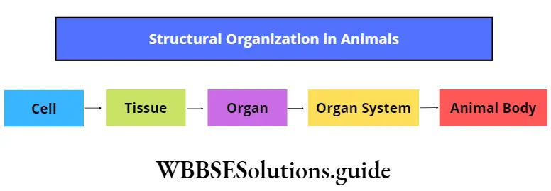
Cells of animal bodies that are similar in origin, structure, and function, are organized to form structures, known as animal tissues. Cells in the embryo are organized into three specific layers called germ layers. Each germ layer has a specific name.
“structural organization in animals notes for class 11”
The outermost layer is called the ectoderm, the middle layer is called the mesoderm, and the innermost layer is called the endoderm. All the tissues and organs of the body arise from either of these three layers.
Body parts originating from different germ layers
- From ectoderm: Epidermis of skin, nails, hair, sweat gland, sebaceous gland, scales of fish, feathers of a bird; brain and spinal cord, lens and cornea of eyes; epithelium of nasal cavity and mouth cavity; mammary gland; enamel present in teeth, etc.
- From mesoderm: Muscles; connective tissue, blood vessels, lymphatic vessels, blood cells; ducts within the excretory and reproductive system; adrenal cortex; bones, cartilages, tendons, ligaments; testes, ovary; kidney, spleen, etc.
- From endoderm: Epithelium of digestive and respiratory tract; liver, pancreas, prostate gland, thymus gland, thyroid gland, parathyroid gland; tympanic membrane of ear and epithelium of auditory canal, etc.
Types of tissues: The bodies of all vertebrates and most of the invertebrates are made of a variety of tissues.
However, all the tissues may be grouped into four main types. These are epithelial tissue, connective tissue, muscular tissue, and nervous tissue.
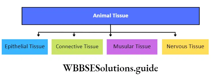
Pseudostratified Epithelial Tissue Definition: The epithelial tissue which has a single layer of cells but appears multi-layered, due to the irregular shape of the cells, is known as pseudostratified epithelial tissue.
Pseudostratified Epithelial Tissue Position: It is found in the trachea, nasal passage, epididymis, large duct of salivary gland, male urethra, etc.
“detailed notes on structural organization in animals”
Pseudostratified Epithelial Tissue Structure:
- All the cells touch the basement membrane but all do not reach the free surface. As a result, it appears that more than one layer of cells is present.
- The nuclei of the cells lie in different planes. This again makes the epithelium appear as stratified.
- Longer cells are columnar in shape with oval nuclei.
- Shorter cells have round or cone-shaped nuclei.
- These shorter cells can replace the degenerated longer cells.
- The free surface of longer cells may or may not have cilia. (For example, the inner surface of the nasal passage has ciliated pseudostratified epithelium while in salivary glands, the pseudostratified epithelium has no cilia.)
class 11th chapter 7 biology notes
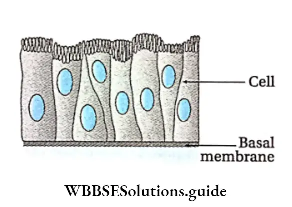
Some other types of cells belonging to epithelial tissue
Serous cells: Serous cells are present at the distal end of certain glands such as salivary glands, acinus of the pancreas, etc.
These cells contain a large spherical nucleus and secretory granules called zymogen granules.
“tissues, organs, and organ systems in animals notes”
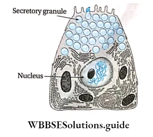
Mucus-secreting cells: The most predominant mucus-secreting cells are goblet cells that are glandular in nature.
These are present within the digestive tract, along with the simple columnar epithelial cells.
These cells are highly polarised with the nucleus and other cellular organelles are situated towards the base of the cells.
The apical portions contain secretory vesicles which secrete a glycoprotein named mucin. Mucin can transform into mucus under mechanical stress. Mucus helps in lubrication and defense as well.
“structural organisation in animals short notes “
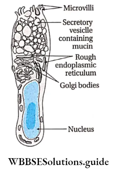
Myoepithelial cells: These cells are usually found in glandular epithelium as a thin layer above the basement membrane.
They are found in various exocrine glands such as sweat glands, mammary glands, lacrimal glands, and salivary glands. They can contract and release the secretions of the respective exocrine glands.
Function: Secretion of saliva, reabsorption of ions, and protection of visceral organs are the main functions of this tissue.
Connective Tissue
Connective Tissue Definition: The tissues that are derived from embryonic mesoderm, present throughout the body to support and bind other tissues in vertebrates are called connective tissues.
Connective Tissue Origin: Connective tissue develops from mesoderm.
“animal tissue types and their functions explained”
Maintains structure of the body: It forms a supporting framework of cartilage and bones in the body.
Coordination: It connects different organs and systems of the body.
” class 11 structural organisation in animals”
Support: It fills up spaces within the organs and tissue layers, hence providing support to the organs.
Temperature regulation: Connective tissue such as blood, helps to regulate the temperature of the body.
Storage: It also stores several nutrients such as glucose, certain proteins, salts of different ions, etc.
Protection: It protects the body from pathogens (bacteria, fungus, flu, etc.).
Structural Components
Three main components of connective tissue are—cells, intercellular medium or matrix, and fibers. Apart from these, blood vessels are also present in this tissue.
The matrix is made up of proteoglycans associated with mucopolysaccharides like chondroitin, hyaluran, etc. The three components of connective tissue are described below.
Cells: The constituent cells of connective tissue are different in terms of structure, function, and origin. Some of the connective tissue cells are described as follows.
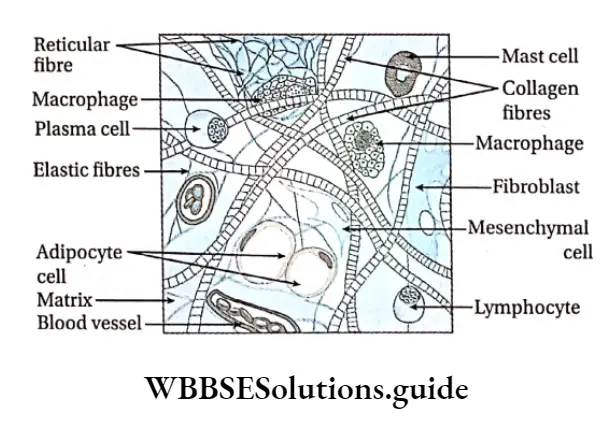
” structural organisation in animals new ncert pdf”
Fibroblast or Fibrocytc Definition: The spindle-shaped, branched cells of connective tissue that help in the formation of the matrix and fibers are called fibroblasts or fibrocytes.
Fibroblast or Fibrocytc Characteristics:
- Cells are large, flat, star-shaped, and branched,
- Cytoplasm is distinct with large and oval nuclei,
- Small granules and fat droplets are present in the cytoplasm.
Fibroblast or Fibrocytc Function: These cells mainly secrete collagen and elastin proteins. These proteins form white collagen fibers and reticular, elastic fibers respectively.
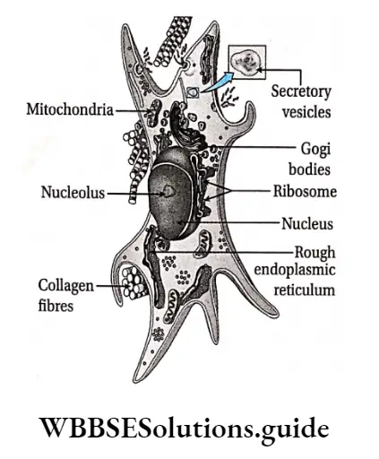
Some important facts about fibroblast tissue are called adipocytes or fat cells.
- Fibroblast cells do not undergo cell division. However, if the number of fibroblasts required for growth or regeneration is high, then these cells divide by mitosis to form more cells.
- Fibroblast cells play a major role in wound healing.
- Myofibroblast is a special type of cell that has the characteristics of both, smooth muscles and fibroblasts. It plays an active role in wound healing.
Mast cell Definition: The polygonal and calculated secretory cells of the connective tissue that secrete heparin, serotonin, histamine, etc., are called mast cells.
Mast cell Characteristics:
- Cells are large, round, or oval with dense, granular cytoplasm,
- The nucleus is small and oval in shape,
- Cells are capable of forming pseudopodia-like projections.
Functions:
- Heparin secreted by the mast cells, prevents blood coagulation within blood vessels.
- Serotonin secreted by mast cells helps in maintaining blood pressure by constriction of the blood vessels,
- Histamine secreted by these cells dilates blood vessels during allergic reactions.
Plasma cell Definition: The round or oval cells of the connective tissue that produce antibodies and contain an eccentric (away from the center) nucleus, are called plasma cells.
Plasma cell Characteristics:
Cells are oval or round with non-granular cytoplasm,
The Chromatin material of the nucleus is oriented in the form of the spokes of a cartwheel. Hence, these are also called cartwheel cells.
Plasma cell Functions: These cells synthesize 7-globulin protein (a plasma protein made up of immunoglobulins acting as antibodies) that help in defense.
Adipocyte Or Fat Cell
Adipocyte Characteristics:
- Cells are large, round, or oval with a single distinct nucleus,
- They store fat droplets within the cell cytoplasm which push the nucleus to the periphery. This gives the cell a signet ring-like appearance
Adipocyte Functions:
- The cells store food in the form of fat that can be used for energy production,
- They help in thermoregulation.
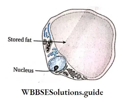
Pigment Cell Or Melanocyte Definition: The pigment-containing cells of connective tissue which determine the color of skin and hair, are known as pigment cells or melanocytes.
They are also called chromatophores.
“structural organization in animals NEET notes PDF”
Pigment Cell Or Melanocyte Characteristics:
- Cells are irregular with cytoplasmic processes,
- Cells contain yellow, brown, or black pigment granules.
Pigment Cell Or Melanocyte Functions:
- They produce melanin pigment, that imparts color to skin and hair,
- This pigment also provides protection from harmful UV rays.
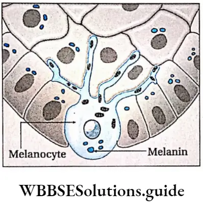
Macrophage Or Histiocyte Definition: The irregular-shaped cells of connective tissue that contain lysozyme and have phagocytic functions are known as macrophages or histiocytes.
Macrophage Or Histiocyte Characteristics:
- Cells are small, irregular in shape with short protoplasmic processes,
- The cytoplasm is granular and vacuolated with a single oval nucleus.
- Cells contain Golgi bodies and lysosomes.
Macrophage Or Histiocyte Functions:
These cells have phagocytic and pinocytic properties. They engulf foreign particles and microbes by their phagocytic action and destroy them by secreting proteolytic enzymes.
They remove dead and damaged cells from wounds etc., by digesting them
Mesenchymal cell Definition: The immature cells of the connective tissue that transform into other types of connective tissue cells like osteoblasts, myocytes, adipocytes, etc., when required, are known as mesenchymal Cells.
Mesenchymal cell Characteristics:
Cells are star-shaped with cytoplasmic processes. These cytoplasmic processes continue with the adjacent cells to form syncytium (multinucleated structure) with reticular fibrils (small fibers made up of reticulin protein).
These cells occur in embryonic connective tissue.
Mesenchymal cell Function: Mesenchymal cells do not have a specific function. However, they act as reserve cells that transform into other connective tissue cells when required.
Wandering cell or hematogenous cell: Different kinds of cells of the circulatory system such as erythrocytes, and leucocytes are found in the special fluid connective tissue. They move from one part to another with the circulatory fluid and perform various functions.
Mononuclear phagocyte system
The mononuclear phagocyte system or MPS (also called the Reticuloendothelial system or Macrophage system) is a part of the immune system.
It consists of phagocytic cells of reticular connective tissue (a type of connective tissue made up of fibers called reticulin).
The phagocytic cells primarily include monocytes and macrophages, which are important for the immune system. They accumulate in lymph nodes and spleen.
Matrix Definition: The amorphous, viscous, colorless, transparent ground substance of the connective tissue is known as a connective tissue matrix.
Components: The matrix is made up of three chemical components. These are—
- Glycosaminoglycan (acid mucopolysaccharide),
- Proteoglycan, And
- Glycoprotein.
Matrix Functions:
- The matrix gives protection and support to the connective tissue.
- It is present in the intercellular spaces and acts as a lubricant, helping in the easy movement of the cells.
- It also serves as a medium for the exchange of metabolites between the cells.
Fibre Definition: The proteinaceous, elastic, reticular components of connective tissue that provide structural support are called fibers.
“earthworm, cockroach, and frog morphology and anatomy notes”
Fibre Functions: They provide mechanical support to the connective tissue cells.
Fibre Characteristics:
- The fibers are yellow, branched, elastic, and present as single units
- Elastic fibers are made up of three types of proteins—elastin, melanin, and oxytocin. These fibers make up the elastic fiber system. Out of the three types of fibers, elastin fibers are the predominant one.
- A single fiber branches out. Several such branches connect to form a reticular or net-like structure,
- The bundles of fibers are straight and not wavy.
Functions:
- They provide elasticity to the tissue.
- These fibers are present in blood vessels and help maintain blood pressure through constriction and dilation of blood vessels.
Reticular fibre
Reticular fiber Definition: The proteinaceous, branched, non-elastic fibers of connective tissue that are arranged to form a mesh is called reticular fibers.
Reticular fiber Characteristics:
- The fibers are short, thin, and branched and form a network or mesh,
- They are composed of reticulin protein,
- The fibers are non-elastic in nature.
Reticular fiber Functions:
- They provide support to the organs and protect them from mechanical shocks,
- They also form the cytoskeleton of some structures of the body (such as the lymphatic gland),
- They hold the cells in position within the matrix.
Classification of connective tissue
Connective tissue is categorized into two types— connective tissue proper and specialized connective tissue. They are described as follows—
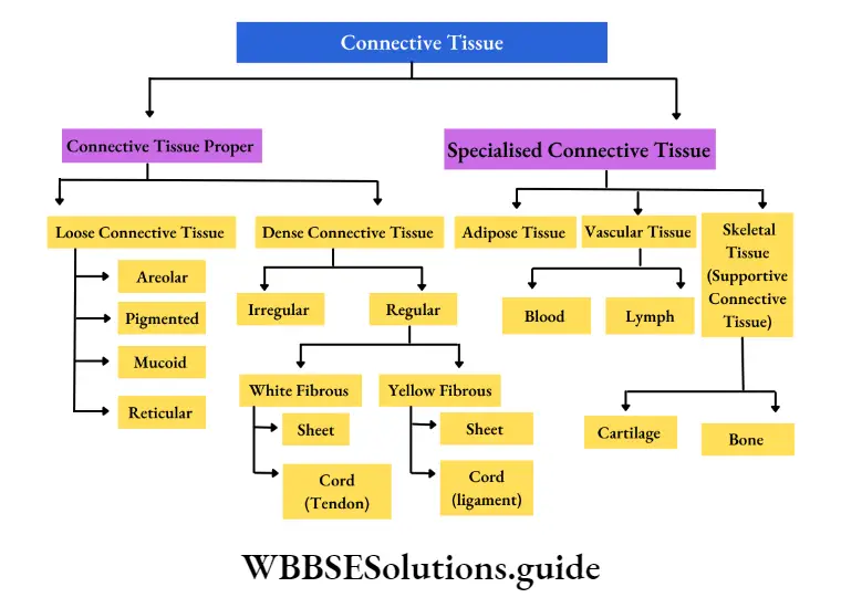
Connective Tissue Proper Definition: Connective tissue proper is a fibrous tissue consisting of cells, fibers, and intercellular ground substance or matrix which constitute the framework of many organs.
Connective tissue proper is characterized by a soft matrix.
It is of two types—
Loose connective tissue and dense connective tissue.
Loose connective tissue: The type of connective tissue that has less loosely arranged fibers and soft, gel-like ground substance or matrix is called loose connective tissue.
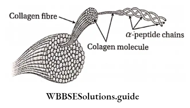
It may again be divided into the following types—
Areolar connective tissue: The white, translucent, loose connective tissue which consists of many connective tissue cells in between the meshwork of fibers in its matrix is called areolar connective tissue.
Areolar connective Position: It is the most abundant type of connective tissue and is present surrounding the nerves and blood vessels, between muscular and nervous tissues, beneath the skin.
Areolar connective Structure: It is a soft, transparent tissue consisting of numerous spaces or cavities.
“difference between epithelial, connective, muscular, and nervous tissue”
It contains different types of cells like—fibroblasts, macrophages, mast cells, plasma cells, pigment cells, and some fat cells. It contains white collagenous and yellow elastin fibers.
The spaces between the fibers are called areolae, hence called areolar connective tissue. Cells and fibers remain embedded within the matrix.
Areolar connective Functions:
- It serves as the binding material as it helps in binding various structures of the body like skin with the muscles, etc.
- It is present around the blood vessels and hence, holds them in position.
- Different types of cells present in it perform different types of functions which are already mentioned earlier.
- For example, macrophages and plasma cells, present within this tissue, play a defensive role against infection, fibroblasts play an important role in fibrosis (formation of excess fibrous connective tissue in an organ or tissue) during tissue repair, etc.
Pigmented connective tissue: The loose connective tissue which is similar to areolar tissue and consists of a large number of pigment cells called chromatophores is called pigmented connective tissue.
Pigmented connective tissue Position: It is present in the dermis of the skin, iris, and choroid of the eye, etc.
Pigmented connective tissue Structure: The cells are irregular in shape. Pigment cells are of two types—melanocytes or melanophores and lipophores.
The melanocytes are branched and synthesize melanin and lipophores synthesize lipochrome.
Pigmented connective tissue Functions:
- The pigments absorb harmful solar radiations, hence protect the skin,
- Pigments present in the choroid prevent internal reflection of light in the eye, etc.
Mucoid connective tissue: The jelly-like connective tissue that contains an excess matrix, containing hyaluronic acid, and very less fibers is called the mucoid connective tissue.
Mucoid connective tissue Position: Mucous connective tissue forms the main component of the umbilical cord. This tissue is commonly called Wharton’s jelly. It is present in the pulp of milk teeth. It is present in the vitreous humor of the eye.
Mucoid connective tissue Structure: It is composed mainly of a ground substance with few cells and fibers. Cells present are mostly large fibroblasts. A few macrophages and lymphocytes are also found. The matrix is largely made up of mucopolysaccharides (hyaluronic acid and chondroitin sulfate).
Mucoid connective tissue Functions:
- It provides insulation and protection to the umbilical cord. It also generates new cells that help to form the internal structure of the umbilical cord,
- It regulates the internal pressure of the eye.
Reticular connective tissue: The fibrous connective tissue in which fibers are anastomosing (attached) and arranged in an irregular manner forming a network, is called reticular fibrous connective tissue.
Reticular connective tissue Position: It is present in lymph nodes, liver, spleen, thymus, and bone marrow.
Reticular connective tissue Structure: This tissue comprises star-shaped reticular cells called stellate cells. The cytoplasmic processes of the stellate cells form a network. The fibers are fine, and branched, forming a network made up of protein, reticulin. The ground substance, the matrix, fills up the intercellular spaces.
Reticular connective tissue Functions: It provides structural support to organs like the liver, lymph nodes, etc. Reticular cells play a defensive role due to their phagocytic nature.
Dense connective tissue. The type of connective tissue that has a greater number of fibers and is mainly responsible for providing mechanical strength is called dense connective tissue.
It is of two types—
- Dense irregular connective tissue, containing bundles of fibers arranged irregularly.
- Dense regular connective tissue, containing bundles of fibers arranged parallel or along the same plane. Dense regular connective tissue can be of the following types
White fibrous connective tissue: The dense, regular, fibrous connective tissue, primarily made up of white collagenous fibers is known as white fibrous connective tissue.
White fibrous connective tissue Position: It is present in the tendons, ligaments, cornea of the eye, dura mater of the brain and spinal cord, etc. It is also present in the periosteum and perichondrium (of bone and cartilage respectively).
White fibrous connective tissue Structure:
- The tissue is tough and appears shiny. It is made up of more or less regular bundles of wavy white fibers.
- Fibers are made up of collagen protein.
- The bundles may be branched but the individual fibres are unbranched.
- Relatively few fibroblast cells and matrix are present in between the bundles.
White fibrous connective tissue Functions: It provides protection to various organs against mechanical stretch, pressure, and stress. The organs can withstand pressure due to the elastic properties of these fibers.
White fibrous connective tissue Types: Two types of white fibrous connective tissue are sheets and tendons. In Sheet form, the white fibrous bundles are arranged in a criss-cross manner. The fibroblasts are present in the areolae.
Sheets of white fibrous connective tissue form tough coverings. In the tendon, the bundles of white fibers run parallel to one another.
The fibroblasts are present in single rows between the bundles. Tendons connect skeletal muscles to bones.
Sheets of white fibrous connective tissue form tough coverings. In the tendon, the bundles of white fibers run parallel to one another. The fibroblasts are present in single rows between the bundles. Tendons connect skeletal muscles to bones.
Yellow fibrous connective tissue: The dense, regular, fibrous connective tissue almost entirely made up of yellow elastic fibers is known as yellow fibrous connective tissue.
Yellow fibrous connective tissue Position: It is present in the larynx, bronchi, lungs, arteries, ligaments, etc.
Yellow fibrous connective tissue Structure: The tissue is made up of a branched network of yellow elastic fibers. These fibers do not have wave-like structures. These fibers are made up of elastin protein.
Fibroblasts and matrix are present in between the elastic fibers. The yellow fibers join to form a reticular structure. The fibers are individually branched and are arranged linearly.
Yellow fibrous connective tissue Functions:
The tissue provides maximum elasticity and extensibility to the structures where it is present,
In the arteries, it helps to maintain normal circulation of blood and blood pressure due to their contraction-expansion property. In the lungs, it helps in the expansion and contraction of the lungs.
Yellow fibrous connective tissue Types: Two types of yellow fibrous connective tissue are found. They are—sheets and ligaments. Sheets are commonly found in yellow fibrous connective tissue.
“important points on structural organization in animals for exams”
These are found in various organs like walls of bronchi, bronchioles, lungs, etc. They help in undergoing stretching.
Ligaments appear as thick cords and fibroblasts are scattered. Ligaments connect bones to bones and help in the movement of the neck, fingers, etc.
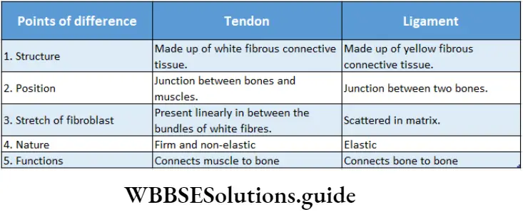
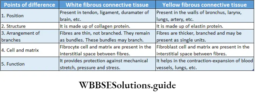
Specialized connective tissue Definition: The tissue formed by special cells of the connective tissue that have undergone a transformation to carry out specific functions is called specialized connective tissue.
Specialized connective tissue is of three types—
- Adipose tissue,
- Vascular tissue and
- Skeletal tissue
Adipose tissue Definition: The specialized connective tissue that consists of abundant fat-storing cells, on a framework of loose fibers and the excess matrix is known as adipose tissue.
Position: It is found around the heart, folds of the peritoneum, the kidney, in orbits behind eyeballs, beneath the skin, mammary glands, and in the mesenteries (the structure that attaches the body wall to the organs) around the viscera.
Adipose tissue Structure: This type of connective tissue contains distended, oval, or round, flat cells called adipocytes. Adipocytes can be monolocular when fat is present in the center as a large globule or can be polylocular when fat is present as several fat globules.
The peripheral nucleus and cytoplasm form a signet ring-like structure due to the deposition of fat bodies at the center of the cell. White fibers are present as bundles. A vasculated thick matrix is present.
Adipose tissue Functions: It protects the internal organs from mechanical injury, and maintains the proper shape of organs. It mainly stores fat and prevents the loss of heat from the body.
Adipose tissue is called a special tissue because it performs the following special functions
Stores energy: In terms of storage of energy (in the form of triglyceride), adipose tissue plays the most important role.
The muscles and liver also help in storing energy (in the form of glycogen). Triglyceride has a more calorific value than glycogen.
Adipose tissue Insulator: The adipose tissue layer beneath the skin insulates the body (because fat is a bad conductor of heat). This property helps to maintain body temperature.
Structural component: Adipose tissue is an important structural component of the body.
In a normal male, adipose tissue constitutes about 15-20% of body weight.
While in a normal adult female, this value is 20-25% of body weight.
Shock absorber: A thick layer of adipose tissue is found beneath the skin. This deposition of adipose tissue acts as a shock absorber and protects the body from physiological damage.
Formation of organ system: This tissue fills the gaps between other tissues at the time of organ formation.
Formation of organ system Types: Based on the position, structure, color, and other features, adipose tissues are of two types— Unilocular adipose tissue and multilocular adipose tissue.
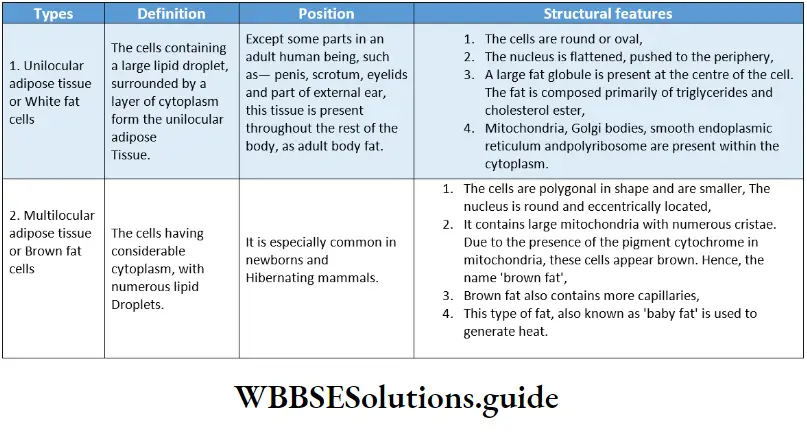
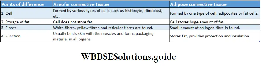
Vascular Tissue Definition: The type of mobile connective tissue that consists of a fiber-free fluid matrix and specialized living cells and is responsible for transport in the body, is called vascular connective tissue.
Vascular connective tissue is of two types—blood and lymph.
Vascular Tissue Blood: The red-colored, viscous, slightly alkaline, opaque, fluid connective tissue that flows through the heart and blood vessels is called blood.
Vascular Tissue Position: Blood is present within the vessels and chambers of the heart in vertebrates. In invertebrates, it is found in the members of phyla annelida, arthropoda, mollusca, etc.
Vascular Tissue Structure: It consists of blood cells suspended in a pale yellow fluid matrix called plasma.
The matrix is viscous and colloidal in nature and present in excess quantity.
Blood is mainly red in color due to the presence of hemoglobin protein (Haemoglobin is red when oxygenated and blue-red when deoxygenated).
On the other hand, the blood of crustaceans is blue due to the presence of a protein called hemocyanin (Haemocyanin is blue when oxygenated and colorless when deoxygenated). Insects have colorless blood since their blood lacks any pigment.
Vascular Tissue Functions:
- The blood maintains homeostasis within the body,
- It transports nutrients and oxygen throughout the body,
- It removes wastes from tissues,
- Certain cells (lymphocytes, monocytes, neutrophils, etc.) present in the blood provide immunity.
Vascular Tissue Components: Components of blood.
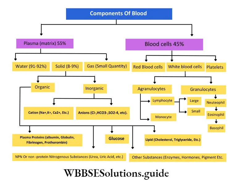
Lymph: The colorless fluid which is made up of plasma and white blood cells, flows through lymph vessels and ultimately drains into venous blood is called lymph.
Lymph Position: It is present mainly in the lymph vessels and also between the blood capillaries and tissues.
Lymph Structure: It is formed of two parts—Plasma—the matrix of lymph and is fluidity in nature.
It contains less proteins, calcium, and phosphorous but more glucose than blood. Leucocytes—derived from either lymph nodes or blood capillaries by the process of diapedesis.
Lymph Functions: Lymph carries food and oxygen from the blood to the tissues and excretory products, hormones from the cells to the blood.
Skeletal connective tissue Definition: The connective tissue in which the matrix is dense, solid, and mineralized and which forms the endoskeleton of vertebrates is called skeletal connective tissue.
Skeletal connective tissue is divided into two types—
- Cartilage and
- Bones
Skeletal connective tissue Cartilage: The firm, elastic, non-vascular skeletal connective tissue comprising cells and solid matrix is known as cartilage.
Skeletal connective tissue Position: It is found in the tip of the nose, external ear or pinna, tracheal rings, intervertebral discs, bony joints, endoskeleton of cartilaginous fish, etc.
Skeletal connective tissue Structure: The cartilage is covered by a tough, dense layer of cells and fibrils, called the perichondrium. Cartilage is mainly composed of cells called chondrocytes.
The other components of cartilage are fibers and intercellular ground substance or matrix. The cells are large, oval, or round with small cytoplasmic processes and a round or oval nucleus.
The chondrocytes are released into the matrix from the perichondrium. They are present in groups of two or four and remain embedded in the lacunae of the matrix. Each group of cells is enclosed within a capsule.
The matrix is tough, transparent, and is called chondrin. It is made up of chondromucoid (complex formed by chondroitin sulfate and collagen protein) and chondroalbuminoid (complex formed by chondroitin sulfate and albumin protein).
The matrix also contains a very fine network of elastin and collagen fibers.
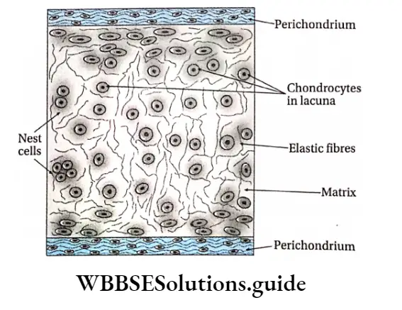
Skeletal connective tissue Functions:
- It provides mechanical support and elasticity to delicate structures of the body such as the knee, hips, etc.
- It also prevents mechanical erosion at the joints and the intervertebral discs.
- In cartilaginous fish, it forms the endoskeleton of the body.
Skeletal connective tissue Types: Based on the type of cells, matrix, and fibers, it is of four types.
These are described in the following table—
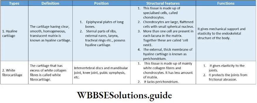
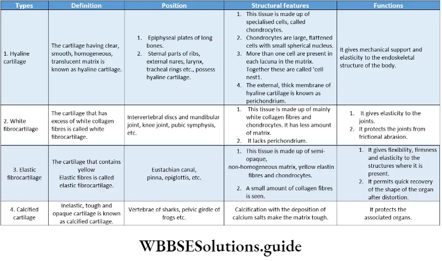
Bone: The hardest connective tissue comprising of i calcified matrix and which forms the skeleton of the body is known as bone.
Bone Position: Bones are found throughout the endoskeleton of the vertebrates.
Bone Structure: The bone is covered by a tough, fibrous sheath called the periosteum. It is composed of bone cells, collagenous fibers, and intercellular ground substance or matrix.
The cells are large, oval, or round with cytoplasmic processes. They are found in the lacunae of the matrix and have a round or oval nucleus. The matrix is made up of 40% organic and 60% inorganic substances.
The organic content of bones consists of a collagen-like protein called ossein, a mucoid derived from ossein protein called osseomucoid, and osteocollagenous fibers. Vitamins J A, D, and C are also present in the bones.
The inorganic content of the bones includes—
- Calcium salts such as calcium carbonate, calcium phosphate, calcium fluoride and
- Magnesium salts such as magnesium phosphate and a small amount of magnesium sulfate and hydroxides.
Bone Functions:
- Bones form the endoskeleton of vertebrates. They provide mechanical support to the structural framework of the body,
- They provide protection to delicate organs like the brain, heart, lungs, kidneys, etc.
- They act as a storehouse of minerals, mainly calcium and phosphorus,
- Bones are associated with muscles. The muscles act as a lever system for the movement of bones at the joints,
- They produce reticuloendothelial cells to protect the body from various diseases.
Types of bone cells: Based on their position, function, and structure, three types of bone cells are found.
Types of bones: On the basis of density and compactness, bones are two types—compact bones and spongy bones.
The histological structure of compact bone has been discussed under a separate head later.
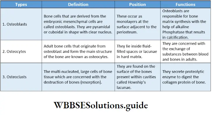
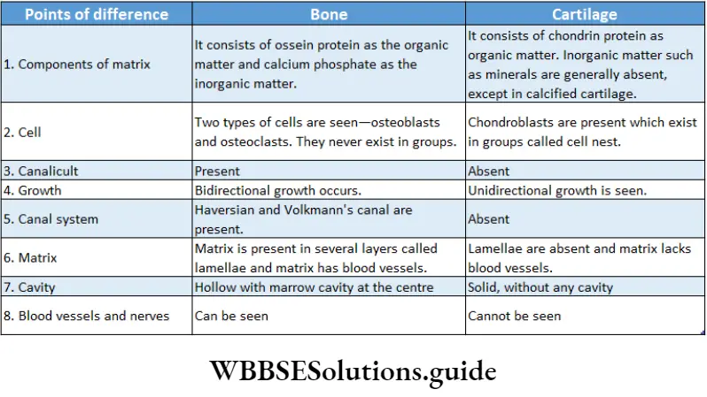
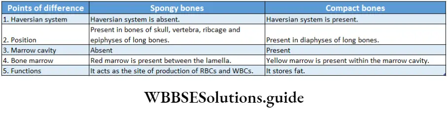
Histological structure of compact bones: Under the electron microscope, the following structures of a compact bone are seen.
Haversian system: The system formed by the numerous longitudinal canals, along with the Haversian lamellae, lacunae, canaliculi, and osteocytes, is called the Haversian system or Osteon of mammalian bones.
The haversian system is the structural and functional unit of compact bone. Its components are discussed below.
“structural organization in animals short notes for quick revision”
Haversian canal: Each Haversian system has a central canal or fine duct that runs lengthwise through the compact bone.
This canal is called the Haversian canal. It contains blood vessels, nerves, lymphatic vessels, and marrow cells.
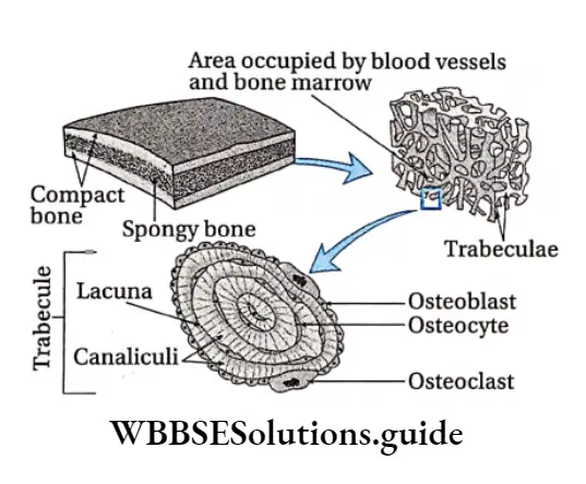
Lamellae: Each Haversian canal is surrounded by 6-18 concentric layers of the matrix. These concentric layers are called Haversian lamellae.
Lacunae: Between two consecutive lamellae as well as within the lamellae, small irregular fusiform (spindle-shaped) spaces are found, arranged in circles. These spaces are known as lacunae.
Canaliculi: Very fine wavy branched channels that radiate out from the periphery of lacunae are called canaliculi.
Osteocytes: Each lacuna has a branched bone cell or osteocyte. Its cytoplasmic processes or appendages extend through the canaliculi.
Cementing line of Ebner: The thin boundary of an osteon or Haversian system is called the cementing line of Ebner. The term was coined by Von Ebner.
Volkmann’s canals: In addition to the Haversian canal, another set of transversely (slanting) or horizontally arranged small canals are present in the periosteum.
These are called Volkmann’s canals. These canals contain blood vessels and nerves and help to interconnect the Haversian canals of adjacent Haversian systems.
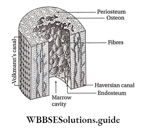
Ground or Interstitial lamellae: The lamellae found between the Haversian systems are known as the ground or interstitial lamellae.
Periosteal or Circumferential lamellae: The lamellae that are concentrically arranged below the periosteum and outside the endosteum are called periosteal or circumferential lamellae.
Endosteal lamellae: The lamellae that are present concentrically around the endosteum are known as endosteal lamellae.
Collagen
It is the most abundant protein of connective tissue. Collagen contains amino acids such as glycine, proline, hydroxyproline, and arginine.
These amino acids are arranged in different patterns, forming the triple helix.
The most common arrangements in the amino acid sequence of collagen are glycine-proline-X and glycine-X-hydroxyproline, where X is any amino acid other than glycine, proline, or hydroxyproline.
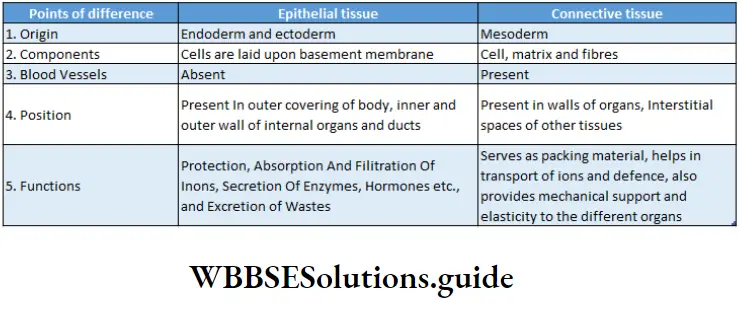
Structural Organisation In Animals Notes
- Autonomic nervous system: Part of the nervous system; responsible for the regulation of involuntary physical processes within the body.
- Collagenous: Made up of collagen protein.
- Central nervous system: Part of the nervous system, comprising the brain and the spinal cord.
- Ectodermal: Originated from ectoderm (outermost germ layer).
- Oastln: Highly elastic protein found in connective tissue.
- Elaunin: Component of elastic fibers formed from deposition of elastin protein.
- Hyaluronic acid: An anionic, non-sulfur-containing mucopolysaccharide found in connective, epithelial, and neural tissues.
- Keratin: A type of protein found in nails, hair, etc.
- Lacinia and Galea: The Apex of each stipe has two lobes—the inner one is called Lacinia and the outer one is called Galea.
- Lacunae: Fluid-filled spaces within the matrix of connective tissue.
- Microvilli: Minute finger-like projections arising from the cell membrane, that increase the surface area.
- Mucopolysaccharides: Long chains of unbranched polysaccharides.
- Mucoproteins: A glycoprotein composed mainly of mucopolysaccharides.
- Olfactory mucosa: Epithelial tissue present in the upper region of the nasal cavity.
- Oxytalan: A type of fibers found in the dermis, tooth sockets, etc.
- Periplanetin: A type of peptide (a compound containing two or more amino acids) found in insects.
- Phagocytic: Cell eating.
- Phenotype: Characteristics of an organism that can be observed externally.
Pinocytic: Cell drinking. - Podical plates: Ventrolateral plates arising from the tenth abdominal segment of an insect.
- Proteoglycans: Compounds formed by binding proteins with mucopolysaccharides, found mainly in connective tissue.
- Proteolytic enzymes: Enzymes that bring about proteolysis (breakdown of proteins)
- Protopodite: Basal segments of a biramous limb or appendage.
- Pulsatile: Having periodic vibrations or pulses
- Regenerative medicine: It is a branch of medical science, concerned with the restoration of the structure and function of damaged tissues and providing solutions for organs that have become permanently damaged.
- Reticuloendothelial: Related to non-circulating and circulating phagocytic cells (macrophages and monocytes) associated with the immune response.
- Sclerotin: A type of protein found in the cuticle or tough outer covering of the insects.
- Sclerites: Chitin or calcium-containing plates that form the exoskeleton in insects.
- Segmental arteries: Arteries present in each segment of the body of an insect.
- Sloughed off: Allowed to fall off or degenerate.
“structural organization in animals notes PDF download”
Points of Remeber
- The bodies of all higher animals are made of four types of tissue—epithelial tissue, connective tissue, muscle tissue and nervous tissue.
- Epithelial tissue forms the outermost surface of the body. lt also forms the outer covering of the internal organs.
- This tissue is of two types—simple epithelium and compound or stratified epithelium. Besides, there is another type of epithelial tissue called the pseudostratified epithelial tissue.
- Fibroblasts are large, flat, and spindle-shaped cells that are found in areolar connective tissue.
- Histiocytes are irregular, large cells that are found in all areolar connective tissue.
- These cells are phagocytic in nature and engulf the microbes, thereby protecting the body. So, they are known as macrophages.
- Tough connective tissue containing calcium-rich matrix is called bone.
- Bone is formed of three types of cells—osteoblasts, osteoclasts, and osteocytes.
- All bones are covered by an outer layer of vasculature connective tissue. This outer layer is known as periosteum.
- Each layer of bone or the region between two layers of bone is occupied by small cavities. These are called lacunae.
- Thin, wavy tubules originate from each lacunae and are called canaliculi.
- The connective tissue which is elastic and softer than bone is called cartilage.
- The structure which is inelastic, composed of white fibers, and which connects the muscles with bones, is called a tendon.
- The connective tissue which is elastic, composed of yellow fibres, and connects bone with bone, is called ligament.
- White blood corpuscles can move like Amoeba and so can come out of blood vessels. This property of white blood cells is called diapedesis.
- The membrane surrounding muscular cells is called sarcolemma. The cytoplasm of muscle cells is called sarcoplasm.
- The endoplasmic reticulum in the cytoplasm of muscle cells is called sarcoplasmic reticulum.
- Four proteins of muscle cells are—actin, myosin, tropomyosin, and troponin.
- The numerous thin fibers that are arranged parallel in the sarcoplasm are called myofibrils.
- Myofibrils are made up of smaller fibers that are visible under a microscope. These are called myofilaments.
- The main components of nervous tissue are—nerve cells or neurons and neuroglia.
- Neuroglia divide by mitosis. Dead neurons are replaced by another type of supporting cell called neuroglia.
- The part of the central nervous system where myelinated axons are seen is called the white matter. Cell bodies are not located in this region. The white matter is located in the central part of the brain and the peripheral part of the spinal cord.
- The part of the central nervous system that contains cell bodies, dendrites, and the initial segment of the axon is called the grey matter. It is located in the periphery of the brain and the central part of the spinal cord.
- The island-like structures formed by clusters of nerve cell bodies, scattered in the white matter of the central nervous system are called nuclei.
- The cell body of numerous neurons of the central nervous system forms a cluster that is covered by a membrane to form a structure outside the CNS. The structure formed is called a ganglion.
- 30 species of cockroaches are found in human habitats. Among them, 4 species have been designated as pests.
- The most abundant cockroach in the world is the German cockroach. It is followed by the American cockroach.
- The head of the cockroach is hypognathous. It means that the head remains bent ventrally.
- The head bears a pair of antennae, compound eyes, and fenestrae. The mouth is surrounded by movable mouth parts—one labrum, a pair of mandibles, a pair of maxillae, one labium, and a hypopharynx. The adductor and abductor muscles help in chewing.
- Thorax has three segments, each segment bears a pair of jointed appendages that help in locomotion.
- It also contains 1 pair of large wings and 1 pair of small wings. The large wings keep the small wings covered. The small pair of wings help in flying.
- The coelom of the cockroach is not covered by the peritoneum but is filled with a fluid called hemolymph. Hence, the coelom is called hemocoel.
- Respiration in cockroaches is carried out by hollow, branched tubules called trachea. The trachea opens outside the body with ten pairs of spiracles. Branches of the trachea are called tracheoles.
- Spiracles are covered by ring-like sclerite called peritreme.
- The colorless fluid in hemocoel that helps in circulation in cockroaches is called hemolymph.
- Haemolymph lacks respiratory pigment. The colorless granule present in hemolymph is called hemocyte. The cockroach has an open circulatory system.
- The heart of a cockroach is neurogenic. It has 13 chambers.
- Beneath the antennae, two accessory hearts are present in cockroaches that produce periodic vibrations. These are known as pulsatile hearts.
- The main excretory organ of cockroaches is the Malpighian tubule, the excretory substance is uric acid.
- The sense organs of cockroaches are compound eyes, bristles of antennae, etc.
- The endocrine glands of cockroaches are the intercerebral gland, prothoracic gland, corpora cardia, and corpora allata. These endocrine glands secrete hormones that control various physiological functions.
- A cockroach is a uricotelic animal.
- A cockroach undergoes 12 moultings to transform into a developed cockroach from a nymph.
- Cockroaches undergo incomplete metamorphosis.
Cell Cycle And Cell Division Notes
Cell Cycle And Cell Division Introduction
Cell Division Notes
All over the world, we can see various types of organisms.
All of them have originated from a single cell. Again, these single cells arise from pre-existing cells.
The process, by which new cells arise from pre-existing ones, sustain life and bring variation among organisms, is called cell division. It is of three types—amitosis, mitosis and meiosis.
A mature cell which undergoes cell division is called a mother cell or parent cell and the newly formed cells are known as daughter cells.
In the case of unicellular organisms, a single mother cell divides to form two daughter cells. Multicellular organisms, like human beings, are made up of billions of cells.
| Class 11 Biology | Class 11 Chemistry |
| Class 11 Chemistry | Class 11 Physics |
| Class 11 Biology MCQs | Class 11 Physics MCQs |
| Class 11 Biology | Class 11 Physics Notes |
Cell cycle and cell division notes for NEET PDF
Such a large number of cells originate from a single-celled zygote which undergoes repeated divisions to develop into an embryo. The embryo divides multiple times to develop into a complete organism.
The cell division continues in a newly formed daughter cell till it reaches its mature form, in a cyclic and controlled manner. This is called the cell cycle. It has two phases—interphase and M phase.
But, after a certain period of time, a ‘suicide’ programme is activated within the cells. It leads to cessation of cell division, fragmentation of the DNA, shrinkage of cytoplasm, membrane change and cell death without lysis or damage to neighbouring cells. This is known as apoptosis or programmed cell death.
Hence, the birth of young ones, their complete development and eventual death, all depend on the processes of cell cycle and cell division.
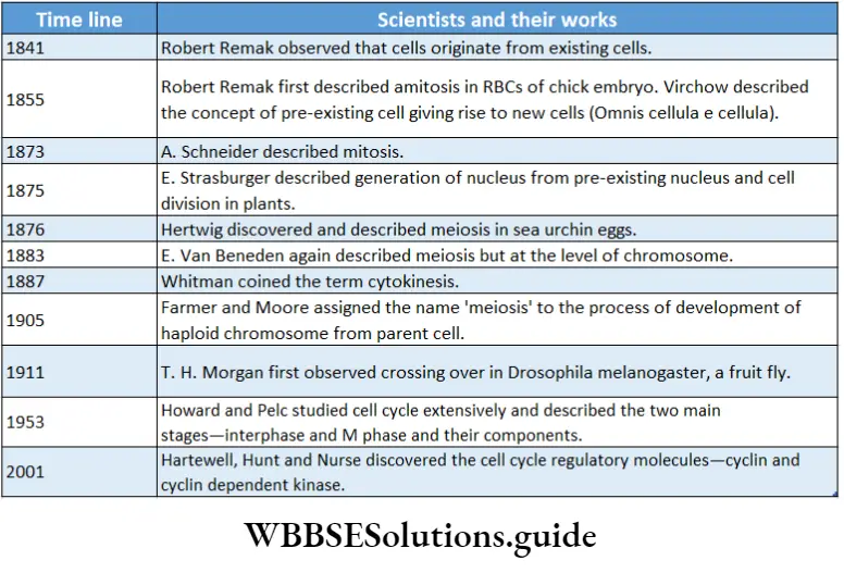
Cell Cycle
Cell Cycle Definition: The orderly sequence of events by which a cell duplicates its DNA, synthesises its other components and produces daughter cells is known as the cell cycle.
In an organism, cell division takes place all the time in different parts of the body.
Class 11 biology cell cycle and cell division notes with diagrams
A daughter cell, after division, grows, develops and divides again by passing through a series of stages collectively known as the cell cycle. The time period between two successive divisions is known as the generation time.
Phases Of Cell Cycle
There are two phases of the cell cycle in a proliferating somatic cell—interphase and mitotic phase.
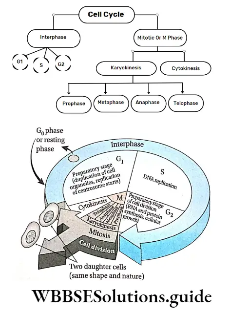
Interphase
Interphase Definition: Interphase is a metabolically active stage between two mitotic divisions during which the cell prepares itself for another division.
Previously, the interphase stage was considered as a resting phase of the cell cycle.
But now, it has been regarded as the metabolically most active phase of the cell cycle. It is involved in different types of synthetic activities like synthesis of DNA, RNA, protein, etc.
The word ‘interphase’ is derived from two different words— Inter is a Latin word, that means ‘between’ and phasis is a Greek word, that means ‘state’. Interphase is also known as ‘enter mitosis’.
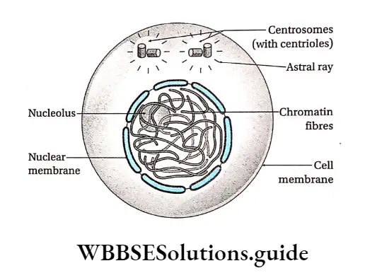
Characteristics:
- It is the longest phase of the cell cycle. It lasts for approximately 22-23 hours (about 75-95% of the total span of a cell cycle).
- This stage exists between two successive cell divisions.
- In this phase, DNA replication takes place. RNA, histones and other nuclear proteins are also synthesised.
- Centrosome duplication to form two centrosomes in animal cells is seen. Each centrosome contains two centrioles. Hence, this phase is also known as the preparatory phase.
- The size and volume of the nucleus increase. Chromosomes undergo a condensation-decondensation cycle during this period. They cannot be seen under a light microscope at this stage.
- Cells spend most of their time in this intermediate non-mitotic state.
- During interphase (in the S phase), 4 copies of each gene form instead of the normal 2 copies in a diploid cell.
- Energy-rich ATP is synthesised and stored in cells. So, this phase is also known as the energy phase.
Phases of interphase: it is divided into three phases, G1-S-G2. The G1 and G2 phases stand for ‘gap phase 1’ and ‘gap phase 2’ respectively. The S phase stands for the ‘synthesis phase’ and in this phase, DNA replication occurs.
Short notes on cell cycle and cell division for quick revision
Within the G0 phase, sometimes, the cell stops dividing and enters into a sleeping stage or quiescent stage of the cycle called the G0 phase.
Different phases of the interphase have been discussed under separate heads.
G1 phase: The first gap phase of interphase, i.e., the phase between the M phase of the previous cell cycle and the S phase of the new cell cycle which is metabolically active, is called the G1 phase (growth phase I).
Characteristics:
- It is usually the longest period of the cell cycle. It lasts for about 11-12 hours (45-50%) of the total cycle.
- In some embryonic cells that are rapidly dividing, G1 may last only for a few minutes.
- It is a metabolically active phase.
- In this phase, carbohydrates, lipids, and all types of proteins and enzymes (DNA polymerase, RNA polymerase), that are required for DNA replication in the S phase, are synthesised.
- In this phase, three types of RNAs and structural proteins are also synthesised, but not DNA.
- Each chromosome is elongated, and thin and contains one DNA molecule. Therefore, the chromosome is monad.
- Different cellular organelles—mitochondria, Golgi bodies, plastids, ER, centrioles and ribosomes, increase in number. In this phase, ATP is stored in cells which is essential for the proper progression of cell division. So, the G1 phase is known as anaphase.
- Some cells, like nerve cells, never leave G2 and remain arrested in a stage called the G0 phase.
Monad and dyad
A chromosome with one chromatid is known as a monad. Chromosomes with two chromatids are known as dyads.
Cdk (Cyclin-dependent kinase)
The fate of a cell is determined by the regulatory enzyme; cyclin-dependent kinase, present at checkpoints of a cell cycle.
S phase: The phase of interphase that lies between two gap phases and is responsible for DNA replication and synthesis of histone proteins is called the S phase or synthesis phase.
S phase Characteristics:
It is the second phase of the interphase (next to the G1 phase).
Generally, the duration of the S phase is 7-8 hours. It occupies 35-40% of the total cell cycle. However, this duration varies in different stages stands for the ‘synthesis phase’ and in this phase, DNA replication occurs.
Within the G2 phase, sometimes, the cell stops dividing and enters into a sleeping stage or quiescent stage of the cycle called the G0 phase. Different phases of the interphase have been discussed under separate heads.
G2 phase: The first gap phase of interphase, i.e., the phase between the M phase of the previous cell cycle and the S phase of the new cell cycle which is metabolically active, is called the G2 phase (growth phase I).
G2 phase Characteristics:
It is usually the longest period of the cell cycle. It lasts for about 11-12 hours (45-50%) of the total cycle.
In some embryonic cells that are rapidly dividing, G1 may last only for a few minutes.
It is a metabolically active phase. In this phase, carbohydrates, lipids, all types of proteins and enzymes (DNA polymerase, RNA polymerase), that are required for DNA replication in the S phase, are synthesised.
In this phase, three types of RNAs and structural proteins are also synthesised, but not DNA.
Each chromosome is elongated, and thin and contains one DNA molecule. Therefore, the chromosome is monad.
Different cellular organelles—
Mitochondria, Golgi bodies, plastids, ER, centrioles and ribosomes, increase in number. In this phase, ATP is stored in cells which is essential for proper progression of cell division. So, the G1 phase is known as antiphase.
Some cells, like nerve cells, never leave G1 and remain arrested in a stage called the G1 phase.
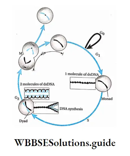
M Phase Or Mitotic Phase
M Phase Or Mitotic Phase Definition: The shortest phase of a cell cycle which occurs after the completion of interphase and is involved in cell division is called the M phase.
M Phase Or Mitotic Phase Characteristics:
- It is the dividing phase of the cycle.
- Cell division occurs either by mitosis or meiosis.
- During this phase, the components of the cell that are synthesised in interphase, (chromosome, DNA, cellular organelles, cytoplasm, etc.), are distributed among the daughter cells.
- It occupies 5-12% of the total cell cycle, i.e., less than 1 hour or 1 hour.
- The phase includes the breakdown of the nuclear membrane, the condensation of chromosomes, their attachment to the mitotic spindle and the segregation of chromosomes to the two poles.
- It occurs in two phases—nuclear division or karyokinesis followed by cytoplasmic division or cytokinesis.
G0 phase (Quiescent phase)
- In some cells, at a certain point of the G2 phase, the cell cycle stops, i.e., the cell enters an inactive phase. This phase is known as the G0 phase.
- Mainly, a cell enters the G0 phase due to the absence of cell cycle-controlling factors (e.g. nitrogenous and energy-rich compounds).
- However, the cell is found to be metabolically active and viable but is not proliferative in this phase.
- Cells do not divide at this phase but act as reserve cells. Only under favourable conditions, do these cells re-enter the cell cycle and start division.
- Most cells do not re-enter the cell cycle. They grow and differentiate to play their roles in an organism’s body. For example, nerve cells remain in the G0 phase permanently.
- Mature brain cells become arrested in G0 and do not normally divide again during a person’s lifetime.
- Some cells de-differentiate to re-enter the cell cycle from the GQ phase and thus resume division. Examples are the parenchyma cells of plants and fibroblasts of animals.
Importance Of Cell Cycle
The importance of the cell cycle is as follows—
- Due to the cell cycle, amounts of DNA, RNA and protein increase in the cell.
- However, after the generation of daughter cells, these molecules are distributed equally.
- The number of cellular organelles increases as a result of the cell cycle.
- Daughter cells are produced due to the cell cycle and they are supplied with the required components.
Control Of Cell Cycle
The transformation of cells from one phase of the cell cycle to another is controlled by some external factors.
These factors control the cell cycle at certain points. In most cases, the first factor is present in the G1 phase.
A number of regulatory systems of the cell which monitor the progress in the cell cycle and can inhibit subsequent stages in the event of failure to maintain normalcy are called cell cycle checkpoints.
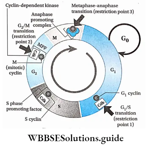
Cell cycle checkpoints
There are three main checkpoints in a cell cycle. They are as follows—
G1-S checkpoint: This is the major checkpoint that determines the transition of the cell from the G1 to the S phase. If the DNA of a cell gets damaged or the cell has not attained proper volume, then the cell is arrested at the G0 phase.
The cell is not allowed to proceed to the S phase. Rather, the cell is directed to move to the G0 phase.
In yeasts, this checkpoint is known as START and in animal cells, it is known as restriction point or commitment point.
G2-M checkpoint: This checkpoint occurs at the G2 phase and it regulates the entry into the M-phase.
Unless all DNAs have been replicated in the S phase and the cell has attained proper volume, the cell is not allowed to enter the mitotic phase of the cell cycle from the G2 phase.
M checkpoint: This checkpoint occurs during the M-phase. Spindle fibres are formed in the mitotic phase during metaphase.
The chromosomes should align properly at the mitotic plate in order to trigger the separation of chromatids. This checkpoint stops the cell from undergoing mitosis unless these conditions are fulfilled.
Chemical regulation of checkpoints
The cell cycle is regulated chemically by a variety of regulatory proteins, namely cyclin and cyclin-dependent kinases (Cdks). They are the key components of the cell cycle and associate with one another.
Cyclins are proteins that get their name from their cyclically fluctuating concentration in the cell.
These undergo structural modification in different phases of the cell cycle.
Cell cycle and cell division chapter summary with important points
Animal cells have four types of cyclins—cyclin A, cyclin B, cyclin D and cyclin E. Cdk is present in all phases of the cell cycle but as an inactive protein kinase.
The cyclin-dependent kinases associate with cyclin to form active Cdk-cyclin complexes.
On the basis of the mode of activities in specific events in animal cells, cyclin–
CDK complexes are classified into three types-
Gl/S-cyclin-Cdk complex: Gi/S-cyclin-Cdk complex has two structural components—cyclin D-Cdk 4/6 and cyclin E-Cdk 2. The transition from Gx to S is promoted by Cdk.
CdK becomes active in the presence of G2 cyclin and ATP which causes the transition of Gx to the S phase
Once the Gj cydins activate the Cdks, the levels of cyclins degrade due to proteolysis.
S-cyclin-Cdk complex: The component of this complex is cyclin A-Cdk 2. During the S phase, cyclin A is synthesised and associates with Cdk to form the S-cyclin-cdk complex. This complex controls DNA replication.
G2/M-cyclin-Cdk complex (MPF): The component of this complex is cyclin B-Cdk1 which binds with ATP to become active. The complex so formed is also known as the M phase promoting factor or the Maturation promoting factor.
This factor was first discovered in mature unfertilised eggs of frogs. MPF activation stimulates the G2-M transition. It controls the supercoiling of chromosomes, contraction of the nuclear membrane, arrangement of spindle fibres, etc., at the M phase.
At the end of the M phase, proteolytic degradation of cyclin B causes the termination of MPF activity. Cyclin B is marked for destruction by the Anaphase Promoting Complex (APC).
APC is the ubiquitin-protein ligase that targets cyclin B and destroys it to enable the transition of a cell from metaphase into anaphase.
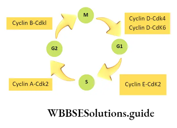
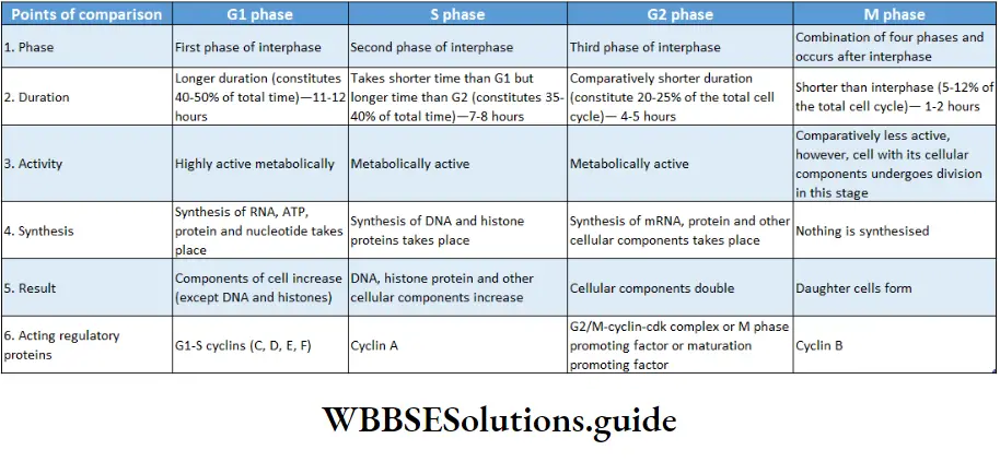
Uncontrolled cell cycle and cell division in animal cells If, for any reason, cell cycle checkpoints do not function, cell division becomes uncontrolled.
The mass of a cell that develops due to unregulated cell division, is known as a tumour.
In the animal body, when the tumour cells spread to other parts of the body from their origin, via blood vessels and form new tumours, then it is known as malignant or cancerous.
Malignant cells have uncontrolled growth and show genetic and cellular changes. These changes enable the cancer cells to spread to distant locations from their original site.
This phenomenon or characteristic is called metastasis. The tumour which remains at the original site is known as benign. Most benign tumours do not cause serious problems.
Different types of uncontrolled cell proliferation
The proliferative growth of cells occurs due to various physiological factors and the presence or absence of effectors.
Some examples of proliferative cell growth are—
Hyperplasia: Overgrowth of a particular tissue or organ due to an increase in the rate of cell division of that region is known as hyperplasia. It happens due to increased hormone secretion that leads to an increase in metabolic activity of the cells of that tissue or organ.
Example development of breasts in females during pregnancy, and the thickening of endometrium in aged women.
Hypertrophy: It causes an increase in the size of an organ due to the enlargement of cells. This may occur due to any infection or increase in the metabolic activity of cells.
For example swelling of striated muscles during heavy exercise, and swelling of the endothelium of the alimentary canal due to infection by worms.
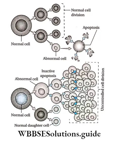
Metaplasia: It causes the transformation of mature cells into abnormal ones due to infection or injury.
For example during lung infection and in the case of smokers, the columnar epithelial cells of the bronchus transform into squamous epithelial cells.
In certain precancerous conditions, the normal existing epithelium is replaced by an epithelium from a nearby tissue by the process of metaplasia.
For example in Barrett’s oesophagus or Barrett’s oesophagitis, the normal squamous cells of the oesophageal lining are replaced by secretory cells that migrate from the stomach lining (metastatic Barrett’s epithelium).
Dysplasia: The production of abnormal tissue or cells, due to uncontrolled and abnormal cell division, that displays a transitional state between benign and pre-malignant growth is known as dysplasia.
Neoplasia: The uncontrolled, abnormal growth of cells in the body is known as neoplasia (neo meaning ‘new’). It occurs due to the presence of any effector.
Neoplastic growth may be of two types—benign or malignant. Benign tumours are confined or restricted to a particular site and are not harmful whereas malignant tumours are harmful and undergo metastasis.
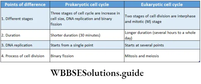
Chromosomes
- The thread-like, self-replicating structures of the nucleus, which is made up of nucleoprotein and which transmit hereditary properties generation after generation through cell division, are known as chromosomes.
- During cell division, chromosomes are formed from chromatin fibres of the nucleus. Chromosomes were first observed by W. Hofmeister in 1848. However, it was named ‘chromatin1 by W.
- Flemming in 1879. Later, in 1888, W. Waldeyer coined the term ‘chromosome’, Generally chromosomes are thread or stalk-like structures. During the anaphase stage of cell division, they appear like V, ‘L’, ‘J’ or T.
- They are 0.5-30 /rm in length and 0.2-3.0 /im in diameter. Their structure becomes clearly visible at metaphase.
Chromosomes occur in homologous pairs in the body cells—
- One member of each pair is derived from the female parent (mother) and the other from the male parent (father).
- Gametes usually contain only one set of chromosomes. This number is called ‘haploid’ (n).
- The haploid set of chromosomes is known as the genome, while the number of chromosomes in somatic cells is ‘2n’. Hence, they are diploid.
Cell Division
Cell Division Definition: The fundamental and active biological process by which a parent cell, after replication of its components, divides to produce its daughter cells is called cell division.
Cell division is essential for growth, development and regeneration.
Types: There are three types of cell division—
- Amitosis,
- Mitosis and
- Meiosis.
Amitosis is the direct division which involves simple cleavage of the nucleus into two daughter nuclei.
The cytoplasm constricts and two daughter cells, each with a nucleus, are produced. Mitosis is essentially a duplication process.
It produces two genetically identical daughter (progeny) cells from a single parent (dividing) cell. Meiosis, on the other hand, is quite different.
It recombines the chromosomes to generate daughter cells that are distinct from one another and the original parent cell as well. Meiosis produces male and female gametes that undergo fertilisation to give rise to offspring.
So, basically, mitosis is for growth and maintenance, while meiosis is for sexual reproduction.
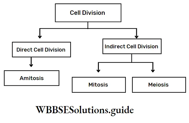
Conditions: Scientists say that normal cell division depends on certain conditions.
These are as follows—
Minimum growth: A newly formed cell does not undergo division immediately. It must attain a minimum level of maturity before it gains the ability to divide.
Karyoplasmic index: The ratio between the volume of the cell cytoplasm and the nucleus is known as the karyoplasmic index. Declination of this ratio leads to cell division.
Amount of nuclear material: When the DNA content of the nucleus doubles, then cell division takes place.
Cell volume and metabolism: With the increase of cell content due to a rise in metabolic activity, the volume of the cell increases without an increase in surface area. This causes cell division.
Mitogen: Some chemical factors which induce cell division are called mitogens. Examples are cytokines in plants, epidermal growth factor (EGF), platelet-derived growth factor (PDGF), etc.
Reproduction: In the case of unicellular organisms, cell division is an inevitable occurrence, as it is the means of reproduction. This is the only way by which they continue their existence.
Importance: The importance of cell division is—
Increase in number: Due to cell division, the number of cells increases which is essential for the development of the body.
Repair and healing: In multicellular organisms, cells continuously undergo wear and tear. These damaged cells are continuously replaced by new cells.
Reproduction: Unicellular and primitive multicellular organisms multiply and reproduce by cell division. Cell division produces gametes which help in reproduction in higher organisms.
Transmission of hereditary characters: Due to cell division, daughter cells acquire characteristics of the parent cell and also develop some new characteristics.
Variations in the offspring allow them to adapt to changing environments. Variation often leads to the evolution of new species.
Constant chromosome number of species: in sexually reproducing organisms, cell division occurs during gamete formation to maintain a constant chromosome number of a species.
Amitosis
Amitosis Definition: The simplest type of cell division in which two daughter cells are formed by simultaneous and direct division of both nucleus and cytoplasm by forming a constriction in the cell, without the formation of spindle fibre is known as amitosis (or direct cell division).
The amitotic type of cell division was first described by German biologist Robert Remak (1841). But, the term was proposed by histologist W. Flemming (1882).
The word is derived from two Greek words amitos and osis which mean ‘without thread’ and ‘state’ respectively.
Amitosis Characteristics:
- It is the simplest type of cell division in which the nucleus divides by constriction.
- Nuclear and cytoplasmic cleavage occurs without spindle formation.
- Maximal condensation of chromatin into chromosomes does not occur and thus chromosomes are not visible.
- The nuclear membrane does not disappear at all.
Site of occurrence: Amitosis is observed in bacterial cells, protozoans, mammalian cartilage cells, and foetal membrane cells.
Amitosis takes place in those cells which do not require equal distribution of cellular substances of parent cell to daughter cells, such as the formation of the large nucleus in Paramoecium, and the division of internodal cells in Chara.
Drawbacks Of Amitosis
Due to amitosis, chromatin material is unequally divided among daughter cells which causes structural and functional abnormalities in daughter cells.
In amitosis, there is no possibility of genetic recombination and there is a possibility of expression of unwanted recessive lethal genes.
Amitosis Process:
- The nuclear membrane remains intact.
- The nucleus increases in length and two constrictions appear at the centre. As a result, the nucleus appears like a dumbbell.
- The constrictions of the nucleus gradually grow deeper and meet each other. Then the nucleus ultimately divides into two nuclei without the formation of spindle fibre.
- Cytoplasmic division occurs along with nuclear division.
- The cell membrane shrinks and invaginates towards the centre of the cell and forms a constriction between the two nuclei. Ultimately, the cell divides to form two daughter cells.
Amitosis Importance:
- It is a simple type of cell division without any complexity.
- It completes within a short duration and cell number increases rapidly in this process.
- Lower organisms like yeast, bacteria and protozoa reproduce mainly by this process.
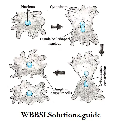
Mitosis
Mitosis Definition: The indirect process of cell division by which the somatic parent cell divides once to produce two daughter cells, which are identical in shape and contain an equal number of chromosomes as the parent cell is called mitosis.
Walther Flemming (1879) first described and coined the term mitosis in 1882 in Salamander. Schneider (1879) also described the various stages of mitosis.
Mitosis Short Notes
The biochemical aspects of this process were explained by Cockraum and MacCanley (1960). The word ‘Mitosis’ is derived from two Greek words—mitos which means ‘thread’ and osis meaning ‘state’.
It is a type of equational division, that produces two identical daughter cells having the same genetic constituent (same chromosome number) as the parent cell.
Mitosis Characteristics:
The nucleus with chromosomes and cytoplasm of the cell divides only once.
The nucleus, cytoplasm, organelles and cell membrane of the parent cell are distributed among two daughter cells, containing roughly equal shares of these cellular components. Hence, it is called an equational division.
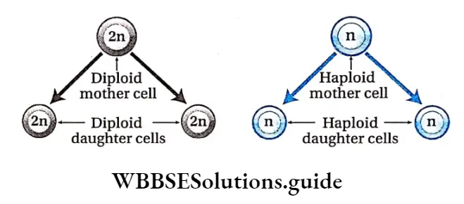
Site of occurrence:
- Mitosis occurs in almost all eukaryotic somatic cells.
- In some single-celled organisms, mitosis forms the basis of asexual reproduction.
- In advanced multicellular organisms, mitosis occurs during the formation of the embryo from the zygote, the development of a complete individual from an embryo and the subsequent growth and development of the organism.
- In some special parts of advanced organisms, ‘mitosis occurs throughout life. For example, mitosis occurs in meristematic tissue (plants) found in root tips, shoot tips, buds and leaf primordia. In the case of animals, this is found in cells of skin and bone marrow.
- Gamete mother cells also undergo mitotic cell division to increase their number in gonads.
- It occurs in the damaged organs of plant and animal bodies during wound healing.
Duration of mitosis: Duration of mitosis varies in different species.
In actively dividing animal cells, the whole process takes about one hour.
But generally, it takes 30 minutes to 3 hours. The time span of different phases of mitosis actually depends on different external and internal conditions of the dividing cell and it is very much tissue-specific.
Process Of Mitosis
Though mitosis occurs in several tissues of plants and animals, it is best to study the phases of mitosis from the stained squash of the root tip. Acetocarmine is commonly used for staining chromosomes of root tip cells.
The process of mitosis occurs in two stages—karyokinesis and cytokinesis.
Karyokinesis
Karyokinesis Definition: The division of the nucleus at the time of cell division is called karyokinesis.
It is a process of indirect nuclear division. The nucleus passes through a sequence of events and forms two daughter nuclei from a single division.
Different phases: Karyokinesis is conventionally divided into four phases—prophase, metaphase, anaphase and telophase. These have been discussed below under separate heads.
Prophase
Prophase Definition: The first and the longest-running stage of karyokinesis where chromatin condenses to transform into chromosomes that eventually unwind to form two chromatids with the dissolution of the nucleolus and nuclear membrane, is called prophase.
Prophase Characteristics: Prophase is divided into three subphases.
The characteristics of each subphase are—
Early prophase:
The nucleus is diffused, and granular in appearance.
In the interphase, the refractive index of the chromatin fibre is almost the same as that of the nucleoplasm. Hence, the chromatin is not visible.
As the nucleus enters into prophase, a refractive index of chromatin changes from that of nucleoplasm due to dehydration of the nucleus. As a result, chromatin fibres become visible.
Refractivity and viscosity of cytoplasm also increase.
Chromatin begins to coil and condense to form thin, thread-like chromosomes. The sister chromatids which were intertwined during the G2 phase become untangled during chromatin condensation.
Condensation occurs due to the coming together of scaffolding proteins and folding of individual chromatin through spiralisation.
Due to spiralisation, chromosomes appear short, thick and stacked on each other like woollen balls. This stage is known as the supreme stage.
In animal cells and in the cells of some lower organisms, each of the centrosomes gives rise to two daughter centrioles by replication.
They associate with a newly formed centrosome followed by the shifting of a pair of centrioles to one pole and the other pair to the opposite pole.
In animal cells, each centrosome produces fine, thin microtubular fibrils forming a star-shaped body called aster.
The microtubular fibrils radiating out of the surface of the aster are known as astral rays.
Middle prophase:
- The ends of the chromosomes become more visible. They undergo further thickening and shortening.
- Chromosomes are seen to be composed of two chromatids attached by centromere.
- The pair of centrioles move further to opposite ends of the cell.
- Astral rays increase in length.
- At this stage, the number of chromosomes (containing a pair of chromatids) is considered to be equal to the number of centromeres.
- The nucleolus remains associated with a specific secondary constriction of the chromosome.
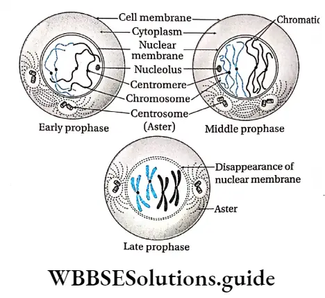
Late prophase:
- The nuclear envelope disintegrates into small vesicles as the centrosomes continue to move towards opposite poles. Nucleolus disappears.
- Chromosomes become thicker as a result of further condensation and coiling.
- Two centrosomes occupy two poles with radiating microtubules.
- Mitotic spindle begins to form from centrosomes by forming spindle microtubular fibrils.
Prometaphase
- The transitional stage between the end of prophase and the beginning of metaphase is called prometaphase.
- In this shortest phase, the nuclear membrane disappears completely, thus cytoplasm and nucleoplasm merge. In plant cells, centrosomes are absent.
- So mitotic spindle is developed from the cytoplasmic and nuclear sap and is known as the anastral spindle.
- In animal cells, the poles of the spindle are formed by two asters at two poles. So this type of spindle is called an amphiastral spindle.
Metaphase
Definition: The second, short durational stage of karyokinesis in which chromosomes with two clearly visible, chromatids are arranged along the equator of the metaphase plate is called metaphase.
Metaphase Characteristics:
- The two chromosomal fibres attached to each chromosome and connected to opposite poles start to contract.
- Thus, the chromosomes are arranged at the equator, on a specific plane of the spindle fibres. It is known as the metaphase plate.
- Shorter chromosomes are arranged at the interior while the longer chromosomes are arranged at the periphery of the metaphase plate. This arrangement of chromosomes at the metaphase plate is called congression.
- Spindle fibres bind themselves to the chromosomes through the kinetochore at the metaphase plate.
- The centromere remains connected with spindles while the arms of chromosomes remain suspended.
- The chromatids of the chromosomes are clearly visible.
- At the end of metaphase, the centromere of a chromosome splits and the two sister chromatids separate.
- Metaphase chromosomes can be stained and they show distinctive.
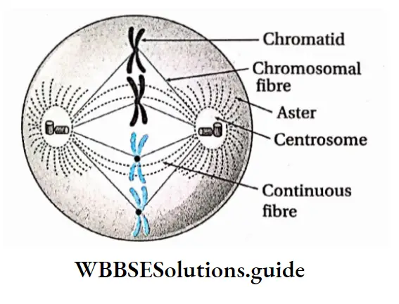
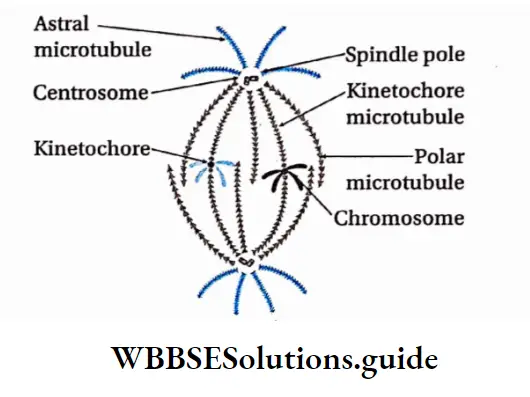
Anaphase
Anaphase Definition: The phase in karyokinesis in which chromosomes with single chromatids separate and move from the metaphase plate to the respective poles is called anaphase.
Characteristics:
- Centromeres divide separating the pair of chromatids.
- Each sister chromatid with one centromere is called a monad chromosome. At this stage, the number of chromosomes becomes double that of the parent cell.
- Each daughter chromosome binds with the spindle fibres through the kinetochore, located in the centromere.
- Before the onset of anaphasic movement of the monad chromosomes, a third type of fibre appears between the two separating centromeres known as interzonal fibres.
- Due to chromosomal repulsion, contraction of chromosomal fibres and expansion of interzonal fibres between daughter chromosomes, they move to opposite poles in equal halves. This movement of chromosomes is known as anaphasic movement. As the daughter chromosomes are now pulled towards the spindle poles, centromeres are found to lead the path while the arms of chromosomes trail behind.
- The daughter chromosomes become short and thick, hence, clearly visible.
- Poles of the spindle apparatus are pushed apart as the cell elongates. When an equal number of chromosomes reach their respective poles, the chromosomal fibres disappear. The number of chromosomes at the poles is equal to the number of chromosomes in the parent cell before division. So, equational division takes place. Anaphase results in the distribution of one complete diploid complement of genetic information to each daughter cell.
- In animal cells, the cell constricts from the middle and so the spindle fibres aggregate to form a structure, called a stem body. The stem bodies extend cylindrically, pushing the daughter chromosomes at the poles.
- At anaphase, the chromosomes with one chromatid bind with spindle fibres through the centromere and take various shapes— metacentric chromosome as V, submetacentric as T, acrocentric as T and telocentric as ‘I’.
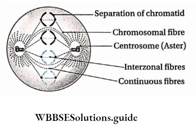
Some Facts About Anaphase
Reasons For Anaphasic Movement:
- The daughter chromatids separate due to the shortening or depolymerisation of the kinetochore microtubules from the poles of the chromosomal or centromeric fibres.
- It also involves simultaneous elongation of the continuous fibres due to polymerisation of the polar microtubules.
- Extension of interzonal fibres between two daughter chromosomes.
- Repulsion between daughter chromosomes that causes their sliding movement.
- Some facts about anaphase
Reasons for anaphasic movement:
- The daughter chromatids separate due to the shortening or depolymerisation of the kinetochore microtubules from the poles of the chromosomal or centromeric fibres.
- It also involves simultaneous elongation of the continuous fibres due to polymerisation of the polar microtubules.
- Extension of interzonal fibres between two daughter chromosomes.
- Repulsion between daughter chromosomes causes their sliding movement fragments of the nuclear membrane and ER.
- Nucleoli reappear in the nucleolus organiser regions (NORs) of SAT (satellite) chromosomes in both nuclei. Regeneration of nuclear membrane.
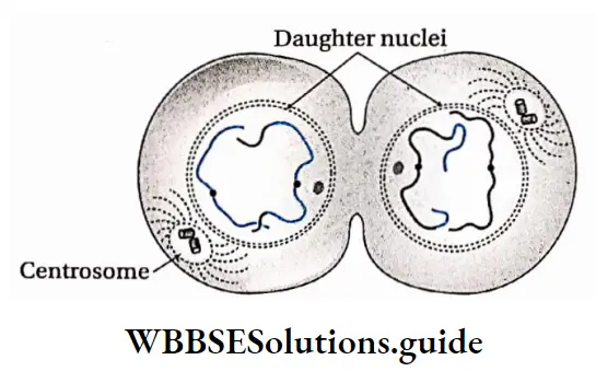
The density of cytoplasm and its refractive index decrease and chromosomes become invisible as chromatin fibre. Thus reconstruction of daughter nuclei is completed.
In animal cells, spindle fibres completely disappear. In plant cells, spindle fibres disappear from the poles but remain in the equatorial region. Golgi complex and endoplasmic reticulum are regenerated.
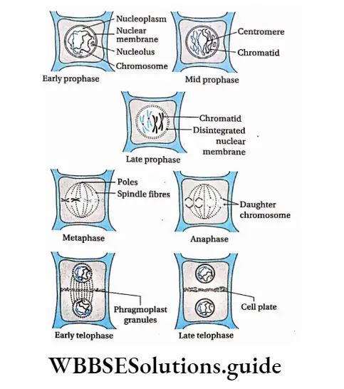
Types Of Mitosis
The different types of mitosis encountered in different organisms are discussed as follows—
Promitosis or intranuclear mitosis: It is a type of primitive intranuclear mitotic division which occurs without the formation of aster and disappearance of the nuclear membrane.
Thus, the nucleus divides within the nuclear membrane. For example, Amoeba, Yeast, etc., are divided by this process.
Eumitosis or extranuclear mitosis: This is the typical mitosis. This division involves the disappearance of the nuclear membrane at the end of the prophase.
Spindle forms and chromosomes are arranged at the equatorial plane by binding with spindle fibres. Example mitosis in plant and animal cells.
Endomitosis or Endoduplication or Endopolyploidy: The process in which repeated reduplication of chromosomes occurs during interphase without nuclear division (formation of daughter nuclei or daughter cells) is known as endomitosis or endoduplication or endopolyploidy.
Here, the segregation of chromosomes does not take place. Example polytene chromosomes in the salivary gland of Drosophila sp.
Paramitosis: The type of mitosis in which the spindle forms and the nuclear membrane disappears but the chromosome does not undergo coiling is known as paramitosis. Example division in Dinoflagellata.
Handwritten notes on cell cycle and cell division PDF download
Free nuclear division: The type of mitosis in which nuclear division is not followed by cytoplasmic division leading to a multinucleated condition is known as free nuclear division.
Plant cells containing more than one nucleus are called coenocytes and animal cells containing more than one nucleus are called syncytiums.
For example, the liquid endosperm of coconut, Mucor, Rhizopus, bone marrow cells and bone cells of animals contain more than one nucleus.
Cryptomitosis: The type of mitosis in which chromosomes coil, the nuclear membrane disappears, and typical spindles form but chromosomes are not arranged at the equatorial plate is called cryptomitosis.
Example division in parasitic protozoa like Plasmodium, and Heptazoon.
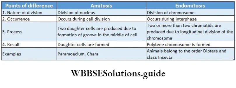
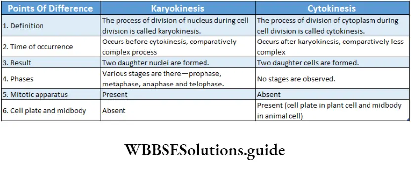
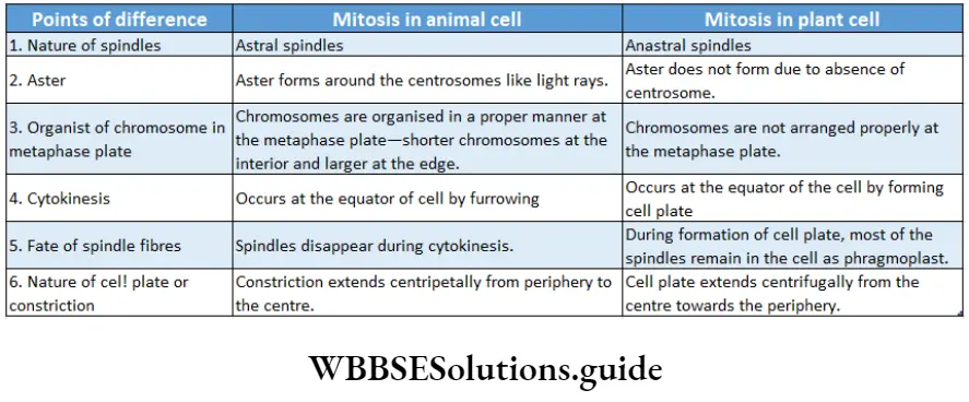
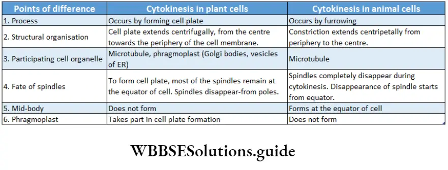
Significance of mitosis
The significance of mitosis is as follows—
Overall growth and development of the organism: The embryo forms from a single-celled zygote by mitosis and it divides multiple times to develop into a complete organism.
Thus, it is essential for increasing the number of cells in multicellular organisms.
Reproduction: It is a method of multiplication or reproduction in some unicellular organisms such as Amoeba, Paramoecium, etc.
Genetic stability: Due to mitosis, the number of chromosomes and genetic constitution like the total amount of DNA, RNA remain conserved in the daughter cell with respect to the parent cell.
Daughter cells are genetically almost identical to the parent cell and no variation in genetic material can therefore be introduced during mitosis.
So, mitosis results in genetic stability within the population of cells.
Repair and regeneration of worn-out parts:
Damaged cells are regularly replaced by new cells because of mitosis. It helps to heal wounds.
Replacement of older cells: By mitosis, older cells are replaced by newer cells.
Maintenance of surface-volume ratio:
The metabolic activity of a cell reduces when cell volume increases. Due to mitosis, the surface-volume ratio of a cell is maintained.
Maintenance of nucleoplasmic ratio: In mitotic cell division, the nucleoplasmic ratio of the cell is maintained.
Formation of gametes: Haploid or gametophytic organisms (n) produce gametes by mitosis.
Vegetative propagation: Mitosis helps in vegetative reproduction and micropropagation. New tissue or organs are formed by the process of mitotic cell division.
Meiosis
Meiosis Definition: The indirect process of cell division in which the chromosomes of parent cells divide once, but the nucleus divides twice to form four daughter cells, each containing half the number of chromosomes compared to the parent cell is called meiosis.
Van Beneden (1883) first reported that the reduction in chromosome number occurred during cell division. T. Bovery (1887) described the process in the gonads of Ascaris. J.B. Farmer and Moor coined the term ‘meiosis’.
chromosomes occur only once leading to a reduction in the chromosome number to half. The word ‘meiosis’ is also derived from two Greek words—meion means ‘to less’ and osis means ‘state’.
It is also known as reductional division because the chromosome number becomes haploid in the daughter cells produced. In all the sexually reproducing organisms meiosis occurs to produce haploid gametes from diploid cells.
Meiocytes And Meiospores
The diploid cells in which meiotic cell division occurs, are known as meiocytes. Example spermatocyte, and oocyte.
The haploid spores obtained by meiotic cell division, are known as meiospores.
Meiocytes And Meiospores Characteristics:
It is an indirect division of cells and occurs in several stages.
The process involves DNA replication during the ‘S’-phase of interphase followed by two successive nuclear and cytoplasmic divisions.
A diploid parent cell (2n), after division, produces four haploid daughter cells (n). The division takes place in two stages—meiosis 1 and meiosis 2.
During meiosis I, due to crossing over and chiasma formation between the non-sister chromatids, genetic recombination occurs.
The chromosomes divide once during meiosis 2.
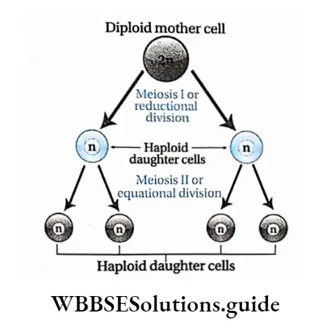
The two daughter cells produced after meiosis I, carry a haploid number of chromosomes as compared to the diploid number of chromosomes in the parent cell.
Site of occurrence: Meiosis occurs in the meiocytes. In plants like bryophytes, pteridophytes, gymnosperms and angiosperms, it occurs in the sporophytes, i.e., spore mother cells of anthers of stamen and ovules of the ovary.
ln animals, it occurs in the primary gametocytes, i.e., primary spermatocytes of the testis and primary oocytes of the ovary. ln lower plants like algae and fungi, it occurs in the diploid zygote.
Meiocytes And Meiospores Types: The process of meiosis is generally similar in both plant and animal cells but cytologists classified meiosis into three types—gametic or terminal meiosis, sporic or sporogenic or intermediate meiosis and zygotic or initial meiosis.
Gametic or terminal meiosis: Meiosis occurs during gamete formation in almost all animals.
The diploid primary gametocytes (spermatocytes and oocytes) undergo gametic meiosis to produce haploid gametes (sperm and ovum).
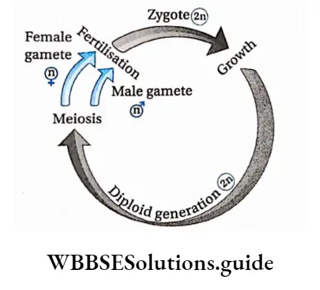
Sporic (sporogenic) or intermediate meiosis: In plants like bryophytes, pteridophytes, gymnosperms and angiosperms, the sporophytes reproduce by spore formation.
Haploid spores are produced from diploid spore mother cells by the process of sporadic or intermediate meiosis.
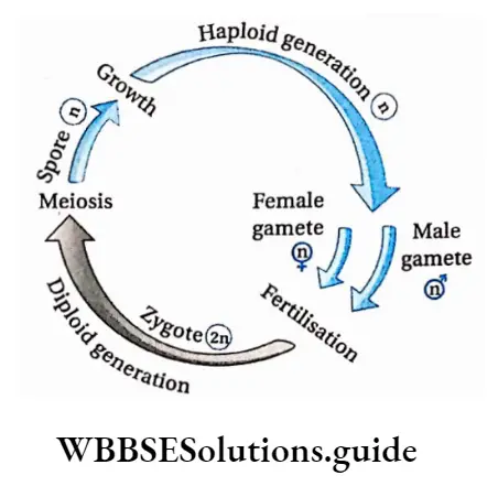
Zygotic or initial meiosis: When meiosis takes place immediately after the formation of the zygote, it is called zygotic or initial meiosis. It occurs in lower plants like algae (Spirogyra), certain fungi (Mucor) and in some sporozoan protozoa (Plasmodium).
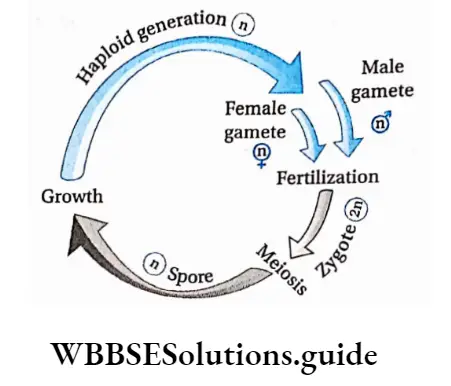
Outline Of Different Stages Of Meiosis
Meiosis is a specialized and complex type of cell division. It occurs only in diploid reproductive cells during the formation of haploid gametes or sex cells.
The process of meiosis consists of two complete divisions of a diploid cell—
1st meiotic division heterotypic cell division or reductional cell division.
2nd meiotic division is homotypic cell division or equational cell division. The process of meiosis starts in the interphase (like mitosis).
The DNA replication (=duplication) takes place at the ‘S’ phase of interphase.
Some biochemical mechanisms like protein synthesis take place at the G2 phase. The short period between meiosis I and meiosis II is called interkinesis.
DNA replication does not occur at this stage. Meiosis I is called heterotypic or reductional division because the two daughter cells so formed, have half the number of chromosomes of that of the parent cell.
Meiosis II is called homotypic or equational division because the two daughter cells have equal chromosome numbers as that of their parent cell.
Both these meiotic divisions have several subphases. These phases and their subphases are discussed under separate heads
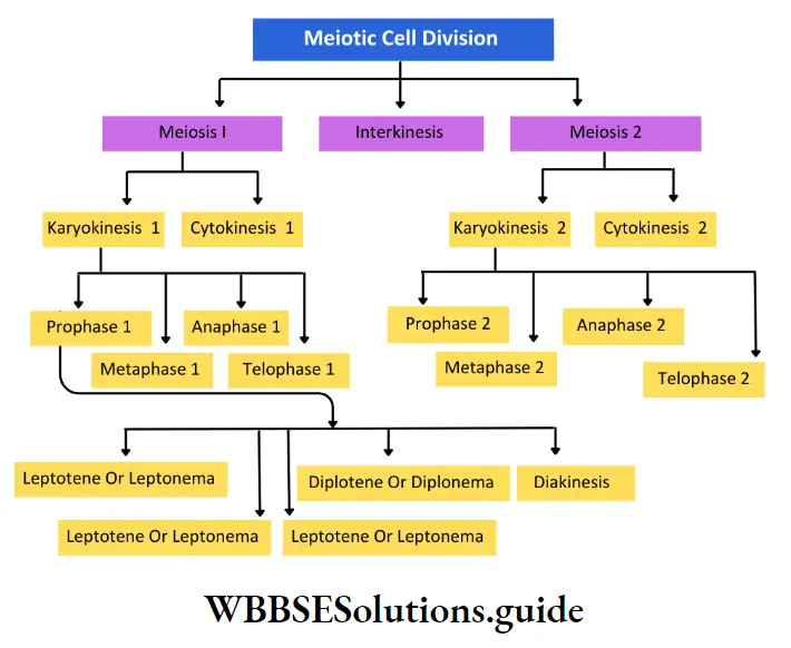
Meiosis Or Meiotic Division
Meiosis I starts after the end of the interphase. Before the first meiotic division, the meiocyte swells up. After meiosis I, two haploid cells form.
This phase is divided into two stages—karyokinesis I and cytokinesis I.
Karyokinesis
It is the process of the first nuclear division. It has been m divided into four phases—
- Prophase I,
- Metaphase I,
- Anaphase I and
- Telophase I.
It is the process of the first nuclear division. It has been divided into four phases—prophase I, metaphase I, anaphase I and telophase I.
Prophase I: The first phase of the first meiotic division in which a progressive sequence of chromosomal changes occurs through spiralisation, formation of synapsis and tetrad, genetic recombination and terminalisation of chiasmata is known as prophase I.
Leptotene or Leptonema
Leptotene or Leptonema Definition: The subphase of prophase I where chromosomes appear as thin threads with bead-like chromomeres visible most of the time, along the length of the chromosome is called leptotene or leptonema.
The characteristics of leptotene are as follows—
- The size of the nucleus increases. Viscosity and refractivity of the nucleus increase due to dehydration of nucleoplasm.
- The reticular form of chromosomes opens up and the chromosomes become thread-like. Condensation and spiralisation of chromosomes begin. As a result, they become more clearly visible as long single threads.
- Homologous chromosomes exist in pairs.
- Although the chromosomes are dyads, but, due to their thin structure and more coiling, they appear as monads.
- Threads of DNA, wrapped with nuclear proteins and histones, gradually become visible.
- These threads often have chromomeres that appear as “bead-like” swellings along their length.
- In plant cells, chromosomes clump at one side of the nucleus like a bouquet, called synizesis.
- In some other groups of plants, chromosomes in this subphase come in contact at one point to diverge again.
- This point of contact is known as a synthetic knot. In animal cells, the terminal end of chromosomes remains bound to the nuclear membrane through an attachment plate.
- They appear like multiple loops. This arrangement is known as the bouquet stage.
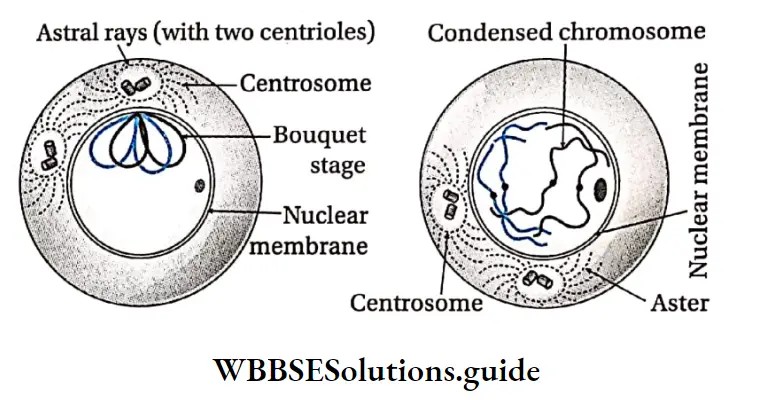
- These chromosomes are also known as polarised chromosomes.
- As leptotene progresses, chromosomes coil up more and become visible.
- In animal cells, the centrosome divides and astral rays start forming spindle fibres. At the end of this subphase, the two asters move apart from each other.
- At this time, components of the synaptonemal complex start gathering.
Homologous chromosomes
In a diploid cell (2n), chromosomes of identical shape, length and characteristics, exist in pairs. These chromosomes in a pair are known as homologous chromosomes.
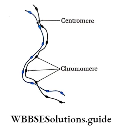
- For example, if the gene that determines the length of fingers, is present at a locus of one chromosome, then its homologous chromosome must carry the same gene at the same locus.
- Either both the genes would code for longer fingers or one would code for longer and the other for short fingers. But they must carry the same character-determining gene at a particular locus.
- Zygote (2n) is formed by the union of sperm (n) and ova (n). Zygote obtains two homologous chromosomes, one each from male and female gametes.
Zygotene Or Zygonema
Zygotene or zygonema Definition: The subphase of prophase I of meiosis I in which homologous chromosomes form synapsis is called zygotene or zygonema.
The characteristics of zygotene are as follows—
The homologous chromosomes get attracted towards each other and pair up lengthwise.
This temporary pairing of homologous chromosomes is known as synapsis and the paired chromosomes are known as bivalents.
Central transverse fibre is connected with a recombination nodule which contains enzymes helping in crossing over.
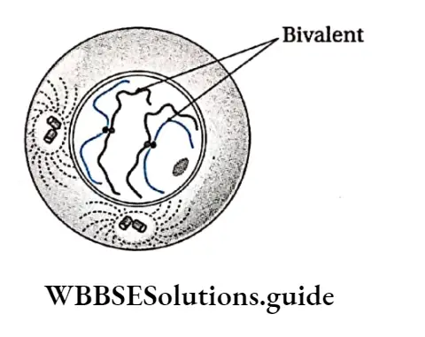
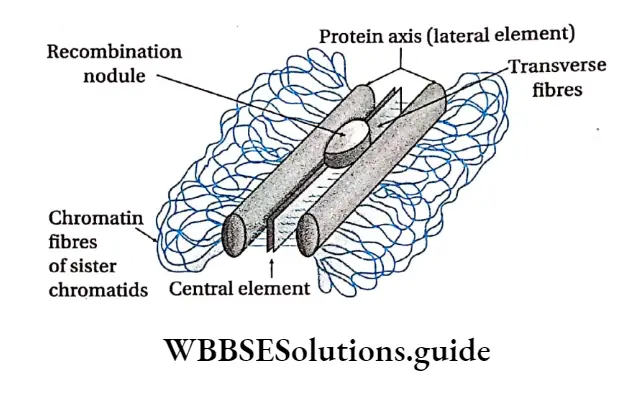
Pachytene
Pachytene Definition: The subphase of prophase in which the paired homologous chromosomes form a tetrad structure, followed by the exchange of genetic material among the non-sister chromatids through the process of crossing over, is called pachytene.
The characteristics of pachytene are as follows—
- This stage is of longer duration.
- Although the longitudinal replication of each chromosome has already taken place in the ‘S’ phase, the two chromatids remain invisible till zygotene.
- Due to continuous molecular packaging and condensation of each bivalent, two chromatids of each chromosome now become visible. The four chromatids of each bivalent are called tetrads.
- The two chromatids of the same chromosome are known as sister chromatids. The chromatids of two different chromosomes of the homologous pair are known as non-sister chromatids.
- Rounded or nodular structures begin to appear along the parts between the non-sister chromatids that occur at these nodules.
- In the recombination nodule, the recombinase complex mediates crossing over.
- The recombinase enzyme complex consists of two enzymes having opposite functions—endonuclease and ligase.
- The two non-sister chromatids of the same homologous pair undergo one or more transverse breaks.
- The breaks are followed by the interchange of chromatid segments between the non-sister chromatids of the homologue. This process is known as crossing over.
- According to Stern and Hotta (1969), chromatid break occurs due to the action of the endonuclease enzyme.
- Ligase helps in joining the broken ends after the exchange. Due to crossing over, the exchange of genetic character between homologous chromosomes and gene recombination takes place.
- In animal cells, asters move away from each other.
- The synaptonemal complex between two homologous chromosomes disintegrates. The nucleolus remains associated with the NOR of a specific chromosome.
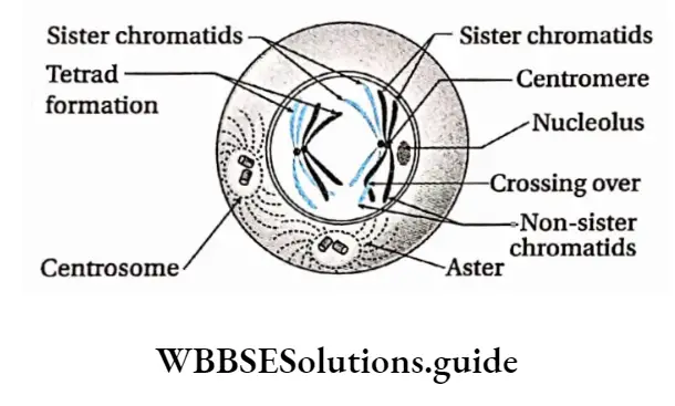
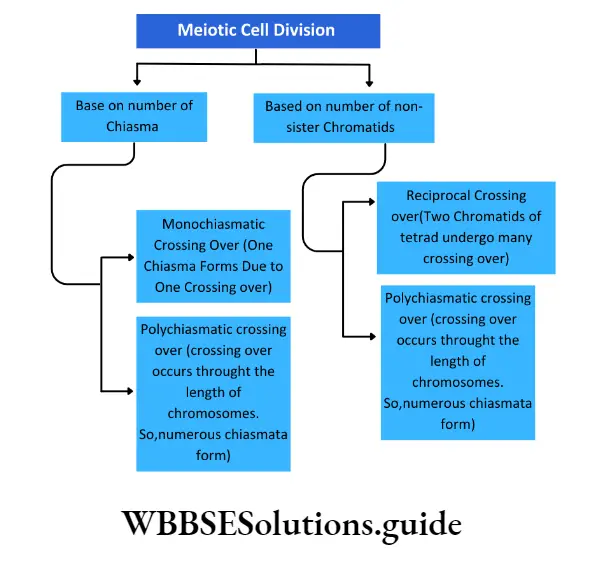
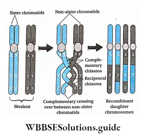
Diplotene Or Diplonema
Diplotene Definition: The subphase of prophase I in which paired chromosomes begin to repel each other along with the formation of chiasmata is known as diplotene.
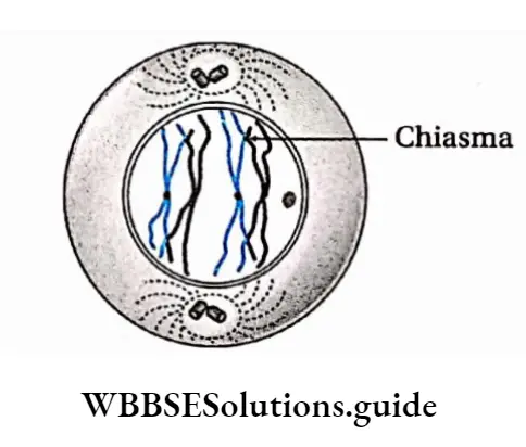
Such points become visible as ‘X’ like structures called chiasmata (singular-chiasma; Greek chiasma means ‘cross’). The position of chiasma may be interstitial, subterminal or terminal.
As the bivalents separate from each other, the chiasma begins to shift towards the terminal end of the chromosome. Thus, the terminalization of chiasmata begins.
Rotation of the arms of chromosomes, with single chiasma, is usually initiated in the late diplotene, forming cross-like (X Structures.
Nucleus Is Still Present. Asters Present In Animal Cells Separate Further Away From Each Other.
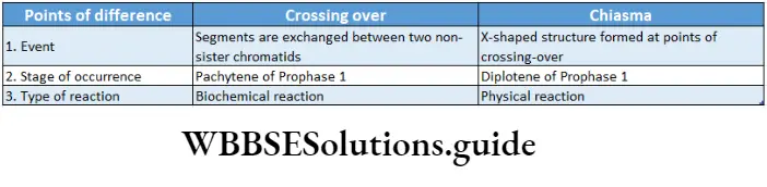
Diakinesis
Diakinesis Definition: The subphase of prophase I in which condensation of bivalents increases with fully terminalized chiasmata is known as diakinesis.
The characteristics of diakinesis are as follows—
- Bivalents are fully contracted, condensed, shortened and deeply stained.
- Rotation and terminalisation continue pushing the chiasmata at the extreme ends of the bivalents forming cross and ring-like structures.
- The shape of bivalents depends on the number of chiasmas—cross-like when the number of chiasmas is one, ring-like when the number of chiasmas is two and like a chain of loops when the number is more than two.
- Nucleolus disappears.
- The nuclear membrane starts disintegrating.
Metaphase 1: The phase of meiosis 1 when the maximally condensed bivalents are arranged at the equatorial plate is known as metaphase 1.
Phases of the cell cycle and types of cell division notes
Characteristics:
In this stage, the structure of the mitotic apparatus is completed. X-shaped, ring-shaped, diamond-shaped maximally contracted and condensed bivalents are arranged at the equator of the spindle.
Amphiastral spindles and anastral spindles form 1 in animal cells and in plant cells respectively.
Homologous pairs of chromosomes (bivalents) are so: arranged at the equator of the spindle that their chromatids lie over the equator while their centromeres are bent in the direction of the poles. This special arrangement of the chromosomes makes a double metaphase plate at random.
The chromosomes of a bivalent remain bound to chromosomal fibres belonging to two poles through the centromeres. Kinetochores of the chromosomes bind to spindle fibres from opposite 1 poles.
The centromere of each chromosome is directed towards the opposite poles.

Prometaphase 1
It is a transitional phase between diakinesis and metaphase of meiosis I when the bivalents are maximally condensed in the form of Y or ring.
Characteristics of Prometaphase:
- X-shaped, ring-shaped, diamond-shaped bivalents reach their maximum contraction and condensation.
- The nuclear membrane completely disappears.
- Spindle formation begins.
- In animal cells, the microtubules are arranged in the form of a spindle apparatus in between the two centrosomes occupying the two opposite poles.
- In plant cells, a spindle arises from the cytoplasmic microtubules in the absence of centrosomes.
Anaphase I: The phase of meiosis I at which the homologous chromosomes separate and move to the opposite poles when pulled by the spindle microtubules is known as anaphase I.
Characteristics:
- The chiasmata joining the; homologous chromosomes dissolve allowing the separation of the maternal and paternal homologs,
- Spindle fibres pull the homologous pair towards opposite poles of the spindle.
- The smaller homologue separates more quickly than the larger one because the latter has more number of interstitial chiasmata that take time to dissolve.
- The separated homologs, each consisting of two chromatids united by a centromere, move towards the opposite poles.
- Thus, each pole receives half of the total number of chromosomes, reducing the chromosome number to haploid in the daughter cells.
- This movement of chromosomes towards opposite poles is known as anaphasic movement.
- Anaphasic movement takes place through the elongation of the continuous fibres and contraction of the equatorial zone. The spindle also elongates vertically.
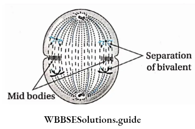
Telophase 1: The phase of meiosis I when the nuclear membrane is reorganised around each group of daughter chromosomes, that spiralised into thin elongated threads forming a reticulum, is known as telophase I.
Characteristics:
- Telophase I is marked by the presence of chromosomes which are half in number than that of the parent cell, at the poles.
- The nucleolus and the nuclear membrane reappear.
- Water diffuses in the nucleus, so chromosomes are not clearly visible.
- The chromosomes undergo deserialization and become elongated into thread-like structures forming a nuclear reticulum.
- Spindle fibres disappear.
- At the end of telophase, two daughter nuclei with a haploid number of chromosomes (n) are produced.

Cytokinesis
Cytokinesis Definition: The phase of meiosis during which the cell cytoplasm divides into two after the division of the nucleus, leading to the formation of two daughter cells is known as cytokinesis.
Cytokinesis Characteristics:
Cytokinesis can be of two types—simultaneous or successive. In the former separate cytokinesis, I cannot be distinguished.
- In the latter, distinct cytokinesis can be seen after both meiosis 1 and meiosis 2.
- In animal cells, a constriction appears in the equatorial region of the parent cell.
- The constriction gradually deepens to form a narrow furrow and divides the cell into two equal halves. The daughter cells thus produced, have a haploid number of elongated dyad chromosomes.
- In most plant cells, daughter cells are produced by the formation of phragmoplast or cell plate.
- Small vesicles or phragmosomes from the Golgi body appear in the equatorial region. They fuse with each other to form the cell plate or phragmoplast.
- The cell plate forms the middle lamella on which the primary cell wall and secondary cell wall are deposited.
Interkinesis or intrameiotic interphase: The short period between the end of telophase of meiosis I and the beginning of prophase of meiosis 2, where replication of DNA does not take place, is known as interkinesis or intrameiotic interphase.
Cytokinesis Characteristics:
- RNA and proteins are synthesised, but DNA replication does not take place. Hence, the S phase is absent.
- Many plants skip telophase I and enter prophase 2.
- Very little ATP is synthesised.
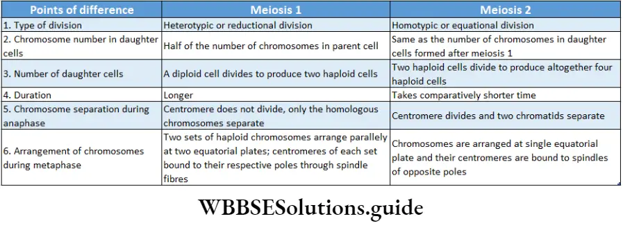
Meiosis 2 or meiotic division 2
The events of meiosis 2 are analogous to those of a mitotic division, although half the number of chromosomes participate in this division.
It results in the formation of two daughter cells from each of the daughter cells formed after meiosis
They contain an equal number of chromosomes as that of their parent cells.
So, this division is also called homotypic or equational cell division. Meiosis 2 occurs in two stages—karyokinesis 2 and cytokinesis 2.
Karyokinesis 2
This is the nuclear division which occurs in four phases—prophase 2, metaphase 2, anaphase 2 and telophase 2.
Prophase 2: The phase of meiosis 2 during which elongated spiralised chromosomes with two chromatids joined at the centromere, become visible due to dehydration and condensation, is known as prophase 2.
Characteristics:
Due to dehydration, chromosomes become visible as they condense. Each chromosome consists of two chromatids. The chromatids are joined together in the region of centromere.
The chromatids coil thickens and becomes short and visible.
The nuclear envelope and nucleoli disappear and the spindles form. In animal cells, the centrosome divides into two, which move towards opposite poles. Asters form again and spindles form between asters.
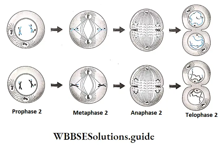
Metaphase 2: The phase of meiosis 2 during which the shortest and thickest chromosomes in the daughter cells arrange themselves at the equatorial plate, is known as metaphase 2.
Characteristics:
- Spindle formation between the two centrioles is completed.
- The shortened and condensed chromosomes become oriented at the
equatorial plate. - The kinetochore of the sister chromatids becomes attached to the spindle microtubules.
Anaphase 2: The phase of meiosis 2 during which the sister chromatids separate and move to the opposite poles when pulled by the spindle microtubules, is known as anaphase 2.
Characteristics:
- The centromere of the chromosome divides into two. As a result, the two sister chromatids (monad) separate.
- The chromatids are pulled to the opposite poles due to the shortening of the chromosomal spindle fibres.
- Interzonal fibres form between the daughter chromosomes. Expansion of interzonal fibres and contraction of chromosomal fibres result in anaphasic movement.
- Telophase 9: The phase of meiosis 2 during which the nuclear membrane is reorganised around each group of chromatids or daughter chromosomes at the poles, that spiralised into thin elongated threads forming a reticulum, is known as telophase 2.
Characteristics:
- Chromatids reach the opposite poles and clump together to form chromosomes.
- Chromosomes begin to uncoil and decondense. They become elongated into indistinct thread-like structures forming a nuclear reticulum.
- The nuclear membrane and nucleolus form again in each of the daughter nuclei.
- The spindle fibres and astral rays usually disappear.
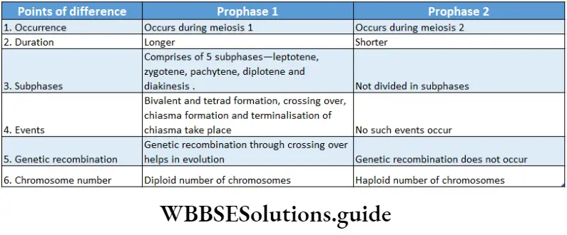
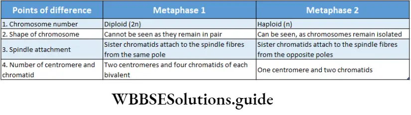
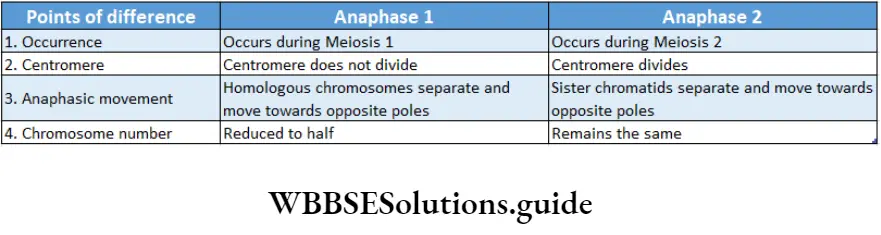
Cytokinesis 2
Cytokinesis Definition: The phase of meiosis 2 during which the cell cytoplasm divides into two after the division of the nucleus, leading to the formation of four daughter cells is known as cytokinesis 2.
Characteristics:
Cytoplasm divides for the second time and two haploid (n) daughter cells of meiosis I, give rise to four haploid (n) daughter cells.
Cytokinesis occurs in animal cells by cleavage and in plant cells by the formation of cell plates.
Significance Of Meiosis
Sexual Reproduction: Meiosis Is responsible for the formation of haploid gametes in sexually reproducing organisms.
Maintains chromosome number: During fertilisation, the nuclei of the two gametes fuse and produce a zygote.
Thus, fertilisation doubles the chromosome number in the zygote. Meiosis halves the number of chromosomes in the gametes to maintain the chromosome number constant and stable for each species.
Source of variation: As a result of crossing over between two homologous chromosomes during prophase I, the exchange of genetic material occurs.
This leads to new combinations of alleles in the chromosomes of the gametes which result in genetic recombination.
Maintains chromosome number: During fertilisation, the nuclei of the two gametes fuse and produce a zygote. Thus, fertilisation doubles the chromosome number in the zygote.
Meiosis halves the number of chromosomes in the gametes to maintain the chromosome number constant and stable for each species.
Source of variation: As a result of crossing over between two homologous chromosomes during prophase I, the exchange of genetic material occurs.
This leads to new combinations of alleles in the chromosomes of the gametes which result in genetic recombination.
Meiosis in plant cell
In plant cells, meiosis occurs in two phases—meiosis I (reductional division) and meiosis II (equational division).
In both the phases karyokinesis is followed by cytokinesis.
Meiosis 1 has two stages—karyokinesis I and cytokinesis I.
Meiosis 2 also has two stages—karyokinesis 2 and cytokinesis 2. At interphase, the nuclear membrane, nucleoplasm and chromatin fibres of the nucleus of the plant cells become visible.
Meiosis 1 starts after interphase. It is followed by a short interkinesis phase after which meiosis 2 takes place. The different stages of meiosis.
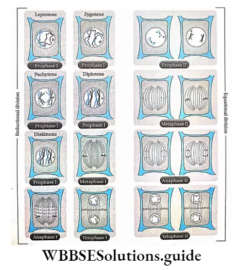
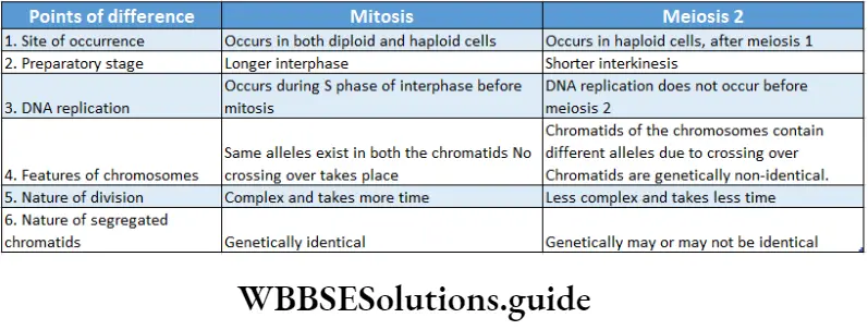
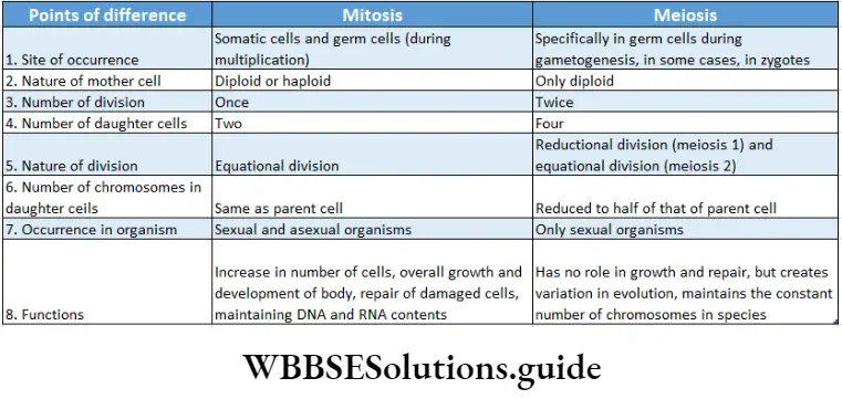
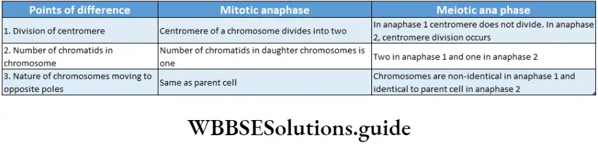
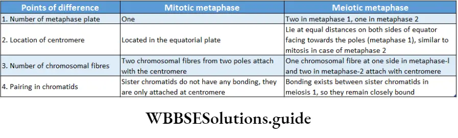
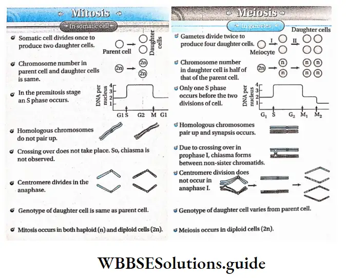
Cell Cycle And Cell Division Notes
- Cyclin: A protein involved in the cell cycle. It goes through cycles of synthesis and degradation during the cell cycle and activates cyclin-dependent protein kinases.
- Cyclin-dependent protein kinase (CDK): Protein found in eukaryotic cells which remains active when associated with cyclin. It regulates the cell cycle.
- Cyclin: A protein involved in the cell cycle. It goes through cycles of synthesis and degradation during cell cycle and activates cyclin-dependent protein kinases
- Cyclin-dependent protein kinase (CDK): Protein found in eukaryotic cells which remains active when associated with cyclin. It regulates the cell cycle.
NEET cell cycle and cell division revision notes with MCQs
Point Of Remember
- The cell generates from an existing cell by division.
- The phase between two successive cell divisions is known as interphase.
- The cell cycle occurs in two main phases, one is the M phase and the other is the I phase or interphase.
- During the Gx phase of interphase, RNA, protein, amino acid, ATP and nucleotides are synthesised.
- DNA and histone proteins are synthesised during the S phase of interphase.
- A mature cell that is no longer capable of undergoing mitosis enters the G0 phase and remains there permanently. This type of cells is known as post-mitotic cells. A neuron or nerve cell is an example.
- The cell cycle is controlled by cyclin and cyclin-dependent kinase.
- Cell division is of three types—amitosis, mitosis and meiosis.
- Amitosis is called direct nucleus division because in this process—
- Spindles do not form,
- Cells divide directly and
- No stages of cell division are found.
- Amoeba and bacteria divide by amitosis (binary fission) to achieve asexual reproduction.
- Alkaloid colchicine, obtained from the plant Colchicine autumnal (Liliaceae), inhibits spindle formation and is known as mitotic poison.
- Mitosis is known as equational division because it results in the formation of two identical daughter cells with an equal number of chromosomes as in the parent cells.
- In plant cells, spindle apparatus forms from microtubules of cytoplasm.
- In animal cells, the spindle apparatus forms by division of centriole and aster. So, spindles of animal cells are known as amphiaster.
- Contraction of the spindle, repulsion between two split centromeres and formation of interzonal fibres together help the daughter chromosomes to move towards the opposite poles (anaphasic movement).
- Presentation of all the chromosomes. pairs of a species
in a diagram or photograph is called a karyogram. - Cytokinesis occurs in plant cells by cell plate formation and in animal cells by furrowing or cleavage.
- Mitosis can be intranuclear or extranuclear. Again, mitosis can skip karyokinesis and can cause endoploidy or endomitosis by duplication of chromosomes.
- Three types of meiotic cell divisions occur. They are—
- Zygotic or initial meiosis,
- Gametic or terminal
- meiosis and
- Sporogenetic or intermediate meiosis.
- Segments of chromatids are exchanged in crossing over. This causes genetic variability.
- Crossing over starts at the pachytene subphase but becomes visible in the diplotene subphase.
- Subphase pachytene in prophase I of the first meiotic division is the longest phase.
- During anaphase I, homologous chromosomes separate and move away from each other. This process is known as disjunction.
- A bivalent contains four chromatids, which are together known as tetrad and this stage is called the tetrad stage.
- Excessive growth of a tissue or organ due to the increase in volume or shape of its constituent cells is known as hypertrophy.
- The process of programmed cell death is known as apoptosis.
- The crossing over between non-sister chromatids that results in the formation of chromosomes with new properties and new gene rearrangement is known as recombination.
- Malignant cells spread to different parts of the body from their primary origin through blood and lymph. This is known as metastasis.
- Two chromatids of a chromosome are known as a dyad.
- Two chromatids of one chromosome in a bivalent are known as sister chromatids. Two chromatids of two different chromosomes of a bivalent are known as non-sister chromatids.
Biomolecules Notes
Biomolecules Introduction
Biomolecules
The body of an organism is just like a chemical factory. Numerous chemical reactions are continuously occurring inside our body.
Such reactions constitute the metabolic pathways of the various biochemical processes like respiration, photosynthesis, etc.
These reactions involve different molecules and enzymes. Many of these molecules such as carbohydrates, proteins, and lipids are the major constituents of the living body.
The collection or sum total of all these molecules present within a cell is called the cellular pool. All such molecules that are involved in the maintenance and metabolic processes of the living body are called biomolecules.
Different molecules found in a living body—Biomolecules
Biomolecules occur naturally in living organisms. All forms of life have biomolecules as their constituents.
Biomolecules consist mainly of carbon and hydrogen with nitrogen, oxygen, sulfur, and phosphorus.
The biomolecules are divided into two types—
- Inorganic and Organic.
- Inorganic molecules
These molecules either contain fewer carbon and hydrogen atoms or none at all.
They remain dissolved in the cellular fluid and form a colloidal solution, For example, different minerals (in the form of ions and salts), water, gaseous products, etc.
Biomolecules notes for NEET PDF
Inorganic molecules are of two types—
- Water And
- Inorganic Salts.
Water: About 90% of the cellular protoplasm is made up of water.

Water is required for the following functions—
- Solvent: Water is used as a solvent in different organic and inorganic cellular components.
- Medium for metabolic reactions: Several metabolic reactions take place in aqueous medium.
- Temperature regulation: Water plays an important role in regulating the body temperature mainly in higher animals and also in plants.
- pH regulation: The balance between the acidic and basic components in the body is maintained by water.
Inorganic salts: Inorganic salts can carry out the following functions—
- Structure of body parts: Different body parts such as bones, teeth, etc., have inorganic salts as one of the major components.
- Chemical reactions: Inorganic salts also take part in different chemical reactions within the body.
- Formation of organic molecules: Organic biomolecules such as DNA, RNA, etc., have inorganic salts as one of the major components.
| Class 11 Biology | Class 11 Chemistry |
| Class 11 Chemistry | Class 11 Physics |
| Class 11 Biology MCQs | Class 11 Physics MCQs |
| Class 11 Biology | Class 11 Physics Notes |
Organic molecules
In these molecules, the number of carbon and hydrogen atoms is more compared to inorganic molecules.
Different sugars, lipids, amino acids, proteins, nucleic acids, etc., fall under this group.
They are divided into two types—
- Macromolecules And
- Macromolecules.
Micromolecules: The molecules, that have a simple structure, low molecular weight, and are mostly water soluble, are called micromolecules.
Class 11 biology biomolecules notes with diagrams
Examples: Monosaccharides, amino acids, nucleotides, etc.
Macromolecules: The larger molecules that are made up of smaller monomeric units (micromolecules), linked by chemical bonds, through polymerization reaction, are called macromolecules.
Examples: Proteins (polymer of amino acids), nucleic acids (polymer of nucleotides) etc.
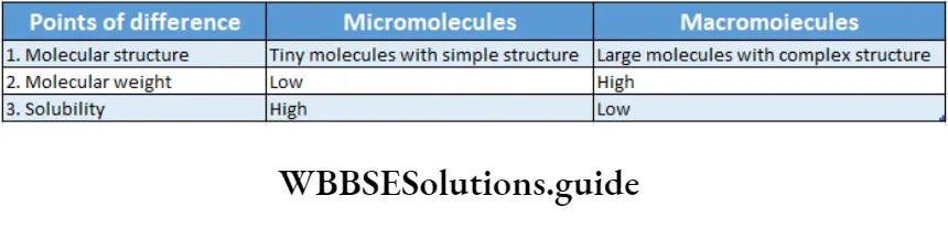
Different Essential Elements Of —Living Bodies
Among elements, about 22 are essential for the body.
According to their requirement, the elements are divided into two categories—
- Macroelements And
- Microelements.
Macroelements
These elements are required by the body in large amounts. Also, deficiency of any of them may inhibit the growth and physiological functioning of the body.
About 95% of the cellular components are made of these elements. In our body, oxygen, carbon, hydrogen, nitrogen, etc., are examples of macroelements.
Biomolecules Notes Significant Biomolecules
Carbohydrates, lipids, proteins, and nucleic acids are four main macromolecules.
Carbohydrates
Carbohydrates Definition: Carbohydrates are polyhydroxy aldoses, ketoses, and their derivatives that yield- such compounds in hydrolysis where the ratio of carbon to hydrogen to oxygen atom is 1:2:1.
General formula: Cn(H20)n
Nomenclature: French term ‘Hydrate de carbone’, gives rise to the word ’carbohydrates1.
It is a class of compound called ‘saccharide’ derived from the Greek word ‘saccharin’ meaning sugar.
Sources: The natural sources of carbohydrates are plants—grapes, mango, sugarcane, beet, potato, maize, wheat, rice, vegetables, etc.; animals—milk, liver, fish, meat, etc.
Classification of carbohydrates: All carbohydrates are classified into three basic types— monosaccharide, oligosaccharide, and polysaccharide.
Biomolecules Chart
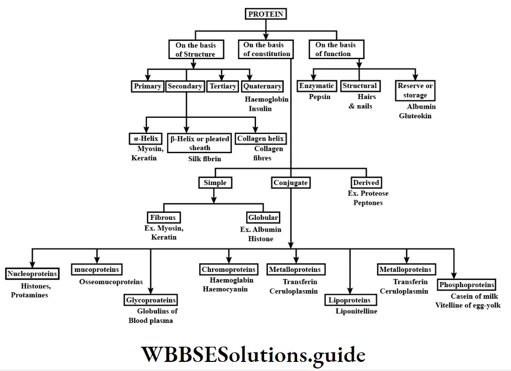
More facts on carbohydrates
Free fructose is not generally available in the body but it is available in free form only in semen. Levulose is present in honey.
In some carbon compounds hydrogen and oxygen may be present in a 2:1 ratio but they are not considered carbohydrates. For example, acetic acid (CH3C00H) etc
In a DNA molecule, the two intertwined strands are not parallel, i.e., antiparallel. One strand is aligned 5′-3′ while the other is aligned 3′-5′.
The diameter of the DNA helix is about 20A.
In this structure, the helix makes a turn at every 3.4 nm length of DNA, and the distance between two neighboring base pairs is 0.34 nm. Hence, there are about 10 base pairs per turn.
The intertwined strands make two grooves of different widths, referred to as the major groove and the minor groove, which may facilitate binding with specific proteins.
Each DNA molecule has a sugar-phosphate backbone, with nitrogenous bases attached to them.
The nitrogenous bases of one DNA molecule form a hydrogen bond with the adjacent base belonging to the other DNA molecule.
A binds with T with a double bond & G binds with C with a triple bond. This keeps the helical structure of the DNA stable, with its diameter constant.
Biomolecules Notes
In a single strand of DNA, the amount of A is not equal to that of T, while the amount of G is not equal to that of C. On the other hand, in the case of double-stranded, A + G = T + C.
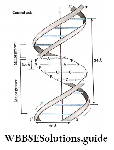
Types of DNA: Most organisms have regular double-helical forms of DNA. Some bacteriophages and animal viruses have single-stranded DNA.
In prokaryotic organisms (bacteria, etc.), the DNA molecule is not linear, rather it is circular.
DNA can be classified according to the following characters—
- The number of nucleotides in each turn,
- The bond angle between the nitrogenous base pairs,
- The diameter of dna double helix,
- The direction often of double helix structure, i.e., Right or left.
There are three major forms of DNA helices—A-DNA, B-DNA, and Z-DNA. The structure of B-DNA was proposed by Watson and Crick, which has been discussed in detail in this chapter.
The comparisons between the three forms of DNA.
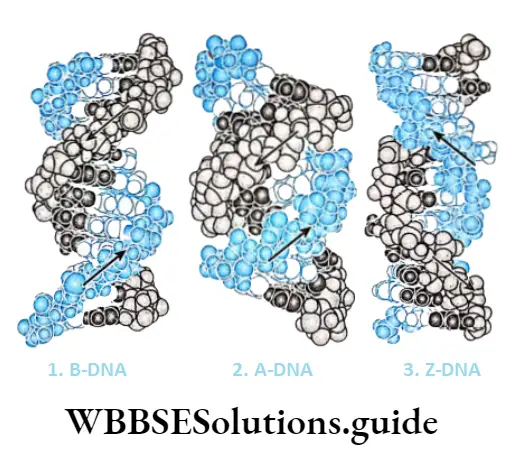
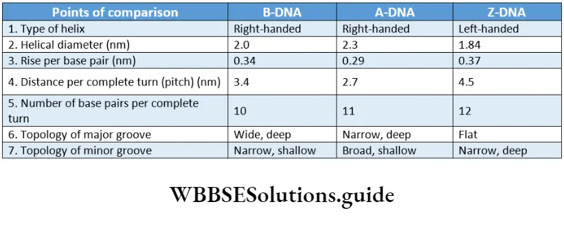
Ribonucleic acid (RNA): RNA is a type of nucleic acid that is comprised of a single-stranded polynucleotide chain and responsible for protein synthesis.
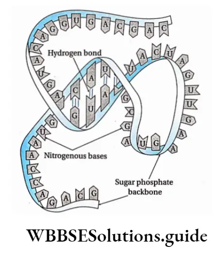
Occurrence: The non-genetic RNA is present in all prokaryotic and eukaryotic cells. It occurs both in the nucleus as well as in the cytoplasm.
In some viruses, like TMV, influenza virus, retrovirus (HIV), etc., RNA acts as a genetic material and is known as ‘Genetic RNA’.
Quantity: The quantity of RNA depends on the metabolic reactions of the cell. During protein synthesis, the amount of RNA increases in cells.
Structural features:
Cellular RNA, both genetic and non-genetic, is single-stranded and may fold upon itself to form hairpin-like structures entirely or at certain.
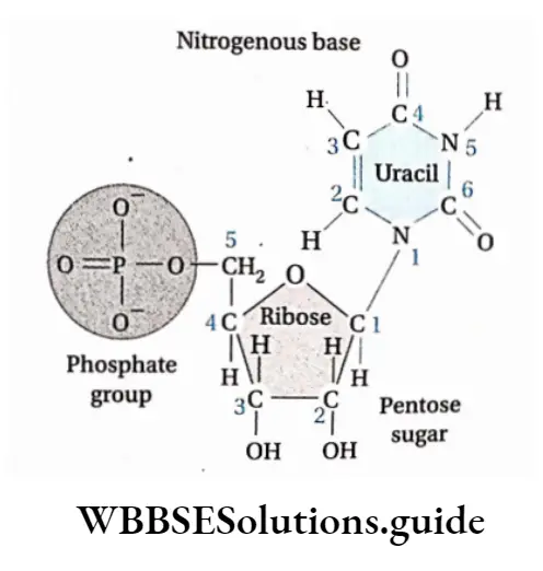
- Regions Rheovirus has double-stranded genetic RNA.
- The strand is unbranched but may be folded in some cases so that the structure is complex.
- They are made up of several nucleotides linked by 3′-5′ phosphodiester bonds.
- Every nucleotide is made up of a ribose sugar, a nitrogenous base, and a phosphate. The nucleotides are called ribonucleotides.
- The nitrogenous bases present in RNA are adenine, guanine, cytosine, and uracil.
- The main function of RNA is protein synthesis. It is the main genetic material in many viruses.
- RNA is of different types. They are discussed under separate heads below.
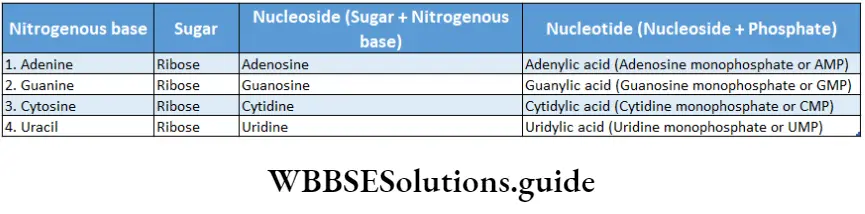
Types of RNA: The different types of RNA have been discussed below.
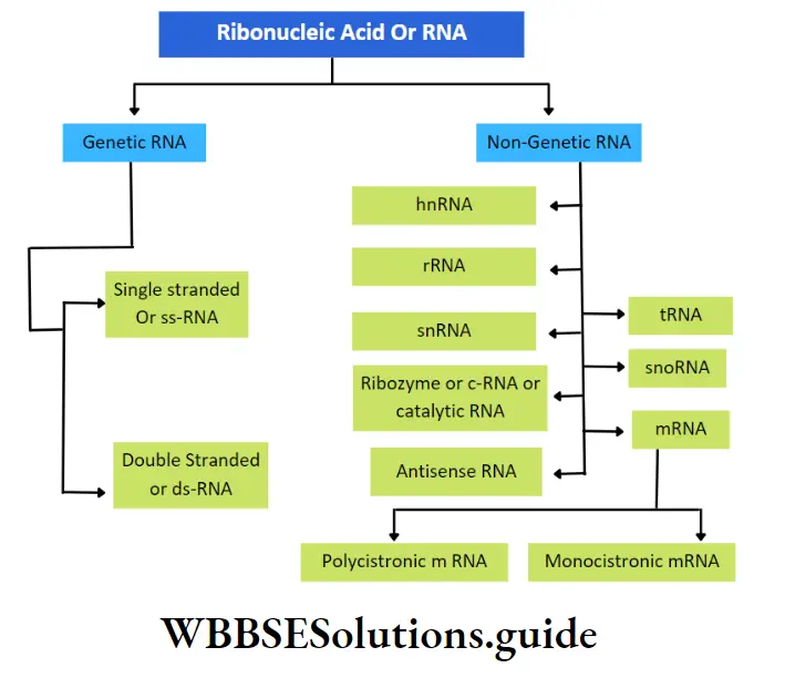
Genetic RNA: The RNA that acts as genetic material in the absence of DNA, is called genetic RNA.
For example, Tobacco Mosaic viruses (TMV), wound tumor viruses, etc. contain genetic RNA.
This type of RNA is again of two types—
Single-stranded RNA: The RNA molecule made up of a single strand is called single-stranded RNA. E.g., TMV (plant virus), influenza (animal virus).
Double-stranded RNA: The RNA molecule made up of two strands is called double-stranded RNA. E.g., Wound tumor virus (plant virus), Rheovirus (animal virus), etc.
Non-genetic RNA: The RNA presenting the prokaryotic and eukaryotic cells, where DNA is the main genetic material, is called non-genetic RNA. These are synthesized from DNA by transcription.
In both prokaryotic and eukaryotic cells, three principal types of RNA are found. They are messenger RNA (mRNA), transfer RNA (tRNA), and ribosomal RNA (rRNA).
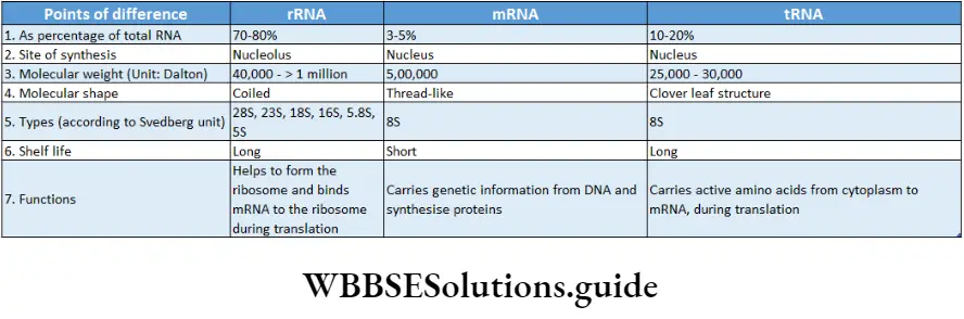
Genetic material: DNA is the biological molecule that stores all the genetic information of the cell. In some viruses, RNA may function as the molecule that stores genetic information.
Transmission of hereditary characters: DNA functions as the molecule, that carries the genetic information from parents to offspring.
Synthesis of protein: Coded genetic information is transferred to the mRNA from DNA by the process of transcription. These codes are used to synthesize proteins by translation with the help of rRNA and tRNA.
Thus, information in DNA or genes is expressed in the form of proteins. Messenger RNA (mRNA) is the nucleic acid that carries information from the nucleus to the cytoplasm on which proteins are made.
Biomolecules Notes
The rRNA is a constituent of ribosome. This type of RNA helps in binding mRNA to ribosome and also in decoding the codons.
They also help in catalyzation of amino acid assembly into a polypeptide chain. The tRNA transports amino acids to the growing end of a polypeptide chain at ribosomes.
Role in metabolic activity: DNA indirectly controls the metabolic activity of the cell through the synthesis of necessary protein molecules, like enzymes.
Role In gene mutation and variation: Changes in the sequences of base pairs of DNA alter the arrangement of nucleotides in mRNA.
This causes alterations of codons which lead to changes in proteins. This induces variations in organisms leading to mutations. Mutation when leads to evolution can give rise to new species.
Biomolecules Class 11 Notes
Concept Of Metabolism
Definition: The sum total of all chemical reactions constantly occurring in the living organism, that are necessary for maintaining the living state of cells and that of the organism, is termed as metabolism.
The metabolic reactions take place as a series of linked reactions. These multi-step processes are called metabolic pathways.
Metabolic pathways of a living system can be categorized into two types—
- Anabolic pathways and
- Catabolic pathways.
Anabolic pathways
These pathways are constructive in nature and result in the formation of complex structures from simpler ones. Anabolic pathways consume energy and hence, are endergonic in nature.
For example, amino acids are linked together to synthesize proteins. Protein synthesis requires energy input and is an example of the anabolic pathway.
Catabolic pathways
These pathways are destructive in nature and lead to the breakdown of complex substances into simpler ones.
Catabolic pathways are made up of exergonic reactions because they release energy.
For example, glycolysis is a catabolic pathway where glucose breaks down to produce two molecules of pyruvic acids.
The energy released during catabolic pathways is stored in the form of chemical energy in the high-energy bonds of ATP (Adenosine triphosphate) molecules.
ATP acts as a link between exergonic and endergonic reactions. This is because ATP is hydrolyzed to release the chemical energy needed for various endergonic (energy-consuming) reactions.
Living Stage
A living system exists in a steady state, characterized by particular concentrations of all the biomolecules present within the living system.
The steady state is in a non-equilibrium state because the living system is working continuously. It prevents itself from reaching the state of equilibrium so that it is able to perform work.
The rate of formation, utilization, and transformation of biomolecules is called turnover. The rate of turnover of molecules in a metabolic pathway is called metabolic; flux.
For being in the state of metabolic flux, the metabolites tend to reach equilibrium. The equilibrium state is not reached because the end products of the metabolic pathways are utilized again.
Thus, the living organism tends to attain a steady state by metabolic flux. This takes place in the presence of energy, which is produced as a result of different metabolic reactions.
So, we can conclude that, without metabolism, a living state cannot exist.
According to Lehninger and others, biomolecules are normally present as essential organic components of living systems, and their essential organic compounds are collectively called metabolites.
These are mainly of two types—
Primary metabolites: They are formed as intermediates and produced via normal metabolic pathways in almost all living organisms, e.g., sugar, amino acids, nucleotides, lipids, proteins, etc.
Secondary metabolites: They are formed especially by the alteration of primary metabolites, mainly in the plant’s body, e.g., alkaloids, pigments, aromatic compounds, terpenoids, etc.
Enzymes
Enzymes Definition: Enzymes are protein molecules acting as biological catalysts that are able to accelerate the rate of chemical reactions in cells. While they remain unchanged at the end of the process.
Enzymes Discovery:
- Jon Jakob Berzilius (1835) was the first to term the activity of enzymes ‘catalytic’.
- Louis Pasteur (1850) concluded from his studies that fermentation of sugar to alcohol is catalyzed by a vital force in living yeast cells, which he termed as ‘ferments’
- The term ‘enzyme’ was coined by W. Kuhne in 1878.
- It is derived from the Greek words, En= in and Zyme = yeast.
- In 1897, Edward Buchner was the first to isolate the enzyme, zymase, which was able to ferment sugar. (f)J.B. Sumner first purified and crystallized the urease enzyme from jack bean.
- After this, John Northrop (1930) isolated many enzymes like ‘pepsin’, and ‘trypsin’ in crystalline form. Sumner and Northrop were awarded the Nobel Prize in Chemistry along with Wendell Stanley in 1946.
- Thomas Cech and Sidney Altman (1983) were awarded the Nobel Prize in chemistry for the discovery of the catalytic property of RNA.
Properties of enzymes: The properties of enzymes are discussed in separate heads below.
Reversibility: Most of the enzymes are able to catalyze reversible reactions.
This property of the enzyme is dependent on various factors. For example, fumarase catalyses the conversion of malic acid to fumaric acid at pH 7.8 and the reverse reaction occurs at pH 6.2.
Biomolecules chapter summary with important points
\(\text { Malic acid } \frac{\text { Fumarase }(\mathrm{pH}: 7.8)}{\text { Fumarase }(\mathrm{pH}: 6.2)} \text { Fumaric acid }\)Reusability: Enzymes remain unchanged and are released after the completion of the biochemical reactions. The unchanged enzyme is reused again.
Solubility: Enzymes are proteinaceous in nature and are soluble in water, mild glycerol, sodium chloride solution, and alcohol.
Proteinaceous nature: Enzymes are generally globular proteins. All the enzymes have a specific amino acid sequence.
Usually, they are colloidal in nature and have high molecular weights. For example, the molecular weight of bacterial ferredoxin is 6000 Da.
Carry charge: Enzymes are charged molecules.
Due to the presence of amino acids, each enzyme has a charge. The charge depends on the pH of the solution.
Biocatalysis: The enzymes show catalytic properties. They cannot initiate or stop a reaction, but only accelerate the rate of reactions. They also remain unchanged at the end of the reaction and, thus, can be reused as minute quantities are required for every reaction.
Buffering capacity: Enzymes have a buffering capacity (acid-base). They are amphoteric molecules that behave both as acids and bases.
Isoelectric point or pH (I): Each enzyme has a specific isoelectric point. It is a specific pH at which the net charge of protein equals zero so that it does not move in an electric field.
pH temperature sensitivity: Due to the proteinaceous nature, enzymes are extremely sensitive to the pH and temperature. Their structure and activity depend on these two factors.
Most enzymes have an optimum pH which ranges between 4 to 9 and the optimum temperature range for the activity of the enzymes is 37°-40°C.
At very low temperatures, the enzymes are not active. At high temperatures of 50°-60, °C usually enzyme activity is usually damaged, however, few enzymes are activated at higher temperatures (e.g., Taq polymerase from Thermus aquaticus).
For most enzymatic reactions, a 10° C rise in temperature doubles the rate of reaction. While the temperature decreases by 10° C the rate becomes half.
This measure of the rate of change of a biological or chemical system as a consequence of the change of temperature by 10° C is called Q10 (temperature coefficient).
\(\begin{array}{r}\mathrm{Q}_{10}=\frac{\text { Rate of the reaction at temperature }(\mathrm{t}+10)^{\circ} \mathrm{C}}{\text { Rate of the reaction at temperature } \mathrm{t}^{\circ} \mathrm{C}} \\
\text { [Mostly, value of } \mathrm{Q}_{10}=2 \text { to 3] }
\end{array}\)
Denaturation: When enzymes are heated or exposed to extremely high temperatures, the non-covalent bonds between peptide chains break giving rise to the primary structure of the enzyme which is a protein.
This unfolding of protein is due to the loss of secondary, tertiary, and quaternary structures. Due to this effect of denaturation, the loss of activity takes place due to the loss of the active site.
Biomolecules Class 11 Notes
Specificity: Enzymes catalyze specific reactions. Each enzyme is specific for each reaction.
This catalysis occurs in the special site where the reactants (known as substrates in enzymatic reactions) bind and react to form the products.
This site is known as the active site of the enzyme, which is specific for the substrates. For example, sucrase catalyzes the hydrolysis of sucrose only. Similarly, protease breaks down proteins.
Activity: The activity of the enzyme refers to the rate of reaction at which it binds to the substrate. Enzymes lower the activation energy of the reactions.
The measure of enzyme activity, i.e., specific activity is usually expressed as n moles of substrate transformed to products per minute per mg of the enzyme under optimal conditions of measurements.
NEET biomolecules revision notes with MCQs
It can catalyze the conversion of about 104-105 units of the substrate into products, in one minute.
Substrate concentration: The activity of the enzymes depends on the concentration of the substrate. The activity of the enzymes increases up to a certain concentration of the substrate, after which it attains a maximum velocity.
Location:
- It is present in all living cells. They may remain within the cells in inactive form. These are called proenzymes or zymogens. They are converted to active enzymes only in the presence of certain factors.
- Some of the intercellular enzymes remain within certain organelles such as nucleus, mitochondria, plastid, etc. They may also remain within the cytosol or attached to the plasma membrane, etc.
- Extracellular enzymes like pepsin are synthesized within the cells of some organs but are secreted outside the cells, where they show their action.
Chemical nature of enzymes: All enzymes are protein in nature but ribozyme is a complex of the ribosomal RNA (rRNA) and protein. This catalyzes the synthesis of protein from the mRNA template. Generally, the enzyme is of two types-
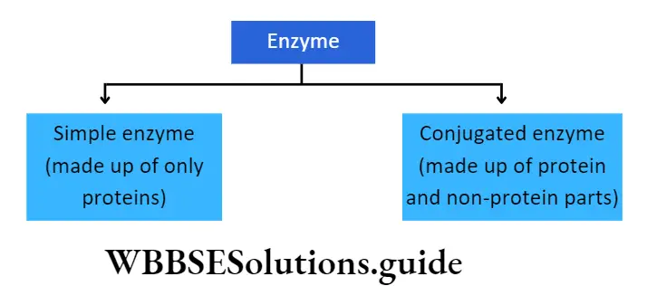
Simple enzyme: It comprises only a protein part called apoenzyme. E.g., pepsin, trypsin, amylase.
Conjugated enzyme: It comprises a protein part or apoenzyme with a non-protein part or cofactor. A cofactor can be inorganic or organic in nature. Conjugated enzymes are also called holoenzymes.
Apoenzyme: The protein part of the enzyme is called apoenzyme. The inactive form of the apoenzyme is known as a proenzyme or zymogen.
The proenzyme may contain several extra amino acids in the protein. These additional amino acids are removed by proteolysis.
This allows the final specific tertiary structure to be formed before it is activated as an apoenzyme.
The apoenzyme is thermolabile. It is destroyed under high temperatures and on application of heat. The apoenzymes have one or more active sites to which co-factors attach and form the holoenzyme.
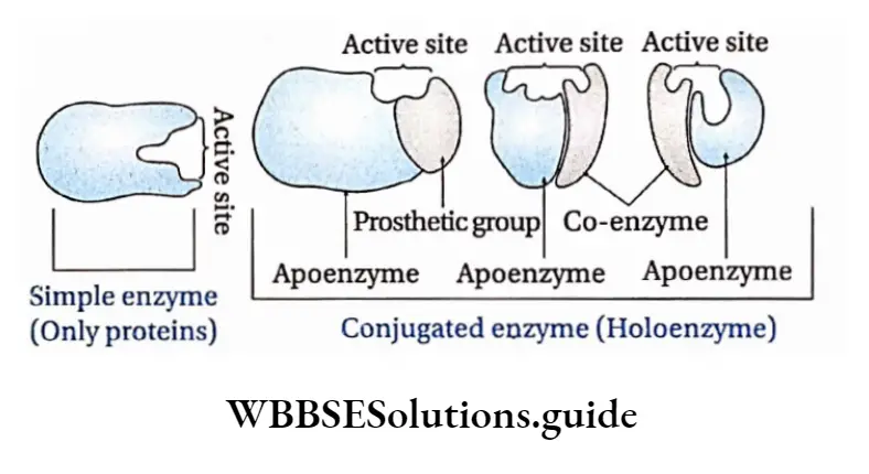

Cofactor: The nonprotein part of the enzyme responsible for the catalytic activity is called cofactor. Cofactor is thermostable. There are three recognized categories of cofactors. These are of the following types
Inorganic ion: Inorganic ions are alternately called metal activators. The different types of inorganic ions like Cu2+, K+, Fe2+, Mg2+, Zn2+, Mn2+, etc., play an important role in activating the catalytic reaction. For example, zinc is a cofactor of the enzyme carboxypeptidase.
Prosthetic group: The organic nonproteneous cofactor or inorganic factor that remains firmly attached to the protein part of the enzyme through a covalent bond and assists in the catalytic activity, is known as the prosthetic group.
For example, in peroxidase and catalase, which catalyze the breakdown of hydrogen peroxide to water and oxygen, haem is the prosthetic group and it is a part of the active site of the enzyme.
Co-enzyme: The organic non-proteinaceous, heat-stable part of an enzyme that remains loosely attached to the apoenzyme, is called a coenzyme.
Usually, they attach only at the time of catalysis. E.g., NAD (Nicotinamide adenine dinucleotide), NADP (Nicotinamide adenine dinucleotide phosphate).
This is derived from nicotinic acid and can exist in both oxidized and reduced forms.
Biomolecules Class 11
Metalloenzyme
The enzymes which contain metal ions, that are directly bound to the protein or to enzyme-bound non-protein components, are called metalloenzymes. E.g., Carbonic anhydrase is a zinc metalloenzyme.
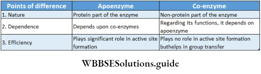
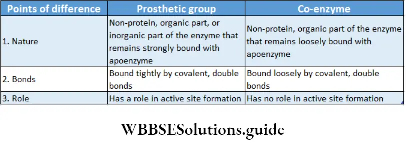
Structure of the enzyme: Enzymes are proteins and are made up of long chains of amino acids. The linear chains may fold to form tertiary structures.
Different enzymes have different sequences of amino acids.
The active site is a location on the enzymes that are formed when the enzymes fold into their functional shape.
The substrate binds to the enzyme at the active site. The complex formed by the union of the enzyme and its substrate is called the enzyme-substrate complex.
Once the substrate has been chemically modified, meaning the reaction ends, it becomes a product.
That product then gets released from the active site of the enzyme.
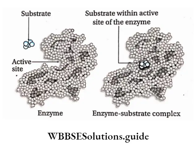
Nomenclature and classification of enzymes
- Enzymes react upon specific substrates. They are named according to the substrate with the suffix-ose or by adding ‘lytic’ to the name of the substrate.
- Enzymes can be named according to the reactions they catalyze. For example, oxidase (this type of enzyme catalyzes oxidation reactions). Enzymes can also be named according to the type of substrate of the reaction they catalyze.
- In such cases, ‘in’ may be added to the name of the substrate. E.g., pepsin, trypsin, etc.
- Enzymes may also be named according to their source. For example, papain is obtained from papaya.
- According to a modern process of naming, enzymes may have two parts in their names.
- The first part denotes the substrate while the second part denotes the reaction catalyzed by the enzyme. Example: glutamate-pyruvate transaminase.
Nomenclature and classification of enzymes based on International rules
- In 1961 the commission of enzymes of the International Glutamic Acid + Pyruvic Acid Union of Biochemistry and Molecular Biology (IUBMB) framed certain rules for the nomenclature and classification of enzymes.
- The commission recognized six different classes of enzymes, which were further divided into subclasses.
- The enzymes are now named according to their systematic position and are given the commission number. In this pattern of naming the first number indicates the class to which the enzyme belongs and subsequent numbers indicate sub-class in that order.
- For example, nitrate reductase, or nitrate oxidoreductase, is named E.C.1.7.1.1. Here, E.C. stands for enzyme commission.
The number 1, denotes the class of enzyme—oxidoreductase.
Here, the number, 7 means that it belongs to the subclass of enzymes that acts on another nitrogenous compound as electron donors.
The next number denotes that the enzyme has NAD+ or NADP+ as an acceptor of electrons.
The last number 1 indicates the above enzyme as the first enzyme of the series. This enzyme helps in the reduction of nitrate to nitrite.
The main six classes of enzymes are discussed under separate heads below.
E.C.l or Oxidoreductases: The enzymes that catalyze oxidation and reduction reactions of their substrate, i.e., the enzymes that catalyze the transfer of H or O atoms or electrons from one molecule to another are known as an oxidoreductase.
E.g., Oxidases (cytochrome oxidase), reductase (nitrate reductase), dehydrogenase (lactate dehydrogenase), etc.
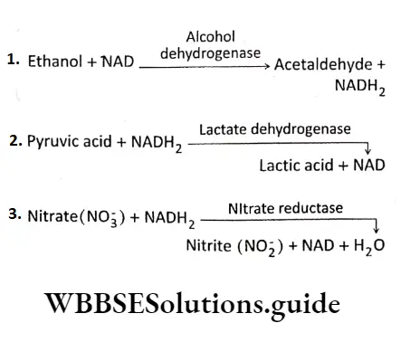
E.C.2 or Transferases: Transferases are the enzymes that catalyze the transfer of a functional group from one molecule to another. E.g., transaminase (leucine Transaminase), Phosphotransferase(2′-Phosphotranferase),etc.
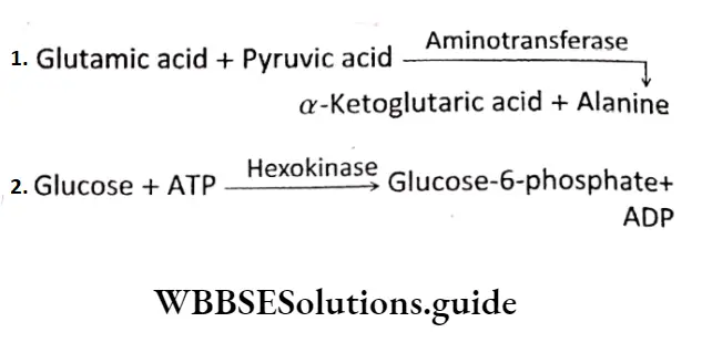
E.C.3 or Hydrolases: The enzymes which catalyze the breaking down of specific bonds of various compounds by the addition of water molecule are known as hydrolases.
In hydrolysis reactions, C-O, C-N, and C-S bonds are cleaved by the addition of H20 in the form of OH- and H+ to the atoms forming the bond. Example Esterase (acetylcholine esterase), glycosidase (protease), peptidase (trypsin), etc.
\(\text { Sucrose }+\mathrm{H}_2 \mathrm{O} \longrightarrow \text { Sucrase } \longrightarrow \text { Glucose }+ \text { Fructose }\) \(\text { Protein }+\mathrm{H}_2 \mathrm{O} \stackrel{\text { Protease }}{\longrightarrow} \text { Peptides }\)E.C. 4 or Lyases: Lyases are the enzymes that catalyze the cleaving of C-C, C-O, C-N, and C-S bonds without hydrolysis or oxidation. E.g., decarboxylase (pyruvate decarboxylase), aldolase (fructose bisphosphate aldolase), etc.
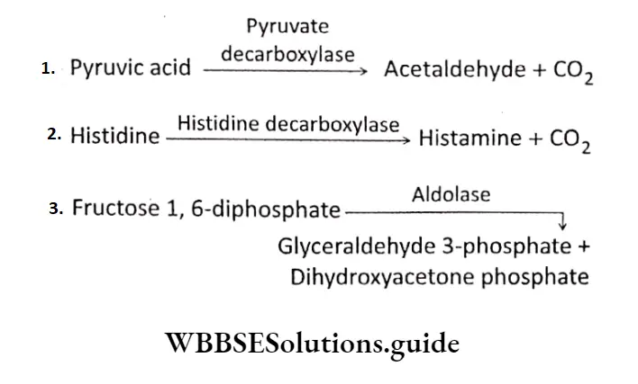
E.C.5 or Isomerases: Enzymes that catalyze the isomerization reactions that cause intramolecular rearrangement of atoms in the substrates and thus form one isomer from another, are known as isomerases. Examples are isomerase (phosphohexo isomerase), epimerase (4-hydroxyproline epimerase), etc.
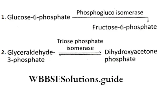
E.C.6 or Ltgasc (Synthetase): Ligases are the enzymes that catalyze the synthesis of C-C, C-S, C-O, and C-N bonds in reactions coupled with the cleavage of high-energy phosphate bonds in ATP or some other nucleotide.
Examples: carboxylase (pyruvate carboxylase), synthetase (glutamine synthetase, aminoacyl tRNA Synthetase), ligase (malate CoA Ligase), Etc.
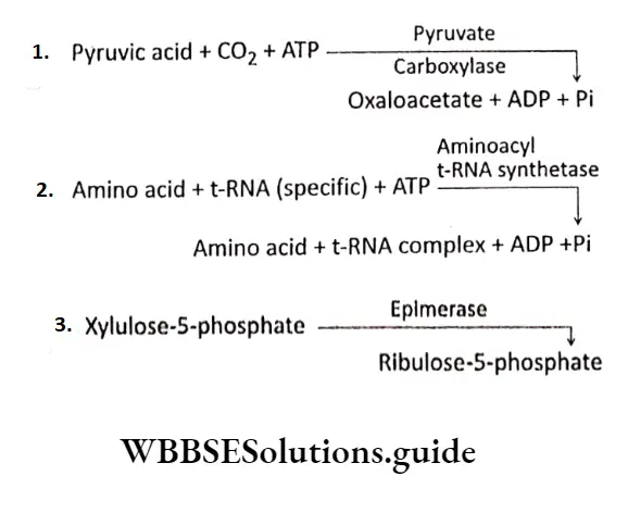
Mechanism Of Enzyme Action
The action of an enzyme depends on different factors. Enzymes are biocatalysts in nature.
All the reactions in a living organism’s body are catalyzed by enzymes.
But to begin the reactions, some amount of energy is required to initiate the reaction. Enzymes enhance the rate of chemical reactions by lowering their activation energy.
Biomolecules Class 11 Activation energy
The activation energy of a reaction is the amount of energy in calories required to bring all the molecules in one mole of a substance at a given temperature to the transition state at the top of the energy barrier.
At this point, there is equal probability for reactants to undergo a reaction to form products or to fall back and remain unreacted.
Michaelis and Menten described the activity of the enzymes with respect to activation energy. They observed that reactions that are not catalyzed by enzymes, occur at a slower rate and require higher activation energy.
For example, hydrolysis of casein requires 20600 kcal/mol in the absence of any enzyme, while only 12600 kcal/mol is required in the presence of the enzyme.
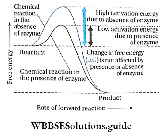
Collision theory
According to this theory, the rate of a chemical reaction is proportional to the number of collisions between reactant molecules.
So, the more often the reactant molecules collide, the more often they react to each other and the faster is the reaction rate.
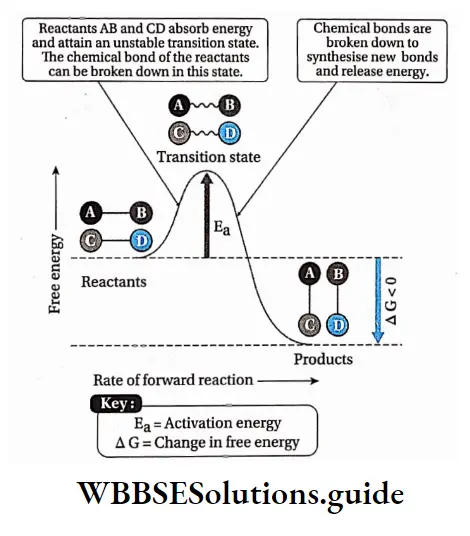
The point in the chemical reaction where there is the maximum value of energy is called the transition state.
The reactant molecules collide with each other, if they hit with enough energy to go through the transition state, they will react and form new molecules.
In the transition state, new bonds are formed while old ones are broken.
Biomolecules Class 11 Models Of Enzyme Action
Two models have been proposed regarding the mode of enzyme action:
Lock and Key hypothesis: According to this hypothesis, the enzyme active site has a configuration complementary to the shape of the substrate so that the enzyme and substrate can recognize and fit each other. This model was proposed by Fischer (1890).
According to this model, Each enzyme [E] has a specific active site or catalytic site into which only the substrate [S] fits.
The enzyme [E] and the substrate [S] both possess complementary conformation, which fits exactly into one another just like a key fits into a lock.
The active site of the enzyme is rigid and fixed, which further helps in the binding.
After binding, the enzyme [E] and the substrate [S] form the ES complex.
This [ES] complex immediately breaks down into the product [P] and enzyme [E] and the product is released from the catalytic site of the enzyme.
\(\mathrm{E}+\mathrm{S} \rightleftharpoons \mathrm{ES} \longrightarrow \mathrm{P}+\mathrm{E}\)Special groups like -NH2, -COOH, etc., help in binding the enzyme and the substrate. Co-enzymes and activators may also influence the binding.
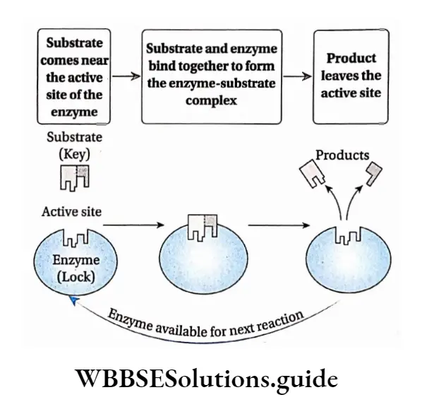
Biomolecules Class 11 Notes
Limitations of the model: Fischer explained enzyme specificity and rigidity of the binding sites of the enzymes.
However, he could not explain the allosteric behavior of enzymes (binding to a molecule at a site other than the active site) which emphasizes that the active site of an enzyme changes its conformational ensemble in order to bind with the substrate.
Induced fit hypothesis: Koshland et al. (1959) suggested another model of enzyme action with some modifications to the lock and key hypothesis.
According to this model, the active site contains two groups—the buttressing region and the catalytic region.
But the tressing region binds the substrate and forms a complex. Catalytic region weakens the bond of the substrate by electrophilic and nucleophilic processes and leads to the formation of the product maximum velocity or Vmax
This hypothesis proposes that the enzymes and their active sites are not rigid, but rather physically more flexible.
When a substrate combines with the buttressing region of an enzyme, it induces conformational changes in the enzyme structure.
This brings the catalytic region opposite to those bonds of the substrate that are to be weakened. The amino acids in the active site are modified into specific structures.
This enables the enzyme to bind with the substrate and perform its catalytic activity effectively.
The substrate changes into a product and gets released from the buttressing site due to structural changes in the site. As a result, the product is released.
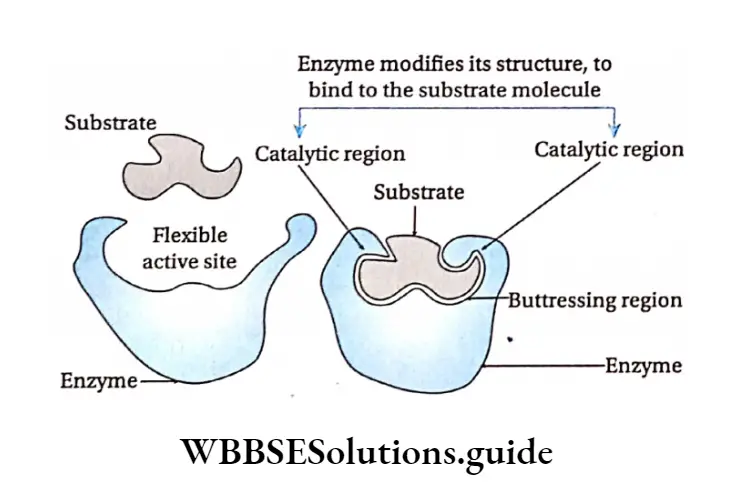
Regulation Of Enzyme Action
The action Of enzymes is regulated by different factors.
Factors affecting enzyme action
Substrate concentration: The catalytic activity of the enzyme is influenced by substrate concentration.
The rate of enzyme action increases with increasing substrate concentration.
If the substrate concentration is increased gradually from a very low concentration, the enzyme activity at first rises proportionately.
If this is plotted on a graph it will show a steep rise in the initial velocity (V0) With further increase in the substrate concentration, the initial rise in velocity of enzyme action falls and ultimately reaches a point where this velocity neither increases nor decreases. This point is called the maximum velocity or Vmax.
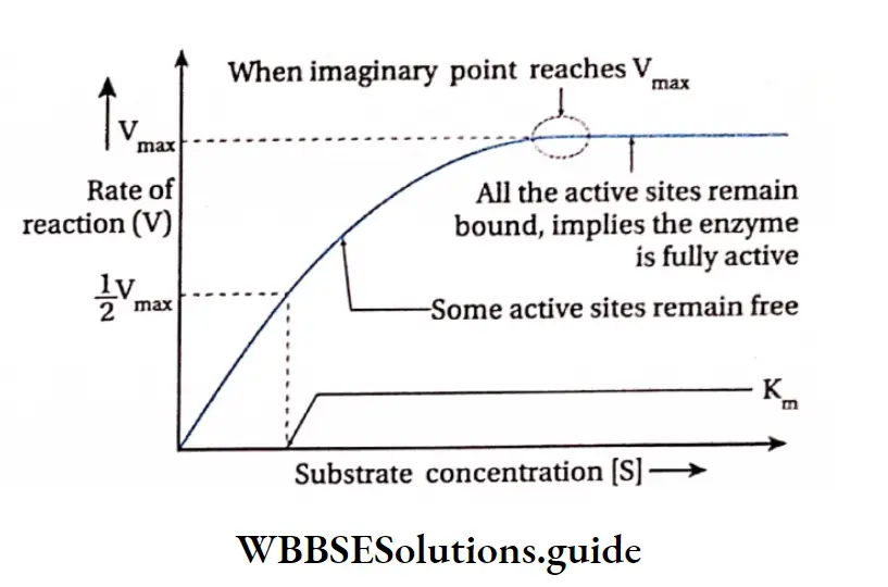
The initial reaction rate, V0 can be described by Michaelis-Menten equation as proposed by Leonor Michaelis and Maud Menten (1930).
\(\mathrm{V}_0=\frac{\mathrm{V}_{\max }[\mathrm{S}]}{\mathrm{K}_{\mathrm{M}}+[\mathrm{S}]}\)where, Vmax= Maximum velocity
S = Substrate concentration
KM = Michaelis-Menten constant
V0 = Initial velocity
The substrate concentration at which half the maximum velocity, -y is attained by an enzyme catalysed reaction is called KM or Michaelis-Menten constant.
When initial velocity (V0) of an enzyme catalysed reaction is half of the maximum velocity (vmax) i.e., V0 \(v_0=\frac{v_{\max }}{2},\) hen the equation changes as follows—
\(\text { When, } v_0=\frac{v_{\text {max }}}{2}\) \(\frac{V_{\max }}{2}=\frac{V_{\max }[S]}{K_M+[S]}\) \(\text { or, } \quad \frac{1}{2}=\frac{[S]}{K_M+[S]} \text { or, } K_M+[S]=2[S]\)or, Km =[s]
Thus the Michaelis-Menten constant [KM] is the amount of substrate concentration [S] when the initial velocity of an enzyme action is half of the maximum velocity \(\left[\frac{V_{\max }}{2}\right]\).
The KM value is an indicator of the affinity that an enzyme has for its substrate. Therefore, the value of KM is different for different enzymes. The KM value of the protease, which acts on a variety of proteins, varies with the type of protein.
An equation with a high KM indicates that the enzyme does not bind efficiently with the substrate, and Vmax will only be reached if the substrate concentration is high enough to saturate the enzyme.
Temperature: Enzymatic reactions usually take place between 20°-40°C, but at high temperature of 60’-70°C they get denatured as they are thermolabile. Generally the enzymatic action is maximum at an optimum temperature of 37C.
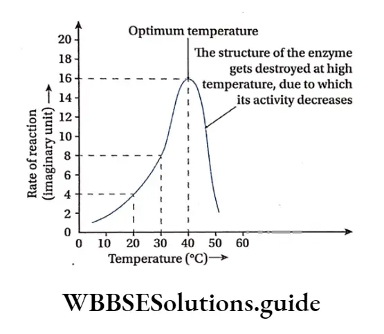
Enzyme concentration: The increase in the concentration of enzyme increases the rate of enzyme action within the cell.
At constant temperature and pH, a high level of substrate concentration increases the rate of enzyme action proportionately.
Product concentration: A high concentration of accumulated product inhibits an enzyme to catalyse a forward reaction.
This type of inhibition is known as feedback inhibition or product inhibition.
pH: Enzymatic action is sensitive to pH. Each enzyme shows its maximum activity at a particular pH known as optimum pH. Some enzymes are active in acidic medium while others in alkaline medium. Each enzyme can act efficiently at its optimum pH. E.g., pepsin acts at an optimum pH of about 2.0 while trypsin acts at an optimum pH of 8.5
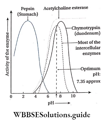
Methods of regulation of enzyme action
Enzyme action can be accelerated or inhibited by allosteric modulation and feedback regulation.
Allosteric modulation: The property of an enzyme by which an effector molecule binds to any site other than the active site of the enzyme, resulting in acceleration or inhibition of enzyme action is known as allosteric modulation.
Allosteric site: Some enzymes possess a second site of activity other than the active site, called the allosteric site. Such enzymes are known as allosteric enzymes.
Allosteric enzymes do not obey Michaelis-Menten kinetics. The effector molecule or allosteric modulator binds to the effector or allosteric site while the substrate molecule binds to the active site for the reaction to take place.
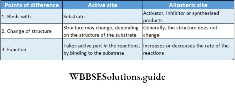
Allosterism: Some modulator molecules upon binding to the allosteric sites of the enzymes influence the enzyme activity. This phenomenon is called allosterism.
Compounds that can speed up the enzyme action are known as allosteric activators or inducers while those which slow down the reaction rate of an allosteric enzyme are known as allosteric inhibitors.
Types of allosteric: According to the property there are two types of allosteric—
Positive allosteric: When an effector or inducer molecule that binds with the allosteric site of an enzyme, enhances the binding capacity or the functional efficacy of the active site, then it is known as positive allosteric. In this case, the reaction proceeds in the forward direction resulting in formation of the product.
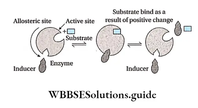
This in turn increases the enzyme activity and hence the rate of the reaction increases. An example of positive allosterism is the binding of oxygen molecules to hemoglobin.
Negative allosteric: Sometimes, when an inhibitor molecule binds with the allosteric site of an enzyme, the conformation of the active site changes. As a result, the substrate cannot bind to it. This is known as negative allosterism.
In this type, the reaction is inhibited so, the product is not formed. This, in turn, reduces the rate of the reaction. For example, when 2,3-bisphosphoglyceric acid binds to an allosteric site of hemoglobin, the affinity towards oxygen decreases in all subunits of the protein.
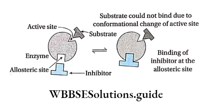
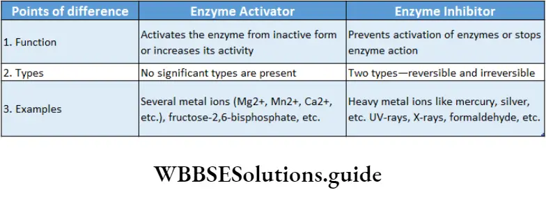
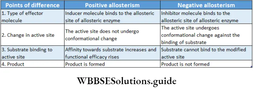
Feedback regulation: When the concentration of the end product of a series of enzymatic reactions regulates the activity of the enzymes involved, the phenomenon is called feedback regulation.
When a large amount of product is produced, it binds to an allosteric site of the first enzyme in that reaction series, thus, inhibiting its activity. When the concentration of the end product decreases, it frees the allosteric site and the enzyme reverses back to its active state.
For example, the amino acid isoleucine is synthesized through a series of biochemical reactions from the amino acid threonine. An adequate amount of isoleucine as an end product in the cell, inhibits the activity of the enzyme threonine deaminase.
It is caused by feedback inhibition which prevents the further synthesis of isoleucine. A decrease in the concentration of isoleucine in the cell, activates the threonine deaminase enzyme by feedback mechanism.
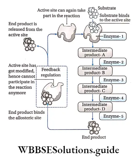
Carbohydrates, proteins, lipids, and nucleic acids notes
Enzyme Inhibitors And Enzyme Inhibition
The substances that inhibit the activity of the enzymes are called enzyme inhibitors and the mechanism by which they do so is called enzyme inhibition.
Types Of Enzyme Inhibition
Enzyme inhibition is of two types—
- Irreversible and
- Reversible.
Irreversible inhibition: The method of inhibition by which the activity of the enzymes is inhibited permanently and cannot be restored, is called irreversible inhibition.
Characteristics: An irreversible inhibitor binds to the active site and changes its structure permanently or destroys the protein structure of an enzyme.
It stops the enzymatic activity and the enzyme cannot be recovered.
Heavy metal ions, SH groups, radiations like UV rays, X-rays, are responsible for irreversible inhibition.
For example, salts of cyanides inhibit the activity of cytochrome oxidase essential for cellular respiration, resulting in cell death.
Reversible inhibition: The method of inhibition by which the activity of an enzyme is inhibited temporarily is called reversible inhibition.
Characteristics: In this type of inhibition, some inhibitor molecules may attach to the active site by non-covalent interactions.
It may also involve feedback inhibition or modification of the active site by positive modulator, etc.
When these inhibitors are released from the active site, the enzyme reverses back to the functional state.
Types: There are three types of reversible inhibition—competitive, non-competitive and uncompetitive.
Competitive inhibition: Competitive inhibitors are molecules that are structurally similar to the substrates and compete with the substrates to bind at active sites.
This leads to slowing down of the reactions. The mechanism is called competitive inhibition. Competitive inhibitors increase KM value, thus decrease substrate affinity of the active site. Vmax remains unchanged.
For example, malonate is the competitive inhibitor of succinate dehydrogenase. Malonate binds to the active site of succinate dehydrogenase, preventing succinate from binding to it.
Non-competitive inhibition: Non-competitive inhibitors have no structural similarity with the substrate but may form an inhibitor-enzyme complex at the allosteric site of the enzyme.
It inactivates an enzyme by changing the structure of the active site. Thus, the substrate can no longer recognise or bind to the active site of the enzyme.
This prevents the enzyme-substrate complex formation. Non-competitive inhibitors lower the Vmax of an enzyme but keep the value of KM unchanged.
For example, non-nucleoside reverse transcriptase inhibitors or NNRTIs (like Etravirine) can bind to the reverse transcriptase enzyme of HIV, inhibiting the synthesis of viral RNA without interfering with substrate binding. This inhibitor is used as an anti-HIV drug.
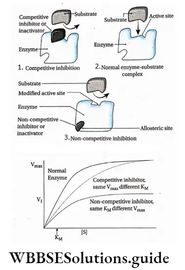
Uncompetitive inhibition: Uncompetitive inhibitors bind to the enzyme after the formation of the enzyme-substrate complex.
It binds close to the active site. This stops the action of the enzyme by altering the structure of the enzyme-substrate complex.
As a result, KM decreases. The Vmax also decreases as a result of the removal of activated complexes.
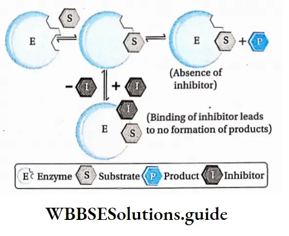
Significance Of Enzyme Inhibition
- Excess products can be regulated by feedback inhibition.
- An idea about the different metabolic reactions within the body can be derived.
- Structure and activity of the enzymes can also be explained by these theories.
- Different medicines and pesticides can be designed applying the knowledge of enzyme inhibition.
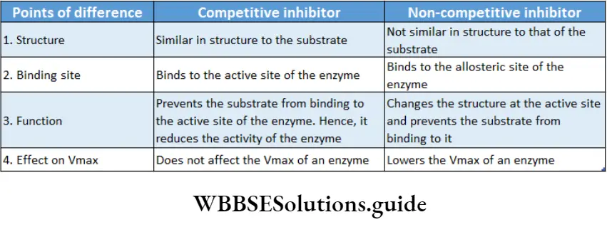
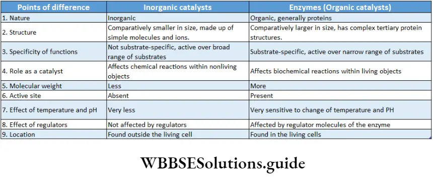
Denaturation of enzymes
High temperature, high energy radiation (UV rays, X-rays, etc.), heavy metals (Ag+, Hg 2+, As+), salts, concentrated acids, alkali, etc., affect the active site of the enzymes. This in turn changes the structure of the proteins, breaking the hydrogen bonds. This finally destroys the enzyme integrity permanently.
Isoenzyme or Isozyme
- The different forms of an enzyme, differing only in certain amino add sequences but catalyzing the same reaction, are called isoenzymes.
- Generally, different isoenzymes are found within different cells and tissues.
- Due to slight differences in the molecular structure, they have different properties (like favorable pH, affinity towards the substrate, the effect of inhibition, etc.).
- For example, the lactate dehydrogenase enzyme of human cells has five isoenzymes.
- An enzyme, a-amylase, of wheat endosperm has 16 isoenzymes. Alcohol dehydrogenase of maize has 4 isoenzymes.
Allozyme
Allozymes or allozymes are variant forms of an enzyme that are coded by different genes of the same locus. E.g., DNA polymerase.
Proenzymes or Zymogens
Proenzymes or zymogens are biologically inactive substances which are metabolized into enzymes. For example, HCl of gastric juice alters the inactive pepsinogen to an active pepsin enzyme.
Biomolecules Notes
- Amphiphilic: A molecule having both hydrophilic and hydrophobic parts.
- Calorific value: The energy contained in food or fuel, determined by heat produced by the complete combustion of its unit quantity. This is expressed in joules per kilogram.
- Chiral molecule: A molecule that is non-superposable on its mirror image.
- Culture medium: It is a solid or liquid mixture of various substances, designed to support the growth of microscopic organisms, plants, etc., in artificial environmental situations.
- Furfural: It is a heterocyclic (ring structure) aldehyde compound having a chemical formula of C5H4O2. It is a colorless oily liquid, which quickly darkens when exposed to air.
- Glycocaiyx.’The glycocalyx is a glycoprotein-polysaccharide covering that surrounds the cell membranes of some bacteria.
- Hexosamine: It is an amino sugar produced by the addition of an amine group to a hexose. Example glucosamine (based upon glucose), fructosamine (based upon fructose), etc.
- Fmmunog robin: Any of several classes of structurally related proteins, found in serum, other body fluids, and tissues, that help in fighting diseases is called immunoglobin or antibody.
- Melanin: A pigment, produced by melanocyte cells, that gives color to human skin, hair, and eyes.
- Mucopolysaccharides: These are heteropolysaccharides composed of hexosamines and repeating units of monosaccharides.
- Mutation: A relatively permanent, heritable alteration in the genetic material involving a physical or a biochemical change.
- Myelin sheath: It is a covering made of myelin, a fatty colorless substance, around the axon(long slender projection) of some neurons and acts as an electrically insulating layer.
- Neurotransmitters: The chemicals that enable transmission, i.e., conduction of electrical impulses through the nerve cells and across chemical synapses or junctions between adjacent nerve cells.
- Non-polar molecule: If electrons are shared equally by the atoms forming a molecule, then there is no resulting charge and the molecule is non-polar.
- Nucleosome: A structural unit of an eukaryotic chromosome, consisting of a length of DNA coiled around a core of histone proteins.
- Polar molecule; A polar molecule has a partial positive charge in one part of the molecule and a complementary negative charge in the other part.
- Polarized light: The light waves in which vibrations occur in a single plane.
- Spectrophotometer: An apparatus for measuring the intensity of light in a part of the spectrum, especially as transmitted or emitted by particular substances.
- Stereoisomer: Each of two or more compounds differing only in the spatial arrangement of their atoms.
- Sugar acids: A monosaccharide containing -COOH group. Thermolabile: A substance that can be destroyed by the application of heat.
- X-ray diffraction: It is a rapid analytical technique primarily used for the identification of crystalline substances and crystalline phases in a material and can provide information on its atomic and molecular structure. It is also called X-ray crystallography.
- Zwitterion: A neutral molecule having both positively and negatively charged groups.
Points of remember
- Sugar acids: A monosaccharide containing -COOH group.
- Thermolabile: A substance that can be destroyed by the application of heat.
- X-ray diffraction: It is a rapid analytical technique primarily used for the identification of crystalline substances and crystalline phases in a material and can provide information on its atomic and molecular structure of it.
- It is also called X-ray crystallography.
- Zwitterion: A neutral molecule having both positively and negatively charged groups.
- Most enzymes are conjugated proteins.
- The activity of an enzyme can be determined by its turnover number. It is the maximum number of chemical conversions of substrate molecules per second that an active site will execute for a given enzyme concentration.
- The activity of an enzyme depends on the nature and concentration of substrate and products, temperature, and pH.
- The iodine test is performed to determine the presence of starch. Iodine reacts with starch to produce a deep blue color.
- To determine the presence of reducing sugars, Benedict’s test is performed.
- Copper sulfate reacts with the reducing sugars to produce a brown/red-colored precipitate of Cu20. This determines the presence of reducing sugars.
- To distinguish between the aldose and the ketose sugars, Seliwanoff’s test is done, if a red-colored precipitate is obtained by a reaction between the sample solution and Seliwanoff’s reagent (HCL+ resorcinol), the sample sugar is a ketose sugar.
Neural Control And Coordination Notes
Neural Control And Coordination Introduction
As you are reading this book, Think about the organs That are Functioning with functioning within you right now! Your eyes are sensing the light reflected off this page.
Your brain is decoding the meanings of the words you are reading. Think about the processes that are running within your body. You are breathing, perhaps perspiring, feeling the chair on which you are sitting, and your heart is beating.
All of these are being regulated by an organ system inside your body. It is the neural system or nervous system.
The nervous system controls various metabolic and physiological functions of the body.
Different organs of the body are directly connected to the nervous system. The actions of these organs are coordinated by the nervous system. This is known as neural coordination.
Read and Learn More: WBCHSE Notes for Class 11 Biology
Neural System
All organ systems of animals are controlled and coordinated effectively by the neural system or the nervous system. The neural system allows the organism to interact with its environment, appropriately.
This system functions at the cellular level through nerve cells or neurons.
It receives sensory information, interprets it, and generates appropriate responses using organs like muscles. The neurons set up the neural system in a number of discrete anatomical locations in the brain.
These systems send and receive impulses from the sense organs and other receptors of the body.
Thus, neural control provides the basis for body functions such as perception of senses, attention, memory, emotions, rational thinking, etc.
| Class 11 Biology | Class 11 Chemistry |
| Class 11 Chemistry | Class 11 Physics |
| Class 11 Biology MCQs | Class 11 Physics MCQs |
| Class 11 Biology | Class 11 Physics Notes |
Morphology of leaf structure and its modifications
Neural system of different animals
- Sponges (Porifera) do not have any neural system, Hydra (Cnideria) has a neural system that is composed of a network of neurons.
- Planaria (Platyhelminthes) have two nerve cords that converge to form a rudimentary brain.
- The neural system of earthworm (Annelida) is composed of a single nerve cord and paired nerve ganglia.
- The neural system of arthropods is better organized, It consists of the brain, ganglia, and neural tissue.
- The neural system of vertebrates is highly developed.
The nervous system of humans and other advanced vertebrates consists of three parts—
- Brain,
- Spinal cord and
- Nerve.
The brain and spinal cord form the central nervous system while nerves form the peripheral nervous system.

Neurons As Structural And Functional Units Of Neural System
Though neurons are the main structural and functional components of the nervous system some other cells also take part in maintaining the proper functioning of this system.
The cells of the nervous system can be divided into two broad categories—
- Nerve cells (or neurons) and
- Supporting cells called neuroglia (or simply glia), are discussed below
Neuron
The human brain alone contains over 100 billion neurons. Each neuron can have up to 10,000 connections to other neurons in the brain.
Types of leaves and their modifications with examples
Neuron Definition: The Structural And Functional units of the nervous system which are capable of Sending and receiving nerve impuluses is called a neuron.
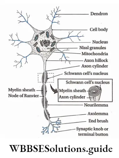
” class 11th biology chapter 21 notes”
Different Parts Of A Neuron
A typical neuron consists of a cell body (perikaryon or soma) and cellular processes called dendrons and axons. Different parts of a neuron are discussed below.
Cell Body or soma or perikaryon or neurocyton: The swollen part of a typical neuron, containing cytoplasm and nucleus, from which cellular processes emerge, is known as the cell body.
Different parts of the cell body are as follows—
- The cell membrane forms the external boundary of the neuronal cell body and its processes. It is also known as a neurilemma.
- The nucleus is located at the center of the cell body. The contents of the nucleus are enclosed within a double-layered nuclear membrane.
- The cytoplasm is known as neuroplastic.
Neuroplasm contains—
- Microtubules—These are hollow rod-like filaments that are composed of a globular protein named tubulin.
- Neurofilaments— These are thinner than microtubules but thicker than microfilaments. They are a major Component Of the neuroral Cytoskeleton.
- Microfilaments— These Are Thinner Than The Neurofilaments. They consist of two intertwined strands, each of which is a polymer of actin protein subunits. They help in the maintenance of cell shape, changing of cell shape, and in cytoplasmic streaming.
- Neurofibrils— Neurofibrils are filamentous structures or microscopic fibrils that are found running in every direction through the nerve cell. They provide the neuron support and help to give it a shape,
- Nissl granules— These are the granular material present throughout the entire cell body and proximal portions of the dendrites. However, it is not present in the axon hillock (a portion of the soma from which the axon arises). The Nissl granules consist of ribosomes and endoplasmic reticulum.
- Centrosome— Nerve cells are incapable of division. So, it is assumed that neurons lack centrosomes. However, under the electron microscope, a non-functional centrosome has been observed,
- Other organelles—The cell organelles, commonly found in animal cells are also present within the neuroplasm. These include the Golgi complex, endoplasmic reticulum, mitochondria, lysosomes, etc.
- Storage material—The cytoplasm of a neuron contains glycogen, lipids, etc., as storage material They consist of two intertwined strands, each of which is a polymer of actin protein subunits. They help in the maintenance of cell shape, changing of cell shape, and in cytoplasmic streaming.
- Neurofibrils— Neurofibrils are filamentous structures or microscopic fibrils that are found running in every direction through the nerve cell. They provide the neuron support and help to give it a shape,
- Nissl granules— These are the granular material present throughout the entire cell body and proximal portions of the dendrites. However, it is not present in the axon hillock (a portion of the soma from which the axon arises). The Nissl granules consist of ribosomes and endoplasmic reticulum.
- Centrosome— Nerve cells are incapable of division. So, it is assumed that neurons lack centrosomes. However, under an electron microscope, a non-functional centrosome has been observed,
- Other organelles—The cell organelles, commonly found in animal cells are also present within the neuroplasm. These include the Golgi complex, endoplasmic reticulum, mitochondria, lysosomes, etc.
- Storage material—The cytoplasm of a neuron contains glycogen, lipids, etc., storage material.
Cellular processes: The cytoplasmic, thread-like appendages that emerge from a cell body are known as cellular processes. These projections are of two types—dendrons and axons.
Dendron: The short cellular processes emerging from the cell body of a neuron that receives impulses from another neuron is called dendron (from the Greek dendron = tree branch).
Dendron Structure:
- The number of dendrons can be from one to many,
- Branches of dendrons are known as dendrites,
- Nissl granules, ribosomes, smooth endoplasmic reticulum, neurofilaments, micro-filaments, microtubules, and mitochondria are present in the dendrites,
- Dendron lacks myelin or medullary sheath.
Dendron Function:
- The dendrite is the impulse-receiving part of the neuron,
- Dendrites increase the surface area for receiving signals from other neurons.
Axon: A long cellular process emerging from a neuron that transmits nerve impulses from the cell body to another neuron is called an axon.
Dendron Structure:
- In most cases, a neuron consists of one axon. Apoiar neuron lacks any axon,
- The plasma membrane of the axon is called the axolemma and its cytoplasm is called axoplasm.
- The axoplasm does not contain the Nissl substance or Golgi apparatus. But the axoplasm does contain mitochondria, microtubules, and neurofilaments,
- Depending upon the presence or absence of myelin sheath around axons, they are either myelinated or non-myelinated, respectively.
- In myelinated axons, the part that lacks myelin forms a ridge due to the fusion of neurilemma and axolemma. This region is called the node of Ranvier. Generally, nodes of Ranvier are distributed at a distance of 1-3.
- At their distal ends, the axons branch extensively. Their terminal ends, which are mostly enlarged, are called synaptic terminals (synaptic buttons)
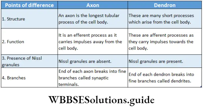
Neurons are classified into different categories on the basis of certain features which are as follows-
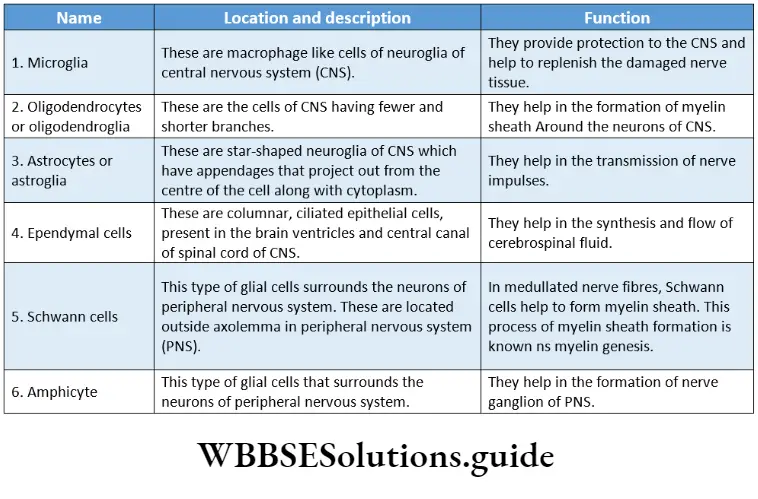
Detailed structure of a leaf with labeled diagram
Neuroglia Or Glial Cell
Neuroglia Or Glial Cell Definition: The cells of the nervous system other than neurons that are incapable of generating and transmitting nerve impulses but provide structural and functional support to the neurons are called neuroglia or glial cells.
Neuroglia Or Glial Cell Characterises:
- Neuroglia are incapable of transmitting nerve impulses.
- These cells can divide (unlike nerve cells).
- The brain contains more neuroglial cells than neurons.
- The appendages of some glial cells (astrocytes) are thinner at their terminal end and form special structures called end-feet that remain associated with blood capillaries.
Ganglion
The region outside the CNS where there is an aggregation of numerous nerve cell bodies, is known as ganglion.
Ganglion Example: 31 pairs of nerves have originated from the human spinal cord. Two main nerve roots—dorsal nerve root and ventral nerve root, combine to form spinal nerves.
But before their meeting point, a thinner region is seen where numerous neurons remain associated with each other. This region is known as the dorsal ganglion.
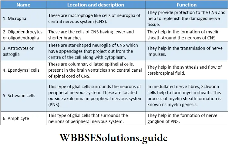
Nerve
Nerve Definition: A cord-like structure, comprising a collection of neurons, blood vessels, lymph vessels, and adipocytes enclosed by a layer of connective tissue, that conveys impulses throughout the body is called a nerve.
Structure Of Nerves
Nerves emerge from the brain and spinal cord and branch out to almost all parts of the body. The nerves are composed of nerve fibers or neurons.
These nerve fibers are bundled together like strands of an electric cable enclosed within a connective tissue covering.
Connective tissue coverings: On the outer side of the nerve, there is a covering made up of connective tissue.
- Blood vessels, lymph vessels, and adipocyte cells are found within it.
- This covering consists of three layers—endoneurium, perineurium, and epineurium.
Endoneurium: It is the innermost layer that covers up the individual nerve fibers. This layer is relatively thin.
Perineurium: A collection of nerve fibers that are individually bound by endoneurium, is again surrounded by another connective tissue layer called the perineurium.
The bundle of nerve fibers bound together by perineurium is called fasciculus (plural: fasciculi).
Epineurium: Several fasciculi are again bound together by another layer of connective tissue. It is called epineurium. It is the outermost layer of connective tissue layer covering the nerve.
This layer is usually thick and rich in blood vessels, lymph vessels, adipocytes, and collagen fibers.
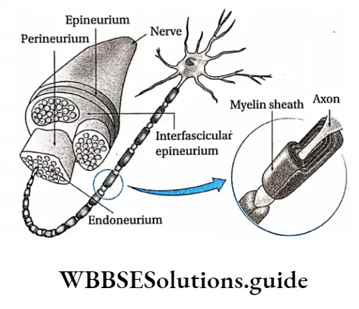
Parts of a leaf and their functions in plants
Nerve fibers: A nerve may have a few to more than a million nerve fibers. These are actually axons of different neurons. Several of these fibers remain bound together beneath the connective tissue coverings.
Types Of Nerves
Nerves are of different types. Their classification on the basis of several criteria has been discussed below
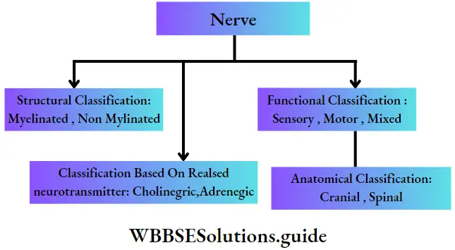
Structural classification of nerves: Based on the presence or absence of myelin sheath, nerves are classified as—
Myelinated nerve: Medullated or myelinated nerves are nerves with large diameters. The nerve fibers of medullated nerves remain wrapped with a protein-lipid sheath called myelin sheath, through which nerve impulse is conducted rapidly.
This type of nerve is made of nerve fibers consisting of three main components. These components are—
- Axis cylinder— It is the central core of the axon of a neuron. There is a thin covering called an axolemma that surrounds the axon. The axolemma is again enclosed by an outer covering called a myelin sheath.
- Myelin sheath—It is an external covering of the axon. It is made up of protein and lipids. It provides insulation to the nerve fiber and hence prevents the spreading of nerve impulses to adjacent tissues. In the nerves of the peripheral nervous system (PNS), the myelin sheath is produced by a type of glial cell called Schwann cell. Nodes of Ranvier are present in this type of nerve.
- Neurilemma—There is another covering external to the myelin sheath, which is called neurilemma.
Example: This type of nerve fiber is found in the white matter of the central nervous system.
Non-myelinated nerve: Non-medullated or non-myelinated nerves are nerves of small diameter. The nerve fibers of these nerves are devoid of the myelin sheath. Through these nerves, a nerve impulse is conducted very slowly. This type of nerve fiber is covered with an external neurilemma. They are devoid of nodes of Ranvier.
Example: This type of nerve fiber is found in the grey matter of the central nervous system.
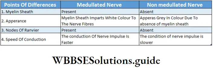
Functional classification of nerves: Based on the functions carried out by nerve fibers, nerves are classified into three kinds. These are—
Sensory nerves: These nerves contain sensory fibers. Sensory nerves carry impulses from sense organs to the brain or the spinal cord. These nerve fibers are also called afferent nerve fibers. The nerve impulse conducted by sensory nerve fibers is called sensory impulse.
Examples of sensory nerves are—the olfactory nerve, optic nerve, auditory nerve, etc fibers.
Sensory nerves carry impulses from sense organs to the brain or the spinal cord. These nerve fibers are also called afferent nerve fibers. The nerve impulse conducted by sensory nerve fibers is called sensory impulse.
Examples of sensory nerves are—the olfactory nerve, optic nerve, auditory nerve, etc.
Anatomical Classification: Based on the anatomical origin of nerves, these are classified as—
Cranial nerves: These nerves originate from the brain. They may be entirely motor-sensory or mixed.
The human nervous system consists of 12 pairs of cranial nerves.
Spinal nerves: It is always of a mixed type originating from the spinal cord. The human nervous system consists of 31 pairs of spinal nerves.
Classification based on neurotransmitter: Based on neurotransmitter released, nerves are classified as—
Adrenergic nerves: These nerves synthesize and release adrenaline and nor-adrenaline.
Cholinergic nerves: These nerves synthesize and release acetylcholine (ACh).
“class 11 neural control and coordination “
Nervous System In Humans
The nervous system in the human body is an organ system that coordinates the functioning of all the systems of the body and helps the body to respond to external stimuli.
Functions Of Nervous System
The main functions of the nervous system in humans and other animals are described below.
Response to external stimuli: The human nervous system is equipped to sense and respond to continuous changes within the body and in its external environment.
Control and coordination: The nervous system works along with the endocrine system to communicate, integrate, and coordinate the functions of various organs and organ systems in our body. It also helps the body to respond to the external stimuli.
Higher mental functions: It also acts as a seat of self-consciousness, intelligence, thoughts, and emotions.
Maintenance of balance with the environment: Humans And Other higher animals sense the changes in the environment and Act Accordingly, Both with the help of the nervous System.
Control of reflex action: Grey matter of the spinal cord, within the central nervous system, controls the reflex actions of the body.
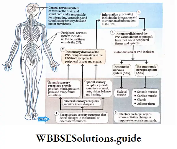
Mechanism Of Action Of The Nervous System
The action of the nervous system is accomplished in some stages. These are—
Receiving stimulus: Mechanical, chemical, or physical changes that excite the body or body parts, are known as stimuli.
Stimulus is received by the receptors present in the eyes, ears, nose, tongue, skin, and other parts of the body.
Transmission of impulse to the central nervous system: The received stimulus is transmitted to the central nervous system as a sensory nerve impulse, by sensory nerves.
Sending instructions from the central nervous; system to the target site: The sensory information is analysed in the brain and spinal cord of the central nervous system.
Instruction for necessary action is then sent as a motor impulse, through the motor nerves, to the effector organizer
The accomplishment of action: As per instruction from the central nervous system, actions are completed in the effector area or organizer:
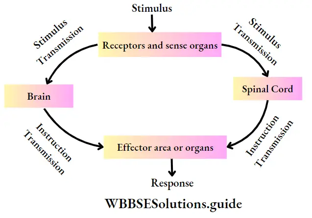
Divisions Of Human Nervous System
The human nervous system comprises the following divisions—
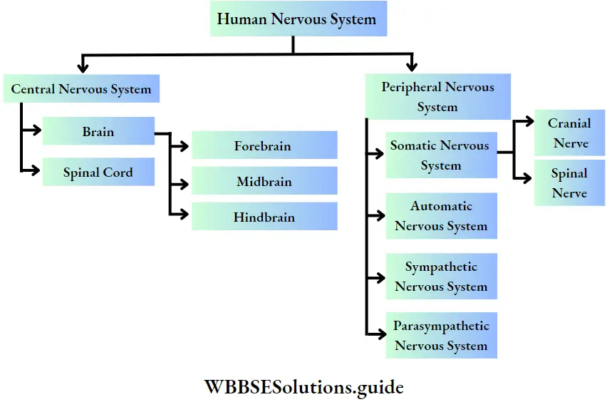
” neural control and coordination notes pdf download”
Central Nervous System
Central Nervous System Definition: The structural and functional center for the entire nervous system comprising the brain and spinal cord is called the central nervous system or CNS.
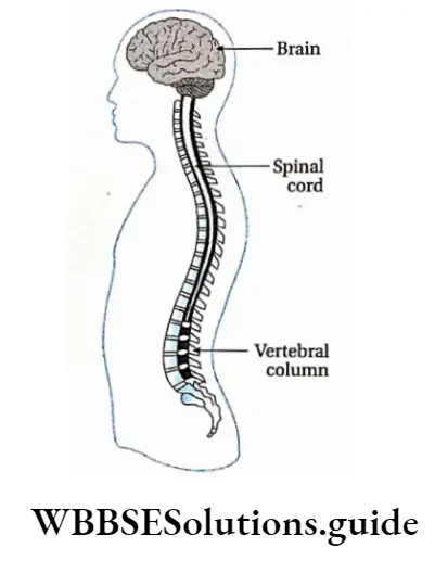
Central Nervous System Characteristic features:
- The components of the CNS are the brain and the spinal cord.
- The brain is located within a bony cavity called the cranium or brain box. The dorsal part of the brain bears an opening called the foramen magnum.
- Through this opening, the spinal cord enters the vertebral column. The spinal cord is present within the neural canal of the vertebral column. It runs upto the end of the vertebral column.
- Outside the CNS, three layers of protective tissues are present, which are referred to as meninges.
- These meninges surround the brain and the spinal cord providing additional layers of protection to. The three layers of meninges are—
- Dura mater,
- Arachnoid mater and
- Pia mater.
- The space between the dura mater and the arachnoid mater is called subdural space.
- The small space between the arachnoid mater and the pia mater is called subarachnoid space. Within this space cerebrospinal fluid (CSF) is present.
- At the end of the spinal cord, the pia mater extends upto the coccyx like a thin thread, termed filum terminale. This part is composed of fibrous connective tissue.
- Human CNS has grey matter and white matter beneath the layer of meninges. In the CNS of the brain, the white matter lies beneath the grey matter.
- But in the spinal cord, the grey matter lies beneath the white matter. CNS of vertebrates comprises four brain ventricles. The spinal cord consists of one central canal or neurocoel.
- Note that brain ventricles (chambers within the brain) and central canal or neuronal are completely filled by cerebrospinal fluid (CSF).
- Within CNS, grey matter is present like an island in white matter. This is known as the nucleus.
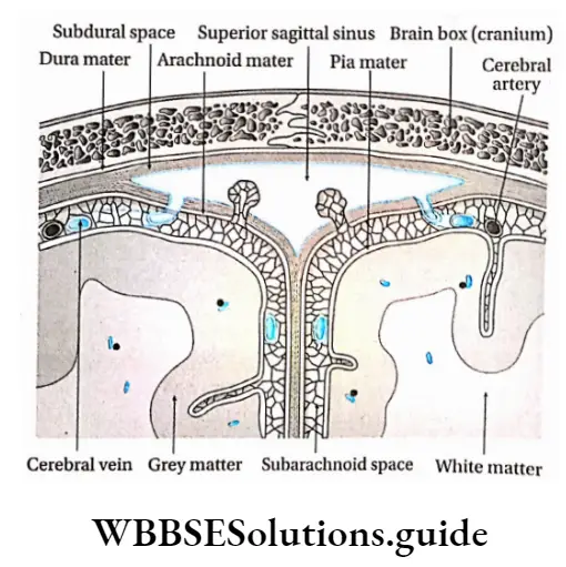
Central Nervous System Structure: The central nervous system is mainly composed of two parts—the brain and the spinal cord. These are described below in detail.
Brain Ventricles
- The human brain contains four connected cavities called brain ventricles. The cerebrum of the forebrain has two cerebral hemispheres.
- The first and second ventricles are located there laterally and are called lateral ventricles.
- The diencephalon of the forebrain includes two parts—the hypothalamus and epithalamus.
- The third ventricle is present between the epithalamus and the hypothalamus and is connected to the lateral ventricles by an interventricular pore, known as the foramen of Monroe.
- The fourth ventricle is located within the cerebellum and pons of the metencephalon of the hindbrain.
- The fourth ventricle is connected to the third ventricle by a tubule called the aqueduct of Sylvius.
- The fourth ventricle remains connected to the central canal of the spinal cord. Two holes are present in the fourth ventricle—the foramen of Lusaka and the foramen of Magendie.
- Through these pores, the cerebrospinal fluid moves upward to enter into the subarachnoid space.
Brain
Brain Definition: The swollen part of the central nervous system located in the brain box, having cavities filled with CSF and Procted By meninges, is called Brain.
Brain Location: The brain is located in the cranial cavity of the human skull.
Brain Structure: The brain of vertebrates is made up of 100 billion neurons and a large number of neuroglia.
The approximate weight of an adult human brain is 1.36 kg. The different parts of the human brain are shown below in the given chart.
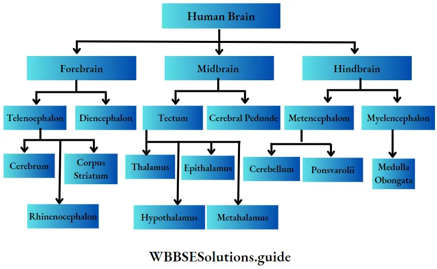
Forebrain or Prosencephalon
The forebrain has two subdivisions—the telencephalon and the diencephalon.
Telencephalon: Telencephalon is again subdivided into three parts namely, cerebrum, corpus striatum, and rhinencephalon.
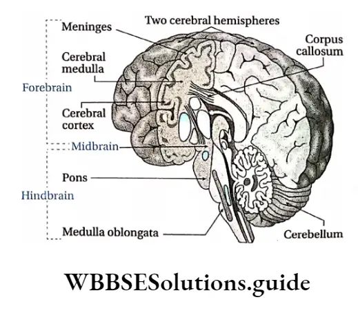
Brain Structure: Cerebrum is divided into two symmetrical semicircular parts called cerebral hemispheres.
The surface area of the cerebral hemisphere is greatly folded, accommodating several layers of neurons.
This area is called the cerebral cortex. Each fold is known as gyrus (plural: gyri) and the groove or depression between them is called sulcus (plural: sulci).
Deep and wide sulci are called fissures. Fissures divide the cerebral hemisphere into a number of lobes.
These are—
- Frontal lobe,
- Parietal lobe,
- Occipital lobe,
- Temporal lobe.
The fissure between the frontal and parietal lobes is called the fissure of Rolando or central sulcus.
The fissure between the parietal and temporal lobes is known as the Sylvian fissure. The fissure between the parietal and occipital lobe is called the parieto-occipital fissure.
Beneath the cerebral cortex, there lies the cerebral medulla. It is made up of white matter which is mainly composed of nerve fibres.
The two cerebral hemispheres are linked by a bundle of longitudinal nerve fibers called the corpus callosum. The cerebrum contains pyramidal (Betz), stellate, and spindle neurons.
Brain Functions:
The cerebrum controls the action of different voluntary muscles, the movement of eyelids and eyeballs,
Different areas of the cerebral cortex control our ability to speak (Broca’s area) and write (Wernicke’s area),
The temporal lobe contains the auditory cortex and is involved in auditory perception,
Similarly, the occipital lobe contains the visual cortex and is responsible for visual processing.
Different areas of the cerebral cortex are also responsible for the perception of other sensations like taste, smell, touch, pressure, pain, heat, and cold.
The cerebrum is the center for memory, intelligence, judgment, conscious thought, and other activities associated with thinking and feeling.
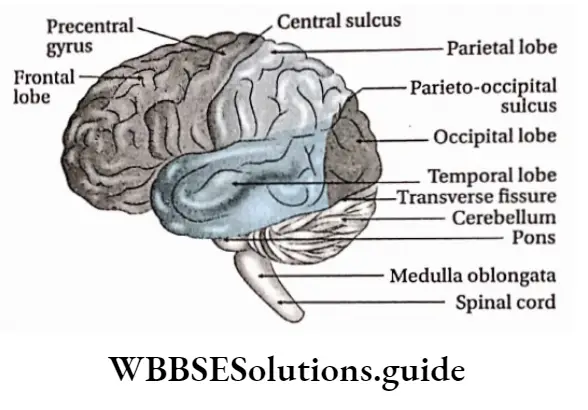
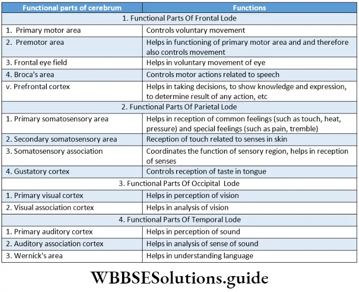
Corpus striatum: It is made of grey matter suspended within white matter within the cerebrum.
This part of the brain constitutes a structure called basal ganglia. The Corpus striatum helps in the modification of functions of the motor region in the cerebrum.
Rhinencephalon: The ring-like structure around the brain stem is known as rhinencephalon. The rhinencephalon helps in sensing smell.
Limbic System
The system that is composed of parts of the telencephalon, diencephalon, and mesencephalon and is expanded at the periphery of the cerebral cortex and diencephalon, controlling human emotions, feelings, inspiration, etc., is called the limbic system. The term limbic’ means border or boundary.
The parts of the diencephalon that are associated with the limbic system are—the hypothalamus and the nucleus of the frontal thalamus.
The parts of the telencephalon such as the amygdala, hippocampus, septal nuclei, para-olfactory lobe, and limbic cortex form the structural components of the limbic system. Other than these, reticular formations of the midbrain are also associated with the limbic system.
In human beings, the primary function of the limbic system is to control emotions such as fear, hopelessness, anger, depression, happiness, etc.
Diencephalon: It is located between telencephalon and mesencephalon. The diencephalon is situated between the two cerebral hemispheres.
It has four parts, namely—hypothalamus, epithalamus, metathalamus, and thalamus.
Hypothalamus: It is a small cone-shaped structure of the brain lying below the thalamus. It makes up the floor of the third cerebral ventricle projects downwards from the brain and ends in the pituitary gland.
Location: The hypothalamus is located below the thalamus, just above the brain stem, and anterior to the third ventricle.
Structure: Hypothalamus consists of numerous neurosecretory cells. Near infundibulum (pituitary
Limbic System Structure: Hypothalamus consists of numerous neurosecretory cells. Near the Infundibulum (pituitary stalk) of the hypothalamus, two optic nerve crosses each other.
This region of crossing is known as optic chiasma. The hypothalamus also consists of numerous hypothalamic nuclei.
Limbic System Function:
- The hypothalamus plays a significant role in the regulation of body temperature (thermoregulation),
- The hypothalamus regulates our appetite and feeding behavior,
- The hypothalamus also helps in the maintenance of water balance in our body.
- The hypothalamus also helps to maintain the normal functioning of the cardiovascular system.
- The hypothalamus regulates the action of the autonomic nervous system,
- The neurosecretory cells situated in the hypothalamus secrete neurohormones.
- These act as releasing factor or inhibitory factor, that controls the secretion of different hormones from adenohypophysis or anterior pituitary
- The hypothalamus helps to regulate circadian rhythm. Different functions of our body like sleep, wakefulness, etc., take place in a cyclic way following the day-night cycle.
- This rhythm inside our body is called circadian rhythm,
- The hypothalamus is also involved in the expression of emotions like rage, fear, and extreme anxiety.
Epithalamus: It is a small region in the diencephalon.
It is located in the posterior (dorsal) segment of the diencephalon (superior and posterior to the thalamus). It is connected to the pineal gland and epiphysis by a stalk.
The functions of the epithalamus are—
- It controls the function of the pineal gland.
- It plays an important role in receiving smell impulses and analyzing them.
Thalamus: It is a mid-line symmetrical structure of two halves, within the brain, situated near the center of the brain, superior to the mid-brain.
The thalamus consists of a large mass of grey matter and is located on both the lateral walls of the third ventricle.
The thalamus acts as a relay station for sensory impulses such as pressure, temperature, touch, pain, etc. From there, the impulses are retransmitted to the different areas of the cerebral cortex.
Metathalamus: It is a small region located at the extreme dorsal end of the brain. It consists of the two, paired, lateral and medial geniculate bodies. The metathalamus lies between the epithalamus and the hypothalamus.
The functions of the metathalamus are—
The medial geniculate body of the metathalamus acts as a relay station of audition i.e., it helps us to hear.
The lateral geniculate body of the metathalamus acts as a relay station of vision i.e., it helps us to see.
Midbrain or Mesencephalon
The midbrain is located between the diencephalon and mid-region of the hindbrain. It is the smallest part of the human brain. It is almost not visible from the outside. It consists of two parts, namely— tectum and cerebral peduncle.
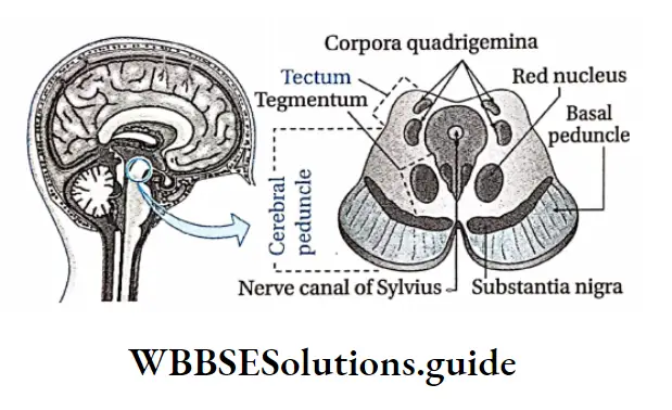
Tectum: The tectum of the midbrain is located in the dorsal region of the aqueduct of Sylvius. The primary structure of the tectum includes two swollen regions— superior colliculi and inferior colliculi.
The superior colliculi forms part of the visual system. The inferior colliculi is a part of the auditory system.
The structures appear as four small bumps on the brain stem. These bumps are together known as corpora quadrigemina.
Simple and compound leaf types with examples
Cerebral peduncle: The cerebral peduncles (or crus cerebri) are located anterior to the aqueduct of Sylvius in the midbrain.
From the interior, three layers are the visible tegmentum, substantia nigra, and basal peduncle.
Due to the presence of melanin pigment, the substantia nigra appears black or brown.
Function Of Midbrain:
- It coordinates the actions of different regions of the midbrain, forebrain, and hindbrain.
- The tectum region of the midbrain coordinates the reflex actions related to visual perception and olfaction.
- The midbrain plays an important role in controlling the body temperature.
- The red nucleus of the midbrain controls muscle tone.
- The midbrain controls the actions of different voluntary muscles.
Hindbrain Or Rhombencephalon
It is located at the posterior end of the midbrain. It consists of two parts metencephalon and myelencephalon.
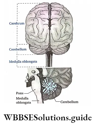
Metencephalon: The Metencephalon is located in the posterior region of the hindbrain.
It has two parts— the cerebellum and the pons.
Cerebellum: Cerebellum is a major structure located at the top of the brain stem (where the spinal cord meets the brain) and at the back portion of the brain, lying under the occipital and temporal lobes of the cerebral cortex.
The cerebellum is the most important and largest part of the human hindbrain. It is also known as the ‘little brain’ or the ‘tree of life’.
Cerebellum Structure: Cerebellum is made up of two hemispheres called cerebellar hemispheres. Two cerebellar hemispheres are connected by a mid-region named vermis. The cerebellum contains Purkinje cells.
Cerebellum Functions:
- The cerebellum plays an important role in controlling body balance,
- The cerebellum controls the movement of the hands and legs. It receives impulses from the cerebral cortex. Depending on these impulses, it controls the movement of the hands and legs.
- The cerebellum controls voluntary movement.
- The cerebellum exerts control over the reflex arc involving muscles. In this way, it helps to maintain normal muscle tone.
Pons Varolii:
- Pons Varolii is a region in the brain stem that lies between the midbrain (above) and medulla oblongata (below) and in front of the cerebellum.
- The ventral part of the pons is convex and the dorsal part is plain. Pons is connected to 5, 6, 7, and 8 cranial nerves.
The main functions of pons Varolii are as follows—
- Pneumotaxic and apneustic centers of pons act as respiratory centers for the regulation of respiration.
- Pons serve as a relay station between several areas of the brain.
- The nerve center of pons Varolii conducts contraction of the detrusor muscle of the urinary bladder, in order to carry out urination.
Myclencephalon or medulla oblongata:
- The medulla oblongata is a cone-shaped structure located in the hindbrain, anterior to the cerebellum, below the pons, and above the foramen magnum. It is the lowest part of the hindbrain.
- The medulla oblongata constricts at the posterior end and appears like a cone. Its length is approximately 2.4 cm. The spinal cord arises from its posterior end.
- Cranial nerves 9, 10, 11, and 12 remain connected to it. Medulla oblongata consists of different nerve centers, such as—the vasomotor center, respiratory center, vomiting center, and salivary center.
Pons Varolii Functions:
- Medulla oblongata controls visceral reflexes such as—sneezing, coughing, vomiting, secretion of saliva, engulfing, etc.
- Medulla oblongata plays an important role in controlling respiration.
- Medulla oblongata plays an important role in controlling the action of the cardiovascular system including the heartbeat.
CSF Or Cerebrospinal Fluid
Cerebrospinal Fluid Location: CSF is present in the neuronal of the central nervous system and central canal of the spinal cord.
It is also present in the subarachnoid space of the meninges.
Cerebrospinal Fluid Origin: The first and second ventricles of the human brain have ciliated epithelium on their wall.
The wall has enough capillary-rich area known as choroid plexus. Plasma of the blood, in the capillaries of the choroid plexus, enters the brain ventricles by diffusion and forms CSF.
CSF content: The total CSF content of a human body is approximately 150 ml.
Components: CSF is clear, and slightly alkaline in nature (pH—7.33). The various components of CSF are—water, plasma proteins, glucose, mineral salts or ions, creatinine, etc.
Cerebrospinal Fluid Functions:
- It acts as a shock absorber and protects the brain from mechanical injury,
- It maintains the shape and structure of the brain by maintaining an outward pressure,
- It transports nutrients to the tissues of the brain and spinal cord,
- It moistens the brain and spinal cord and enables the exchange of substances within I nerve cells,
- It has white blood corpuscles that protect the brain from infections,
- CSF provides buoyancy to the brain.
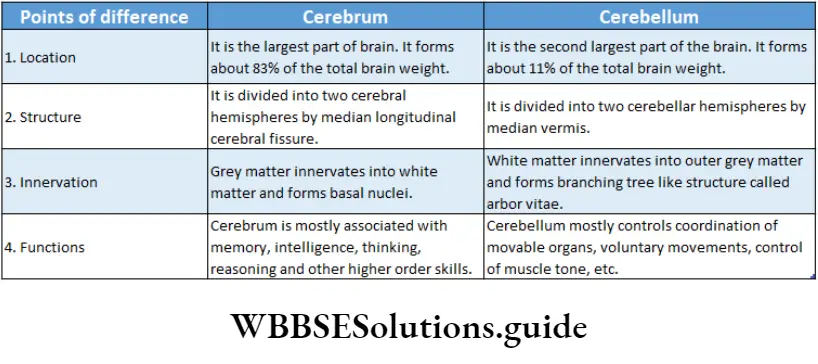
Spinal Cord
Spinal Cord Definition: The long, thin, tubular bundle of neurons and supporting cells that originate from the posterior end of the medulla oblongata of the brain enters the vertebral column, and runs through it is known as the spinal cord.
Spinal Cord Location: The human spinal cord originates from the medulla oblongata of the brain and is present within the neural canal of the vertebral column.
Spinal Cord Length and breadth: It is elongated and about 45 cm long in men and around 43 cm in women. It is enlarged in the cervical and lumbar regions.
Spinal Cord Coverings:
Just like the brain, the spinal cord also has an outer covering of meninges.
From outside to inside, layers of meninges are—dura mater, arachnoid mater, and pia mater.
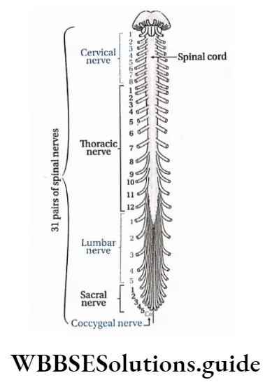
Spinal Cord External structure:
- The human spinal cord continues upto the conus medullaris (tapering posterior end of the spinal cord) near the first or second lumbar vertebra, terminating in a tapering fibrous extension, known as the filum terminale.
- It is hollow, elongated, and cone-shaped.
- The posterior and anterior sides of the spinal cord bear longitudinal grooves.
- The posterior groove is known as the posterior median sulcus and the anterior groove is known as the anterior median fissure. Along these two grooves, the spinal cord divides into two symmetrical parts.
Internal structure: The transverse section of the human spinal cord shows the following structures—
Central canal: The transverse section of the spinal cord shows two symmetrical hemispheres.
Its center constitutes the central canal. The canal and its ducts are together known as neurocoel.
The central canal is lined by ciliated epithelial tissue known as ependyma. Through this canal, the CSF flows.
Grey matter: Grey matter is present in the center of the spinal cord like ‘H’ or like the wings of a butterfly. In each segment of the spinal cord, grey matter is distributed in three grey columns—the posterior grey column or dorsal horn,
Anterior grey column or ventral horn and Lateral grey column or lateral grey horn. Two nerve roots originate from the human spinal cord. The dorsal nerve root originates from the dorsal horn and the ventral nerve root originates from a ventral horn.
White matter: White matter envelopes grey matter and is present at the periphery of the spinal cord. In each half of the spinal cord, white matter is distributed in three columns. These are—
- Posterior white column,
- Anterior white column and
- Lateral white column
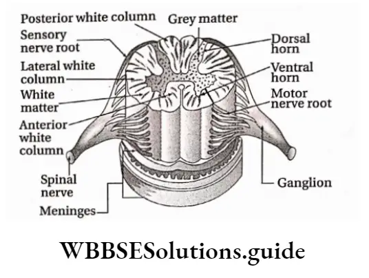
Functions:
- Spinal cord functions as the central control center for reflex actions.
- It partially controls the functions of the autonomic nervous system.
- Controls muscle tone.
- Through afferent nerve fibers, impulses from the peripheral nervous system are transmitted to the brain.
Again, motor impulses are transmitted from the brain to effector organs by efferent nerve fibers, many of which pass through the spinal cord.
This means the spinal cord maintains coordination between the peripheral system and the brain.
Peripheral Nervous System
Peripheral Nervous System Definition: The part of the nervous system that is constituted of nerves is called the peripheral nervous system.
Peripheral Nervous System Structural characteristics: The peripheral system is of two types—the somatic nervous system which is constituted of 31 pairs of spinal nerves and 12 pairs of cranial nerves and the autonomic or visceral nervous system which is constituted of sympathetic and parasympathetic nerves.
These nerves innervate various visceral organs of the body. These nerves carry sensory impulses from these organs to the CNS.
Leaf modifications in different plants with examples
Also, they carry motor impulses from the CNS to the effector organizer As a result, a response is being given by that organ.
Cranial Nerves
Cranial Nerves Definition: The nerves that originate directly from the brain are known as cranial nerves. These nerves are of three types—sensory or afferent, motor or efferent, and mixed types.
Note that, sensory nerves or afferent nerves originate from receptor organs or sense organs and meet at the brain. But, efferent nerves or motor nerves originate from the brain and meet at the effector organizer
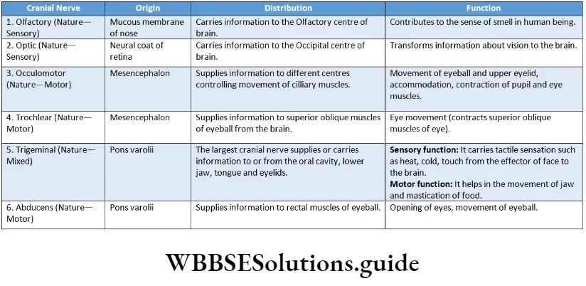
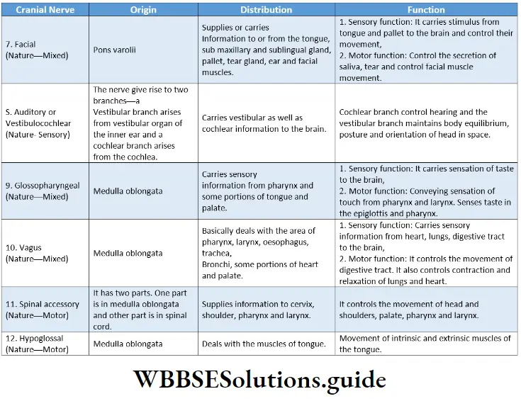
Spinal Nerves
Spinal Nerves Definition: The nerves that emerge from the spinal cord are known as spinal nerves.
The human body consists of 31 pairs of spinal nerves. They are named as per their origin
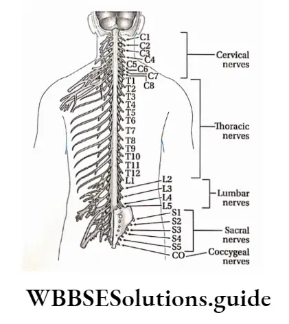
Features
- Dorsally, a branch of the spinal nerve called the dorsal root divides into six to eight nerve rootlets that enter the spinal cord.
- There is swelling, a little away from the rootlets. It is called the dorsal root ganglion.
- It contains cell bodies of afferent neurons.
- Ventrally, another six to eight rootlets leave the spinal cord and converge to form the ventral root.
- The ventral and dorsal roots merge, penetrate the dural sac, and enter the intervertebral foramen to form the spinal nerve proper.
- Spinal nerves are mixed nerves, with a two-way traffic of afferent (sensory) and efferent (motor) signals.
Function: Within the grey matter of the spinal cord, the sensory nerve enters carrying the sensory impulses.
These impulses are translated into motor impulses in the grey matter. Finally, the motor impulses leave the grey matter by traveling through the motor nerve.
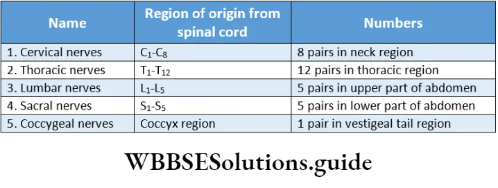
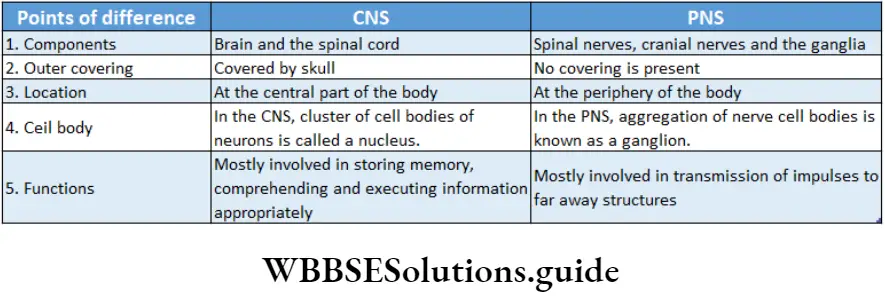
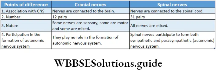
Divisions of the peripheral nervous system Based upon the consciousness of our body movements PNS can be classified into the somatic nervous system (SNS) and autonomic nervous system (ANS).
The somatic nervous system controls our voluntary movement. On the other hand, the autonomic nervous system works on its own, self-controlled and independent of our voluntary efforts.
Visceral Or Autonomic Nervous System
Visceral Or Autonomic Nervous System Definition: The part of the nervous system that controls bodily functions that are not controlled voluntarily such as breathing, heartbeat regulation, digestive processes, etc., is known as the autonomic nervous system.
It consists of a pair of chains of ganglions (a ganglion is a group of cell bodies of neurons outside the CNS) and nerves, found on either side of the backbone.
Two antagonistic and anatomically separate divisions comprise the autonomic nervous system.
These two are called the sympathetic and parasympathetic divisions, With a few exceptions, the two subdivisions influence and act upon the organs of the body with differential effects.
Functions of the autonomic nervous system:
The general functions of ANS are as follows—
General functions: The autonomic nervous system maintains homeostasis in the body. It controls the actions of cardiac muscles and muscles of different visceral organiser
Due to the antagonistic or opposite actions of the sympathetic and parasympathetic nervous system, a balance is maintained.
Fight Or Flight Response
The ‘fight or flight’ response was first described by an American physiologist, Walter Bradford Cannon to describe the physiological reaction that occurs within animals in response to a perceived attack or threat. In this response, the animal reacts to the threat by choosing either to fight or to flee from the area.
Rest and digest the response
The ‘rest and digest response occurs when the body is at rest, especially after feeding. In this response, energy is conserved as it slows heart rate, increases intestinal and glandular activity, and relaxes sphincter muscles in the gastrointestinal tract, thereby facilitating the process of digestion.
Functions of sympathetic division:
The sympathetic nervous system controls catabolic reactions associated with the release of energy.
It functions along with the adrenal medulla. Together they constitute the sympathetic-adrenal medullary system, It increases heart rate, blood pressure, etc., dilates the pupil, reduces the secretion of saliva, secretion of sweat, expands the bronchioles of the lungs, and also expands the gall bladder, urinary bladder, etc.
Functions of the parasympathetic nervous system:
The parasympathetic nervous system controls the anabolic reactions of the body. It enables digestion, absorption, storage of food, etc., and stores energy in the body.
The parasympathetic nervous system controls normal regular physiological activities of the body. It helps to secrete various digestive juices and thus helps in digestion. It also helps to absorb the digested food.
It increases the secretion of tears, reduces blood pressure, and contracts the urinary bladder.
Sympathetic Nervous System
The division that accelerates various activities of the body like heartbeat, breathing, etc., is called the sympathetic nervous system. The response given by this system is a ‘fight or flight’ response.
Sympathetic nerves originate from the thorax and lumbar region. During anger, fear, etc., the activity of the sympathetic nervous system is increased.
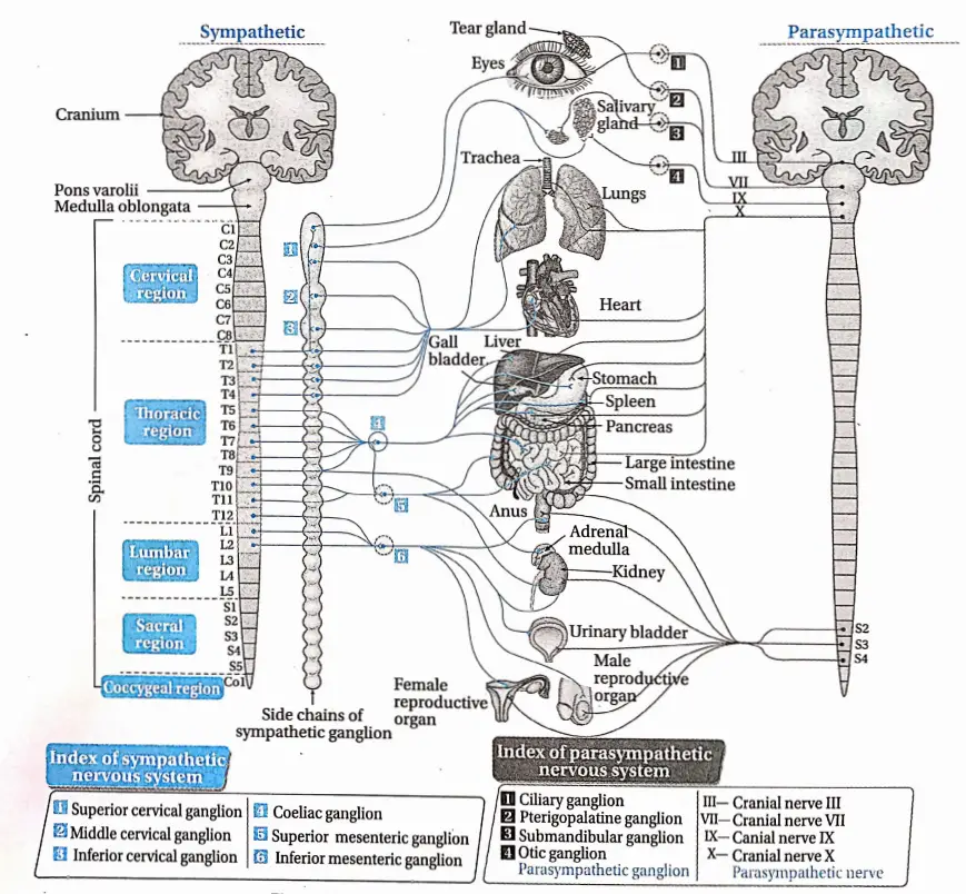
Origin: Sympathetic nerves originate from the lateral horn of grey matter of the first thoracic vertebra to the third lumbar vertebra.
Components: The four components of the sympathetic nervous system are—
Chain of lateral ganglia: This chain is composed of 21 ganglia which extend from the cervical to the sacrum region.
Pre-ganglionic fibers: These fibers are axons of neurons that are located in the spinal cord.
Collateral ganglia: Coeliac ganglion, superior mesenteric ganglion, and inferior mesenteric ganglion are the three collateral ganglia of the sympathetic nervous system which are located in the abdominal cavity.
Parasympathetic Nervous System
The part of the autonomic nervous system that decreases or slows down various activities of the body like heartbeat, respiratory process, etc., is called the parasympathetic nervous system.
The parasympathetic nervous system is anatomically of craniosacral type. The response given by this type of nervous system is Vest and digest’ response.
The nerves of the parasympathetic nervous system originate from the cranium and sacral vertebral segments of the spinal cord and are called the craniosacral nervous system.
Generally, the action of parasympathetic nerves increases when the body is at rest.
Parasympathetic Nervous System Origin: Some of the parasympathetic nerves originate from the brain by fusing with cranial nerves 3, 7, 9, and 10.
The rest of the parasympathetic nerves originate from the sacral vertebra 2nd, 3rd, and 4th (S2, S3, and S4) of the spinal cord.
Components: The three components of the parasympathetic nervous system are—
Pre-ganglionic fiber: These are the axons of neurons that are located in the midbrain, brain stem, and sacral segment of the spinal cord.
Parasympathetic ganglia: These ganglia are located very close to the peripheral tissue.
They do not form a chain of ganglia. The pre-ganglionic fibers synapse with the neurons of parasympathetic fibers.
Post-ganglionic fiber: These are the axons of neurons that are located in the sympathetic fiber.
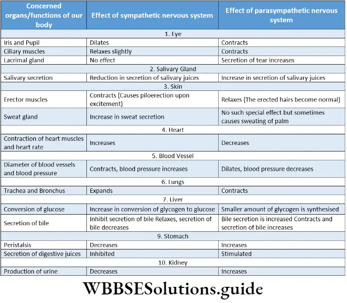
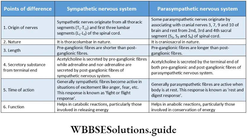
Nerve Impulse
A nerve impulse refers to an electrical or chemical signal that is transmitted along a nerve fiber.
The neurons of the nervous system are not connected directly i.e., they lack any cytoplasmic connections between them.
So nerve impulses are transmitted from one neuron to another by synapse.
So before going into the details about the process of transmission, we have to know about synapses which are as follows.
Synapse
The word ‘synapse’ came from ‘synaptein’, which Sir Charles Scott Sherrington et a!., had coined from the Greek word ‘syn-‘ (‘together’) and ‘haptein’ (‘to clasp’).
Synapse Definition: The microscopic gap present between the terminal end of the axon of one neuron and the dendron or axon or cell body of another neuron, through which nerve impulses are transmitted by neurotransmitters or electrical ions from the neuron to the post neuron is called a synapse.
Synapse Structure:
- In a neuron, an impulse travels along the length of the axon until it reaches the far end, called the axon terminal.
- The axon terminal branches profusely and gives a brush-like appearance hence called end brush.
- The terminal portion of each of these branches is swollen. It is called the synaptic knob or terminal knob.
- The axon terminal ends very close to the dendron or cell body or axon of another neuron, with which it forms the synapse.
- Here, the first neuron is called a presynaptic neuron while the second one is called a postsynaptic neuron.
- The nerve impulse is transmitted from the presynaptic neuron to the postsynaptic neuron.
- The membrane of the presynaptic neuron along the synapse is called the presynaptic membrane.
- The membrane of the postsynaptic neuron along the synapse is called the postsynaptic membrane. There is a gap between pre- and postsynaptic neurons. It is called the synaptic cleft.
- In the cytoplasm of the presynaptic axon terminal, there are small membrane-bound vesicles called synaptic vesicles which contain neurotransmitters.
- These substances act as chemical messengers and help to carry the neural signal from one neuron to another.
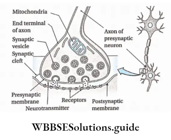
Types of synapses: On the basis of the medium of transmission of impulses synapses can be classified into three types, such as—
- Chemical synapse: Impulses are transmitted by neurotransmitters.
- Electrical synapse: impulses are transmitted by ions.
- Conjoint synapse: Impulses are transmitted by both chemical as well as electrical signals.
On the basis of structure, synapses can be classified into four types, such as—
Axodendritic synapse: The synapse between the axon of the presynaptic neuron and the dendrite of the postsynaptic neuron.
Axosomatic synapse: The synapse between the axon of the presynaptic neuron and the cell body or soma of postsynaptic neurons.
Axoaxonic synapse: The synapse between the axons of both, pre and postsynaptic neurons.
Dendrodendritic synapse: The synapse between the dendrites of both, pre and postsynaptic neurons.
On the basis of participating nerve cells, synapses can be classified into 3 types—
One-on-one: In this case, one neuron forms a synapse with another neuron.
Many with one: In this case, the terminal end of a number of neurons forms a synapse with one neuron.
One with many: In this case, the terminal end of one neuron forms a synapse with many neurons.
Functions: Synapses have four main functions. They are as follows—
They transmit information between neurons or neurons and their target cells.
They filter out low-frequency impulses.
They act as valves to ensure that impulses pass across them in one direction only.
They also act as junctions allowing impulses to be divided up along many neurons or merge into one.
Generation Of Nerve Impulse
The generation of nerve impulses in nerve fibers depends on the distribution of Na+ and K+ in and out of the axolemma. Again, the distribution of Na+ and K+ is controlled by protein channels.
(such as Na+ and K+ channels) and carrier proteins like Na+/K+ -ATPase pump, in the axolemma. The different stages of the generation of nerve impulses are described below.
Resting Potential: It is the potential difference across the membrane of a resting neuron i.e., when nerve impulses are not transmitted.
At this stage, the axonal membrane is nearly impermeable to Na+ while comparatively more permeable to K+.
The ‘membrane is also impermeable to negatively charged proteins present in the axoplasm. As a result, inside the axoplasm, there is a high concentration of K+ and negatively charged proteins and a low concentration of Na+.
During resting conditions, this potential difference across the axonal membrane is called resting membrane potential. This state of the resting membrane is called the polarised state.
The average value of resting membrane potential is -70mV.
Action Potential: When a nerve receives a stimulus, a sequence of events occurs.
Due to the conduction of impulse in a region, Na+ channels of that region open.
This causes an influx of Na+ inside the axoplasm. This creates an increase in positive charge in the axoplasm.
Thus the polarisation across the membrane, as found under resting conditions is now reversed. The reversal of polarity is called depolarization. The potential difference across.
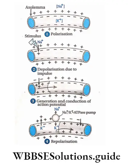
The membrane under this condition, is called action potential. Its value is +35mV.
After depolarisation, Na+ channels are closed. The wave of this reversed polarity of depolarisation (action potential) moving down an axon is called a nerve impulse or spike.
Repolarisation: When the action potential of the axon becomes +35 mV, the K+ channel of the axolemma opens. Due to this reason, K+ from the intracellular matrix moves out to extracellular fluid. As a result, electric potential is reduced.
Gradually, the positive charge in the extracellular fluid increases compared to td axoplasm.
After some time, the concentration of K+ increases in extracellular fluid so much that the K+ channel closes. At this time, the Na+/ K+-ATPase pump opens.
Through this pump, three Na+ move out from the axolemma, and two K+ move inside the axolemma.
In this way, after some time, the concentration of Na+ and K+ becomes the same as before (as in resting condition) on either side of the axolemma. Thus, nerve fibers again attain resting potential.
Conduction Of Nerve Impulse
The process of conduction of nerve impulses through medullated, and non-medullated nerve fibers and synapses follows various methods as described below.
Conduction of nerve Impuluses in Non-medullated nerve fibres: Nerve impulse or action potential is transmitted in small steps. In each step, repolarisation and depolarisation take place. This is known as continuous conduction.
Conduction of nerve impulses in medullated nerve fibers: Myelin sheath contains fat. Since fat is a poor conductor of heat and electricity, it cannot transmit impulses.
Only nodes of Ranvier allow transmission of impulses and ion exchange occurs at these points. So, depolarisation of nodes of Ranvier occurs.
The depolarised node of Ranvier triggers an increase in the permeability of ion channels leading to depolarisation in the adjacent node of Ranvier. Therefore, nerve impulses are transmitted by jumping from one node to the next.
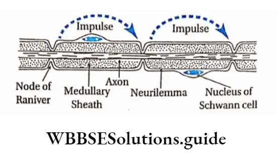
So, it is faster in the medullated nerve fibers than the non-medullated nerve fibers.
This type of conduction is called saltatory conduction because it has been derived from the Latin word saltare which means ‘leaping’.
Synaptic transmission: Synaptic transmission is the process whereby electrical impulses pass through a synapse with the release of neurotransmitters.
Transmission of nerve impulses in the synapse region is a chemical process.
The sequence of events that lead to postsynaptic changes is as follows—
- As the action potential reaches the axon terminal of a presynaptic neuron, depolarisation occurs which causes voltage-gated Ca2+ channels to open. This leads to an influx of Ca2+ into the synaptic knob from the cisternal.
- Ca2+ influx in the axoplasm of the synaptic knob continues. This causes an increase in concentration of Ca2+ and eventually induces vesicles filled with neurotransmitters, to migrate towards the presynaptic membrane. There the vesicles fuse with the presynaptic membrane.
- The presynaptic membrane and vesicles now form a continuous membrane, so that the neurotransmitter i.e., acetylcholine, is released into the synaptic cleft also by exocytosis. There may be other types of neurotransmitters such as adrenalin, dopamine, etc., but the following discussion is given with respect to acetylcholine.
- Acetylcholine diffuses through the synaptic cleft and binds with receptor site on the postsynaptic membrane.
- As a result, acetylcholine-dependent sodium ion channels open, and movement of sodium ions takes place through the postsynaptic membrane. This creates a change in the membrane potential of the post-synaptic membrane.
- The postsynaptic membrane and synaptic cleft contain the enzyme acetylcholinesterase. This enzyme hydrolyses acetylcholine into acetate and choline. This causes repolarisation of the postsynaptic membrane.
- The entire cycle repeats again.
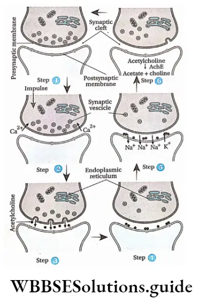
Neuromuscular transmission: The junction between a motor neuron and a muscle fiber by which the nerve can communicate with the muscle fiber is called the neuromuscular junction.
Through these junctions, neurotransmitters are transmitted from the neuron to the muscle. This type of transmission is known as neuromuscular transmission.
In this case, the terminal end of the axon extends and appears like a plate, which is located near the sarcolemma.
At the neuromuscular junction, the plate-like structure formed by the axon terminal is known as the motor end plate.
The extended part of the terminal end motor neuron is known as sole feet or end feet. The axoplasm of this part contains more vesicles.
These vesicles contain neurotransmitters like acetylcholine. This portion also contains endoplasmic reticulum, mitochondria, etc. Sarcolemma of muscle fibers are thick and contain grooves.
The following events occur at the neuromuscular junction—
The axon of the motor neuron carrying a vesicle containing acetylcholine reaches the motor end plate with the help of Ca2+ ions.
Acetylcholine from these vesicles is released between the membrane of the sole feet and muscle fibers. This causes depolarization of the end plate and creates action potential.
As a result, impulses from the end plate enter muscle fibers and cause contraction of muscle fibers.
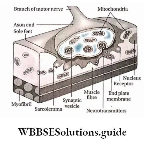
Neurotransmitters
The organochemical substances that transmit nerve impulses from the presynaptic region to the postsynaptic region at the synapse are known as neurotransmitters.
They are of two types—excitatory and inhibitory.
Excitatory: These are capable of depolarising the postsynaptic membrane and transmitting nerve impulses. Examples—are acetylcholine, and noradrenaline.
Inhibitory: These prevent the transmission of nerve impulses in the synaptic region. Example—GABA (y-amino butyric acid), is Screted from the axon end of some neurons in CNS.
Reflex Action And Reflex Arc
Some neurons are organized to enable the human body to react rapidly at times of danger, even before consciously being aware of the threat. Under such conditions, impulses from sense organs are not transmitted to the brain but to the grey matter of the spinal cord.
Venation in leaves and its types with diagram
There it is analyzed fast and action impulses are transmitted to the effector organiser These sudden, unlearned, involuntary responses to certain stimuli are called reflex action. The neural path along which reflex action takes place is known as the reflex arc.
Reflex Action
Reflex Action Definition: A spontaneous, automatic, involuntary response to a stimulus that occurs without any conscious effort or thought and requires the involvement of CNS, is known as a reflex action.
Examples:
- Drawing back of the hand when pricked with a needle.
- Sudden closing of eyes when flashed with a high-intensity light.
- Secretion of saliva on seeing or smelling food.
Types Of Reflex Action
The types of reflex actions are described below.
Unconditioned or inborn reflexes: The reflexes which are automatic, instinctive, unlearned reaction and can be evoked even immediately after birth, is called inborn or unconditioned reflex action.
These reflex actions do not need previous exposure to the stimulus.
So these are inborn and inherited reflex actions. The reflex arc of this reflex action is stable (that is, it follows a simple reflex arc. These types of reflex actions cannot be changed or forgotten easily.
Types: On the basis of the type of organs involved with inborn reflex actions, these are divided into three types. These are as follows—
Superficial reflex action: The types of reflex action in which stimuli are exerted on superficial structures, mainly in skin or mucous membranes, are known as superficial reflex action.
Example:
- Sudden exposure to bright light causes us to close eyes due to pupillary reflex,
- Sudden exposure of the hand to fire causes it to draw it back.
Simple And Complex Reflex Arc
If a sensory neuron connects directly to a motor neuron in the special cord then it is known as a simple reflex arc.
However, in some reflex arcs, the sensory neuron connects to the motor neuron through association neurons, and such reflex arcs are known as complex reflex arcs.
Effect Of Cerebellum On Reflex Action
The role of the cerebellum is to finely tune the reflex arc. This role of the cerebellum is gradually archived and adjusted through the process of learning.
Based on past experiences, the cerebellum rectifies any motor error in the reflex arc and uses this prediction to adjust motor output, thereby helping in the process of reflex action.
Visceral reflex action: The type of reflex action in which stimuli are exerted on the visceral organs of the body is called visceral reflex action.
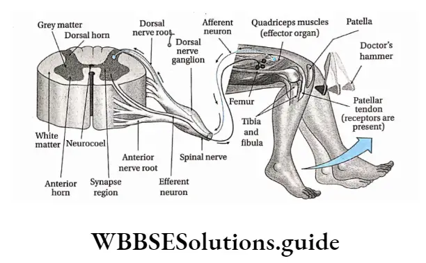
Example:
Digestive reflexes such as secretion of digestive juices after engulfing food and movement of the bowel after digestion.
Cardiovascular reflexes such as regulation of heartbeat, blood pressure, etc.
Acquired or conditioned reflex action: The reflex actions that are acquired by training after birth and are subjected to changes are known as acquired or conditioned reflex actions.
These actions are acquired by humans and other vertebrates after birth. These actions depend on repeated conditioning. These actions are subjected to be forgotten or changed as per need.
Example:
- Walking, writing by a child.
- Riding bicycle.
- Reciting poem.
- Actions shown by animals in circuses, etc.
Pavlov’s experiment
The famous Russian scientist Ivan Petrovich Pavlov is known as the father of conditional reflex action.
He has performed an experiment to prove how an unconditioned reflex action can generate a conditioned reflex action. His experiment was as follows
Deep reflex action: The type of reflex action in which stimuli are exerted on some underlying structures, mainly in muscles and tendons, is known as deep reflex action.
Example:
Knee jerk or patellar tendon reflex—When the knee is hit by a doctor’s hammer, the thigh and lower portion of the leg jerk,
Biceps jerk reflex—Suddenly hitting the biceps causes stretching in the tendon due to which elbow folds.
An unconditioned stimulus (food—meat) was presented to a hungry dog every day. It caused salivation (an unconditioned response).
At the same time, a conditioned stimulus (ringing a bell) was given. The dog learned to associate the conditioned stimulus (bell) with the unconditioned stimulus (food).
Eventually, after some days, it was observed that ringing of the bell alone caused salivation. This is because the dog adapted to the stimulus of a ringing bell that brought food.
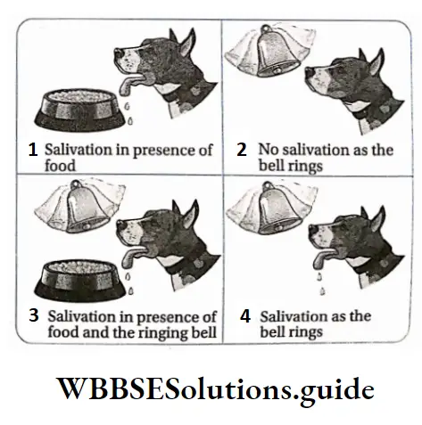

Reflex arc Definition: The path through which reflex action takes place and extends between the sensory organ to the effector organ through the grey matter of the spinal cord is called reflex arc.
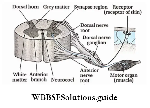
Different components of a reflex arc and their functional roles
The reflex arc is constituted of five parts that are described below.
Receptor: To accomplish a reflex action, the reflex arc must be directly linked with a receptor organ. Reflex action occurs when sense organs or receptors are excited by a stimulus.
Sensory or afferent neuron: These neurons of a reflex arc remain connected with the receptor. They carry impulses from the receptor to the grey matter of the spinal cord.
Grey matter of spinal cord: Grey matter of the spinal cord acts as the controller or center of reflex action. In the grey matter of the spinal cord, sensory impulses are analyzed and interpreted.
Effector organ: Effector organs are capable of responding to a stimulus to perform a particular action. Generally, muscles, glands, etc., participate as effector organs.
Sense Organs
Sense Organs Definition: The organs that are specialized to acquire and process sensory stimuli from different sources, both external and internal are called sense organizer
Sense Organs Types: Humans and other advanced vertebrates have different types of sense organizers The human body consists of five main types of sense organizer
These are—
- One Pair Of Eyes
- One Pair Of Ears
- Nose
- Tounge
- Skin
Sensory Receptors
Sensory Receptors Definition: The sensory nerve endings, a cell or group of cells, that respond to various kinds of stimulation are called receptors.
They are transducers that convert various forms of energy in the environment (internal or external) into action potentials in the afferent neurons. The awareness that results within the body is called sensation.
The sense organs—the eyes, ears, skin, mouth, and nose—have receptors that detect sensations. The sensory neurons present within them transmit the information to the central nervous system where it is interpreted as sight, sound, touch, taste, or smell.
Types of receptors: Receptors are classified into different types, as depicted in the chart given below.
On the basis of the location of the receptors, it can be classified into three types—
Exteroceptors: These receptors are located near the surface of the body. They can sense changes in the surrounding environment. Generally, they include receptors for touch, heat, cold, etc.
Interoceptors: These receptors are located within the body. They are stimulated by the internal stimuli which are generated inside the body.
There are several types, such as—chemoreceptors, stretch receptors, baroreceptors, etc.
Proprioceptors: The receptors which can detect the position and posture of the body.
They are located in skeletal muscle, bones, tendons, ligaments, etc. On the basis of the type of the received stimuli, receptors can be classified into five types, etc
Mechanoreceptors: These are receptors that are stimulated by mechanical forces, such as touch, pain, equilibrium, etc., received on muscles, tendons, ligaments, and joints. They comprise both exteroceptors and interoceptors.
Several types of mechanoreceptors are discussed in the following table.
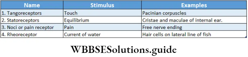
Telereceptors: Receptors sensing light and sound, which provide information about the distant environment, are called telereceptors.
Telereceptors, on the basis of their location, have been divided into three types which have been discussed below.
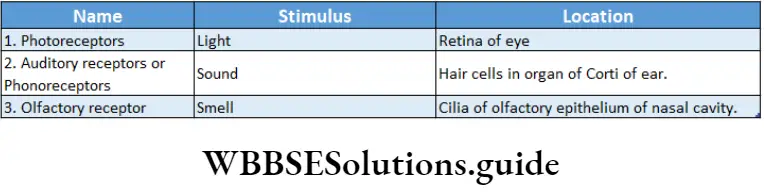
Chemoreceptors: These are receptors that are stimulated by chemical changes or by chemicals that are either inhaled or tasted (gustatory receptors).
They also modify hydrogen ion and carbon dioxide concentrations in blood. For example, chemoreceptors of the carotid artery can sense changes in blood partial pressure of 02 and C02 in the blood
Thermoreceptors: These are receptors that are sensitized by heat. For example, Ruffini’s end organs, are the end organ of Mazzoni.
Galvanoreceptors: These are receptors that are sensitized by gravity. For example, the organs of some fish have this type of receptor.
Human Eye
Our eyes are sense organs for vision. They also help in the perception of color.
Human beings have two simple eyes, present ventrally to the cranium and above both sides of the nose.
They are placed within the eye orbits. We have binocular vision (which involves two eyes to view an object). In the case of frogs, fish, etc., monocular vision (which involves one eye to view an object) is observed.
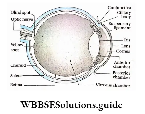
Eyeball
On the ventral portion of the brain box and above both sides of the nose, there are two cavities or orbits called the eye cavities within which eyeballs are located. These are supported by muscular tissue.
Each eyeball is almost spherical and has a diameter of 2.5 cm. The eyeball remains in position within the cavity with the help of six muscles. Eyeball actually is a fluid-filled sac-like structure.
It is composed of three parts—
- Coats,
- Eye chambers and
- Refractory media
- Coats
The human eyeball is covered with three layers or coats which are—
- Fibrous coat,
- Pigmented coat and
- Neural coat.
Fibrous coat: The outermost layer of the wall of the eyeball is made up of fibrous connective tissue. This layer can be divided into two—
- Sclera and
- Cornea.
Sclera: This is the outer covering layer of the eyeball. The sclera is present in the posterior portion, comprising about five-sixth part of the external layer.
The sclera is a comparatively tough, translucent, and thick layer that is composed of white collagenous connective tissue. The posterior region of the eyeball from where the optic nerve emerges lacks sclera.
Cornea: It is the bulged, transparent anterior region of the eyeball. It covers the l/6th part of the fibrous coat of the eyeball. On the outer surface of the cornea, there is a thin and clear layer called the conjunctiva.
Functions of fibrous coat:
The sclera provides shape to the eyeball and forms a protective rigid layer for the eyeball.
Being transparent, the cornea acts as a refractive medium and permits light to pass through easily and fall on the lens. It acts as a convex lens to converge the diverging rays.
Pigmented coat or vascular coat: The middle layer of the human eyeball contains melanin pigment in its cells.
So, this layer appears brown. This layer is known as the pigmented layer. It is calculated and so, known as vascular coat. The different parts of this layer are discussed below.
Choroid: The melanin-containing brownish part beneath the sclera, in human eyeball is known as choroid.
Ciliary body and suspensory ligament: The thin layer that is present in the terminal region of the choroid, at the junction of the sclera and cornea of the human eyeball is known as the ciliary body. The thin fibers projecting from the ciliary body are known as suspensory ligaments.
Iris: The pigmented part present in front of the ciliary body, suspensory ligament, and lens of the human eyeball is called the iris. It controls constriction and dilation of the pupil.
At the center of the iris, a spherical opening is present, known as the pupil.
Iris contains two types of muscles—
Circular Muscles And Radial Muscles.
Functions of pigmented or vascular coat:
Due to its deep brown color, the choroid absorbs excess light and prevents its repeated reflection. Thus, it helps to form a clear image.
Suspensory ligaments hold the lens in its proper position.
While seeing objects nearer or farther, we cannot move the lens forward or backward, but with the help of ciliary muscles, the curvature of the lens can be changed. As a result, the focus of the lens can be changed as required. This phenomenon is known as accommodation.
The pupil dilates and constricts due to the action of circular muscles and radial muscles of the iris. In this way, the iris modifies the diameter of the pupil and controls the incident rays on the lens. The color of the iris is considered the color of the eye.
Neural coat or retina: The most internal layer of the human eye is the posterior part of the lens and the inner portion of the choroid where the optic nerve is found. This layer, made up of nervous tissue and connective tissue is known as the neural coat or retina
Structural features of the retina:
- Retina, present posterior to the lens, is extended dorsally to ciliary bodies.
- The different cells of the retina are arranged in 10 layers.
- The most external layer among the 10 layers is pigmented and remains associated with the choroid.
- There are two types of pigmented cells in this layered cells and cone cells. These cells are also described later.
The different neuronal cell layers of the retina bear four types of cells—bipolar cells, ganglion cells, horizontal cells, and amacrine cells.
All the nerve cells in the most internal retinal layer form the optic nerve.
The retina also contains one type of glial cell called Muller cell that helps in maintaining its structural integrity.
A depressed region is observed at the center of the retina. It is yellowish and so, known as yellow spot or macula lutea.
The clearest and brightest image is formed in this region. Its central region is depressed known as fovea centralis. It is the most photosensitive region of the retina.
The junction between the origin of the optic nerve and the posterior end of the retina lacks any photosensitive cells (rod and cone cells).
As a result, the image does not form in that region. This part of the retina is known as the blind spot or optic disc.
Different cellular layers of the retina: The retina consists of the following 10 layers—
Pigment epithelium: This outermost layer of the retina consists of pigmented cells. This is a non-neural layer that absorbs excess light which has passed through the other retinal layers and is immediately next to the choroid.
Photoreceptor layer: This layer includes the outer external segments of the photoreceptor cells, rods, and cones.
External limiting membrane: Although this appears to be a membrane, actually it is not. It is formed by intercellular junctions—the zonula adherent—binding the apical aspect of the Muller cells.
External nuclear layer: This consists of the cell bodies of the rods and cones.
External plexiform layer: This is a synaptic area that contains the terminals of the rods and cones.
Inner nuclear layer: This consists of the soma of the retinal interneurons (horizontal, bipolar, and amacrine cells). It integrates and modulates the activity of photoreceptors.
Inner plexiform layer: This is a synaptic area containing the terminals of the bipolar, amacrine, and ganglion cells.
Ganglion cell layer: This consists of the soma of the ganglion cells (multipolar neurons).
Optic nerve fiber layer: This layer is composed of the axons of the ganglion cells that gather to form a thick bundle, the optic nerve.
Internal (inner) limiting membrane: This membrane is composed of a basal lamina interposed between the vitreous body and the Muller cells.
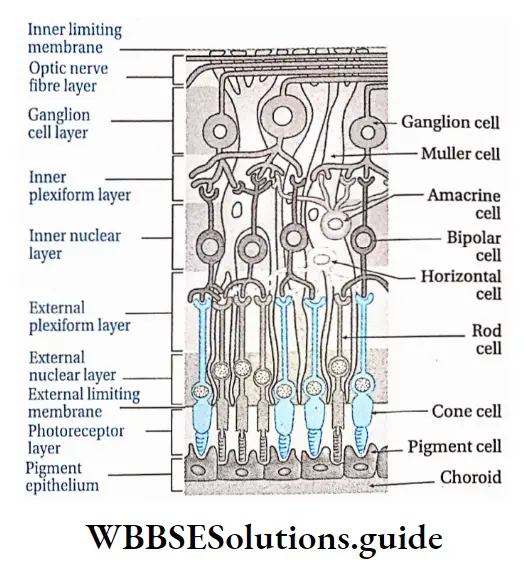
Photoreceptor cells of the retina: The human retina contains two types of photoreceptor cells—rod cells and cone cells. These are discussed below.
Rod cells: These cells are rod-shaped. They contain the pigment rhodopsin. Vitamin A is required for the synthesis of rhodopsin. The human eye contains 115-120 million rod cells.
Functions:
Rod cells help to see objects in dim light at night, which means they can absorb low-intensity light,
Rod cells generally form the black and white vision.
Cone cells: These cells are cone-shaped. They contain the pigment iodopsin. The human eye contains 6.0-6.5 million cone cells.
Functions:
Cone cells are capable of absorbing high-intensity light, so they help us to see objects in bright light,
They help in the perception of color and thus are responsible for color vision. The inactivity of cone cells leads to color blindness.
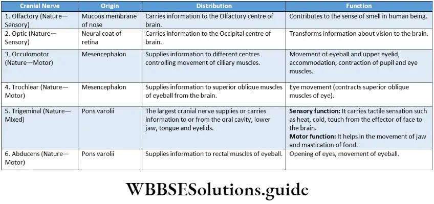
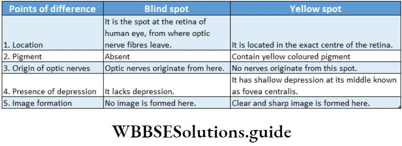
Functions of retina
The retina of the human eyeball acts as the photoreceptor organ. Rod cells of the retina help to see objects in dim light. On the other hand, cone cells help to see objects in bright light.
Light from different sources reflects on the lens and creates an image on the retina. This reflection is transmitted to the cerebrum by optic nerves. There it is translated and a sensation of vision is created.
The yellow spot of the retina, especially the fovea centralis, forms a clearer image of the object.
Rod cells of the retina cause black-and-white vision and cone cells causes colored vision.
Eye chambers
The human eyeball contains three chambers which are as follows—
- Anterior chamber: The chamber between the iris and cornea of the human eyeball is known as the anterior chamber. This chamber is filled with fluid aqueous humor.
- Posterior chamber: The thin chamber between the lens and iris of the eyeball is known as a posterior chamber. This chamber is also filled with aqueous humor.
- Vitreous chamber: The large chamber at the posterior part of the lens, between the lens and the retina is known as a vitreous chamber. This chamber is filled with fluid vitreous humor.
Refractory Media
All those parts of the eyes that convert parallel or divergent light rays into convergent light rays are known as refractory media. The refractory media of human eyes are—lens, aqueous humor, vitreous humor, and cornea.
Lens: It remains suspended from ciliary bodies by the suspensory ligament in the human eyeball. It is the main refractory medium of human eyes.
The lens is elastic in nature and bi-convex in shape. It is composed of transitional columnar epithelial tissue. It is as clear as crystal. It contains the protein crystallin. It is surrounded by a capsule.
Functions:
It is the main refractory medium of human eyes.
It helps to focus on objects based on distance. While seeing a nearby object, ciliary muscles contract and the lens becomes thick. While seeing distant objects, ciliary muscles relax, thus reducing the thickness of the lens.
Aqueous humor and vitreous humor: The clear fluid substance present in the anterior and posterior chambers of the human eye is known as aqueous humor.
On the other hand, the fluid substance present in the vitreous chamber of the human eye is known as vitreous humor.
Both aqueous and vitreous humor is made of salt, sugar, and protein, vitreous humor is viscous i.e., jelly-like.
Functions:
- Both aqueous and vitreous humor maintain intraocular pressure and inflate the eyeball. This is how they maintain the shape of the eyeball.
- They act as refractory media of the eyes.
- Aqueous humor provides nutrients to the lens, cornea, iris, etc.
Cornea: It is the convex, transparent part of the human eyeball which also acts as a refractory medium to some extent.
Accessory Protective Part Of Human Eye
The structure and functions of accessory protective parts of the eyes are described below.
Accessory Protective Part Of Human Eye Eye orbit: In the anterior part of the human cranium, beside the nose, within two hollow cavities the eyeballs are placed.
These two hollow cavities are known as eye orbits. Note that eye orbit has a thick layer of muscular tissue, on which eyeballs are placed. Eyeballs are bound to the eye orbit by six muscles. The optic nerve too, helps to bind the eyeball to eye orbit.
Accessory Protective Part Of Human Eye Functions: Due to the presence of muscular tissue from the interior and bony covering from the outside, the eye orbit protects the eyeball from mechanical injury.
Eye muscles: The six muscles by which eyeball remains bound to the eye orbit are known as eye muscles.
These are—
- Superior rectus muscle,
- Inferior rectus muscle,
- Medial rectus muscle,
- Lateral rectus muscle,
- Superior oblique muscle,
- Inferior oblique muscle.
Functions:
- With the help of these muscles, we can move the eyeballs down and sideways.
- Eyeballs can also be rotated by these muscles.
Eyelids: The human eye is covered by a thin, extended, skin layer on the upper part and lower part.
These are known as upper eyelids and lower eyelids respectively. At the corner of the human eye (near the nose), a small, red, muscular nictitating membrane is present in a vestigial condition.
Accessory Protective Part Of Human Eye Functions:
Eyelids protect the frontal part of the eyeball from dust.
Under bright light eyelids close, thus protecting the eye, rather than the lens, from bright light.
Eyebrow and eyelash: Each eye of humans has two special parts on upper region of the eyelids and the lower region of the forehead.
This part is curved like a bow and is known as an eyebrow. The eyelids are again lined by fine hairs which are known as eyelashes.
Accessory Protective Part Of Human Eye Function: Due to the presence of an eyebrow, rainwater, and sweat cannot enter the eyeball from the forehead.
Conjunctiva: The thin and transparent layer present beneath the upper eyelid and which extends upto the lower lid, covering the anterior part of each human eye is known as conjunctiva.
Conjunctiva is actually made up of squamous epithelium. Inflammation of the conjunctiva is known as conjunctivitis.
Accessory Protective Part Of Human Eye Functions:
It protects the eyeball from dust particles.
Being transparent, it allows light rays to pass through it easily into the eyeball to reach the lens.
Lacrimal gland: Lacrimal gland or tear gland is present in the upper lateral region of each orbit or beneath the upper and outer border of each eyelid. It opens into the upper eyelid by some tubules.
The tear produced in the tear gland is a fluid containing NaHC03, NaCI, etc. Tear also contains a bacteriolytic enzyme called lysozyme.
Accessory Protective Part Of Human Eye Functions:
Tears keep the upper surface of the eyeball moist. If the lacrimal gland gets damaged, secretion of tears is reduced, and conjunctiva contracts thereby causing dryness and burning sensation in the eye.
Tear wash out the dust particles present in the open the surface of the eyeball.
Phyllotaxy in plants – types and examples
Lysozyme, an antibacterial enzyme present in tears protects the eye from bacterial infection.
Physiological Function Of Eye
Vision is the main physiological function of the human eye. In case of the human eye helps us with two types of vision, such as—light vision and color vision.
Light vision: Eyes help us to see objects by providing a stereoscopic vision. The mechanism of vision by which eyes sense and visualize nearby and far away objects is given below.
Mechanism of vision:
- The human eye acts as a camera. Parallel or divergent light rays from objects pass through the conjunctiva, cornea, and aqueous humor and fall on the lens.
- After falling on the lens or incidence, light rays become convergent. These convergent rays pass through the vitreous humor, present behind the lens, and then fall on the yellow spot of the retina.
- Retina contains a photosynthetic pigment, called rhodopsin. Light splits rhodopsin and converts it into two chemical compound named retinene (retinal) and opsin.
- Light also brings changes in the structure of opsin which causes changes in membrane permeability.
- Changes in membrane permeability cause ion channels to open which results in potential difference and generation of action potential.
- This action potential i.e. impulse is carried by the optic nerve to the visual cortex of the cerebrum through the optic tract.
- Here the image is formed. Note, stimulus from the right eye goes to the visual cortex of the left cerebral hemisphere and stimulus from the left eye goes to the visual cortex of the right cerebral hemisphere. In the visual cortex, the stimulus is analyzed.
- By the special capability of the brain, the image becomes elongated. This results in the sensation of vision.
Color vision: Cone cells of the human eye help to determine the color of different objects.
Visual Accommodation
Human eyes cannot change the distance of the lens directly to observe objects nearer or farther. However, ciliary muscles and suspensory ligaments can adjust the curvature of the lens.
As a result, the focal length of the lens changes due to which the image of the object falls on the retina. This phenomenon is known as accommodation.
This is a type of reflex action. Human eyes have good visual accommodation. Light coming from any object located 25cm away can be easily sensitized by eyes.
This is known as the least distance of distinct vision. The ciliary bodies and suspensory ligaments of human eyes together are known as accommodation apparatus.
Some Important Eye Disorders
There are some important disorders related to the eyes which are as follows.
Myopia: It is the most common refractive error of the eye. Myopia is also referred to as short-sightedness as the person cannot see distant objects.
In this condition, parallel rays of light coming from distant objects, instead of focusing on the retina, focus in front of it.
As a result, objects located distantly cannot be seen clearly. It can be corrected by using glasses with a concave surface.
Hypermetropia or Hyperopia: It happens when the eyeball is shorter than normal and the parallel rays of light are brought to focus behind the retina.
It is also referred to as long-sightedness. In this case, nearby objects appear blurry while distant objects are seen clearly. It can be corrected by using glasses with convex lenses.
Astigmatism: Astigmatism is a common condition in which the curvature of the cornea or lens is not uniform.
This causes blurred vision of objects both near and far. This can be corrected by using glasses with cylindrical lenses.
Presbyopia: Presbyopia is an age-related eye condition that makes it more difficult to see near objects very clearly.
After the age of 40, the lens of the human eye becomes hard thereby losing the capacity of contraction-relaxation and thus people find difficult to see, particularly to read. It can be corrected by using glasses with bifocal or convex lenses.
Cataract: Due to aging, the lens becomes swollen, hard, and opaque. Vision becomes feeble and sometimes, is lost. This condition is known as cataract. It can be corrected by replacing the affected lens with an artificial one.
Glaucoma: The defects in the eyes that lead to damage of the optic nerve are known as glaucoma.
It happens due to an increase in intraocular pressure leading to the rupture of the optic nerve.
When blood flow is reduced in the optic nerve i.e., in the case of ischemia, the optic nerve is damaged.
Due to damaged optic nerve, vision is impaired and even blindness may occur.
Strabismus (squint): In this disorder, two eyes do not look exactly in the same direction at the same time.
It is caused due to dysfunction of the extraocular muscle. It is usually corrected by surgical procedures at an early stage.
Trachoma: It is a pathological condition caused by the bacterium Chlamydia trachomatis.
It is characterized by inflammation of the conjunctiva and cornea leading to pain and watering of the eyes.
In extreme cases, it may lead to complete blindness. Trachoma can be treated by antibiotics in the early stages and surgery in the late stages.
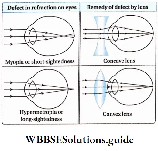
Human Ear
The human ear is the sense organ of hearing and equilibrium. It consists of three main parts—
- External ear,
- Middle ear and
- Inner ear. The ear receives the sound waves and helps in its interpretation.
Parts Of Ear
Different parts of the human ear are discussed under Separte Heads.
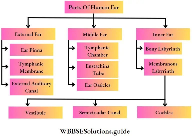
Sound waves can travel through solid, liquid, and gaseous medium. The human ear is the best example to Sound waves that can travel through solid, liquid and gaseous mediums. The human ear is the best example of solid bones in the middle ear and fluid medium in the inner ear.
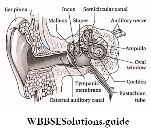
External Ear
The human external ear consists of three parts—pinna, external auditory canal, and tympanic membrane.
External Ear Pinna: The outer ridged cartilaginous part of the ear covered with skin, through which sound waves are received from the external environment is called the pinna.
External Ear Location: The pinna is an expanded cartilaginous portion, covered with skin that projects from both sides of the head.
External Ear Characteristics:
- Pinna is made of elastic cartilage. It has an expanded leaf-like structure, it has a skinny covering on cartilage.
- Along the upper periphery of the pinna, a fold is seen, known as a helix.
- The lower part of the pinna is muscular and so, it is calculated. This flexible lobe is known as the lobule.
- The muscles of the pinna are vestigial, hence, the human pinna cannot be moved.
Functions:
- Pinna collects sound waves from different sources and funnels them to the auditory canal.
- Therefore, pinna acts as the receiver of sound waves.
- Due to the presence of folds in pinna, it inhibits dust, insects, etc., from entering the auditory canal.
External auditory canal: This is the S-shaped passage through which sound waves travel from outside to the tympanum.
Location: External auditory canal or external auditory meatus, a part of the external ear, extends from an opening in the pinna to the tympanic membrane.
Characteristics:
- The external auditory canal is a slightly bent, tubular structure. It runs upward and then downward.
- It is supported by cartilage and temporal bone in the outer and inner portions respectively. The junction of the outer and inner portions is referred to as the osteocartilaginous junction.
- The inner wall of the auditory canal is covered by skin. The sweat gland of this layer has transformed into a ceruminous gland or wax gland which secrete cerumen or earwax.
Functions:
- It conducts the sound waves towards the tympanic membrane. 0 Since it is a bit bent, it protects the tympanic membrane from external mechanical injuries.
- The ceruminous gland in its wall secretes a waxy substance, cerumen that contains lysozyme and immunoglobulin. So, dust particles, insects, and germs cannot reach upto the tympanic membrane.
- Tympanic membrane or tympanum or ear drum: It is the membrane that acts as a partition between the external and middle ear and vibrates in response to sound waves.
Location: It is located in the inner region of the auditory canal i.e., as a separating membrane between the auditory canal and the middle ear.
Characteristics:
It is an elliptical, smooth, and translucent separating membrane. It separates the middle ear from the external ear.
It stretches from the frontal to the posterior end, across the end of the external auditory canal.
Its outer surface is concave and inner surface is
convex. It remains in stretched condition.
The tympanic membrane consists of three layers—the outer layer of stratified epithelium, a middle fibrous layer, and an inner layer of cuboidal epithelium.
Its inner layer is connected with the arm of the ear ossicle, the malleus.
Functions: The tympanic membrane is considered as the eardrum. Vibrating sound waves, transmitted by air strike the tympanic membrane.
This creates vibrations in the membrane. These vibrations are transmitted to the inner ear through ear ossicles in the form of waves.
Middle ear
Inner to the tympanic membrane, within the temporal bone of the human skull, the middle ear is located.
The three parts of the middle ear are—
- Tympanic cavity,
- Eustachian tube,
- Three ear ossicles.
Tympanic cavity: The small space in the middle ear surrounding the ear ossicles is called the tympanic cavity.
Location: It is an irregularly shaped, air-filled chamber, embedded in the inner portion of the temporal bone.
Characteristics:
- The tympanic cavity is an air-filled chamber.
- The inner wall of the chamber is lined by cuboidal epithelial tissue.
Functions:
- The lower part of the tympanic cavity is connected to the eustachian tube.
- It houses three little bones—malleus, incus, and stapes.
Eustachian tube: The tubular canal through which the tympanic cavity communicates with the nasopharynx is called the eustachian tube.
Location: It is located in the middle ear, from the lower portion of the tympanic membrane to the nasopharynx. It is also known as a pharyngotympanic tube or auditory tube.
Characteristics:
- The eustachian tube opens into the nasopharynx through an opening. So, the eustachian tube is also known as pharyngo-tympanic tube.
- Its inner wall is lined by ciliated epithelial tissue.
Functions:
Through this tube air from the mouth cavity and pharynx enters the tympanic cavity. As a result, air pressure on both sides of the tympanic membrane remains the same. So, the tympanic membrane does not burst even in loud sounds.
Due to the presence of ciliated epithelium in the inner wall, dust particles and mucous are present in the air. from the mouth cavity and pharynx, do not reach the tympanic cavity.
Auditory ossicles: These are the three bones in the middle ear, which are articulated from a chain for the transmission of sound from the external ear to the internal ear.
Location: The tympanic cavity of the middle ear contains three ear bones, connected with each other by bony joints. Ligaments bind the joints of these three bones.
Types: Three ear ossicles of the middle ear, from the end of the tympanic membrane, are—malleus, incus, and stapes.
Malleus: Malleus looks like a hammer. It bears a spherical head and an elongated arm. This arm is connected to the inner portion of the tympanic membrane and the spherical head joins with the next ossicle, incus.
Incus: Incus is the second ossicle. It appears like an anvil. Its frontal part has a concave cup-like ridge.
Within it, the head of a malleus is placed and it forms a joint. Its long appendage helps it to join with the third ossicle, stapes.
Stapes: It is the third and smallest ossicle of the human ear. It is also the smallest bone of the human body. It appears like a stirrup. It has a head, neck, two arms, and an oval base.
Its head remains bound to the appendage of the incus. On the inner surface of the tympanic cavity, an oval opening is seen, known as an oval window or fenestra ovalis. The base of the stapes is attached to this opening.
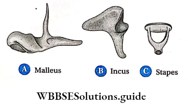
Functions: When sound waves strike the tympanic membrane, the vibrations are transmitted through the ossicles—malleus, incus, stapes, via the oval window to the inner ear.
Inner Ear
The inner ear is located next to the middle ear and is a double-walled structure. It has two main parts— a bony labyrinth and a membranous labyrinth.
Bony labyrinth: Bony labyrinth is located in a cavity, within the temporal bone. The space between the bony and membranous labyrinth is called perilymphatic space. This space is filled with perilymph.
Membranous labyrinth: It is a complex structure, containing canaliculi and membranous sacs.
The fluid present in it is known as endolymph. This part is connected to the auditory nerves.
It is composed of three parts—
- Vestibule,
- Three semicircular canals and
- Cochle
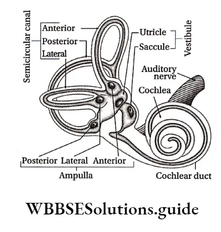
Vestibule: The extended portion of the membranous labyrinth of the inner ear, connected to the three semi-circular canals is known as the vestibule.
It is central sac-like part, which is again formed of two unequal parts— utricle and saccule and is known as the otolith organ.
Structural features:
The vestibule is an endolymph-filled, thin membrane-bound chamber,
This part contains two endolymph-filled sacs. The larger one is called utricle and the smaller one is called saccule.
The endolymph of vestibule contains granules of CaC03 and silica. These are known as otoliths,
Its inner wall is lined by ciliated epithelium and the base of the cells of this part are connected to nerve fibers of vestibular branches of the auditory nerve.
There are 2 sensory spots in the vestibule—the macula of the utricle and the macula of the saccule. The macula consists of sensory hair cells and supporting cells.
Functions: Movement of head causes displacement of the otolith. Thus, sensory cells of the utricle and saccule are stimulated.
This stimulation reaches the cerebellum through auditory nerves. In this way, vestibule of the inner ear maintains body balance with the help of the cerebellum.
Vestibular Apparatus
The three semicircular canals, endolymph containing sacs—utricle and saccule altogether are known as vestibular apparatus. It plays an important role in maintaining body balance.
Utricle And Saccule
Utricle: It is a large sac whose frontal part consists of three semicircular canals, containing ampullae. A duct opens at the dorsal part of the utricle which is known as utricosaccular duct.
Saccule: It is a comparatively smaller sac. The duct opening from the posterior end of the saccule is known as ductus endolymphaticus.
This duct joins with the utriculosaccular duct extends more at the dorsal surface and fuses into a closed sac called saccus endolymaphaticus. Another small duct from the saccule, known as ductus reuniens joins with the cochlear duct.
Three semicircular canals: The frontal part of the membraneous labyrinth contains three bow-like canals. These are known as semicircular canals.
Structural features:
- The three semicircular canals of the inner ear are located in three planes. These planes are located equilaterally.
- The base of each canal is swollen and is known as ampulla (plural: ampullae)
- Due to the presence of the common arm of two semicircular canals, each ear contains five ampullae. Each ampulla contains a sensory spot, called crista ampullaris. Each crista consists of longer hair cells and a gelatinous mass, called a cupula (lacks otolith)
- Endolymph is present between the semicircular canals, ampullae, and vestibule
- The inner wall of an ampulla contains sensory ciliated epithelial tissue.
Functions: The semicircular canal is under the control of the auditory nerve. Displacement of otolith maintains body balance.
Cochlea: The coiled part that appears like a snail and is located posterior to the membraneous labyrinth is known as the cochlea.
Structural features:
The cochlea’s interior is divided into three parallel, fluid-filled compartments by two membranes, the vestibular (Reissner’s) membrane and the basilar membrane,
Each compartment is referred to as a scala (staircase) since each one spiral about an axis or the central pillar of the cochlea like a winding staircase.
They are the (bony) scala vestibuli, the (membranous) scala media or cochlear duct, and the (bony) scala tympani.
The interiors of the scala vestibule and scala tympani are lined by a simple squamous epithelium. Inside scala vestibuli and scala tympani, perilymph is present,
The scala vestibule and scala tympani communicate with each other by a narrow passage, helicotrema. This structure permits perilymph to flow from the scala vestibule into the scala tympani.
At the proximal end of the cochlea, the scala vestibule, and scala tympani communicate with the oval and round window respectively,
The cochlear duct also referred to as the scala media, contains endolymph. In the cross-section of the cochlea, the triangular-shaped cochlear duct appears to be wedged between the scala vestibule and the scala tympani.
The Reissner’s membrane, the roof of the cochlear duct, serves as a division between the cochlear duct and the scala vestibuli. This membrane is composed of squamous epithelium.
Its function is unclear; however it is believed to play a role in the transmission of vibrations from the perilymph of the scala vestibule to the endolymph of the cochlear duct,
The floor of the cochlear duct, which separates it from the scala tympani, is the basilar membrane. The basilar membrane is an elastic structure.
It shows a gradual decrease in stiffness from the oval window (at the cochlear base) to the helicotrema (at the cochlear apex),
The gradual decrease in stiffness permits this membrane to be sensitive to high-frequency vibrations near the cochlear base, and low-frequency vibrations near its apex,
Thus, the Reissner’s membrane is interposed between the scala vestibule and the cochlear duct, and the basilar membrane is interposed between the cochlear duct and the scala tympani.
The receptors associated with the sense of hearing form a structure, the organ of Corti, which rests along the floor of the cochlear duct upon the basilar membrane,
The organ of Corti consists of supporting epithelial cells (pillar cells, cells of Hensen, cells of Claudius, etc.) and neurosensory receptor cells, called ‘hair cells’.
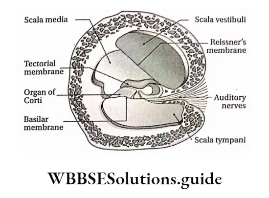
Each hair cell displays numerous stereocilia and a single kinocilium projecting from its apical cell surface,
The free ends of the stereocilia project into the tectorial membrane, an acellular, gelatinous sheet,
The hair cells are the receptors of the auditory system that act as transducers. They serve to convert mechanical energy into electrical energy that can be relayed to the brain-stem,
Hair cells synapse with the peripheral ends (dendritic processes) of the bipolar neurons whose cell bodies are housed in the spiral ganglion.
Leaf morphology important questions for NEET and exams
The central processes of these bipolar neurons form the cochlear nerve, which joins the vestibular nerve to form the vestibulocochlear nerve (VIII cranial nerve).
This nerve courses through the internal acoustic meatus to enter the cranial vault, and then enters the brainstem
Functions: Cochlea is stimulated by sound vibrations produced in the organ of the Corti. This sensation is transmitted to the cerebrum by auditory nerves. In this way, the cochlea helps in hearing.
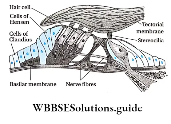
The Physiological Function Of The Ear
Physiological functions of the human ear are as follows— Hearing: The ear enables us to hear external sound.
Mechanism of hearing:
Sound waves from vibrating bodies are transmitted through air to the pinna.
From there, sound waves, through the auditory canal, reach the tympanic membrane. This vibration is transmitted through the ear ossicles of the tympanic cavity to the inner ear via an oval window.
Vibrations are transmitted in the form of waves to the perilymph of scala vestibuli through the fenestra ovalis and then transferred to the scala media through Reissner’s membrane.
The waves in the endolymph cause the basilar membrane to vibrate. This causes the hair cells to bend, pressing them against the tectorial membrane.
As a result of stimulation of the receptor cells, nerve impulses are generated in the associated afferent neurons.
These are conducted by auditory nerves to the auditory cortex of the brain. There it is analysed and we are able to hear.
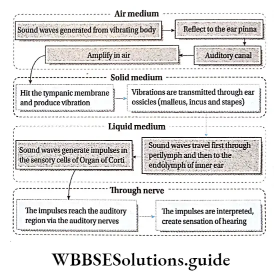
Understanding pitch of sound: The pitch of sound depends on the vibrations of sound waves.
Sounds of different vibrations cause stimulation along different regions of the basilar membrane depending on the pitch of the sound.
So, we can understand sounds of varied pitch. In the case of high-pitched sounds or sounds of high frequency, the receptor cells at the base of the cochlear duct are stimulated.
In the case of low-pitched sound, the receptor cells towards the tip are stimulated.
Understanding loudness of sound: Loudness of sound depends on the distribution of sound. Loudness is expressed by decibels (dB).
The greater the amplitude of sound, the more receptor cells of the organ of Corti in the basilar membrane are stimulated.
Hence, sound can be heard louder. Too loud sound damages the cells of the basilar membrane, causing deafness.
Maintenance of body balance:
- The vestibule and three semicircular canals of the human inner ear maintain the body balance.
- The vestibule and semicircular canals have sensory epithelial tissue. This part contains CaC03 and silica suspended in endolymph.
- These substances are displaced upon a change in body posture, This causes stimulation of cristae and maculae present in the ampulla and vestibule respectively.
- This stimulation is transmitted through auditory nerves of the vestibular branch that join with a cochlear branch to form the vestibulocochlear nerve or auditory nerve.
- The vestibular branch transmits the stimulus first to the vestibular nucleus and next to the cerebellum.
- The cerebellum receives stimuli from proprioceptors of the eyes, skeletal muscles, and joints. These stimuli are then transferred finally to the cerebrum and thus body balance is maintained.
Leaf modifications for storage, support, and reproduction
Notes
- Ampulla: Swollen part of semicircular canal.
- Ganglion: A structure containing a number of nerve cell bodies, linked by synapses and often forming a swelling on a nerve fiber.
- Kinocilium: A special type of cilium on the apex of hair cells located in the inner ear of the vertebrates.
- Motor end plate: The plate-like formation at the nerve terminal where an axon of a motor neuron establishes synaptic contact with the sarcolemma of the striated muscle fiber.
- Neurilemma: The plasma membrane of a neuronal cell body.
- Neurotransmitter: Chemicals that help in the transmission of nerve impulses throughout the nervous system.
- Nuclei: Aggregation of nerve cell bodies within the CNS (not to be confused with the nucleus of a cell).
- Ossicles: A very small bone usually occurring in the middle ear.
- Photoreceptor discs: The outer segment of rods and cone cells lack, ER, Golgi, and mitochondria and are filled with a hundred to a few thousand flattened membrane organelles, called photoreceptor discs.
- Plexus: A structure in the form of a network of nerves.
- Stereocilia: Mechanosensing organelles of hair cells, having a similar structure to microvilli.
- Threshold stimulus: The minimum stimulus required for a reflex action to occur.
Points To Remember
- Neurons are the basic structural and functional units of the nervous system.
- Each nerve cell (neuron) consists of a cell body or soma from which numerous processes arise.
- The human nervous system is divided into two types—central nervous system (CNS) and peripheral nervous system (PNS).
- The cell bodies within the central nervous system (CNS) are frequently clustered into groups called nuclei (not to be confused with the nucleus of a cell).
- The plasma membrane of the axon is called the axolemma, and the cytoplasm contained in it is called the axoplasm.
- Association neurons or interneurons are located entirely within the CNS and serve the associative, or integrative functions of the nervous system.
- The glial cells support neurons structurally and maintain their long-term neuronal integrity.
- The junction between two neurons is called a synapse. It is also called physiological value.
- A nerve is a cord-like structure composed of nerve fibers (axons) and connective tissue.
- A sensory nerve is composed of afferent fibers only a motor nerve of efferent fibers only, and a mixed nerve is composed of both. Most nerves are mixed.
- There are 12 pairs of cranial nerves and 31 pairs of spinal nerves.
- Meninges surround the brain and spinal cord and act as a try layered protective sheath or covering.
- The spinal cord is divided into cervical, thoracic, lumbar, and sacral regions, each served by spinal nerves.
- The autonomic nervous system deals with the visceral organs, like the heart, stomach, glands, and intestines.
- Any changes in the resting membrane potential (which is -70 mV), due to a stimulus of sufficient magnitude, that cause the membrane potential to move towards zero potential (less negative) mean depolarisation.
- A reflex is a quick, involuntary, stereotyped reaction of a gland or a muscle to a stimulus.
- Somatic (spinal) reflexes are responses of skeletal muscles.
- The retina contains the receptors for sight—rod and cone cells.
- The center of the retina contains a special region called the fovea centralis. It is an oval, yellowish area with a depression containing only cone cells.
- The ear is divisible into three parts—the external ear, the middle ear, and the inner ear, each performing a necessary function for the perception of sound.
- The external ear is comprised of the cartilaginous structure—pinna, external auditory canal, and the tympanic membrane.
- The middle ear is embedded in the temporal bone of the skull.
Digestion And Absorption Notes
Digestion And Absorption Introduction
Digestion And Absorption
Your body Requires Energy To Carry Out the Various metabolic processes. This energy is provided by food. It contains nutrients such as carbohydrates, fats, proteins, vitamins and minerals, that are required for overall nutrition and growth of the body.
What happens to the food after eating? But how does the body get these nutrients from food? Here, the digestive system comes into play.
The overall function of the digestive system is to convert the complex food molecules that are ingested, into smaller and simpler molecules.
These simpler molecules can be absorbed by the intestinal cells and are distributed throughout the body via the blood.
These nutrients are utilised for cellular metabolism and also as the building blocks needed for cell growth and repair. In this chapter, you shall study more about digestion and absorption of food.
Digestion and absorption notes for NEET PDF
Digestion
Digestion Definition: Digestion is a physiological process by which enzymes break down complex and insoluble organic components of food, into simpler, soluble substances that can be absorbed and assimilated easily by the cells of the body.
Digestion Can Be Of Different Types Based On—
- Site of Occurrence
- Process Of Digestion.
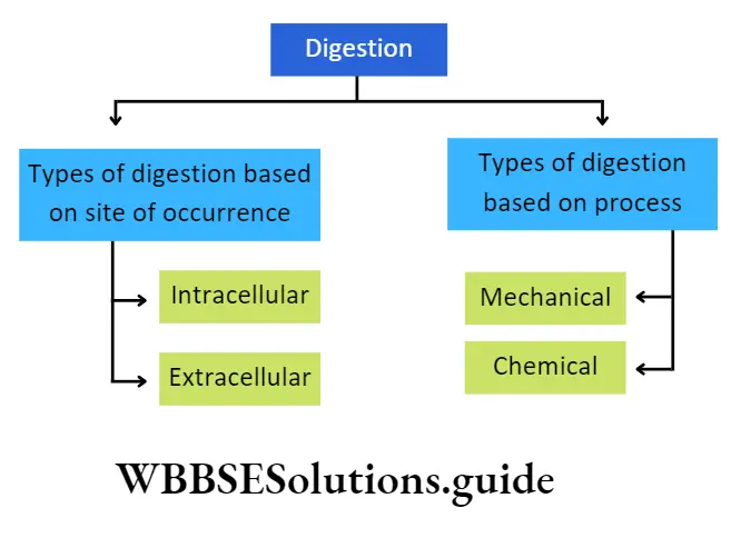
Types Of Digestion Based On Site Of Occurrence: Based on the site of occurrence, digestion can be of two types—intracellular digestion and extracellular digestion.
Intracellular Digestion: The process by which the digestion of food takes place within the cell, is called intracellular digestion. In this process, the cell engulfs the food materials and encloses those inside food vacuoles or endosomes. The cytoplasm, surrounding the food vacuoles, secrete digestive enzymes. This type of digestion is mainly found in protozoa, cnidaria, etc.
Intracellular Digestion Can Be Further Divided Into Two Types—
Heterophagic digestion involves breaking down external substances engulfed by the cell via pinocytosis or phagocytosis.
Autophagic digestion involves consuming components present within the cell.
| Class 11 Biology | Class 11 Chemistry |
| Class 11 Chemistry | Class 11 Physics |
| Class 11 Biology MCQs | Class 11 Physics MCQs |
| Class 11 Biology | Class 11 Physics Notes |
Class 11 biology digestion and absorption notes with diagrams
Extracellular Digestion: The process in which the digestion of food takes place outside the cell is called extracellular digestion.
In this process, the food materials are digested within the alimentary canal, with the help of enzymes containing digestive juices. This type of digestion is found in multicellular organisms such as human beings.
In humans, the mode of nutrition is holozoic.
This Type Of Nutrition Has Five Parts—
- Food intake or ingestion,
- Digestion of food,
- Absorption of nutrients,
- Assimilation of nutrients and
- Egestion or defecation of unabsorbed, undigested food residues.
Types Of Digestion Based On The Process: Based on the process, digestion is of two types— mechanical or physical digestion and chemical digestion.
Mechanical Or Physical Digestion: The process of breaking down larger food particles into smaller particles is called mechanical or physical digestion. This increases the surface area of the food so as to quicken further processes of digestion. It takes place within the mouth by the mastication process.
Chemical Digestion: The process of breaking large particles of food into simple and absorbable form, by the action of the digestive enzymes is called chemical digestion. It takes place in the mouth, stomach and small intestine.
Physiology Of Digestion And Absorption
Absorption
Absorption Definition: Absorption is the process by which digested products and nutrients present in the food move across the epithelium of the alimentary canal into the blood and lymph.
The absorbed nutrients then enter the blood or lymph for circulation to the tissues.
Absorption Site Of Occurrence: Absorption of digested food occurs mainly at three sites of the digestive system— The stomach (a little bit), the Small intestine, and the large intestine.
Absorption Types: The small intestine absorbs the end products of digestion, by two mechanisms of absorption—
- Active absorption,
- Passive absorption.
Absorption Active Absorption: It is an energy-requiring process by which nutrients are absorbed against a concentration gradient from the intestinal lumen by the intestinal epithelial cells.
Passive Absorption: In this process, the nutrients are absorbed along the concentration gradient from the intestinal lumen by the intestinal epithelial cell.
Short notes on digestion and absorption for quick revision
Digestive System
Digestive System Definition: The physiological system in the animal body that is responsible for ingestion and digestion of food, absorption and assimilation of nutrients and digested products and ingestion of undigested food is called the digestive system or alimentary system.
The human digestive system is divided into—
- Alimentary canal or food canal and
- Digestive glands. Different parts of the human alimentary system are given in the following chart.
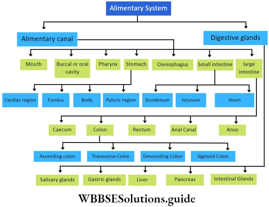
Alimentary Canal
Alimentary Canal Definition: The long, muscular tube-like structure that is part of the alimentary system through which the food passes during digestion is called the alimentary canal.
The nutrients are absorbed from the digested part of food into the blood whereas the undigested or non-essential portion is eliminated as waste. The alimentary canal is also called the digestive tract.
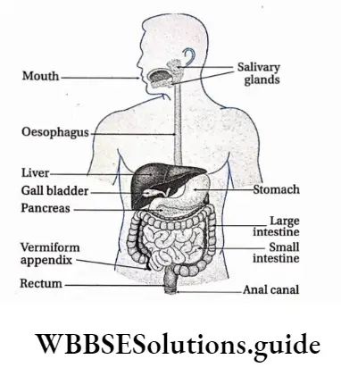
Histological structure human alimentary canal
The histological structure of the human alimentary canal consists of the following layers—
Mucous layer or tunica mucosa: It is the innermost layer of the alimentary canal. The mucus, secreted from the cells of this layer, keeps the inner wall of the alimentary canal slimy. Thus, the wall is protected from mechanical shocks and enzymatic activity of the digestive enzymes.
The glandular cells of the mucous layer secrete several hormones and digestive enzymes. This layer is thrown into circular folds or plicae circulares (or valves of Kerkring or valvulae conniventes) in the intestine.
The intestinal wall also bears numerous finger-like projections called villi. Longitudinal folds observed in an empty stomach are called rugae. The mucous layer itself is made up of three sub-layers.
Short notes on digestion and absorption for quick revision
They are as follows—
Epithelial layer: This is the innermost layer of mucosa. It remains in direct contact with the food passing through the alimentary canal. It consists of different types of cells lined on a basement membrane.
For example, the oral cavity, tongue, pharynx and oesophagus contain squamous epithelial cells. On the other hand, in the stomach, small intestine and large intestine this layer is made up of columnar epithelial cells.
Lamina propria: This is a thin layer of areolar connective tissue that lies beneath the epithelial layer.
This layer contains blood and lymph vessels. The cell population is variable in this layer and can include fibroblasts, macrophages, lymphocytes, eosinophils, etc. These cells provide immunity to the alimentary canal.
Muscularis mucosa: This is the outermost layer of mucosa, made up of smooth muscle fibres. Its contraction produces movement of the mucosa independent of the peristaltic movement of the entire wall of the digestive tract. This layer is absent in the oral cavity and at the pharynx.
Submucous layer or tunica submucosa: This layer remains surrounding the mucous layer and is made up of fibrous and areolar connective tissue. Numerous blood vessels, lymph vessels and nerve fibres are present in this layer. Parasympathetic and sympathetic nerves are present in clusters at different places, forming the submucosal plexus or Meissner’s plexus.
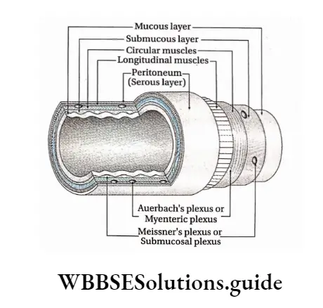
Muscular layer or muscle layer or tunica muscularis: This layer lies next to the submucous layer and is made up of smooth muscle fibres.
This layer is further subdivided into two layers—
- Inner circular
- Muscle layer and
- Outer longitudinal muscle layer.
A thin connective tissue layer containing blood vessels, lacteals and nerve fibres separates these two layers. In the stomach, another layer called the oblique layer is present between these two sub-layers.
Sympathetic and parasympathetic nerves form clusters at different parts within this layer, forming Auerbach’s plexus or myenteric plexus. The muscular layer is responsible for peristaltic contractions. The circular muscle layer forms sphincters at specific locations of the digestive tract.
Serous layer or tunica serosa: It is the outermost layer of the alimentary canal, made up of a layer of simple squamous epithelium (or mesothelium), supported by an underlying areolar connective tissue layer.
This layer is also called tunica adventitia. The parts of the alimentary canal present in the abdominal and pelvic cavity, are covered with a serous layer known as peritoneum. It contains a large number of blood vessels, lymph vessels and lymph nodes.
The peritoneum is made up of two membranes—
- Visceral peritoneum, which protects the internal organs present in the peritoneal cavity.
- Parietal peritonium, that forms the inner lining of the abdominal cavity.
Different parts of the alimentary canal have been discussed separately
Different Parts Of The Human Alimentary Canal
The different parts of the human alimentary canal have been discussed below.
Mouth
The mouth is the opening present at the beginning of the alimentary canal through which the intake of food takes place.
Mouth Location: It is present at the beginning of the alimentary system, just below our nose.
Mouth Structural features:
- The mouth is a transverse slit, bounded by two movable lips.
- The lips are covered with skin on the outer side and mucous membranes on the inner side.
- The inner wall of the mouth is covered with stratified squamous epithelium.
- The small vertical groove present in the middle region of the upper lip is called the philtrum. The philtrum extends and forms another structure called a tubercle.
Mouth Functions:
- The mouth helps in food intake. The lips help in opening and closing of the mouth. They assist in the intake of food as it passes into the mouth.
- As lips have a large number of nerve endings, they are considered tactile organs, and sensitive to touch and temperature.
- The mouth also plays an important role in communication, making sound and speech.
Buccal or oral cavity
The cavity that lies just behind the mouth where the ducts from the salivary glands open is called the buccal or oral cavity.
- Location: It is present just behind the mouth.
- Structural features: The structural features of the different parts of the buccal cavity are as follows.
Mouth Hard and soft palate:
- The concave roof of the mouth is called the palate.
- The anterior bony part of it is called the hard palate. The posterior muscular region that is continuous with the hard palate is called the soft palate.
- The hard palate is covered by a thick mucous membrane. The mucous membrane covering the floor of the nasal cavity is continuous with a soft palate.
- A small projection called the uvula or velum palati, hangs free from the posterior region of the soft palate. During the act of swallowing or deglutition, it prevents pieces of food from entering the nasal cavity.
Mouth Jaw:
- The oral cavity is lined by an upper and a lower jaw. They are hard, bony and contain teeth. The upper jaw is attached to the skull, hence immovable.
- However,- the lower jaw can be moved both sideways and down.
- The jaw bones are covered by gums or gingivae (singular: gingiva) inside the mouth. Gums surround the teeth and provide a seal around them.
Teeth:
- Both the upper and lower jaws have the teeth embedded in them.
- Human adults have 32 teeth. 16 teeth in each jaw. In humans, there are four
types of teeth—8 incisors (cuts food), 4 canines (C) (tears food), 8 premolars (PM) (grinds food) and 12 molars (M) (crushes and grinds food). - The third molar present on both sides of each jaw are together called wisdom teeth. These teeth generally grow at the age of 20-21 years.
- This set of 32 teeth is called permanent teeth. However, there is a set of 20 teeth in children. They are called milk teeth. They develop at about the age of 6 months and remain till they are 5-6 years old.
The nature of dentition and structure of teeth in humans is given below.
- Nature of dentition in human: In human 4 different types of teeth are present, hence, human has heterodont type of dentition,
- Two sets of teeth arise within a lifetime of a human, so it is called diphyodont type of dentition,
- Teeth are embedded into the jaws, so it is called the codon type of dentition.
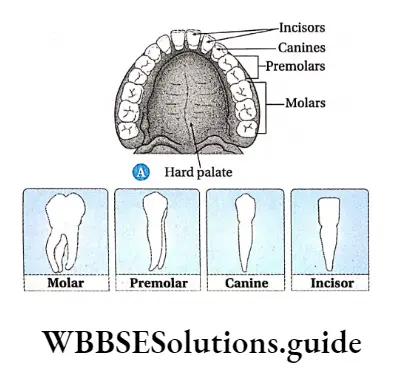
Structure of a tooth: Although the shapes of different teeth vary, they have a similar basic structure.
Its structural features are as follows—
- A tooth is formed of two parts—the exposed part or crown and the part that is embedded in the jaw bone or root.
- The crown is covered with a whitish, hard substance called enamel. It is composed of calcium salts and hydroxyapatite crystals,
- The root is formed by a mineralised matrix called dentin.
- Dentin is covered by a calcified layer called cementum. It anchors the tooth to the bone,
- The central cavity of dentine is filled with soft, gelatinous and vascular connective tissue called pulp. The cavity is called a pulp cavity,
- The portion of the pulp cavity that extends into the root is called a root canal.
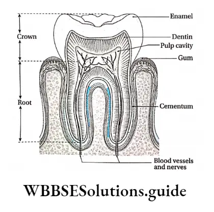
Tongue: The tongue is present on the floor of the mouth.
Its structural features are as follows—
- It is a soft muscular, flattened sensory organ.
- Its anterior two-third part lies in the oral cavity which is free in movement.
- Its one-third posterior part is attached to the floor of the buccal cavity in the pharynx by a fold of the mucous membrane called the lingual frenulum.
- The oral and pharyngeal parts of the tongue are separated by a ‘V-shaped furrow, the sulcus terminalis.
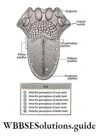
The dorsal surface of the tongue is covered with minute, round-shaped raised structures called lingual papillae.
Each papilla is surrounded by a number of taste buds. The average person has about 10,000 taste buds. Sensory receptors present within these taste buds are responsible for various tastes.
Lingual papillae are of four types—
- Circumvallate papillae are present in a V-shaped row at the posterior region of the tongue. They are 8-12 in number,
- Filiform papillae are the cone-shaped papillae that are the smallest in size. They are the most abundantly distributed at the anterior part of the tongue. They contain receptors for touch instead of taste,
- Fungiform papillae, the mushroom-shaped papillae are distributed on the tip and side of the tongue. Each papilla contains five taste buds,
- Foliate papillae are located on the posterior part of the tongue.
- The tongue has voluntary muscles, hence its movement can be controlled.
Functions: The functions of the buccal cavity—
Digestion: After ingestion, food mixes with the saliva secreted by salivary glands inside the buccal cavity. The food transforms into a soft rounded mass or bolus. The carbohydrates present in the food get metabolised by the action of the salivary enzyme, ptyalin.
Taste: The tongue which is present in the buccal cavity, helps to analyse of texture and taste (bitter, sweet, sour and salt) of the food.
Mastication and swallowing (deglutition) of food: Mechanical processing (mastication) of food occurs inside the buccal cavity with the help of teeth.
Pharynx
Pharynx is the cone-shaped musculo membranous cavity structure, leading from the mouth to the oesophagus.
Pharynx Location: It is present in the posterior region of the buccal cavity.
Pharynx Structural features:
- It is a cone-shaped, small passage where the alimentary canal and air passage cross each other. It is 12-14 cm long. It is divided into three parts.
- The upper part of the pharynx has openings of a pair of internal nares and a pair of eustachian tubes. This part of the pharynx is called the nasopharynx.
- The oropharynx lies behind the buccal cavity and acts as the passage for food bolus.
- The last part of the pharynx or laryngopharynx is connected to the larynx and the trachea (windpipe).
- An aperture called glottis in its anterior portion connects it to the trachea. The glottis is closed by a triangular flap of elastic cartilage called epiglottis. It prevents the entry of food and water into the larynx.
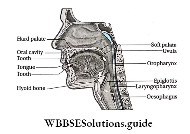
Pharynx Functions:
- The pharynx helps to transfer the food bolus from the mouth to the oesophagus (helps in Deglulition or the act of swallowing).
- The pharynx joins the nasal passage to the larynx and helps in breathing.
Oesophagus
The Oesophagus is the part of the alimentary canal that extends beyond the pharynx, upto the stomach.
Oesophagus Location: From the pharynx, it extends downwards leading to the stomach.
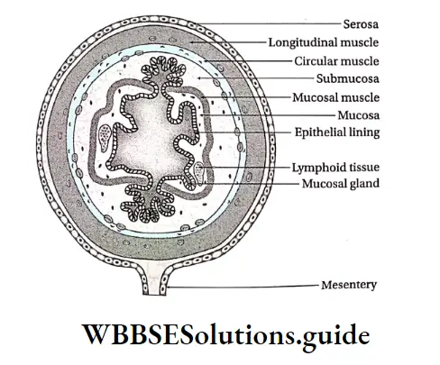
Oesophagus Structural features:
- The oesophagus is about 25 cm in length.
- It is a long and thin tube that enters the thoracic cavity and runs parallel to the windpipe.
- The inner lining of the oesophagus is generally smooth.
- No glands are attached to the oesophagus The oesophagus consists of four layers—the mucosa Submucosa, muscular mucosa and tunica serosa or adventitia (from inside to outwards).
Oesophagus Functions: The oesophagus helps the food bolus to travel down to the stomach, through peristalsis. No digestion of food takes place here.
Stomach
The stomach is the largest part of the alimentary system. It has a sac-like appearance. It is the part of the alimentary canal next to the oesophagus, where digestive juices and hydrochloric acid are released for further digestion of the food.
Stomach Location: It is located below the diaphragm, next to the oesophagus, with the liver on its right.
Stomach Structural features:
- The stomach is a wide, J-shaped organ.
- It is about 25 cm long, while breadth and height both is about 10 cm.
- The stomach has two curvatures. The lesser curvature makes up the right border of the stomach. It is slightly depressed. The greater curvature makes up the left border of the stomach. It is slightly elevated.
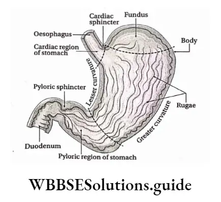
The stomach is divided into four parts—
Cardiac stomach: The part of the stomach that joins the oesophagus is called the cardiac stomach, it is called so because of its location near the heart.
Fundus: The semicircular region of the stomach, that extends a little beyond the level of the cardiac stomach, is called the fundus
Body: The part of the stomach that extends in between the fundus and the pyloric region of the stomach is called the body,
Pyloric stomach or Pylorus: The distal part of the stomach, that extends beyond the body region is called the pyloric stomach or pylorus. It is divided into two parts.
The part of the pylorus that joins the body of the stomach is called the pyloric antrum while the part which joins the duodenum is called the pyloric canal.
The stomach has two openings the cardiac orifice, through which the oesophagus opens into the stomach and the pyloric orifice, through which the stomach opens into the duodenum.
The sphincter muscle present at the junction of the oesophagus and the stomach i.e. at the cardiac orifice, is called the cardiac sphincter.
Digestion And Absorption Of Nutrients
The pyloric orifice is surrounded by a well-defined muscular sphincter, called the pyloric sphincter. It controls the movement of food out of the stomach but prevents reflux of duodenal contents into the stomach.
The stomach wall consists of mucosa, submucosa, muscular and serosa (serous) layers. The inner surface of the stomach, mainly the mucosal region, is thrown into longitudinal folds called rugae. These folds appear only when the stomach is empty.
The inner lining of the stomach has numerous microscopic glands called gastric glands which are present in groups of 2-7, which together link with the gastric lumen through the gastric pits.
Cardiac stomach Functions: The stomach performs the following functions in the human body.
- Digestion: Major enzymatic digestion is initiated in the stomach, particularly that of proteins. The gastric glands secrete digestive juices. Different enzymes are present in these digestive juices that help to digest fats and proteins. HCI creates an acidic environment within the stomach. This allows the enzymes to work properly. The stomach is responsible for the formation of chyme, which is a semi-fluid mass of partly digested food.
- Storage: The stomach acts as a storage for food until it moves into the duodenum.
- Mechanical breakdown: The vigorous contractions of smooth muscles present in the stomach help to grind food materials into smaller bits and also to mix with gastric secretions.
- Absorptive function: Small amounts of salts, water Alcohol, medicine etc get absorbed in the stomach.
- Other secretions: The stomach secretes a substance called Castle’s intrinsic factor. It is required for the absorption of vitamin B12 in the small intestine. Moreover, gastrin hormone is secreted by G cells and stimulates the glands to secrete the digestive juices.
- Protection: HCI released by the oxyntic cells can kill the microbes that enter our body along with food and provide protection against them.
Production of HCI in the stomach
CO2 present in the interstitial fluid diffuses into the parietal cells of the stomach and reacts with water to form carbonic acid (H2CO3).
This reaction is facilitated by carbonic anhydrase. This H2CO3 dissociates into H+ and HCO-3. H+ ions enter the lumen of digestive glands.
Chloride ions already present in the interstitial fluid, enter the digestive glands via the parietal cells. H+ and Cl– react to form HCI.
Small Intestine
The long, narrow and coiled part of the alimentary system that starts just below the stomach, where both digestion and absorption take place, is called the small intestine.
Cardiac stomach Location: It extends from the pyloric valve of the stomach to the large intestine.
Cardiac stomach Structural features:
- The small intestine is a narrow and extended tube-like structure. It has a length of slightly more than 5m and a diameter ranging between 3.5-4.5 cm.
- The walls of the small intestine are made up of four layers, from inside to outwards—mucosa, submucosa, muscularis externa and serosa.
- The entire mucosa layer is lined with numerous, densely packed finger-like extensions called villi.
- The submucosa layer contains Brunner’s glands at the duodenum. It secretes a slimy alkaline substance which neutralises the acidity of the chyme.
- Numerous circular or oblique folds (or folds of kicking) called plicae circulares line the inner wall of the small intestine.
- At the bases of villi, glandular areas called crypts of Lieberkuhn, are present. These contain mucus and hormone-secreting cells.
- It has the following regions—
- Duodenum: It is part of the small intestine. It forms a C-shaped arch with the stomach. Its length is around 25 cm. Its lateral walls have a slightly swollen region called the ampulla of Vater.
- The hepatopancreatic duct (the common duct formed by the pancreatic duct and bile duct) enters the duodenum through this ampulla- The internal wam contains circus folds and villi,
- Jejunum: It is the coiled, middle region of the small intestine. It is about 2 m long. The inner wall is thicker and contains more folds and villi than the duodenum. Glands are absent in this region.
- Ileum: It is the last part of the small intestine. It is about 3 m long. It remains in a coiled condition within the abdominal cavity. The inner wall ileum contains a lesser number of plicae and villi than the jejunum.
Small masses of lymphatic tissues called Peyer’s patches are found on the ileum wall. These tissues produce lymphocytes which prevent infection. It joins with the colon region of the large intestine.
At its junction with the colon, a valve called the ileocecal valve is present. It allows the undigested parts of the food to enter the colon but prevents the reverse.
NEET digestion and absorption revision notes with MCQs
Villi
- Villi Location: The mucosa of the small intestine.
- Villi Structure:
- A mucous layer of the small intestine folds into small finger-like projections called villi.
- Each villi is about 0.5-1 mm long and has an outermost layer of columnar epithelial cells called enterocytes. The free surface of these cells has numerous 1 pm long microvilli and form a brush-like appearance. This is called border structure,
- At the base of the villi crypts of Lieberkuhn are present which contains endocrine cells. These cells secrete intestinal juice.
- Each villus has a central lacteal,
- Several blood capillaries form a network around lacteal. The veins present at the ends of these capillaries join to form the hepatic portal vein.
- Villi Function: The villi as well as the microvilli increase the surface area for absorption in the small intestine.
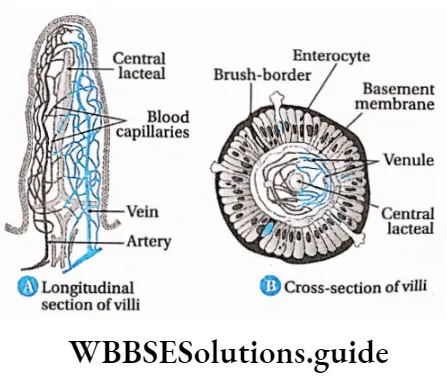
Villi Functions: Functions of the small intestine are—
Digestion
- The small intestine receives bile from the liver. Bile does not contain enzymes. It contains salts like sodium glycolate and sodium taurocholate, which emulsifies fat causing fat droplets to disperse in water.
- It also receives pancreatic juice from the pancreas and intestinal juice from intestinal glands. Both of these juices contain enzymes necessary for the digestion of proteins, carbohydrates and fats.
Absorption: The small intestine is the site for the absorption of nutrients. The circular folds, villi, and microvilli increase the surface area for absorption.
Segmentation and peristalsis: The wall of the small intestine shows two types of movements—segmentation and peristalsis. Segmentation causes small and regular, localised contractions that bring chyme in contact with digestive juices. Peristalsis of the small intestine allows undigested food to move to the large intestine.
Protection: The germs that have survived the effect of HCI in the stomach get killed by the lymphocytes produced at the lymph nodes in the small intestine.
Hormonal secretion: Hormones like cholecystokinin (CCK) and secretin are secreted within the small intestine.
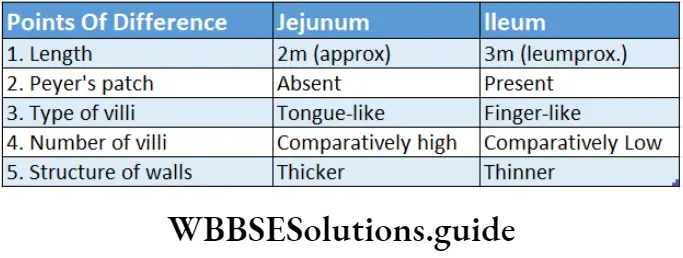
Large intestine
The large intestine is the part of the alimentary tube, present between the small intestine and the anus, where the undigested food remains stored for some time.
Location: The large intestine lies next to the ileum, in the abdominal cavity.
Structural features: The diameter of the large intestine is 6.5 cm and its length is about 1.5 m. The inner wall contains some semilunar folds. These are called plicae semilunaris.
The large intestine is divided into the following parts—
Caecum: It is a small sac-like structure present below the junction of the small intestine with the large intestine.
The caecum has a small (about 8-9 cm length) finger-like blind tube called the vermiform appendix at its lower end. Infection of the vermiform appendix is known as appendicitis. In human beings, it is a vestigial organ.
Colon: It is a wide tube-like structure present between the caecum and rectum. The wall of the colon has longitudinal muscles that form three separate bands called taenia coli.
Contraction of these muscles produces a series of bulges or pouches called the haustra, in the colon. The colon is divided into four parts—ascending, transverse, descending and sigmoid colon.
Rectum: It is the enlarged structure at which the sigmoid colon opens. It is about 13 cm in length.
Anal canal and anus: The rectum opens into a short canal-like structure called the anal canal. It is generally 3.8 cm long. This canal opens outside by an orifice called the anus. Anus is guarded by two sphincter muscles called anal sphincter.
Functions: The large intestine performs the following function in human beings.
Production of faecal matter: The faeces are formed by the action of the different bacteria present in the colon region of the large intestine.
Absorption: In the large intestine, Na+, Cl– and H2O are absorbed from the undigested food matter. Some vitamins and medicines also get absorbed in this region.
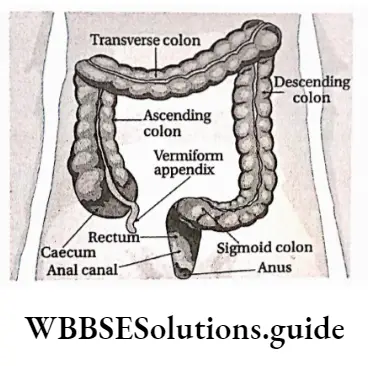
Mucus secretion: The goblet cells present in the large intestine secrete mucus. The mucus softens the faecal mass and makes it slippery.
Storage of faecal matter: The faecal mass is stored in the rectum for some time before egestion.
Egestion of faecal matter: The faecal matter is removed from the body through the anus.
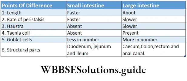
Digestive Glands
Digestive glands Definition: The glands related to the digestive system which secrete digestive juices containing enzymes that convert complex food substances into simpler absorbable forms are called digestive glands.
Digestive glands Types: Digestive glands in humans include salivary glands, gastric glands, intestinal glands, liver and pancreas. The different types of digestive glands, along with their secretions are discussed below.
Salivary glands
The salivary glands are present in the buccal cavity. They secrete saliva. Enzymes present in it help to break down the carbohydrate-rich food.
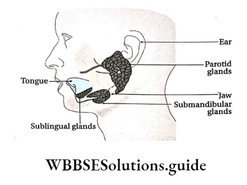
Salivary glands Types: Salivary glands are of three types—parotid, submandibular and sublingual glands.
Parotid gland: The paired parotid glands are the largest of the salivary glands and weigh about 15-30 g.
They are present just below the ears. They open inside the mouth, at the root of the second molar on both sides of the upper jaw, through Stensen’s ducts.
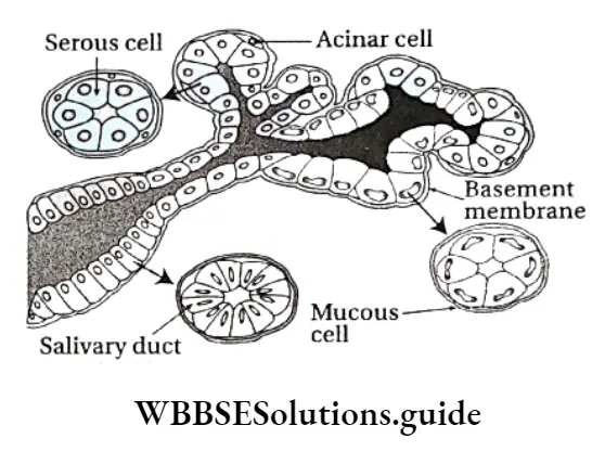
Mumps
- Mumps is a viral disease that affects salivary glands, mainly parotid glands. Children at the age of 5-9 years are more vulnerable to this disease.
- If mumps occurs after adolescence, then it may lead to infection in the testes. This may further lead to sterility. Sometimes pancreas may also get affected by the mumps virus. This is the risk of diabetes mellitus in the patient.
Submandibular gland: The submandibular or submaxillary gland is the second largest salivary gland. It weighs about 7-16 g. The gland is located at the angle of the lower jaw, at the junction of the lower and upper jaws. The duct, by which each submaxillary gland opens into the mouth, is called Wharton’s duct.
Sublingual gland: The smallest of the salivary glands is the sublingual gland, weighing about 2-4 g. It is located below the tongue. It opens into the mouth through several fine ducts called ducts of Rivinus. Structure: The anatomical and histological structures of the salivary glands are discussed below.
Anatomical structure:
- The salivary glands are divided into several lobes. The lobes are separated by interlobular septum.
- Each lobe is further divided into numerous acini (singular: acinus) or alveoli (singular: alveolus).
- Each acinus or alveolus has an outer membrane. The inner surface of the membranes has glandular cells.
- The central portion of the acinus is empty, where the secretions are stored. The ducts that arise from these cavities join to form the salivary duct.
Histological structure:
- Histologically, the salivary glands are made up of two types of cells—
- Serous cells and
- Mucous cells.
- The parotid glands are composed primarily of serous cells. The sublingual glands contain mainly mucous cells. On the other hand, the submandibular glands contain both serous and mucous cells, hence they are called mixed types of glands.
- Mucous cells release a thick secretion called mucus.
A few star-shaped contractile cells are situated in between the basement membrane and the secretory cells called myoepithelial cells which help in the expulsion of saliva from the gland.
Mumps Function: The function of the salivary glands is to secrete saliva.
Saliva: The colourless, watery fluid secreted by the salivary glands, that contains mainly a starch-digesting enzyme called ptyalin (salivary amylase) in the buccal cavity is called saliva.
Characteristics and components: Saliva is a colourless and viscous liquid. The saliva maintains a slightly acidic pH ranging from 6.0 to 7.0. The Components of Saliva Are listed.
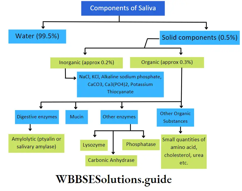
Other Components Of Saliva
- Cellular components: Saliva contains yeast cells, bacteria, parasites, protozoa, polymorphonuclear leucocytes (granulocytes), etc., as their cellular components.
- Gaseous components: Every 100 ml of human saliva contains 1 ml oxygen, 2.5 ml nitrogen and 50 ml CO2 in dissolved condition.
- Function: Saliva plays an important role in the digestion of food. it also has some other functions.
Role in digestion:
Saliva contains the enzyme ptyalin which acts in a slightly acidic medium, in the presence of chloride ions. It breaks down boiled starch, glycogen, etc., into maltose, maltotriose, limited dextrin and isomaltose,
The water present in the saliva helps to break down the protein molecules present in the food.
Non-specific immunity: Human saliva contains an enzyme called lysozyme. This enzyme helps to break down the polysaccharide units of the cell walls of bacteria like Streptococcus spv Staphylococcus sp., etc., that enter the mouth via food. This kills these bacteria and provides protection.
Role in excretion: Saliva acts as a medium to release urea, heavy metals,(Hg, Pb, Bi, As, etc.), isocyanate, excess antibiotics, etc., from the body.
Helps to taste: Saliva aids in moistening dry food. This enables us to feel its taste.
Mechanical and other functions:
- Saliva contains a glycoprotein, called mucin. It mixes with the food and makes it slippery so that swallowing becomes easier,
- It lubricates the mouth and helps in speaking,
- It softens the food and makes chewing easier,
- Saliva has a high concentration of bicarbonate ions. It acts as a buffer and protects the mucous layer inside the oral cavity and teeth from the acids produced by the microbes.
Gastric Glands
Gastric glands are the small tubular glands, present on the mucous layer of the stomach. They secrete digestive enzymes.
Types: Gastric glands are of three types—
- Cardiac gastric glands are located in the proximal part of the stomach,
- Fundic or intermediate gastric glands in the central stomach areas and
- Pyloric gastric glands in the terminal stomach portion.
- Secrete mucus that protects the stomach lining from the digestive action of gastric juice.
- Gastrin cells, also called G cells secrete a hormone, gastrin.
Gastric glands Function:
- Gastric glands secrete gastric juices that help to break down the food and digest it.
- The mucus secreted by these glands helps to protect the tissues of the stomach from the digestive juices.
Gastric juice: The highly acidic fluid, secreted by the gastric glands, that contains digestive enzymes is called digestive juice or gastric juice.
Gastric glands Characteristics and components: The pH of gastric juice is low, ranging from 0.7 to 3.8. About 1200-1500 ml of gastric juice is produced every day. Around 500- 1000 ml is secreted after every meal. The composition of gastric juice is given in the following chart.
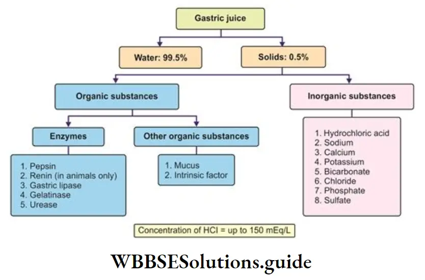
Gastric glands Function: The different components of gastric juice have the following functions—
Role of HCI:
- HCI found in gastric juice converts inactive pepsinogen, and prorennin into their active forms.
- It provides an environment with the optimal pH required for the action of pepsin,
- It converts some of the ingested proteins into acid metaproteins,
- It kills the bacteria that have entered the food.
- It also helps in hydrolysis of sucrose.
Role of mucin:
- Mucin produced by the gastric glands forms a protective barrier on the inner lining of the stomach. It prevents the HCI from damaging the cells of the stomach,
- It acts as a buffer and lowers the acidity of the stomach.
Role of pepsin: Pepsin is a protease, that is the principal enzyme in gastric juice. The precursor, pepsinogen, is converted to the enzyme pepsin when exposed to HCI. This pepsin now hydrolyses proteins, acid metaproteins, etc., into proteoses and peptones.
Role of chymosin: This enzyme also known as rennin is absent in the gastric juice of adults. In children, this enzyme converts milk protein, casein into paracasein, under an acidic medium.
Role of Castle’s intrinsic factor: Castle’s intrinsic factor, present in the gastric juice, helps in the absorption of vitamin B12, in the intestinal lumen. Its absence decreases vitamin B12 absorption.
Role of gastric lipase: Gastric lipase is a fat hydrolysing enzyme. It acts on emulsified fats and hydrolyses them into fatty acids and glycerol.
Role of other enzymes:
Other enzymes present in the gastric juice include—
- Lysozyme kills bacteria by breaking the polysaccharide layer of their cell membrane,
- Urease hydrolyses urea into ammonia,
- Carbonic anhydrase stimulates the oxyntic cells of the stomach to synthesise HCI.
- Gelatinase breaks down gelatin into peptones.
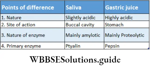
Liver
The largest gland of the body, normally weighing about 1.5 kg, which takes part in digestion and excretion is called the liver. it is present in the upper part of the abdominal cavity, just below the diaphragm, on the right side of the body.
Structure: The anatomical and histological structures of the liver are discussed below.
Anatomical structure:
- The liver is the largest gland (exocrine) in the body.
- It is reddish-brown in colour.
- It has a larger right lobe and a smaller left lobe, separated by a ligament.
- The ligament is called the falciform ligament. It is continuous with the peritoneum.
- The lower part of the liver is further divided into two lobes—quadrate lobe and caudate lobe by transverse fissures.
- The liver is covered by a connective tissue layer, called Glisson’s capsule.
- The lower part of the right lobe has a slightly depressed region, called cystic fossa. It contains a thin-walled, pyriform (pear-shaped) sac-like structure called a gall bladder.
- The duct arising from the gall bladder is called the cystic duct. Two ducts arising from the two lobes of the liver join to form the common hepatic duct.
- When the common hepatic duct joins the cystic duct, it forms the common bile duct.
- The common bile duct joins the pancreatic duct and opens into the duodenum at the ampulla of Vater.
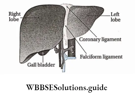
Liver Histological Structure:
- A capsule made of connective tissue surrounds the liver. This capsule invaginate the liver to divide it into numerous hepatic lobules.
- Lobules of the liver are polygonal in shape. They are separated from each other by connective tissue. Each lobule is made up of hepatocytes (hepatic cells).
- Hepatocytes are arranged as plates (hepatic plates) of one cell thickness with vascular channels (sinusoids) on either side. These plates remain surrounding a central vein like the spokes of a bicycle wheel.
- The hepatic artery, hepatic portal vein and a branch of the bile duct are present in the connective tissue, within the liver. Liver Together, they are called the hepatic or portal triad.
- Hepatocytes secrete a fluid called bile into a network of narrow channels between the opposing membranes Of adjacent liver cells. These passageways are called bile canaliculi.
- Large phagocytic cells known as Kupffer cells or Stellate cells are attached to the inner lining of hepatic sinusoids. They act as macrophages. They remove pathogens and debris that may have entered the sinusoids.
- Small spaces called perisinusoidal spaces (also known as the space of Disse) are present in between a hepatocyte and a sinusoid. Transport of proteins and other plasma components from sinusoids to hepatocytes occurs through these spaces.
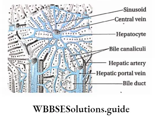
Liver Function: Functions of the liver in humans are given below.
- Secretion: It produces bile, which remains stored within the gall bladder before entering the bile duct.
- Storage: It stores iron (Fe2+) and fat-soluble vitamins such as vitamins A, D, E, and K. Some quantities of fat also remain stored within the liver.
- Metabolism: The liver takes part in the metabolism of carbohydrates, proteins and lipids.
Carbohydrate metabolism: It helps in carbohydrate metabolism and maintains the concentration of glucose in the blood.
It converts glucose to glycogen by glycogenesis (under the influence of the hormone insulin). It converts glycogen into glucose by glycogenolysis (under the influence of the hormone glucagon). The liver also converts fats and proteins into carbohydrates by gluconeogenesis, when required.
Fat metabolism: The liver helps in the oxidation of fats, production of ketones, and synthesis of fats from carbohydrates and proteins. It also helps in the synthesis of phospholipids. It can also regulate the blood cholesterol level by converting some of them to bile acids. These bile acids form bile salts, which help in emulsification of fats.
Protein metabolism: It produces plasma proteins, It also synthesises urea (by ornithine cycle) and uric acid by deamination of amino acids.
Transamination: Synthesis of new amino acids by transfer of amino group from an amino acid to another compound takes place in the liver.
- Role in excretion: The liver acts as an accessory excretory organ. Ammonia produced due deamination of proteins is converted into urea in the liver. It also detoxifies blood by metabolising and eliminating poisonous substances such as toxins, metals, bacteria, etc., from the body. It also excretes bilirubin, produced by the breakdown of haemoglobin.
- Role in synthesis: The liver synthesises plasma proteins, such as prothrombin, fibrinogen, etc., from amino acids. Synthesis of anti-coagulation factor, heparin, takes place within the hepatic cells
- Immunity: It plays an important role in the immunity of the body. Kupffer cells, present in the sinusoids, engulf the bacteria that enter the liver. The liver also produces antibodies that help in the development of the immune system.
- Regulation of body temperature: The metabolic reactions taking place in the liver produce heat, that helps in the regulation of body temperature.
- Bile: It is the greenish-yellow (in the case of humans)coloured, non-enzymatic digestive juice secreted by the 1 hepatocytes of the liver. It enables the breakdown and absorption of fats.
- Characteristics and components: Bile is generally greenish-yellow in colour. It is alkaline in nature, with a pH of about 7.7. Sometimes, the alkalinity is slightly less (pH ranging between 7.0-7.6). An average of 500-1000 ml of bile is produced every day. The components of bile are described in the following chart
- Function: Bile take part in different functions in the human body
1. Role in digestion:
- Bile salts help in the emulsification of fats, i.e., break down fat into small droplets. It increases the surface area for the enzyme lipase to act on. Lipase breaks down the fat droplets into fatty acids and glycerol,
- Bile also contains glycocholic acid and taurocholic acid that stimulate the action of the enzyme lipase,
- Bile also acts as a medium for the reaction between fats and lipase enzymes.
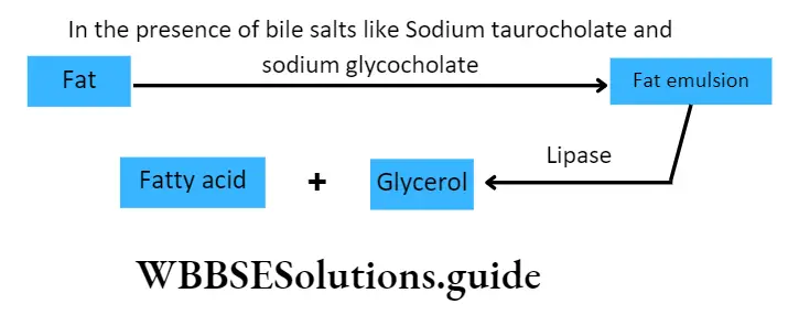
2. Role in absorption:
- Bile also enhances the absorption of fats (digested or undigested), fatty acids, metal ions such as Na+, K+, Ca2+ and the fat-soluble vitamins A, D, E, and K.
3. Role in excretion:
- Bile also serves as the route of excretion for heavy metals such as Znz+ Cu, etc., bile helps in the regulation of body temperature. pigments, toxins, bacteria, cholesterol and lecithin, etc..
4. Choleretic Action:
- Bile contains bile salts that are coloured, non-enzymatic digestive juice secreted by the 1 hepatocytes of the liver. It enables the breakdown and absorption of fats.
5. Laxative action:
- Bile salts act as natural laxatives by stimulating peristaltic movements of the intestine.
6. Role as a lubricant:
- Mucin present in bile, acts as both a lubricant and a buffer.
7. Neutralisation of stomach acid:
- The sodium carbonate present in bile neutralises any excess stomach acid before the chyme enters the ileum. Besides these, bile salts also act as bactericides. They can destroy many microbes present in the food.

Pancreas
The pancreas is the organ that is present below the stomach, in j the abdominal cavity. It has both exocrine (ductal) and j endocrine (ductless) functions.
Pancreas Structure: The pancreas is a slightly pink-coloured light organ. Its length is around 12-15 cm and it weighs around 60 g. It is a slightly flattened and stretched structure.
Its ends are slightly edged. Its narrow end is free, while its broad end has a duct arising from it, called the pancreatic duct. This duct joins the ampulla of Vater, present in the duodenum.
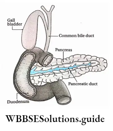
Pancreas is a mixed gland. Its exocrine and endocrine functions are discussed below
Exocrine region of the pancreas:
- The exocrine region I of the pancreas is composed of numerous small, sac-like I structures called acini. They are covered by membranes.
- Each acinus is composed of a single layer of pyramidal glandular cells. These cells are arranged in groups around a central lumen, from which several ducts (ducts of acini) arise.
- These acinar ducts together form a duct of Wirsung or the pancreatic duct. The pancreatic duct, in turn, joins the common bile duct and enters the duodenum at the ampulla of Vater.
- An accessory pancreatic duct called the duct of Santorini is found in some individuals. It rises from the main pancreatic duct and opens in the duodenum just above the common bile duct.
- The blood flow through the ampulla of Vater is regulated by a sphincter muscle called hepato-pancreatic sphincter (sphincter of Oddi).
- The enzyme-containing granules present in the exocrine region are called zymogen granules.
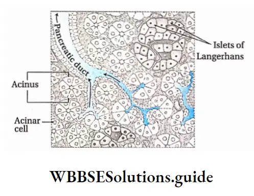
Endocrine region of the pancreas:
- The endocrine cells are arranged in groups, which are interspersed among the acini. These groups are called islets of Langerhans (named after the discoverer scientist P. Langerhans).
- There are five types of endocrine cells found in islets—alpha-beta, delta, epsilon and P-P cells.
- Different hormones (from a, (3, 5, e cells) and pancreatic polypeptide (P-P cells) are secreted by this region. The islets are surrounded by networks of blood capillaries.
- The hormones, that are secreted, enter the blood capillaries and get transported to the target organs.
The pancreas is one of the main digestive glands in humans. It performs the following functions.
- Digestive juice secretion: The enzymes, secreted by the exocrine region of the pancreas, break down. carbohydrates, fats, proteins, and acids in the duodenum.
- Secretion of bicarbonate ions: The exocrine tissue also secretes bicarbonate ions that neutralise the stomach acid in the duodenum.
- Hormone secretion: Insulin, glucagon, and gastrin hormones are secreted by the endocrine region of the pancreas. P-P cells secrete pancreatic polypeptides that regulate the secretory activity of pancreatic cells carbohydrates, fats, proteins, and acids in the duodenum.
- Secretion of bicarbonate ions: The exocrine tissue also secretes bicarbonate ions that neutralise the stomach acid in the duodenum.
- Hormone secretion: Insulin, glucagon, and gastrin hormones are secreted by the endocrine region of the pancreas. P-P cells secrete pancreatic polypeptides that regulate the secretory activity of pancreatic cells.
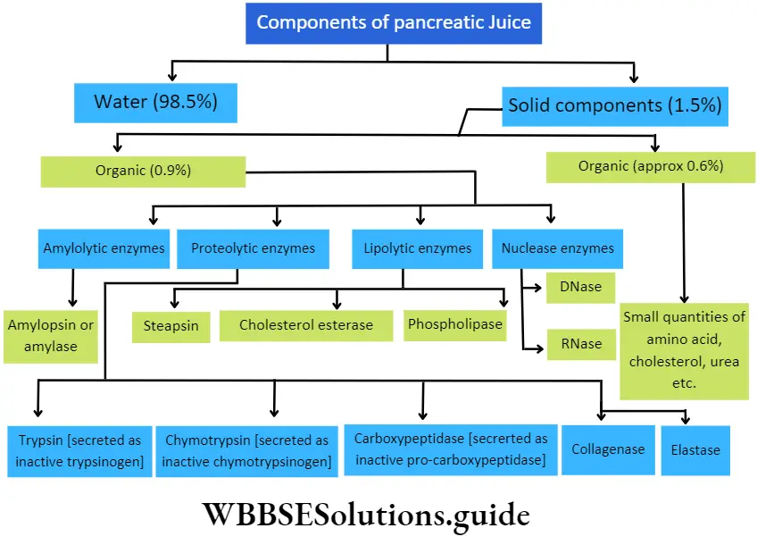
Function: The digestive functions of the pancreatic juice are given below.
- Activation of trypsin and its role: Inactive trypsinogen present in the pancreatic juice gets activated into trypsin, under the influence of enzymes like enteropeptidase or enterokinase.
- Trypsin hydrolyses proteins, acid metaproteins and peptones into polypeptides and amino acids.
- Trypsin activates chymotrypsinogen.
- Activation of chymotrypsin and its role: Inactive chymotrypsinogen present in pancreatic juice gets activated into chymotrypsin, by trypsin. Chymotrypsin helps to convert milk protein, casein, into paracasein which on reacting with Ca2+, precipitates calcium para caseinate as curd, It also helps in hydrolysing other proteins into polypeptides.
- Activation of carboxypeptidase and its role: Inactive procarboxypeptidase present in the pancreatic juice, gets converted into active carboxypeptidase, by the action of trypsin, in the duodenum. Carboxypeptidase hydrolyses long polypeptide chains into shorter ones and releases terminal amino acids from the peptide, within the cavity of the small intestine.
- Activation of elastase and its role: Inactive proenzyme, proelastase is present in the pancreatic juice. It gets activated by trypsin, to form elastase enzyme. Elastin protein present in yellow fibres (found in connective tissue matrix) gets hydrolysed by this enzyme to form amino acids.
- Role of collagenase: Collagenase is a proteolytic enzyme present in the pancreatic juice. It hydrolyses the fibrous protein collagen into amino acids under a slightly alkaline medium.
- Role of steapsin: Lipase or steapsin present in the pancreatic juice, hydrolyses emulsified fats into fatty acids and glycerol. 1 molecule of triglyceride gets hydrolysed into 3 molecules of fatty acids and 1 molecule of glycerol.
- Role of cholesterol esterase: Cholesterol esterase present in the pancreatic juice, hydrolyses cholesterol ester into cholesterol and fatty acids, under the influence of bile. It may catalyse the esterification of cholesterol esters with free fatty acids.
- Role of phospholipase: Phospholipase present in the pancreatic juice, hydrolyses phospholipids into lipophospholipids and fatty acids.
- Role of nuclease: Dnase and RNase present in I pancreatic juice, hydrolyse DNA and RNA j respectively, into specific nucleotides.
- Role of amylase: Pancreatic juice contains an amylolytic enzyme called pancreatic amylase or amylopsin which converts different polysaccharides (glycogen, starch, etc.) into maltose.
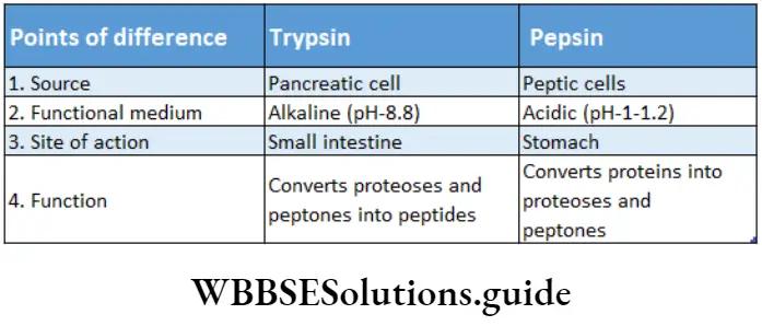
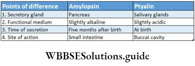
Intestinal Glands
Intestinal glands are the small glands present on the inner epithelium of the small intestine. It secretes enzyme-containing intestinal juice.
Intestinal glands Structure: I small intestine. At the base of the mucosal villi, numerous microscopic intestinal glands are present.
These are called Lieberkiihn’s glands(crypts of Lieberkuhn). These glands are tubular and unbranched and secrete enzymes and mucus.
The submucosa layer of the duodenum has another type of gland called Brunner’s glands. These glands are branched and secrete alkaline watery fluid, enzymes and mucus.
They open into the crypts of Lieberkuhn. The mixture of secretion of these two glands is known as intestinal juice or succus enteric.
Process of digestion and absorption of carbohydrates, proteins, and fats
Intestinal glands Function: The main function of the intestinal glands is the secretion of intestinal juice and mucus.
Intestinal juice or succus entericus: The secretion of intestinal glands, that contains several digestive enzymes is called intestinal juice or succus entericus.
Characteristics and components: Intestinal juice is alkaline in nature. Its average pH is 8.3. An average of 1-2L of intestinal juice is produced every day. The composition of the intestinal juice.
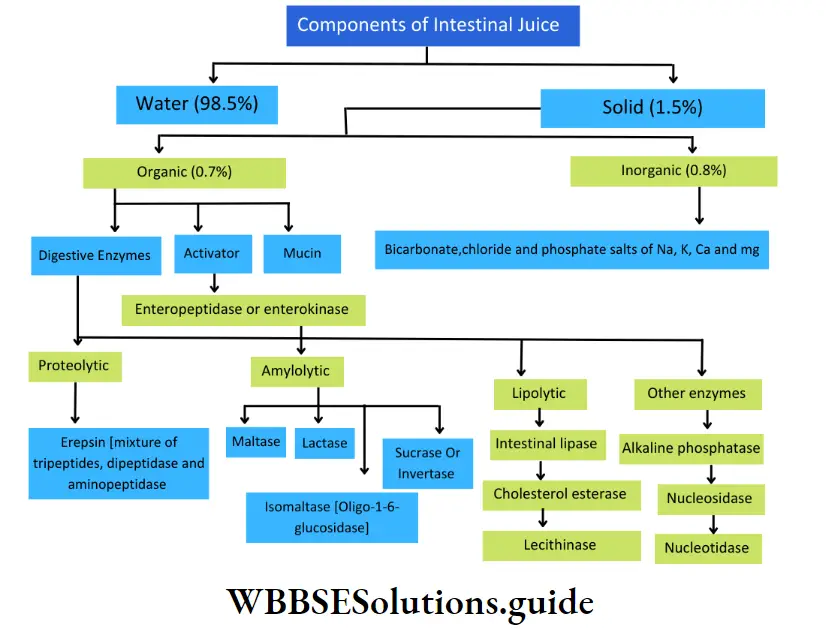
Intestinal glands Function: The functions of the components of intestinal juice are as follows.
- Role of enteropeptidase or enterokinase: Enterokinase, secreted by the intestinal glands into the intestinal juice, activates trypsinogen into trypsin, This enzyme, trypsin, further activates other enzymes.
- Role of erepsin: Erepsin present in the intestinal juice is a mixed enzyme. It is a mixture of aminopeptidase, tripeptidase and dipeptidase. Their functions are as follows—
- Aminopeptidase: It hydrolyses long chains of polypeptides into smaller polypeptides (such as tripeptides and dipeptides),
- Tripeptidase: It hydrolyses specific tripeptides into dipeptides and amino acids,
- Dipeptidase: It hydrolyses specific dipeptides into amino acids.
- Role of amylolytic enzymes: Amylolytic enzymes act on starch and related oligosaccharides and polysaccharides. The most known amylolytic enzymes are or-amylase, amylase and glucoamylase.
The activity of the amylolytic enzymes I are as follows—
Maltase: It hydrolyses maltose into 2 molecules of glucose,
Lactase: It hydrolyses the milk sugar, and lactose into 1 molecule of glucose and 1 molecule of galactose,
Invertase or sucrase: It hydrolyses sucrose/into 1 molecule of | glucose and 1 molecule of fructose,
Isomaltase or oligo-l,6-glucosidase: It hydrolyses isomaltose and branched dextrin into maltose and glucose. It breaks the a-1,6 linkage in dextrin.
Role of lipase: It hydrolyses emulsified fats within the lumen of the small intestine, into fatty acids and glycerol. 1 molecule of triglyceride on complete hydrolysis produces 3 molecules of fatty acids and 1 molecule of glycerol.
Role of lecithinase: Lecithinase removes the fatty acid residue from lecithin. It hydrolyses a phospholipid, lecithin, present within the lumen of the small intestine into fatty acids, glycerol, choline and inorganic phosphate.
Role of nucleotidase and nucleosidase: Nucleotidase (DNase and RNase), hydrolyses nucleotide units of DNA and RNA respectively, into nucleosides. Nucleosidase hydrolyses nucleoside units of DNA and RNA, which leads to the release of basic or nitrogenous components of the chain.
Role of mucin: Mucin proteins are known for providing protection and lubrication to epithelial surfaces of the intestine.
Role of cholesterol esterase: It releases cholesterol from fat-containing food.
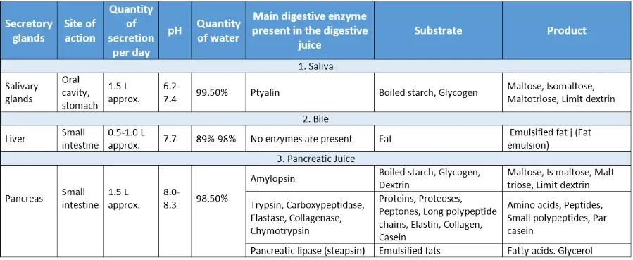
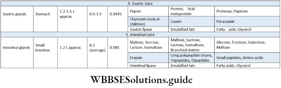
Role Of Digestive Enzymes And Gastrointestinal Hormones In Digestion
Several digestive enzymes and gastrointestinal hormones are secreted by the different glands. Let us study them and their role in digestion in detail
Digestive Enzymes
Digestive Enzymes Definition: The enzymes that act on the complex food substances in the digestive tract and convert them into simple absorbable forms are called digestive enzymes.
Different digestive enzymes and their roles
Different types of digestive enzymes are—
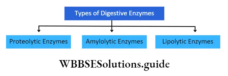
Proteolytic enzymes: The enzymes that hydrolyse the protein molecules present in the food are called proteolytic enzymes or proteases.
Role in digestion: Proteolytic enzymes gradually hydrolyse protein molecules into amino acids, which are absorbed by the cells.
⇒ \(\text { Proteins } \stackrel{\text { Proteolytic enzymes }}{\longrightarrow} \text { Amino acids }\)
Examples: Pepsin, trypsin and erepsin.
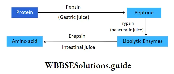
Amylolytic enzymes: The enzymes that degrade or hydrolyse complex carbohydrates present in the food, into simpler products are called amylolytic enzymes.
Role in digestion: Amylolytic enzymes facilitate the breakdown of complex carbohydrates such as fibres starches and sugars into simple sugars, mainly mono and disaccharides and monosaccharides like glucose, fructose, etc. are absorbed by the cell.
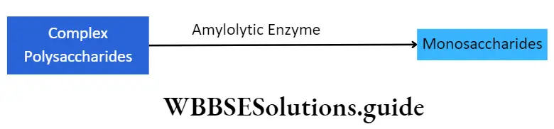
Examples: Salivary amylase (ptyalin), amylopsin, intestinal sucrase, maltase, lactase, etc.
Lipolytic enzymes: The enzymes that hydrolyse fats or lipids present in the food, into fatty acids and glycerol are called lipolytic enzymes or lipases
Role in digestion: Lipases are responsible for the hydrolysis of lipids into fatty acids and glycerol that can be absorbed by the blood as well as the cells.

Examples: Gastric lipase, pancreatic lipase, intestinal lipase, Etc.
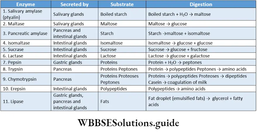
Gastrointestinal Hormones
Gastrointestinal Hormones Definition: The hormones secreted mainly by mucous cells of the digestive tract that control various processes of digestion are called gastrointestinal hormones.
The main gastrointestinal hormones are—
- Gastrin,
- Enterogastrone,
- Secretin,
- Cholecystokinin pan¬creozymin (CCK-PZ),
- Villikinine,
- Duocrinin,
- Enterocrinin,
- Gastric inhibitory peptide,
- Vaso-active intestinal peptide,
- Somatostatin.
These hormones, along with their roles are discussed below.
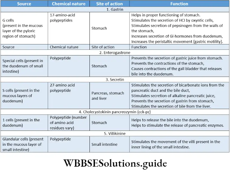
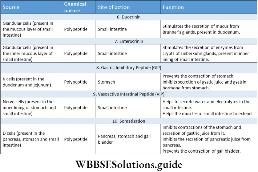
Peristalsis
Peristalsis Definition: The basic propulsive movement of the digestive tract, including its periodic contractions and relaxations, that enable the food to pass through it, is called peristalsis.
Peristalsis Types: The three types of peristalsis are—
Primary peristalsis: It is the peristaltic wave triggered by the swallowing centre in the brain. It is generally slower and mild in nature. The peristaltic contraction wave travels at a speed of 2 cm/s.
Secondary peristalsis: It is generally faster in nature. It travels around 2-25 cm/s. It extends across the entire small intestine. Scientist Alvarez had named it as true peristalsis. Due to its speed, it is also known as rush peristalsis.
Reverse peristalsis: It is slightly different than the other two types of peristalsis. It occurs in the lower part of the duodenum. Due to this peristaltic movement, the food bolus enters the stomach from the duodenum.
Hence, it is called antiperistalsis or reverse peristalsis. Sometimes, this process occurs in the ileum.
Depending on the condition of the food bolus, the receptors on the inner walls of the duodenum get stimulated. This leads to the antiperistaltic movement. Vomiting is caused due to this antiperistaltic movement.
Mechanism of peristalsis: Peristalsis is a propulsive reflex activity that occurs in the digestive tract. Both circular and longitudinal muscle layers are involved in this motion.
Peristalsis is initiated when the outer wall is distended by a bolus or chyme. Bolus of food stimulates mechanoreceptors and chemoreceptors at a specific region in the gastrointestinal tract, ultimately resulting in peristalsis.
The semi-digested food (chyme), after passing through the stomach, enters the small intestine and stimulates the muscles of its inner lining. Due to this, circular muscles of muscularis externa layer, present within the inner lining of the small intestine, contract.
Stimulation at any point in the gastrointestinal tract can cause a contractile ring to appear in the circular muscle. This contractile ring then spreads along the gastrointestinal tract.
This allows the chyme to move downwards. In the next step, these muscles relax and return to their normal state. Due to the alternate contraction and relaxation, chyme gradually moves through the intestine.
Receptive relaxation of the gastrointestinal tract takes place on the distal side of the contractile ring. It allows chyme to be propelled more easily towards the anus. This completes the peristalsis.
Significance of peristalsis: The significances of peristalsis are—
- Transportation of food: In the oesophagus, peristalsis helps to propel the swallowed food or fluid into the stomach. It helps to transport the semi-digested food (chyme) from the stomach to the colon.
- Digestion: Due to peristalsis, different juices like gastric juice, pancreatic juice, bile, etc., can easily mix with the bolus and chyme and stimulate the process of digestion.
- Absorption: It stimulates the absorption of nutrients through the walls of the small intestine, into the bloodstream.
- Blood circulation: Peristalsis or waves of muscular contraction in the gastrointestinal tract also drives the blood flow
- Ejection of faecal matter: Peristalsis occurs about 1-3 times in the large intestine, making the faeces enter the rectum and ultimately helping in its ejection.
Digestion Absorption And Assimilation Of Carbohydrate Protein And Fat
Different substances (such as carbohydrates, protein, fat, etc.) present in food get digested, absorbed and assimilated by the digestive system. These processes are explained below.
Digestion Of Carbohydrates
The process by which complex carbohydrates, present in the food, are broken down into simpler sugars, using amylolytic enzymes is called digestion of carbohydrates.
Carbohydrates comprise 60-80% of our food. An adult human being takes about 300-500 g of carbohydrates every day, which releases about 1200- 2000 kcal of energy.
Carbohydrates present in the food are of three types mainly—
- Monosaccharides (glucose, fructose),
- Disaccharides (lactose, maltose, sucrose),
- Polysaccharides (starch, glycogen, dextrin, cellulose)
Importance of cellulose
Cellulose-containing foods are difficult to digest. But they are an important part of the diet. They provide roughage to the body, which is important for the formation and egestion of faeces.
Roughage is also required for proper peristaltic movements of the intestines and thereby, prevents constipation.
Several bacteria are present within the stomach, small intestine, large intestine, etc., that ferment the cellulose present in the food.
Site Of Digestion
Digestion of carbohydrates is initiated in the mouth by the action of saliva and is completed in the small intestine by I alkaline medium. action of the intestinal juice.
Mechanism of digestion
Carbohydrate digestion by amylolytic enzymes results in the formation of simple carbohydrates, through different steps. These are described in detail below.
Digestion in mouth: The two types of amylolytic enzymes present in saliva are—
- Ptyalin
- Maltase.
Role of ptyalin: This enzyme hydrolyses starch or glycogen into maltose, maltotriose, and isomaltose and limits dextrins in a slightly alkaline medium. But ptyalin can hydrolyse only ar-1, 4-linkages but not ar-1, 6-linkages.
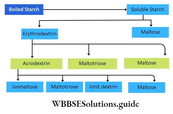
Deglutition Meaning In Biology
Role of maltase: This enzyme acts on maltose to produce two units of glucose.

Digestion in the stomach: There are no carbohydrate hydrolysing enzymes in gastric juice. But HCI present in the gastric juice hydrolyses sucrose molecules into glucose and fructose.
Digestion in the small intestine: Two types of juices, that is, pancreatic juice and intestinal juice, along with bile, are mixed with the chyme in the small intestine.
Digestion by Pancreatic juice: The pancreatic juice contains pancreatic amylase or amylopsin.
It hydrolyses remaining starch, glycogen, dextrin, etc., present in ‘ the food, into disaccharides such as maltose isomaltose, small dextrins, etc., and even glucose. This enzyme functions in the presence of Cl- ions, in an alkaline medium.
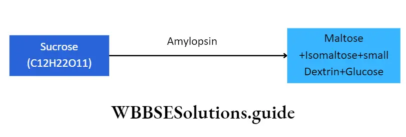
It is to be noted that Cl ions act as activators for both ptyalin and pancreatic amylase.
Digestion by Intestinal juice: The intestinal juice contains the following amylolytic enzymes. These are
- Intestinal amylase
- Isomaltase
- Maltase
- Sucrase
- Lactase.
All these enzymes act in an alkaline medium at pH 8. Their roles in carbohydrate digestion are discussed below.
- Role of intestinal amylase: a-amylase is present in very small quantities in the intestinal juice. The starch and dextrin that somehow escape the action of pancreatic lipase, become the substrate for intestinal lipase. It converts them into disaccharides like maltose, isomaltose, maltotriose, etc.
- Role of isomaltase or oligo-l,6-glucosidase or r-dextrinase: Isomaltase or oligo-l,6-glucosidase or ar-dextrinase breaks ar-1, 6-glycosidic bond of ar-dextrin into maltose and maltotriose. It also hydrolyses isomaltose into glucose.
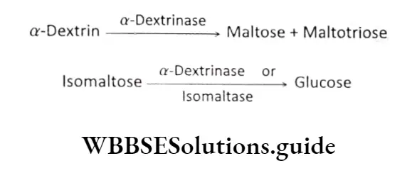
Role of maltase: Maltase present in the intestinal juice hydrolyses disaccharide, maltose, into 2 molecules of monosaccharide, glucose.
1 molecule of maltose (C12H22O11) →Maltase +H2O Glucose (C6H12O6) + Glucose (C6H12O6)
The enzyme hydrolyses trisaccharide, maltotriose into 3 molecules of glucose. The maltase breaks the 1, 4-glycosidic bond between maltose and maltotriose.
1 molecule of maltotriose (C18H32O16) →Maltase 3Molecules of glucose (C6H12O6)
Role of sucrase or invertase: Sucrase or invertase, present in intestinal juice, hydrolyses sucrose into 1 molecule of glucose and 1 molecule of fructose.
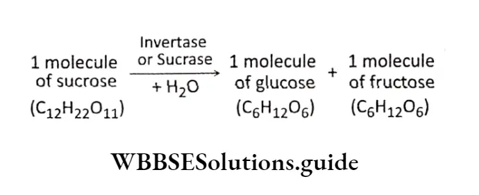
Role of lactase: Lactase, present in intestinal juice, hydrolyses milk sugar lactose, into 1 molecule of glucose and 1 molecule of galactose.
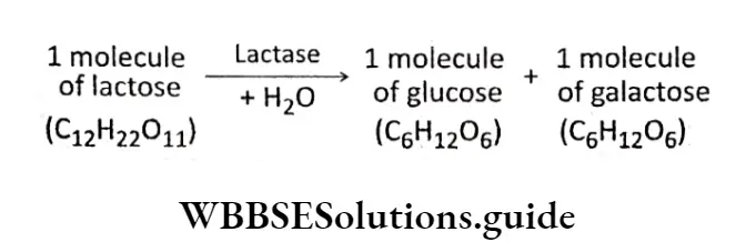
Role of limit dextrinase: Limit dextrinase, present in intestinal juice, hydrolyses dextrins into glucose.
Digestion Of Proteins
The process by which complex protein molecules, are broken down into simpler, absorbable forms (amino acids), using proteolytic enzymes is called protein digestion.
Proteins are required for the proper growth and nutrition of the body. Protein requirement for the body is about 0.5-0.8 g per kg body weight of an adult human being.
For children, this requirement is around 4 g per kg body weight.
The different types of proteins present in the food include—
- Different types of albumin and globulin,
- Milk-protein called casein (phosphoprotein), lactalbumin and lactoglobulin,
- Collagen present in white fibres and elastin in yellow fibres,
- Mucin and
- Nucleoproteins.
Site Of Digestion
Digestion of proteins does not take place in the oral cavity due to the absence of any proteolytic enzymes in saliva. It begins in the stomach and ends in the small intestine.
Mechanism Of Digestion
Protein digestion by proteolytic enzymes results in the formation of amino acids, through different steps. Along with amino acids, some quantities of dipeptides and tripeptides are also generated. These are absorbed by the cells, in their peptide form.
Digestion in the stomach: Protein digestion in the stomach is carried out by the gastric juice.
Gastric juice contains the following proteolytic enzymes—
- Pepsinogen,
- Chymosin,
- Gelatinase.
Besides these, it also contains HCI. Their roles in protein digestion are discussed below.
Role of pepsinogen: It is present in the gastric juice as an inactive proenzyme form. It is initially activated by HCI to form the active enzyme, pepsin.
⇒ \(\text { Pepsinogen } \stackrel{\mathrm{HCl}}{\longrightarrow} \text { Pepsin }\)
Its optimum pH ranges from 1.5-2.2 and hence, can act within the acidic environment inside the stomach.
It is an endopeptidase that cleaves the peptide bond in proteins. It converts metaproteins into proteoses and peptones. It is also capable of digesting collagen, a fibrous protein which is the main constituent of connective tissue in animals.
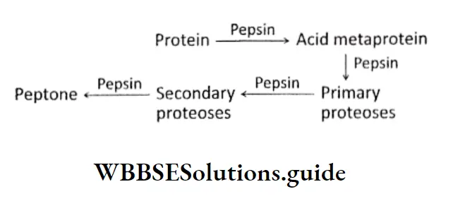
Role of chymosin: An enzyme, called rennin, is present in the digestive juice of cattle, that helps in the digestion of milk.
It is present in a proenzyme form, prorennin, that gets activated into rennin, in the presence of HCI.
Rennin is present as chymosin in the digestive juice of infants but not in adult human beings. It acts on casein, present in the milk, and converts it to soluble paracasein.
This paracasein binds to calcium ions forming insoluble calcium paracaseinate that is finally digested by pepsin.
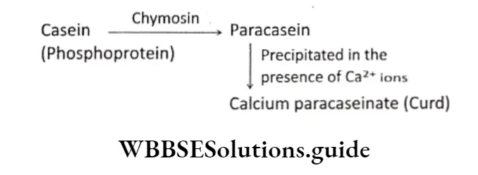
Role of gelatinase: In the presence of HCI in the stomach, gelatinase hydrolyses gelatin into peptones
⇒ \(\text { Gelatin } \stackrel{\text { Gelatinase }}{\longrightarrow} \text { Peptone }\)
Role of hydrochloric acid: It is secreted by the oxyntic cells in the stomach.
Its functions in protein digestion are—
- It causes denaturation and disintegration of proteins into small peptides and amino acids.
- It converts proteins to acid metaproteins, which are easily digested.
⇒ \(\text { Protein } \stackrel{\mathrm{HCl} \text { present in gastric juice }}{\longrightarrow} \text { Acid metaprotein }\)
It activates proenzyme forms of the enzymes into their active forms, such as pepsinogen to pepsin and prorennin to rennin.
It lowers the pH in the stomach, making the medium acidic that is suitable for the action of pepsin.
Digestion in the small intestine: Protein digestion continues in the small intestine under the action of pancreatic juice and intestinal juice. Their roles in protein digestion are discussed below.
Role of pancreatic juice: Trypsin, chymotrypsin, carboxypeptidase, elastase and collagenase are pancreatic proteolytic enzymes. Their roles are as follows
Role of trypsin: It is secreted as trypsinogen. It is an inactive form of trypsin. It is initially activated by enterokinase or enteropeptidase enzyme, present in the intestinal juice.
\(\text { Trypsinogen } \stackrel{\text { Enterokinase }}{\longrightarrow} \text { Trypsin }\)Its optimum pH is 8.0. It hydrolyses proteins, proteoses, acid metaproteins, peptones, etc., into simpler and smaller peptides such as aminopeptides, dipeptides, tripeptides, etc.
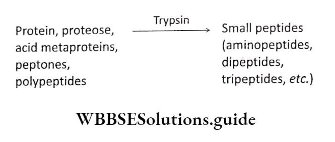
Deglutition Meaning In Biology
Role of chymotrypsin: It is secreted as chymotrypsinogen. It is an inactive form of chymotrypsin.
It is activated by trypsin. Its optimum pH is 8.0. Chymotrypsin converts casein, present in milk, into paracasein in the slightly alkaline medium of the small intestine. Thus, its function is to coagulate milk.
Role of carboxypeptidase: It is secreted in an inactive form called procarboxypeptidase.
It is activated by trypsin. Carboxypeptidase has an optimum pH of 7.4. It hydrolyses long polypeptide chains into smaller ones.
It specifically hydrolyses the amino acid from the carboxyl end of peptide chains, to yield individual amino acids.
⇒ \(\begin{array}{ll}
\text { Long chain } \stackrel{\text { Carboxypeptidase }}{\longrightarrow} & \text { Small peptides + } \\
\text { polypeptides } & \text { Amino acids }
\end{array}\)
Role of elastase: It acts on a protein, named elastin. It hydrolyses the carboxyl group of the terminal amino acid, producing smaller peptide chains.
⇒ \(\text { Elastin } \stackrel{\text { Elastase }}{\longrightarrow} \text { Small peptides }\)
Role of collagenase: It acts on a protein, named collagen. It hydrolyses the collagen fibres, producing smaller peptide chains.
⇒ \(\text { Collagen } \stackrel{\text { Collagenase }}{\longrightarrow} \text { Small peptides }\)
Role of intestinal juice: The main proteolytic enzyme present in intestinal juice is erepsin.
Erepsin is actually a mixture of aminopeptidases, dipeptidases, tripeptidases and enterokinase. These protein hydrolysing enzymes serve a major role in alkaline medium.
Role of aminopeptidase: It is an exopeptidase (peptidase that catalyses the cleavage of terminal peptide bonds).
It acts on the amino terminal of the polypeptides and finally releases an amino, acid. It results in the formation of dipeptides and tripeptides.
⇒ \(\begin{array}{ll}
\text { Long chains of } \stackrel{\text { Aminopeptidase }}{\longrightarrow} & \text { Tripeptide, }+ \text { Amino } \\
\text { polypeptide } & \text { dipeptide } \text { etc. acids }
\end{array}\)
Role of tripeptidase: Tripeptidases act on tripeptides and hydrolyse them into dipeptides and amino acids.
⇒ \(\text { Tripeptide } \stackrel{\text { Tripeptidase }}{\longrightarrow} \text { Dipeptide + Amino acid }\)
Role of dipeptidase: Dipeptidases act on dipeptides and hydrolyse them into two amino acids.
⇒ \(\text { Dipeptide } \stackrel{\text { Dipeptidase }}{\longrightarrow} \text { Amino acid }+ \text { Amino acid }\)
Digestion Of Fats
The process by which fats and lipids present in the food are broken down into simple and absorbable forms like fatly acid and glycerol using lipolytic enzymes, is called digestion of fat.
Fats are the main source of energy. They provide us with the energy for various metabolic processes.
The different fats that are present in the food are—
- Neutral fats or triglycerides,
- Phospholipids,
- Cholesteroids,
- Free cholesterol,
- Fatty acids and
- Glycerol.
About 25% of the fats get converted into fatty acids and glycerol. Some of the fats undergo partial hydrolysis to form monoglycerides and diglycerides.
Site of digestion
Digestion of fats does not take place in the oral cavity due to the absence of any lipolytic enzyme in the saliva. Its digestion begins in the stomach, by gastric juice and is fully digested in the small intestine, by intestinal juice.
Mechanism of digestion.
The key issue in the digestion of fats is that of solubility—lipids are poorly soluble in water. Thus digestion is greatly aided by emulsification (the breaking up of fat globules into much smaller emulsion droplets). It enhances the surface area of the lipid so that water-soluble.
lipase can work on the globule surface of triacylglycerol (TAG). The mechanism of digestion of fats is as follows.
Digestion in the stomach: The fat-digesting enzyme present in gastric juice is gastric lipase.
Role of gastric lipase: The action of gastric lipase is inhibited by the absence of any emulsifying substance and the presence of hydrochloric acid in the stomach.
So, it has mild action over the fats and converts them into fatty acids and monoglycerides.
⇒ \(\text { Fat } \stackrel{\text { Gastric lipase }}{\longrightarrow} \text { Fatty acids }+ \text { Monoglyceride }\)
Digestion in the small intestine: The main digestion of fats takes place in the small intestine. When the chyme enters the small intestine, three alkaline secretions— bile, pancreatic juice and intestinal juice, are mixed with it.
Role of bile: It plays an important role in fat digestion. The chyme mixes with the bile salts due to peristalsis.
The bile salts such as sodium glycocholate and sodium taurocholate have the ability to break down large fat droplets into smaller ones. This process is called emulsification of fats.
It increases the surface area of the fat droplets and decreases the surface tension of the fat droplets to prevent the adherence of the fat droplets together again. This makes the action of the enzyme lipase, easier.
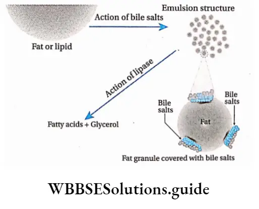
Role of pancreatic juice: The enzymes for fat digestion present in pancreatic juice, are—steapsin, phospholipase, cholesterol esterase. Their roles are discussed as follows.
Role of steapsin: Steapsin present in pancreatic juice, hydrolyses emulsified fats under a slightly alkaline medium. 1 molecule of triglyceride gets hydrolysed into 3 molecules of fatty acids and a molecule of glycerol.
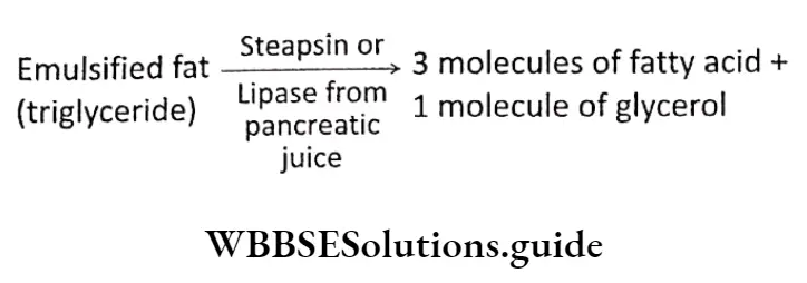
Role of phospholipase: The enzyme, phospholipase, acts upon two phospholipids, lecithin and cephalin
Both lecithin and cephalin release 1 molecule of fatty acid from each of them, producing lysolecithin and lysocephalin, respectively.
Further action of phospholipase B on these products releases another fatty acid molecule and glycerol-phosphoryl choline.
When phosphodiesterase acts upon them, glycerol is released. On the other hand, when phosphatase acts on them, choline and sphingosine are released.
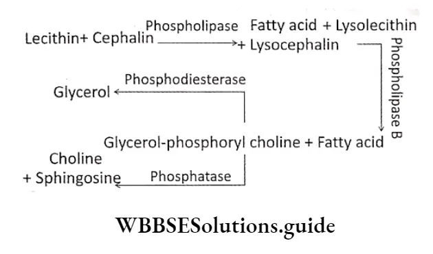
Role of cholesterol esterase: Cholesterol esterase is also termed as cholesterol ester hydrolase.
Cholesterol esterase present in the pancreatic juice, acts on cholesterol ester or cholesteroid to form cholesterol and fatty acids, on hydrolysis under slightly alkaline medium.
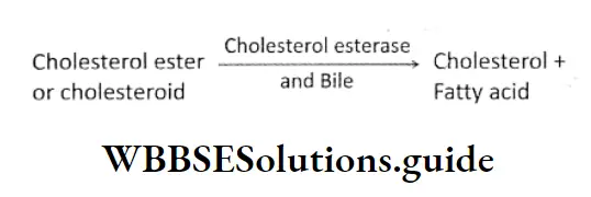
Intestinal juice: The intestinal juice contains the following lipolytic enzymes—intestinal lipase, and lecithinase. Their roles in fat digestion are as follows.
Role of intestinal lipase: It breaks down emulsified fats into glycerol and fatty acids. It produces 3 molecules of fatty acids and 1 molecule of glycerol from each molecule of triglyceride.
Role of lecithinase: It hydrolyses a phospholipid named lecithin into fatty acids, glycerol, choline and inorganic phosphate.
⇒ \(Lecithin \stackrel{\text { Lecithinase }}{\longrightarrow} Fatty acid + Glycerol + Choline + Inorganic phosphate\)
Absorption Of Carbohydrates
The process by which end products of carbohydrate digestion pass through the interstitial mucosa into the lymph or blood, is known as absorption of carbohydrate.
Simple carbohydrates (such as glucose, fructose, and galactose) do not require further digestion.
Complex carbohydrates (such as starch, glycogen, etc.,) are absorbed mostly as monosaccharides (such as glucose, galactose, fructose, levulose).
Some quantities of carbohydrates are also absorbed as disaccharides (such as sucrose, maltose, lactose, etc.)
Human digestive system: structure, enzymes, and absorption mechanism
Site of absorption
Even though some amount of monosaccharides may be absorbed through the mucosa of the stomach, most of the soluble products of digestion are absorbed in the small intestine.
The maximum amount of carbohydrates is absorbed in the duodenum and jejunum, leaving a very small amount for the ileum to absorb. Other than this, some amounts of glucose and galactose are absorbed in the large intestine.
Absorption pathway
The products obtained by the digestion of carbohydrates get absorbed by the microvilli or brush-bordered enterocytes, into the lumen of the small intestine.
These substances enter the network of blood capillaries and then into the hepatic portal system. Finally, they enter into the liver. A small amount of these products pass through the lacteals and flow through the lymph.
Process of absorption
Different types of carbohydrates are absorbed by different processes. These have been discussed below.
Absorption of monosaccharides:
Most of the glucose gets absorbed by this mechanism. This means that their absorption requires the expenditure of energy.
It takes place against the concentration gradient, i.e., occurs even when their concentration in the intestinal
lumen is less than that in the blood. For the active absorption of glucose, carrier proteins and Na+ ions are required.
This mechanism is also known as the active sodium-monosaccharide symport process.
In this case, the carrier protein is sodium-glucose transporter (SGLT-1). Na+ ions are present in the intestinal lumen while carrier proteins are present on the membrane covering the microvilli, located on epithelial cells of the villi.
The carrier protein has 2 separate sites—one for Na+, and the other for glucose. A complex is formed by SGLT-1, glucose molecule, Na+ and carrier protein on the microvilli membrane, towards the intestinal lumen.
This complex passes through the microvilli membrane to the cytoplasmic end of the cells. Here the complex breaks down and glucose molecules and Na+ enter the cells.
It transports 2 Na+ ions (along the concentration gradient) and 1 glucose molecule (against the concentration gradient) to the cytoplasm of the cell. The carrier protein separates from the complex and the same mechanism is repeated.
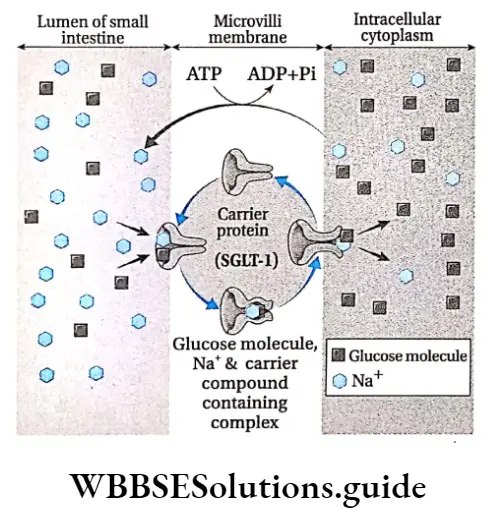
Deglutition Meaning In Biology
Some amounts of glucose and galactose enter the enterocytes by sodium-independent, facilitated diffusion.
During high glucose concentration in the enterocytes, absorption of glucose occurs through a facilitated diffusion process. In this case, a carrier protein named glucose transporter-2 or GLUT-2 is involved.
Some amounts of glucose and galactose also enter the small intestine by simple diffusion.
Absorption of fructose takes place by diffusion. It is passive in nature, i.e., no energy is required for the process and takes place along the concentration gradient.
Among the monosaccharides, pentose and tetrose-like carbohydrates (such as arabinose) get absorbed by simple diffusion.
Absorption of disaccharides: Some amount of disaccharides like sucrose, maltose and lactose get absorbed in the jejunum and ileum. The absorption of these carbohydrates takes place by simple diffusion, which occurs passively, along the concentration gradient.
Absorption Of Proteins
The process by which end products of protein digestion pass through the interstitial mucosa into the lymph or blood, is known as absorption of protein.
Digestion products of proteins are absorbed mainly as amino acids. A small quantity of protein is absorbed as dipeptides or tripeptides.
Site of absorption
Absorption of amino acids occurs in the small intestine. It is rapid in the duodenum and jejunum, but slow in the ileum.
Absorption pathway
The amino acids get absorbed by the epithelium of the villi in the small intestine. They enter the blood flowing through the network of blood capillaries around the villi.
They are then transported to the liver by the hepatic portal vein. From the liver, they are transported to the different organs of the body. A very small amount of amino acid after being absorbed by the villi, gets absorbed into the lacteal and flows through the lymph.
Process of absorption
Amino acids dipeptides and tripeptides are absorbed by different processes. These have been discussed below
Absorption of amino acids:
Active absorption of amino acid in the gastrointestinal tract involves a mechanism similar to that of glucose absorption.
At least four sodium-dependent amino acid transporters are present in the luminal plasma membrane of enterocyte. This process—
- Takes place against the concentration gradient,
- Requires cellular energy and
- Requires carrier proteins (na+-amino acid co-transporter) and na+ ions the carrier protein is present on the microvilli membrane. These transporters bind na+ and amino acid molecules to form a complex.
- This complex passes through the microvilli membrane, towards the cytoplasmic end of the cell.
- On reaching the cytoplasmic end, the complex undergoes conformational change and breaks down to release Na+ and amino acid into the cytoplasm.
- The earner problem separates and repeats the same process with another Na and ammo acid molecule.
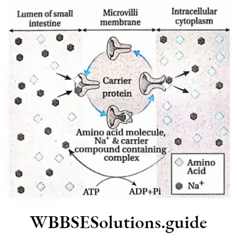
Some of the amino acids get absorbed through the brush-bordered enterocytes by sodium-independent amino acid-transporter.
Once amino acids are in the enterocyte, however, they accumulate and eventually move by facilitated diffusion through the base of the cell and into the blood.
Some amino acids get absorbed within the small intestine by simple diffusion.
Absorption of dipeptides and tripeptides:
Dipeptides and tripeptides are mainly absorbed by the epithelium of the villi and then to blood capillaries.
Absorption Of Fats
The process by which the end product of fat digestion passes through the interstitial mucosa into the lymph or blood is known as the absorption of fat.
About 75% of hydrolysed fat is absorbed mainly as higher fatty acids, glycerol, monoglycerides and cholesterol. Only 25% of the hydrolysed fat is absorbed as fatty acids and glycerol.
Site of absorption
The products of digestion of fats enter the circulatory system from the small intestine, mainly in the jejunum and ileum.
Absorption pathway
- Digested as well as semi-digested fats get absorbed by the epithelium of the small intestine.
- From The epithelium, they get Absorbed into lymph vessels called lacteals. These fats continue to flow within the lymph Present In the Lacteals. The fats next enter the bloodstream from this lymph.
- Only a small amount of fat gets absorbed by the blood in the blood capillaries, surrounding the villi. They are then transported to the liver by the hepatic portal vein.
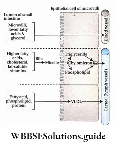
Process of absorption
Fats are absorbed by different processes. These have been discussed below.
Absorption of lower fatty acids and glycerol by portal veins:
- Water-soluble, lower fatty acids and glycerol get absorbed into the epithelial cells of villi from the intestinal lumen.
- From the villi, they enter the nearby blood capillaries. They are then carried to the liver by the hepatic portal vein.
- Some amount of fatty acids, phospholipids and proteins, get absorbed through the villi into the intestinal lumen.
- They combine with each other to form VLDL (very low-density lipoproteins), that enter the lacteal and flow into the lymph. Finally, they enter into the blood flow.
Absorption of higher fatty acids by lacteal: Higher fatty acids being insoluble in water, cannot be directly absorbed into the blood. They are absorbed by the intestine.
In the lumen of the small intestine, higher fatty acids, monoglycerides and fat-soluble vitamins associate to form water-soluble droplets called micelles.
Bile salts and potassium bicarbonate ions help the micelle to penetrate through the microvilli of enterocytes, into the cells.
After entry inside the enterocytes, the bile salts are released and fats (triglycerides) are re-synthesised.
Those triglycerides are then coated with a layer of phospholipids and proteins to form small globules called chylomicrons.
These are about 1 micrometre in diameter. They enter lymphatics (lacteals) instead of blood capillaries due to their larger size. From the lymph, they enter into the bloodstream.
Different hypotheses regarding the absorption of fats
Lipolytic hypothesis:
- This hypothesis was postulated by Verzar and McDougall. According to this hypothesis, the fatty acids and glycerol generated during digestion of fats, are absorbed separately within the body.
- The fatty acids combine with the bile salts to form a complex, which is absorbed by the epithelial cells of the small intestine.
- Especially fatty acids combine with glycerol to synthesise triglycerides that enter the lymph vessels.
Partition hypothesis:
- This hypothesis has been postulated by Frazer. According to this hypothesis, 30% of the fat is hydrolysed in the small intestine to form mono and diglycerides. The rest 70% forms an emulsion of unhydrolysed fats.
- The small hydrolysed fatty acids get absorbed by the portal system. Some part of the hydrolysed as well as unhydrolysed fat gets absorbed by the central lacteal in the villi of the small intestine.
- Finally, they enter the lymph vessels. The unhydrolysed fat is now transferred to the blood vessels through the thoracic duct.
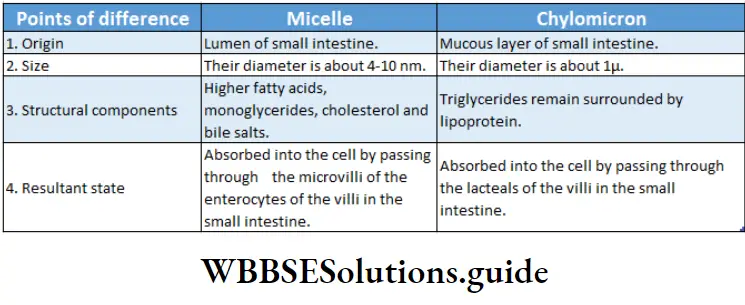
Assimilation Of Carbohydrates
Assimilation is the biological process through which absorbed food materials become the integral component of the protoplasm and extracellular materials.
After digestion of carbohydrates, mainly monosaccharides are obtained. Glucose is the most important monosaccharide obtained, that undergoes various metabolic processes in the body. The metabolic processes of the body that utilise glucose, are as follows.
Glycogenesis
The enzyme-controlled, biochemical process by which glycogen is synthesised within the cell, from glucose and other monosaccharides is called glycogenesis.
Site of occurrence: Liver and muscle fibres
Ribose-5-phosphate and other pentose phosphates are synthesised from glucose and glycogen.
This occurs by a hexose monophosphate (HMP) shunt. These pentose phosphates are used to synthesise DIMA and RNA.
Site of occurrence: Liver, adipose tissue, adrenal cortex, mammary glands, etc.
Glycogenolysis
The enzyme-controlled biochemical process by which glycogen, mainly stored in the liver, gets converted into glucose by several steps within the cell is called glycogenolysis.
Site of occurrence: Liver and cytoplasm of muscle fibres
Normal glucose concentration in the blood
Some amount of glucose is always present in the blood. It is known as blood sugar. An adult human being has 70-100 mg of glucose per 100 ml of blood. This is known as normal blood sugar.
Hyperglycemia, glycosuria and hypoglycemia
Insufficient secretion of the hormone insulin in the body causes diabetes mellitus. Under such conditions, blood sugar level rises (hyperglycemia) and thereby excess glucose gets excreted through the urine (glycosuria).
It is generally seen when there is more than 180 mg of glucose/100 ml of blood. During fasting, the blood glucose level falls, resulting in the condition called hypoglycemia.
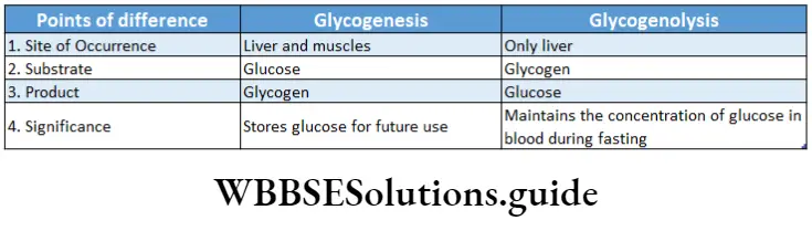
Assimilation Of Proteins
The final products of protein digestion are amino acids. They are absorbed in the small intestine and then pass through the liver before reaching the main bloodstream.
This amino acid are released from the liver to the cells for the synthesis of protein, enzymes, and non-proteinaceous substances (like fat, carbohydrates etc.) and for the production of energy. Excess amino acids get deaminated by the separation of amino groups which combines with CO2 and form urea.
Amino acid pool
The amino acids, resulting from the digestion of proteins, get absorbed in the small intestine and get added to the amino acid pool. The amino acids required for the synthesis of different proteins or non-protein nitrogen (NPN) are taken up from this amino acid pool.
Different uses of amino acids produced from proteins are as follows-
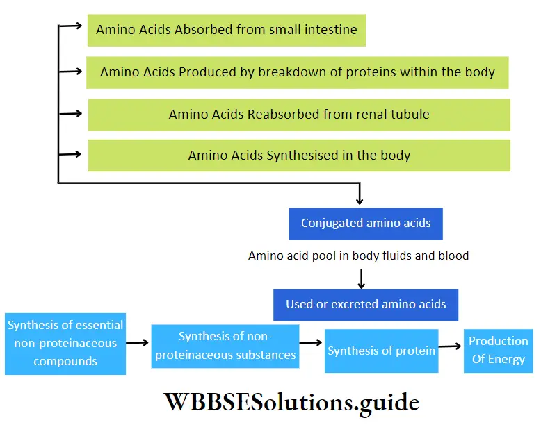
Assimilation Of Fats
Complete digestion of fats or lipid molecules within the alimentary canal produces fatty acids and glycerol.
Partial hydrolysis of lipids produces monoglycerides, diglycerides, etc Fatty acids, glycerol, monoglycerides, diglycerides, etc., get absorbed in the small intestine.
After passing through lacteals, they enter the blood. These components take part in several catabolic and anabolic reactions in the body.
Some important examples are—
- Most of the fats or lipids get stored within the body in several regions like under the skin, mesentery, etc. They prevent the loss of heat from the body.
- A layer of fat is present around the organs, to protect them from mechanical shock.
- Fats or lipids, combine with phosphate and proteins to form phospholipids and lipoproteins. Lipids are essential for the synthesis of steroid hormones.
- Triglycerides or neutral fats are generated from fatty acids and glycerol, mainly in the hepatic cells. This process is known as lipogenesis.
- These are synthesised by intestinal cells and passed into the chyle (milky body fluid consisting of lymph and emulsified fats). Then they go to blood as neutral fat being suspended in plasma.
- Ketone bodies are produced from the fats by ketogenesis. It is used as fuel by the body.
- Energy is released from fatty acids by β-oxidation.
β -Oxidation Of Fatty Acids
The oxidation of fatty adds at β-carbon (third carbon) considering —the COOH group as the first carbon is known as β-oxidation of fatty adds.
Site of occurrence: It takes place in the mitochondria of hepatic cells. It may also take place in cells of the brain, cardiac muscles, kidneys, etc. Additionally, peroxisome also contains the enzyme for this process.
Calorific Value Of Food
Definition: The amount of energy produced by oxidation of lg of protein, fat or carbohydrate, within the cell is called its calorific value.
Unit of calorific value: The solar energy remains stored as chemical energy within the molecules of proteins, fats and carbohydrates. On oxidation, the chemical energy gets converted into heat energy.
This heat energy is utilised by the cells to carry out the various metabolic and physiological processes in the body.
The unit of energy is calorie (cal). It is the heat required to raise the temperature of lg of pure water by 1°C. In biological processes, the unit is generally used in kcal.
1 Kcal=1000 cal
Calorific values of proteins, carbohydrates and fats: Different types of food provide different fat yields of 9.3 kcal (1 kcal = 4.1868 x 10³ joules). One gram of carbohydrate yields 4.1 kcal while, one gram of protein yields 4.3 kcal.
Bomb Calorimeter
A bomb calorimeter is used to measure the calorific values of proteins, carbohydrates and fats. The specific amount of the food sample is placed within a funnel-shaped container inside the instrument.
This sample is oxidised by passing electricity in the presence of oxygen. The energy that is released is measured according to the principle of calorimetry.
Later, scientists Benedict and Atwater determined the amount of energy released by the food inside the cell
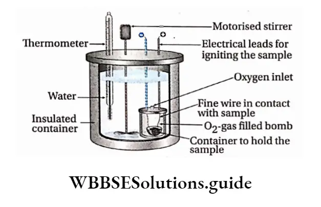
Egestion
Egestion Definition: The process by which remaining undigested, unabsorbed parts of the food are removed from the body, through the anus is called egestion.
Colon during the last phase of digestion. After the nutrients are absorbed in the small intestine, the undigested part of the food passes through the ileocecal valve into the large intestine.
By the peristaltic movement, these substances pass through the ascending, transverse, descending and sigmoid colon, Finally, they reach the rectum where they are stored for some time before egestion.
Composition of faeces: About 1500 ml of chyme enters the large intestine of an adult human being, out of which only 200 ml is excreted as faeces. Faeces are composed of water (75%) and undigested food, fibres, inorganic substances, epithelial cells, etc. It also contains microorganisms, mainly bacteria (5%), secretions of the gastrointestinal tract and mucus secreted by goblet cells,
Egestion of faeces:
- Egestion of faeces occurs as a reflex action. It is excreted from the body through the anus.
- The anus is normally closed by a pair of sphincter muscles. The internal anal sphincter is composed of smooth muscle, and the external anal sphincter is composed of skeletal muscles.
- The external anal sphincter is voluntary in nature, therefore, the opening and closing of the anus are under control.
- Faecal matter enters the rectum due to strong contractions of the sigmoid colon. This causes sudden distension of the walls of the rectum. The receptors in the rectum get stimulated due to this distension.
- This stimulation is sent to the brain, via the nerves. It is interpreted by the brain and the need to defecate is felt. Increased contractions of the colon and rectum help to push faeces through the anal canal, and out of the anus.
Significance of egestion:
- Undigested food, edible fibres (cellulose, hemicellulose, etc.), bacteria, etc., are released from the body by faeces.
- If the egestion of faeces is not regular, then the ability of the colon to undergo peristaltic contractions decreases.
- This leads to the accumulation of faeces in the colon. This condition is known as colonostasis or atonia constipation.
Disorders associated with improper egestion
- Irritable bowel syndrome (IBS): It includes stomach ache, constipation, flatulence in the intestines due to accumulation of gas, etc.
- Inflammatory bowel disease: It includes stomach ache, diarrhoea, wound in the colon, fever, loss of body weight, etc.
- Colorectal cancer: It includes the formation of a malignant tumour (cancerous in nature) in the colon or rectum
- Haemorrhoids or piles: It includes thickening of the inner lining of the rectum or anal canal, which may lead to bleeding and painful egestion of faeces.
Nutritional And Digestive Disorders
Nutritional And Digestive Disorders Definition: The disorders that pertain to the gastrointestinal tract and are caused due to nutritional deficiency or improper functioning of the digestive system are called nutritional and digestive disorders.
Some of these nutritional and digestive disorders are discussed below in detail
Protein Calorie Energy Malnutrition (PCM ) Or Protein Energy
The group of clinical conditions, observed mainly in children, which occur due to deficiency of protein or energy-giving nutrients or both in the diet, are called Protein Calorie Malnutrition (PCM) or Protein Energy Malnutrition (PEM).
Protein Calorie Energy Malnutrition Causes:
- A deficiency of protein-rich food during the foetal stage as well as during infancy and childhood is a major cause of PEM. Besides proteins, deficiency of vitamins, minerals, etc., can also lead to such disorders.
- The inability of the body to digest, absorb and assimilate the nutrients that are generated due to improper functioning of the digestive system is another important cause of such disorders.
- Lack of health consciousness and inadequate breastfeeding by the mother also cause PEM.
Protein Calorie Energy Malnutrition Types: Two types of disorders are caused by PEM—
- Kwashiorkor
- Marasmus.
Protein Calorie Energy Malnutrition Causes:
- Kwashiorkor is caused by protein deficiency. This condition usually occurs at about the age of 12 months to 4 years.
- Marasmus is caused by total calorie/energy depletion. It usually develops in children between the ages of six months and one year.
Protein Calorie Energy Malnutrition Symptoms:
- Symptoms of kwashiorkor include improper growth and weight loss, liver enlargement, oedema, dry or peeling skin, hair discolouration, abdominal distension due to accumulation of fluid, anaemia, diarrhoea, ulcers in the tongue and at the corner of the lips, weak immune system, irritation, moon-like face, etc.
- Symptoms of marasmus include stunted growth and loss in weight. It is also characterised by weak limbs, profound weakness, frequent infections, loss of appetite, diarrhoea, dry skin, loosening of the skin due to degeneration of fats, and thin and discoloured hair.
Deglutition Meaning In Biology
Preventive measures: PEM and PCM can be prevented by the following preventive measures—
- Adequate nutrition, specifically, intake of adequate amounts of protein in the diet must be taken.
- Proper care of the mother should be taken right from the beginning of her pregnancy. Adequate amount of proteins must be there in her diet.
- Breastfeeding for the first six months of the child should be ensured, along with proper nutrition.
Indigestion
The disorder that arises due to improper digestion of food, leading to a loss of appetite and feeling of stomach satiety is called indigestion or dyspepsia.
Indigestion Causes:
- Eating too spicy or fat-containing heavy meals, eating too heavy meals may be an important cause of indigestion.
- Decreased secretion of the enzymes required for digestion also leads to indigestion.
- Indigestion may also be caused due to mental anxiety.
- Food poisoning is another cause of indigestion.
Indigestion Symptoms: Symptoms of indigestion include loss of appetite, feeling of stomach satiety, sour taste, nausea, stomach ache and feeling of discomfort. Sometimes constipation is also a symptom of indigestion.
Preventive measures: Indigestion can be prevented by adopting the following measures—
- Eating very rich, spicy foods should be avoided.
- Too heavy meals must be avoided. Small, regular meals must be taken.
- Enough consumption of water must be ensured. This helps in proper digestion.
- Regular exercise ensures the proper functioning of the digestive system. Hence, exercise should be a part of the daily routine. Mental anxiety must be avoided.
Constipation
The disorder that is marked by difficult or infrequent passage of faeces is called constipation.
Constipation Causes:
- Decreased contractions of the intestines lead to slower movement of bowels through the colon. Hence, constipation occurs.
- Lack of an adequate amount of fibre in the diet causes constipation.
- A low amount of water intake causes constipation.
- Mental anxiety or depression, and lack of physical activity also lead to constipation.
- It may also be caused by tumours or ulcers in the intestines, which block the pathway of the faeces.
- Taking laxatives or medications against constipation can make it chronic.
Constipation Symptoms:
- Symptoms of constipation include very less bowel movements, with the stool being very hard and dry.
- Blood may also be discharged along with the faeces. Loss of appetite and stomach discomfort are its other symptoms.
Vomiting Or Emesis
Forceful oral expulsion of gastric or intestinal content, due to reverse peristalsis caused by accumulation of fluid in the stomach or intestine, is called vomiting.
Vomiting Causes:
- Due to improper absorption of water from the food during digestion, food in semi-liquid or liquid form accumulates in the stomach or small intestine. This stimulates the mucous layer leading to vomiting.
- Vomiting may also be caused by stimulation of the inner walls of the pharynx due to any chemical or foreign body that has entered the body along with food.
- Over-consumption of alcohol or infection in the gall bladder and intestines also causes vomiting.
- In the case of some people, travelling to high altitudes (high altitude sickness) or by sea (sea sickness due to rolling to sea) may initiate vomiting.
Vomiting Symptoms: Symptoms of vomiting include a feeling of discomfort in the stomach, nausea and loss of appetite.
Preventive measures: Vomiting can be prevented by the following measures—
- Particular chemicals or odours that may initiate vomiting for a person should be avoided.
- Consumption of small and light meals must be ensured if vomiting has occurred due to indigestion.
Jaundice
The disorder caused due to the accumulation of bile pigments (bilirubin, biliverdin) in blood is called jaundice,
Jaundice Causes and types: Based on the causes, jaundice can be categorised into three types—
- Haemolytic jaundice is caused by to excessive destruction of RBCs within the spleen. This leads to the rupturing of the plasma membrane of the RBCs, releasing haemoglobin (haemolysis) formation of dark yellow-coloured stool.
- Hepatocellular jaundice is caused due to improper functioning of the liver, when the person is suffering from viral hepatitis or drug-induced hepatitis cirrhosis of the liver, etc. This leads to an accumulation of bile pigments in blood and other cells of the body.
- Obstructive jaundice is caused due to blockage in the bile duct. This leads to less bile secretion and more bile absorption by the hepatic portal vein and lymphatic ducts. As a result, the concentration of bile pigments increases in the blood, colour of the stool becomes pale mud Causing Obstructive jaundice.
Jaundice Symptoms: Symptoms of jaundice include yellowing of skin, mucous membranes and conjunctiva of eyes. Dark yellow or yellowish-green coloured urine is also released in jaundice.
Preventive measures: Jaundice can be prevented by the following measures—
- Since this disease is mainly caused due to improper functioning of the liver, hence, its proper functioning must be checked regularly.
- Bilirubin excretion test, liver function test, etc., are some of the tests that can check liver functioning.
- According to the results of the above tests, proper medical assistance should be taken.
- Water that is consumed must be clean and filtered. Contaminated and impure water consumption can cause jaundice.
Diarrhoea
Gastrointestinal infections caused by a wide range of pathogens including bacteria, viruses and protozoa are called diarrhoea.
Diarrhoea Causes and Types: Diarrhoea can be categorised into the following types—
- Infectious diarrhoea—Diarrhoea that is caused mostly due to contamination by bacteria (such as Campylobacter sp., Salmonella sp., Shigella sp., and Escherichia coli.) and viruses (such as Rotavirus, Calicivirus, and Adenovirus).
- Psychogenic diarrhoea— Tension, fear, etc., are some of the emotions that can cause diarrhoea.
- Ulcerative colitis—In this case, ulcer and inflammation occur in most of the large intestine.
Diarrhoea Symptoms: Symptoms of diarrhoea can range from slightly watery stools to more frequent extremely watery stools.
The frequency of bowels may reach upto 15-20 times a day. Due to the loss of water and minerals like sodium, and potassium salts through the stools, the body gets dehydrated and becomes weak.
Diarrhoea Preventive measures: Diarrhoea can be prevented by the following measures—
- Proper hygiene and cleanliness must be maintained to prevent infection by germs.
- Food should always be kept covered.
- In the case of diarrhoea-affected patients, ORS solutions must be given frequently to replenish the water content in the body.
- In case of severe diarrhoea, intravenous saline must be given after proper medical assistance.
Digestion And Absorption Notes
- Bile salts: A mixture of sodium salts of bile acids, cholic acid and chenodeoxycholic acid, synthesized in the liver as a derivative of cholesterol. It helps in emulsification of fats.
- Bolus: A small rounded mass of chewed food.
- Castle’s Intrinsic Factor: A glycoprotein produced by the parietal cells of the stomach and is necessary for the absorption of vitamin B12. It has been named after American physiologist W.B. Castle.
- Emulsification: A process by which two immiscible substances are mixed or one is suspended in another
- Enterocytes: The tall columnar cells that are found in the luminal side of the small intestine and colon and are capable of absorbing water and nutrients from the digestive tract
- Lymph Node: Each of the small swellings present in the lymphatic system where lymph is filtered and lymphocytes are formed.
- Mucin: A class of glycoprotein present in mucus having water-retaining properties.
Points To Remember
- The major function of the GL tract is the digestion and absorption of nutrients.
- The alimentary canal is the continuous muscular digestive tube comprising of mouth, pharynx, oesophagus, stomach, small intestine, and large intestine.
- Saliva, secreted by salivary glands, helps in swallowing food, carbohydrate digestion and transport of immunoglobulins.
- The pharynx provides a common path for food, fluids, and air.
- The stomach is the part of the alimentary canal that can be distended the most. It is continuous with the oesophagus.
- The inner surface of the stomach consists of long folds called rugae.
- The chyme is prepared by digestion of food in the beginning small intestine.
- Parietal cells produce hydrochloric acid and intrinsic factors, and chief cells secrete pepsinogen.
- Gastrin helps to stimulate the secretion of gastric acid.
- The small intestine is the part of the gastrointestinal tract extending between the stomach and the large J intestine. It has three regions—duodenum, jejunum j and ileum.
- The small intestine is the site where digestion and absorption of nutrients are completed. Digestion of all the contents of food—fats, proteins, and carbohydrates occurs in the small intestine to convert them into soluble molecules that can be absorbed,
- The liver is the largest gland in the body and it produces bile, which is a fat emulsifier.
- Bile is a yellowish-green, alkaline solution. It is composed of bile salts, bile pigments, cholesterol, neutral fats, phospholipids and a variety of electrolytes.
- Bile is received by the small intestine from the gall bladder through the common bile duct and pancreatic i juice is received from the pancreas via the pancreatic duct.
- Bile does not usually enter the small intestine until the gallbladder contracts by stimulation of cholecystokinin (CCK).
- Pancreatic juice mainly contains water and enzymes that break down all categories of foodstuffs and electrolytes.
- Pancreatic secretion helps in the neutralisation of the acids present in chyme. It also has enzymes involved in the digestion of carbohydrates, fats and proteins.
- The pancreas is stimulated by secretin to secrete a bicarbonate-rich fluid for neutralising acidic chyme.
- Glycerol and fatty acids enter the epithelial cells of the villi and are packed as lipoprotein droplets. These droplets then enter a lacteal.
- The large intestine comprises of caecum, colon, rectum and anal canal. The diameter of the large intestine is larger than the small intestine.
- The large intestine absorbs water from undigested food residues and eliminates the latter as faeces.
- When the vagus nerve gets damaged, peristalsis within the oesophagus decreases drastically. This condition is known as achalasia.
- The large intestine has thick mucosa, that has crypts with a large number of mucus-producing goblet cells.
- Bacteria enter the colon via the small intestine and anus. They colonise in the colon and ferment some of the indigestible carbohydrates.
- On digestion, carbohydrates form maltose, maltotriose, and a-limit dextrins. These are cleaved by enzymes to monosaccharides and are taken up by enterocytes.
- Enterocytes absorb the lipids. These lipids are packed and secreted as chylomicrons into the lymph.
- On digestion, proteins break down into amino acids, dipeptides, and tripeptides. These are taken up by enterocytes and transported into the blood.
Excretory Products And Their Elimination Notes
Introduction Excretion Excretory Organs And Excretory Products
You throw your household waste every day into the dustbin. Even your body produces different types of waste every day. Have you ever thought about what happens to those wastes? How are they expelled from the body? This work is carried out by the excretory system.
Different types of wastes are produced in the body -through various metabolic processes. For example, proteins present in our diet get metabolized to produce ammonia, ureSÿand uric acid.
Carbon dioxide, produced due to respiration, is also a waste product. These waste products need to be removed from the body.
The organ system, that is responsible for the removal of wastes from our body, is called the excretory system.
Excretory products and their elimination notes for NEET PDF
Different types of excretory products or wastes are produced by the metabolic activities of cells. They are removed from the body by the process of excretion, carried out by the excretory organizer
Exteriors: The biological process by which the metabolic wastes (nitrogenous and other harmful substances) are removed from the body is called excretion.
Excretory organs: The organs responsible for producing, collecting, and removing the metabolic wastes (both nitrogenous and non-nitrogenous) from the body are called the excretory organizer

The organ of the excretory system, that produces most of the wastes is called the primary excretory organ.
The organs that take part in excretion though they have other major roles in the body, are called accessory excretory organisers Kidney is the primary excretory organ in our body, whereas the lungs, salivary glands, skin, liver, and large intestine are accessory excretory organisers
| Class 11 Biology | Class 11 Chemistry |
| Class 11 Chemistry | Class 11 Physics |
| Class 11 Biology MCQs | Class 11 Physics MCQs |
| Class 11 Biology | Class 11 Physics Notes |
Excretory products: The unwanted and harmful substances, generated by metabolic activities in the body, are called excretory products. For example, urea, ammonia, uric acid, etc.
Types of excretory products: Waste products produced are generally of the following types—
Nitrogenous waste products: These are nitrogen-containing waste products generated by the catabolism of proteins and nucleic acids. For example, ammonia, urea, hippuric acid, etc., are some of the nitrogenous waste products of animals.

Removal of waste products: Like us, unicellular organisms also produce metabolic wastes and toxic compounds. These wastes are excreted directly from the cells, into the external environment.
Multicellular organisms require different excretory organs to remove the wastes. The wastes produced in the body enter the excretory system through different body fluids, such as blood and lymph. Finally, they are removed from the body through the excretory organs as excretory products.
Class 11 biology excretory products and their elimination notes with diagrams
Importance of excretion:
- The harmful metabolic wastes, if not removed may cause several disorders and malfunctioning of different organs of the body. Excretion serves to remove these and helps the body to function properly.
- Excretion also maintains proper metabolic conditions in the cell. It helps to
maintain the concentrations of ions and water within the cells, thus maintaining homeostasis.
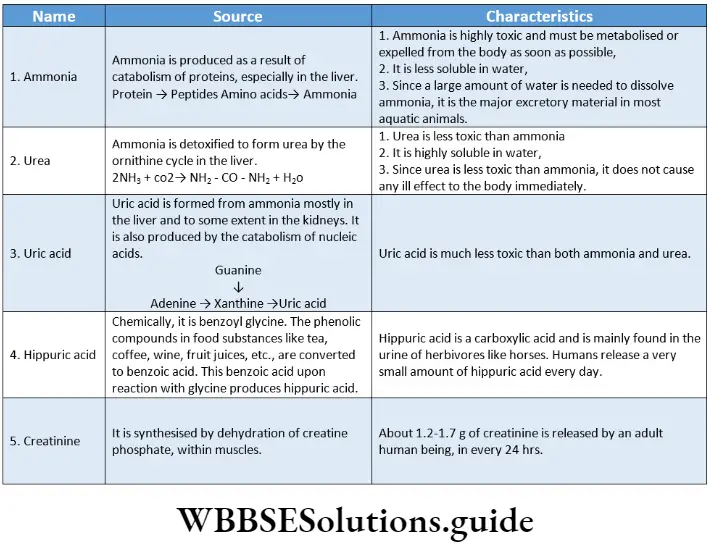
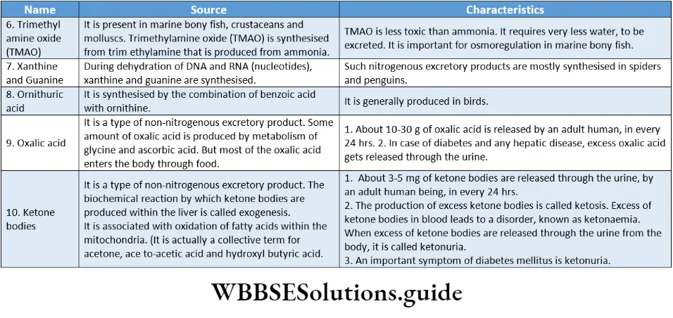
Modes Of Excretion In Animals
Depending on the excretory product, there are Different Modes of excretion in animals.
Principal Modes Of Excretion
Ammonia, urea, and uric acid are the principal excretory products in animals. Based on the type of nitrogenous wastes released, excretion can be of the following types—
- Ammonotelism,
- Ureotelism and cd uricotelism.
Ammonotelism: The excretion of nitrogenous wastes from the body, mainly in the form of ammonia, is called ammonotelism.
The animals, that release ammonia as the main nitrogenous waste product, are called ammonotelic animals.
For example, aquatic animals like sponges, coelenterates, crustaceans, cnidarians, Platyhelminthes, echinoderms, bony fish, tadpoles, salamanders, etc.
Ureotelism: The excretion of nitrogenous wastes from the body, mainly in the form of urea, is called ureotelism.
The animals that excrete urea as the main nitrogenous waste are called ureotelic animals. For example, man, whales, desert mammals like kangaroo rats, camels, terrestrial amphibians, aquatic and semi-aquatic reptiles like alligators, etc.
Uricotelism: The excretion of nitrogenous wastes mainly in the form of uric acid, is called uricotelism.
The animals that excrete uric acid as the main nitrogenous waste product are known as uricotelic; animals. For example, birds, land reptiles, insects, land snails, and some land crustaceans, etc.
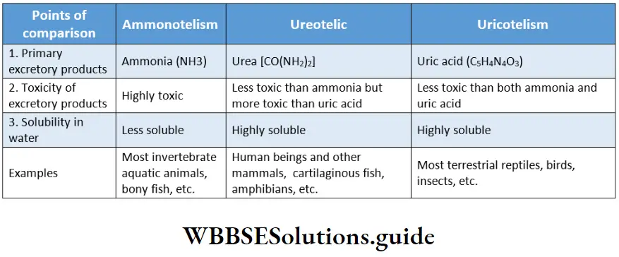
Dual Excretion
Some of the ammonotelic organisms can become partially ureotelic under adverse conditions, especially at times of shortage of water in the environment and in the organism’s body.
For example, Earthworms, when present in moist soil, release ammonia as the excretory product, but release urea, when present in dry soil.
Other Modes Of Excretion
Besides the principal modes of excretion, there are some other modes of excretion as well. They are as follows—
Aminotelism: The excretion of excess amino acids, without being converted to any other substance, is called aminotelism.
Animals showing this mode of excretion, are called aminotelic animals. Some members of Phylum Mollusca and Echinodermata are amniotic.
Guanotelism: The excretion of guanine as the main nitrogenous waste product is called guanotelism.
Animals showing this mode of excretion are called guanotelic animals. Spiders, some bony fish, and some members of Phylum Mollusca are guanotelic.
Short notes on excretory products and their elimination for quick revision
Excretory Organs Of Invertebrates
The excretory system in different organisms varies according to their complexity.
It shows the process of evolution from simple to complex organisms. Freshwater and terrestrial invertebrates have a wide variety of adaptations for maintaining salt and water balance (osmoregulation), and also excreting nitrogenous wastes.
Three well-known invertebrate excretory organs are—protonephridia, metanephridia, and Malpighian tubules.
Protonephridia and flame cells: The tubular excretory structures of Platyhelminthes (flatworms) are called protonephridia. It contains specialized excretory cells called flame cells.
Also found in rotifers, some annelids, and the cephalochordata—Amphioxus/ Branchiostoma.
Nephridia: The simple tubular excretory structure, usually found in annelids, is called nephridia. It removes nitrogenous wastes and maintains a fluid and ionic balance in the body.
Malpighian tubules: The tubular excretory structure of insects is called Malpighian tubules.
Green glands: Green glands or Antennal glands remove excretory products in crustaceAnswer:
Human Excretory System
Human Excretory System Definition: The organ system that is responsible for collecting and removing the metabolic wastes from the human body is called the human excretory system.
The human excretory system consists of a pair of kidneys, a pair of ureters, the urinary bladder, and the urethra.
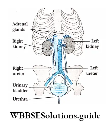
Kidneys
Kidneys Definition: The pair of bean-shaped organs, located in the abdominal cavity, that are responsible for the production of urine are called kidneys.
Kidney Location: The kidneys are located near the rear wall of the abdominal cavity.
They are considered retroperitoneal, which means they lie behind the peritoneum.
Each kidney extends between the 12th thoracic vertebra and the 3rd lumbar vertebra, on both sides of the backbone. The right kidney is situated a little lower than the left kidney.
Kidneys Shape And Size: Each kidney measures about 10-12 cm in length, 5-6 cm in breadth, and 3-4 cm in thickness. The right kidney is slightly smaller than the left one.
Kidneys Weight: The kidney usually weighs 120-170 g. In healthy adult males, each kidney weighs an average of 170 g while in healthy adult females it weighs around 135 g.
Kidneys Structure: Each kidney is covered with a tough capsule made up of fibrous connective tissue. It is known as the renal capsule. The renal capsule is surrounded by a layer of fat, the adipose capsule, and another outer fibrous membrane, the renal fascia.
Adhering to the surface of each kidney, there is a layer of fat (pararenal fat) that protects the kidney from mechanical shock. One side of the kidney is concave in nature. This region has a depression, where renal artery enters while the renal vein and ureter exit the kidney.
This depression is called the hilum. The longitudinal section of a kidney shows three distinctive layers. The outer, granulated layer is called the renal cortex.
Excretory products and their elimination chapter summary with important points
It stretches down through a radially striated middle layer. It is called the renal medulla. A hollow cavity, called the renal pelvis, connects the kidney to the ureter, through which urine passes to the urinary bladder.
The renal pelvis branches into 2-3 compartments, inside the kidney. These are called major calyces (singular: calyx).
The major calyces are further divided into smaller compartments, called minor calyces. The renal medulla is divided into 8-18 triangle-shaped compartments, called renal pyramids.
Several renal pyramids join to form the renal papilla. The renal cortex extends into the renal medulla between the renal pyramids, forming long column-like structures, called renal columns of Bertin.
The kidneys are made up of several tubular structures called nephrons. According to their location, nephrons may be—superficial or cortical and juxtamedullary.
Each nephron is made up of the following parts—Malpighian corpuscle and Renal tubule.
Several renal tubules from adjacent nephrons join to form a larger tubule, called collecting tubule.
These are generally found in the renal medulla. This makes the medulla appear striated. Several collecting tubules join further to form the ducts of Bellini.
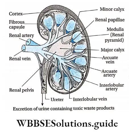
Kidneys Functions: Kidneys in the human body carry out Serval important Functions. They Are-
Kidneys Formation of urine: The primary function of the kidneys is to produce urine. Several nitrogenous and non-nitrogenous wastes get excreted from the body Through urine. urine is actually produced within the Nephrones Present in the kidneys.
Kidneys Maintenance of concentration of H+ and other inorganic ions in blood: Kidneys help to maintain the pH of the blood by releasing hydrogen (H+) ions and reabsorbing bicarbonate (HCO3–) ions.
They also play an essential role in regulating the amount of several important inorganic ions in the body. These include Na+, K+, C–, HCO–3,H+,Ca2+ And PO43-.
Kidneys Maintenance of plasma volume: Kidneys help in the absorption of Na+ ions as well as water. This helps to maintain the volume of plasma.
Kidneys Reabsorption: The nephrons selectively reabsorb certain important ions as well as water. This helps to maintain the proper concentration of ions in the blood.
Kidneys Removal of wastes and harmful substances: Wastes such as ammonia, urea, uric acid, hippuric acid, etc., that are generated within the body, get excreted through urine. Besides these, several other harmful substances such as toxins, drugs, etc., also get removed from the body by urine.
Kidneys Maintenance of acid-base balance in the body: Another important role of the kidneys is the regulation of acid-base balance. Many of the metabolic functions in the body are sensitive to pH.
Thus, the pH of the body fluids must be maintained within a narrow range. This function is carried out by the coordinated action of the lungs, liver, and kidneys.
Controlling blood pressure: The kidneys produce an enzyme, called renin. When the blood decreases, this enzyme is secreted. This enzyme activates the renin-angiotensin system.
This constricts blood vessels and helps to increase blood pressure. It also helps in the absorption of Na+ ions as well as water. This influences the plasma volume.
It again influences the total blood volume, which in turn regulates blood pressure.
Production of RBCs: Kidneys secrete a hormone called erythropoietin in response to low oxygen levels in tissues Erythropoietin stimulates red blood cell formation in the bone marrow. Decreased erythropoietin secretion leads to low erythrocyte production which may cause anaemia.
Homeostasis: Kidneys are able to maintain homeostasis by varying the concentration of urine. They reabsorb water and ions from urine when blood plasma volume is low.
They do not reabsorb ions and water from urine (hence, producing dilute urine) when blood plasma volume is high. This regulates the water content in the blood, which in turn maintains homeostasis.
Secretion of different substances: Kidneys also serve as important glands that secrete renin, calcitriol, erythropoietin, etc.
Renin activates the renin-angiotensin-aldosterone system, which helps to regulate blood pressure and sodium-potassium balance.
Calcitriol, on the other hand, is a metabolite of vitamin D3. It is necessary for normal reabsorption of Ca2+ by the gastrointestinal tract.
It is also important for the deposition of calcium in bones and the synthesis of collagen tissue. Erythropoietin regulates RBC production in bone marrow.
Ureters
Ureters Definition: The muscular tubes that carry urine from the kidneys to the urinary bladder are called the ureters.
Ureter Location: Each ureter descends downwards from the hilum of each kidney, to enter the urinary bladder posteriorly at its inferior surface.
Ureters Structure: The ureters are muscular tubes about 25 cm long and 30 mm in diameter. The lumen of the ureter is narrow and branched.
Ureters Functions:
Both the ureters transport urine produced in the kidney, from the renal pelvis to the bladder.
Peristaltic contractions by the involuntary muscles, cause urine to enter the bladder.
Excretory Class 11 Notes
Since the ureters pass behind the urinary bladder, and enter the bladder at its base, the bladder compresses the openings of the ureters. This prevents the back-flow of urine into the ureters when pressure in the bladder is high during urination.
Urinary Bladder
Urinary Bladder Definition: The pear-shaped, muscular, sac-like organ that collects the urine and stores it till urination is called the urinary bladder.
Urinary Bladder Location: The urinary bladder is located in the pelvic cavity, just posterior to the pubic symphysis.
In males, it is directly anterior to the rectum. In females, it is anterior to the vag*na and inferior to the uterus.
Urinary Bladder Anatomical structure: The urinary bladder is triangular in shape when empty, but appears oval when filled with urine.
It is composed of transitional epithelium. The bladder has three openings—two for the ureters and one for the urethra, which drains out the urine.
The trigone is a smooth triangular area at the base of the bladder bordered by these three openings. Two sphincters are found at the junction of the urethra and the bladder. They are internal and external sphincters.
The internal sphincter is present around the opening of the urethra. Inferior to the internal sphincter, the external sphincter is present. It is composed of skeletal muscle that can be voluntarily controlled. The bladder has several features that allow it to retain urine.
After urine enters the bladder through each ureter, small folds of bladder mucosa act like valves to prevent its backflow.
These folds are made up of transitional epithelial tissues that prevent the reabsorption of urine from the urinary bladder. About 600 ml of urine can be stored in the urinary bladder.
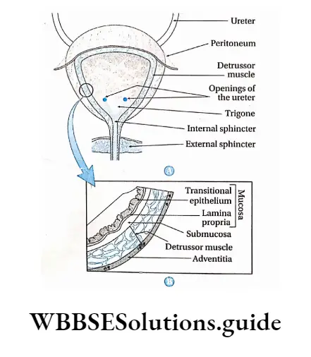
Urinary Bladder Functions: The function of the urinary bladder is to store urine prior to its elimination from the body.
When the urinary bladder gets filled with 300 – 400 ml of urine, an adequate stimulus is generated for its elimination. The bladder then expels urine into the urethra by a process, called micturition.
Urethra
Urethra Definition: The small tube-like structure, extending from the urinary bladder, through which the urine is excreted out of the body is called the urethra.
Urethra Location: It begins at the neck of the urinary bladder and ends at the external urethral orifice.
Urethra Anatomical structure: The urethra is a fibromuscular tube that conducts urine from the bladder (and s*men in males) to the exterior. It is different in structure in males and females.
In males, the urethra is about 20 cm in length. It can be divided into 4 parts. The pre-prostatic urethra is about the first 0.5-1.5 cm in length from the internal urethral orifice.
At this orifice a sphincter muscle is present. The next part extends through the prostate gland. This part is called the prostatic urethra.
At this region, the ejaculatory duct (receives sperms) and several prostatic ducts (receives prostate fluid) meet the urethra.
The urethra now passes the external urethral sphincter, through the perineum at an angle of 90°, before entering the penis.
This portion is called the membranous urethra. Then the urethra enters the penis. This portion is called the penile urethra. Urethra opens to the exterior by a small orifice at the apex of the penis which is called urinogenital aperture or urinary meatus.
In males, the urethra carries urine during urination and sperm during ejaculati*n. The female urethra is about 4-5 cm in length. It extends from the internal to the external urethral orifice.
The urethra is fused with the anterior wall of the vag*na. It ends at the urethral orifice or urinary meatus located between the clitor*s and the vag*na.
There is an external urethral sphincter present near this opening. In females, the reproductive and urinary systems are not connected.
Urethra Functions: The urethra serves as the passageway through which urine is discharged from the body. In males, the urethra also serves as the duct through which s*men is ejaculat*d.
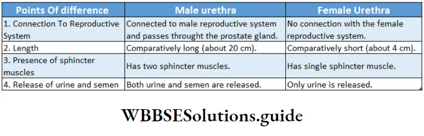
Nephron
Nephron Definition: The structural and functional unit of the kidney is called the nephron.
The name ‘nephron’ comes from the Greek word ‘nephros’, meaning ‘kidney’. Each nephron has two parts a globular renal corpuscle or malpighian body and a tubular structure called renal tubule.
Nephron Location: Most (85%) of the nephrons are present in the renal cortex. The rest (15%) of them are present in the juxtamedullary region of the renal medulla.
Number: Each human kidney has approximately 1.3 million nephrons.
Nephron Types: There are two types of nephrons—
- Superficial and
- Juxtamedullary.
Superficial nephron: The nephron, in which the renal corpuscle remains in the outer region of the cortex and the loop of Henle (a part of renal tubule) is at the outer zone of the medulla, is called a superficial nephron.
This is also called the cortical nephron. About 85% of the nephrons are superficial in nature.
They are mostly located in the periphery of the renal cortex. A few species, including humans, possess very short superficial nephrons whose loop of Henle is short and never enters deep into the medulla.
The efferent arterioles from adjacent nephrons form a network of peritubular capillaries. These are the nephrons that are responsible for the production of urine under normal conditions.
Juxtamedullary nephron: The nephron, in which the renal corpuscle is located in the inner region of the cortex next to the medulla, and the loop of Henle enters the medulla, is called juxtamedullary nephron.
They are generally located in the inner cortex and medulla. They can produce urine during emergency periods.
They differ from superficial nephrons in two important ways—
- The loop of Henle is longer and extends deeper into the medulla.
- The efferent arterioles of adjacent nephrons form not only a network of peritubular capillaries but also a series of vascular loops called the vasa recta around the loop of Henle.
Parts Of Nephron
Nephrons are differentiated into the following regions—
- Malpighian body or renal corpuscle
- Renal tubule.


Malpighian body or Renal corpuscle
Malpighian body or Renal corpuscle Definition: The flattened spherical part of the nephron is called the malpighian body or renal corpuscle.
The Malpighian body is present in the renal cortex only. It has a diameter of about 200 pm.
It consists of a tuft of interconnected capillaries, called glomerulus (plural: glomeruli), that is encased in a hollow sphere called Bowman’s capsule
Glomerulus: A network of capillaries within the Bowman’s capsule that performs ultrafiltration of blood is known as glomerulus.
The afferent arteriole (50 pm in diameter) that leads to the glomerulus has a larger diameter than the efferent arteriole (25 pm in diameter) that leaves it. So the blood pressure in the glomerulus remains very high.
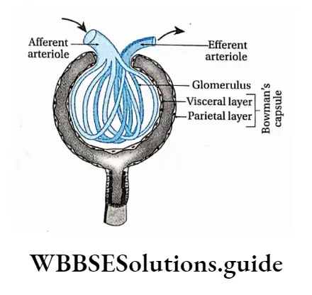
Bowman’s capsule: The hollow, double-layered, cup-shaped part of the renal corpuscle that holds the glomerulus is called the Bowman’s capsule (also called the glomerular capsule)
It is composed of visceral (inner) and parietal (outer) layers
Visceral layer: It is a single layer of squamous epithelium that lies next to the glomerular capillaries. The visceral layer is made of specialized cells, called podocytes.
These podocytes send small projections or foot processes over the length of the glomerulus. The foot processes are called end-feet or pedicels. They interdigitate with one another forming filtration slits.
The size of the filtration slits (generally 25 pm) restricts the passage of large molecules (e.g., albumin) and cells (e.g., RBCs and platelets).
Parietal layer: It lies next to the visceral layer. It is made up of a single layer of simple squamous epithelial cells.
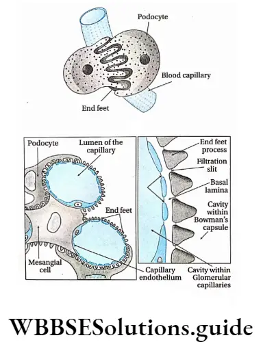
A common basement membrane is present in between the inner layer of the Bowman’s capsule and the endothelium of the glomerulus.
Renal tubule Definition: The tubular part of the nephron, extending from the parietal layer of the Malpighian body, is called the renal tubule.
The renal tubule is about 3 cm in length. It has three parts—The proximal convoluted tubule (PCT), a loop of Henle, and a distal convoluted tubule (DCT).
Proximal convoluted tubule (PCT): The longest and the most convoluted part of the nephron is called the proximal convoluted tubule
Proximal convoluted tubule Characteristics:
- It is about 15 mm long and has an external diameter of about 60 mm.
- The walls of PCT are made up of a single layer of closely-placed epithelial cells called podocytes.
- The cells have projections called pedicels or end feet. These cells interdigitate with one another and are united by apical tight junctions (intercellular junctions between adjacent epithelial cells at the apex of the cells).
- The PCT has a layer of cuboidal cells surrounding the lumen.
- The cells, lining the lumen, have numerous finger-like projections from their free ends. These are called microvilli. They are generally 1-1.3 urn in length, The presence of the microvilli gives them a brush-border appearance.
- Several pinocytic cavities are present within the cell membrane of these microvilli. The microvilli help the lysosome, in protein endocytosis. They serve to increase surface area for reabsorption.
- The upper and lower portions of the cell membrane extend into the cytoplasm as structures, called canaliculi.
- The presence of elongated mitochondria is an important feature of this region. It provides energy for the process of selective reabsorption.
Loop of Henle: The U-shaped portion of the nephron, next to the PCT is called the loop of Henle.
Proximal Convoluted Tubule Characteristics:
- It is about 20 mm in length.
- The loop of Henle (sometimes known as the nephron loop) begins in the cortex, extends into the medulla, and then returns to the cortex.
- Its descending as well as ascending limbs, both have thin and thick regions.
- Like PCT, this region also has elongated mitochondria attached to the basement membrane.
- It has a lesser microvilli
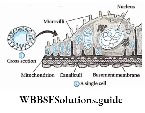
Distal convoluted tubule (DCT): The next convoluted portion of the nephron that arises from the loop of Henle, is called the distal convoluted tubule.
Distal convoluted tubule Characteristics:
- The DCT is about 5 mm long and has an external diameter of 50 mm.
- It is lined by simple cuboidal epithelium, with shorter and fewer microvilli. Hence, there is no distinct brush border.
- A region of this tubule lies very close to the afferent arteriole. This region is called macula densa.
- Macula densa region has tightly packed columnar epithelial cells.
- Cells lining the tubule have mitochondria, attached to the basement membrane. This enables the active transport of ions to take place by the energy supplied by ATP. The plasma membrane has a large number of Canaliculi.
Distal convoluted tubule Collecting duct Definition: The duct that the distal convoluted tubule ends into is called the collecting duct.
Handwritten notes on excretory products and their elimination PDF download
Collecting duct Characteristics:
- The collecting duct is about 20 mm long and pass through both the renal cortex and medulla.
- It is actually not the part of a nephron, rather, the DCTs of several nephrons join together to form one collecting duct.
- Several collecting ducts, carrying urine, join to form the duct of Bellini. This duct enters the renal pelvis.
- The epithelium of the collecting ducts is made up of principal cells (P cells) and intercalated cells (I cells).
- P cells, which predominate, are relatively longer and have few organelles. They are involved in Na+ reabsorption and vasopressin-stimulated water reabsorption.
- I cells, which are lesser in number, have more microvilli, cytoplasmic vesicles, and mitochondria.
- They are concerned with acid secretion and HC03- -transport. Though the collecting duct was previously considered part of a nephron, modern scientists consider it a separate part of the kidney.
Peritubular Capillaries And Vasa Recta
Blood supply in the tubules of nephrons is carried out by peritubular capillaries and vasa recta.
Peritubular capillaries: The network of capillaries, formed by the branching of the afferent arteriole, that runs alongside the nephron and extends over the renal tubule are called peritubular capillaries.
Vasa recta: In the case of juxtamedullary nephrons, peritubular capillaries surround the proximal tubules and distal tubules, as well as Henle’s loop, in a reticulate manner. This is called vasa recta.
Although less than 0.7% of the renal blood enters the vasa recta, these vessels perform many important functions. These include—
Transportation of oxygen and important nutrients to different parts of the nephron.
Delivery of substances to the nephron for secretion.
It serves as a pathway for the reabsorbed water and solutes to enter the circulatory system.
Modification of the concentration of the urine.
Functions Of A Nephron
Nephrons play a very important role in the process of urine formation. Different parts of the nephron perform different functions such as filtration, reabsorption, renal tubular secretion, formation of new substances, etc. These have been discussed in detail in the next section.
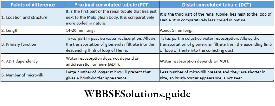
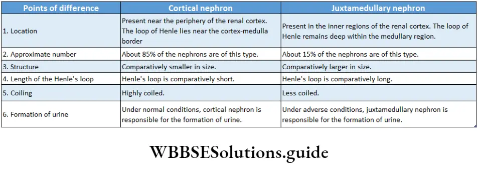
Urine Formation Role Of Different Parts Of Nephron
Both nitrogenous and non-nitrogenous wastes are excreted through the urine. Let us study some more about urine and the role of the nephron in urine formation.
Urine Definition: The transparent, yellowish, acidic fluid, with a characteristic pungent odor, that contains nitrogenous and non-nitrogenous excretory products, is called urine.
Urine Characteristics: The following are the characteristics of urine—
Urine Color: Due to the presence of a pigment, called urochrome or urobilin, the urine attains a yellowish color. This color changes in case of different diseases.
Urine Quantity: About 1-1.5 L of urine is produced per day by an adult human being. This quantity may vary with water intake, external environment, physical and mental conditions, etc.
Urine Chemical properties: It is a transparent and acidic fluid. Its pH ranges from 4.5-8.2. Excess intake of animal proteins may increase the acidity, while a diet rich in vegetables, citrus fruits, etc., may decrease the acidity
Odor: Its characteristic odor is that of aromatic compounds. When urine is left for some time, the urea present in the urine gets converted to ammonia, thereby releasing the pungent smell of ammonia.
Constituents of urine
The urine has normal and abnormal constituents. These constituents have been discussed below-
Normal constituents of urine: Urine contains both water and solid constituents. Most of the urine is water (94-96%), with 4-6% of solid constituents.
The solid constituents can be further divided into organic and inorganic constituents. Inorganic constituents are (in 1.5 I L urine volume in 24 hours)— Cl- (6-9 g), NaCI (10-15 g), P043- (0.8-1.3 g), S042- (0.8- 1.4 g), K+ (2.5-3 g), Na+ (4-5 g), Ca2+ (0.1-0.3 g), Mg2+ (0.1-0.2 g), I (50-250 μg), As (50 μg), Pb (50 μg). Organic constituents are further divided into nitrogenous and non-nitrogenous constituents.
Nitrogenous constituents— urea (25-30 g), ammonia (0.7 g), uric acid (0.7 g), hippuric acid (0.1-1.0 g), creatine (60-150 g), creatinine (1.2-1.7 g), amino acid (150-200 mg).
Non-nitrogenous constituents—ketone bodies (0.002-0.012 g), glucose (0.01-0.04 g), oxalic acid (0.014-0.023 g), ascorbic acid (0.014-0.05 g), phenolic compounds (0.2-0.3 g).
Abnormal constituents of urine: These constituents are not found in urine under normal conditions, but are seen in case there is a disease.
These abnormal constituents are discussed as follows—
Glucose: When the glucose concentration in the blood Exceeds 180 mg/100 mL, it results in the release of glucose in the urine. This condition, marked by the presence of glucose in urine, is called glucosuria.
Other carbohydrates: Due to various reasons, fructose, lactose, and galactose may also be released with I urine. These conditions are called fructosuria, lactosuria, and galactosuria, respectively.
The reason behind the more acidic nature of the urine compared to the blood plasma
The glomerular filtrate generated from the blood plasma has an alkaline pH of 7.4, similar to that of blood plasma.
As the filtrate passes through the renal tubule, reabsorption of the sodium ions from the filtrate takes place. Hence, the alkaline nature of the filtrate decreases.
Simultaneously, cells lining the inner walls of the renal tubule secrete hippuric acid, ammonium ion, hydrogen ion, etc., that get mixed with the filtrate. This makes the urine more acidic in nature (pH of 6.0).
Protein: Exhaustive physical work, intake of a high-protein diet, improper metabolism of proteins, inflammation of kidneys, etc., causes the release of excess protein in urine. This condition is known as proteinuria.
Ketone body: The concentration of ketone bodies in the urine increases in case of diabetes mellitus, pregnancy, or improper metabolism of carbohydrates. This condition is known as ketonuria.
Fat: Due to kidney disorder, the poisonous effect of alcohol, etc., excess fat gets released as abnormal constituents of urine. This condition is known as lipuria.
Pigment: Due to a disorder in liver function, bilirubin and biliverdin are excreted through the urine. This condition is mainly found in cases of jaundice or hepatitis.
Blood: Sometimes blood is found in urine, due to internal wounds, inflammation of kidneys, bacterial infection, etc. This condition is known as haematuria.
Alkaptones: The release of excess alkaptones through the urine, occurs in a congenital or hereditary disorder, called alkaptonuria.
Pus: The presence of pus as an abnormal constituent in the urine is seen in the case of some diseases such as urinary tract infections. This condition is called pyuria.
Role Of Different Parts Of The Nephron In Urine Formation
Urine formation in the nephron is the result of successive processes—ultrafiltration, selective reabsorption, and tubular secretion. The different parts of the nephron carry out these processes. Let us study each in detail.
Role of Malpighian corpuscle
The Malpighian corpuscle performs the following functions—
Ultrafiltration: Ultrafiltration is a variety of membrane filtration in which a difference in pressure or concentration across the semipermeable membrane leads to the retention of colloidal particles while smaller particles and the solvent are forced to move across the membrane.
Water, ions, and small nitrogenous molecules from the blood flowing through the glomerulus are filtered into the Bowman’s capsule by this process.
Ultrafiltration Description: The process of ultrafiltration is as follows—
- The visceral layer of the Bowman’s capsule behaves as an ultrafilter. The cells of the Bowman’s capsule and surrounding capillaries form a selectively permeable membrane.
- This membrane allows only water, ions, small nutrient molecules (such as glucose and amino acids), and nitrogenous waste molecules (primarily urea) to pass through it.
- Blood enters the afferent arteriole and flows into the glomerulus. The high pressure of blood in the glomerulus is called glomerular capillary pressure (GCP). It forces the fluid, containing these molecules and ions, into the capsule.
- Blood cells, platelets, and plasma proteins are too large to pass through. Therefore, they are retained in the capillaries. The charge on molecules as well as their diameters affect their passage into Bowman’s capsule.
- Foot processes have a negatively charged coat (glycocalyx) that limits the filtration of negatively charged molecules, such as albumin. This action is called electrostatic repulsion. All these features of Bowman’s capsule help in the process of filtration of blood.
- The fluid that enters the Bowman’s capsule is an ultrafiltrate of the blood. It is also known as glomerular filtrate or primary urine. The filtrate leaving the Bowman’s capsule is similar to blood plasma in composition, as it passes into the proximal convoluted tubule.
Some factors responsible for ultrafiltration
- The active surface area of the filtration membrane: The active surface area of the filtration membrane depends on the number of active glomeruli and functional blood capillaries present in each glomerulus.
- Permeability of the filtration membrane: Generally, the pores present in the filtration membrane have a diameter of 20-25 nm. Most of the plasma proteins, blood corpuscles, and blood platelets cannot pass through these pores.
However, the colloids present in the blood pressure are called glomerular colloidal osmotic pressure (GOP).
- This pressure along with capsule hydrostatic pressure (CHP) creates a hindrance to ultrafiltration. Both GOP and CHP are known as the opposing pressure. If their total value exceeds Glomerular capsule pressure (GCP) the filtration will occur. The pressure at which the filtration pressure (EFP). The average value of GCP is 75 mm of Hg, while the average values of GOP and CHP are 30 and 20 mm of Hg respectively. The average value of EFP is 25 mm Hg.
- About 1400 L of blood passes through the kidneys every day. The Bowman’s capsule filters about 180 L of fluid from this blood.
- The glomerular filtrate contains wastes such as urea, uric acid, creatinine, excess ions, etc. It also contains glucose, amino acids, etc., some of which get reabsorbed later.
- The amount of glomerular filtrate produced per minute is called the glomerular filtration rate (GFR). The rate of filtration is 125 ml/min. (Jo) Ultrafiltration is driven by Starling forces across the glomerular capillaries. Changes in these forces alter the glomerular filtration rate (GFR). GFR and renal blood flow (RBF) are normally controlled within narrow ranges by a phenomenon called autoregulation.
Storage of ultrafiltrate: The ultrafiltrate is stored in the cavity of Bowman’s capsule for a temporary period.
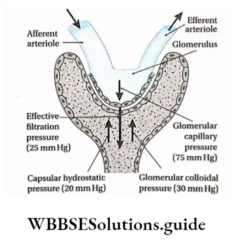
Role Of Renal Tubule
The glomerular filtrate passes through the renal tubule into the collecting ducts.
The renal tubule carries out three functions—
- Selective reabsorption,
- Tubular secretion,
- Synthesis of some substances.
Selective reabsorption: The reabsorption of filtered molecules, such as H+, Na+, and K+ ions from the glomerular filtrate in the tubules to the blood is called selective reabsorption.
About 85% of the 180 L of glomerular filtrate, formed per day, is reabsorbed by this process.
This reabsorption, as well as the regulated reabsorption of the remaining volume of filtrate, occurs by osmosis. It results in the transport of Na+ and Cl– ions across the tubule wall.
A concentration gradient is created between tubular filtrate and the plasma, in the surrounding capillaries. This promotes the osmosis of water back into the vascular system.
Reabsorption in proximal convoluted tubule: The proximal convoluted tubule reabsorbs approximately 67% of the filtered water, Na+, K+, CP, and other solutes.
In addition, the PCT also reabsorbs most of the glucose and amino acids filtered by the glomerulus.
The key element, in proximal tubule reabsorption, is the Na+K+ATPase, which is found in the basolateral membrane. The reabsorption of every substance, including water, is linked in some way to the operation of this enzyme.
Specialized ion pumps transport K+, Na+, and C– from the filtrate into the fluid surrounding the tubule.
Active transport proteins, embedded in the walls of the tubule, reabsorb the amino acids, glucose, and other nutrients out of the filtrate.
Water, chloride ions, and urea are absorbed passively, whereas substances like hormones, amino acids, sulfates, phosphates, etc., are absorbed through active transport.
The coiled structure and microvilli greatly increase the total surface area that is available for the reabsorption of solutes.
The reabsorption of solutes makes the filtrate hypoosmotic to the interstitial fluid. This causes water to flow out of the tubule, into the interstitial fluid by osmosis.
The reabsorption of water in PCT is obligatory in nature. The movement of water is further facilitated by membrane proteins, called aquaporins.
The aquaporins ensure that the maximum water is removed from the tubule during the reabsorption process. The presence of a large number of mitochondria provides the energy to carry out the process of reabsorption.
Reabsorption in a loop of Henle: The fluid, which has a high concentration of urea and other unabsorbed wastes, moves through the proximal convoluted tubule into the descending portion of the loop of Henle.
The descending limb of the loop of Henle is permeable to water, due to the presence of aquaporin-1, in both the apical and basolateral membrane. About 15% of the filtered water is removed from the descending limb.
Therefore, the fluid in the descending limb of the loop of Henle becomes hypertonic, to plasma.
However, the ascending limb is impermeable to water. Na+, K+, and Cl– are co-transported out of the thick segment of the ascending limb.
Bivalent ions like Mg2+, Ca2+, etc., are also reabsorbed actively in this region. This dilutes the solution. In the ascending limb, it becomes more dilute because of the movement of Na+ and Cl– out of the tubular lumen.
Thus, the fluid becomes hypotonic to plasma, when it reaches the top of the ascending limb Hence, this portion of the nephron is called the diluting segment.
Reabsorption in distal convoluted tubule: The distal tubule is relatively impermeable to water. So, water reabsorption is facultative here.
The reabsorption of water is regulated by ADH or antidiuretic hormone (vasopressin). Due to sweating, continued removal of the solute further dilutes the tubular fluid.
Under such conditions, ADH is secreted. It stimulates the cells of the DCT and the collecting duct, which are otherwise impermeable to water, to reabsorb water.
After passing through the distal convoluted tubule, only 3% of water remains in the filtrate.
The salt content remaining in the filtrate is negligible. About 97.9% of the water in the glomerular filtrate gets absorbed in the PCT, loop of Henle and DCT.
Obligatory Water Loss
Although about 180 L of glomerular ultrafiltrate is produced every day, the kidneys normally excrete only 1 to 2 L of urine in a 24-hour period.
The urine volume, however, varies according to the needs of the body.
When a well-hydrated person drinks more water, urine production increases to 16 ml per minute. If this situation continues, 23 L of urine may be excreted per day.
On the other hand, in case of severe dehydration, when the body needs to conserve water, only 0.3 ml of urine per minute, or 400 ml per day, is produced.
A volume of 400 ml of urine per day is the minimum needed to excrete the metabolic wastes produced by the body; this is called obligatory water loss.
Tubular secretion: The secretion of certain substances, such as H+, urea, etc., into the glomerular fluid, when it passes through the DCT is called tubular secretion.
Tubular secretion leads to the removal of waste products from the blood and interstitial fluid.
These waste products are hydrogen ions, creatinine, para amino hippuric acid, certain pigments, and drugs.
H+ ions are actively secreted, while the products of detoxified toxins, produced by the liver, are passively secreted. Many water-soluble drugs, such as penicillin and other medications, along with their metabolites are also secreted into the nephron.
Some amount of ammonia is also secreted into the tubule. H+ secretion is coupled with HCO– reabsorption from the filtrate to the plasma in the peritubular capillaries. The release of certain hormones triggers changes in the salt concentration in the body.
This varies the secretion of K+ and H+ ions into the filtrate in the DCT. Additionally, active secretion of H+ occurs in the collecting ducts. The balance of ions established in the blood, urine, and interstitial fluid, regulates the pH level of the blood and body fluids.
In this way, the kidney acts as a “safety valve.” If the acidity of the body fluid rises, the excess H+ ions are secreted into the collecting ducts. These ions are then excreted through the urine.
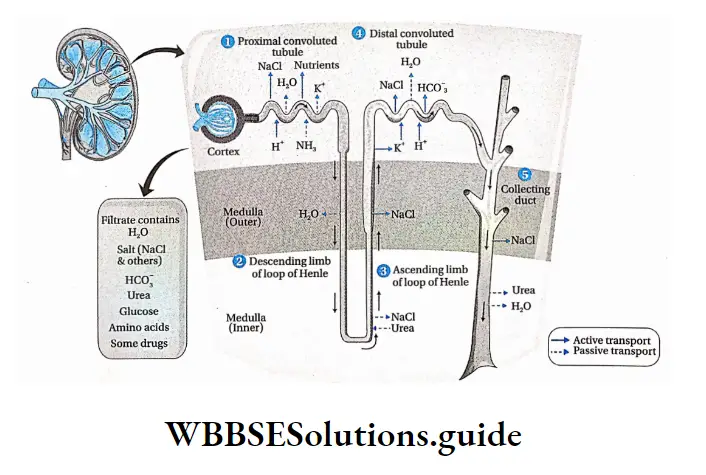
New product formation: Different substances like ammonia, hippuric acid, etc., are synthesized by the cells, lining the renal tubule. These substances are released into the lumen of the tubule and pass out of the body a,ong with the urine.
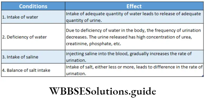
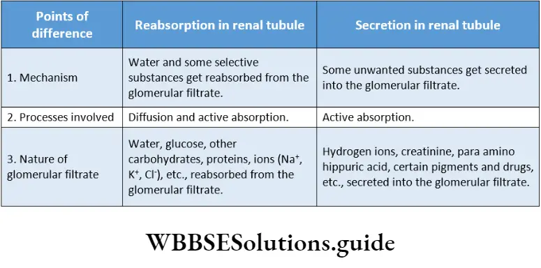
Mechanism For Concentration Of Glomerular Flarte The Countercurrent Mechanism
Mechanism For Concentration Of Glomerular Flarte Definition: The mechanism, present within the nephron that regulates the balance of water, ions, and pH within the blood and interstitial fluid, is called the countercurrent mechanism.
The loop of Henle and vasa recta play important roles in the countercurrent mechanism. In this mechanism, the interplay occurs between the isotonic tubular fluid and the blood flowing in the opposite direction through the vasa recta. The name ‘countercurrent’ indicates the opposite direction of flow of the two fluids.
Mechanism For Concentration Of Glomerular Flarte Types: This system involves two separate types of subsystems.
Countercurrent multiplier: The gradient of increasing osmolarity produced by the activity of the loop of Henle, in the renal medulla, acts as a countercurrent multiplier. This makes the interstitial fluid in the renal medulla, hypertonic.
Countercurrent exchanger: In this system, the hypertonicity of the interstitial fluid is maintained in the renal medulla, by exchanging ions between the blood and the tubular fluid. The Vasa Recta Along The loop of henle acts as a countercurrent exchanger.
Significance of countercurrent mechanism:
Due to the Countercurrent mechanism, the nephrons are able to produce hyper or hypotonic urine. This maintains the osmolarity of the body fluids and blood.
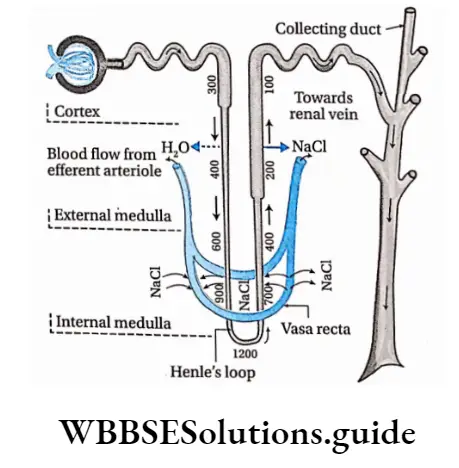
Countercurrent multiplier mechanism in the loop of Henle:
- It is an energy-driven process that generates an osmotic gradient along certain sections of the renal tubule (Henle’s Loop and DCT, collecting duct).
- The loop of Henle is bathed in hypertonic interstitial fluid in the medulla. Its descending limb is permeable to water.
- Thus, water moves out of the tubular fluid making it concentrated. This water readily enters the vasa recta and is carried away. This rapid removal of water from interstitial fluid maintains its hypertonicity.
- As the fluid gradually moves through the descending limb, more water gets reabsorbed, but, urea and other ions such as Na+, and K+ do not get reabsorbed. This increases the osmotic concentration of tubular fluid up to 1200 mOsmol L-1.
- This hypertonic filtrate moves up to the ascending limb of Henle’s loop. Here water does not get reabsorbed as its wall is impermeable to it. But, a large amount of Na+ and Cl” is reabsorbed via active transport. Hence, the filtrate becomes isotonic (with respect to blood).
- Consequently, tubular fluid rises through the ascending limb, along with active reabsorption of NaCI, while, urea passively diffuses into the tubular fluid. The osmotic concentration of the filtrate lowers to about 100 mOsmol L-1.
- Some reabsorption takes place in the DCT due to the action of ADH hormone, this increases the osmotic concentration of the filtrate to about 300 mOsmol L-1.It depends on
- The high permeability of the thin descending limb to water (via aquaporin-1),
- Active transport of na+ and clout of the thick ascending limb, and
- Inflow of tubular fluid from the PCT, and its outflow through the DCT.
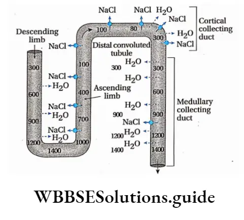
Countercurrent Exchanger Mechanism in the vasa recta:
- Blood enters and leaves the medulla through the vasa recta present at the boundary of the cortex and the medulla. These capillaries are highly permeable to solute and water.
- As the blood descends into the medulla towards the papillae via the descending limb of the vasa recta, it becomes more concentrated by gaining solutes and losing water to the hypertonic interstitial fluid.
- By the time blood reaches the tip of the ascending limb of the vasa recta, it has an osmotic concentration of 1200 mOsmol L-1.
- As this hypertonic blood moves towards the ascending limb of the vasa recta (towards the cortex) solutes diffuse back into the interstitial fluid and the water diffuses back into the blood, making it isotonic.
- Under normal conditions, vasa recta carries away only as much solute and water as is absorbed from the tubules. In this way, the high concentration of solutes is established in the interstitial fluid and the countercurrent mechanism is maintained.
- The ability of the vasa recta to maintain the osmotic gradient of interstitial fluid in the medullary region is flow-dependent.
- A substantial increase in blood flow through the vasa recta will ultimately disrupt the medullary osmotic gradient.
- Alternatively, if blood flow is reduced, the nephron segments within the medulla will receive inadequate oxygen.
- This will disrupt the active transport of ions across the ascending limb of Henle’s loop. As a result, the medullary osmotic gradient will be disrupted.
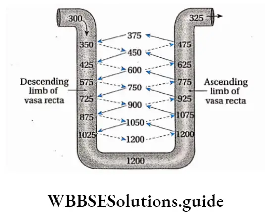
Osmotic concentration
The osmotic concentration of a solution is the concentration of particles that remain dissolved in that solution. It is generally expressed as milli osmoles 1 per liter (mOsmol L-1).
Osmorecepter
There are certain receptors in the body that can sense the change in the osmolarity of the body fluids. These receptors are called osmoreceptors. These are located in the anterior hypothalamus in the brain.
They respond only to certain solutes that are called effective osmoles. Certain solutes such as urea, do not affect the function of the osmoreceptors; These are called ineffective osmoles.
NEET excretory products and their elimination revision notes with MCQs
Role of countercurrent mechanism in producing hypertonic urine:
- As the tubular fluid passes through the descending limb of the loop of Henle, more water gets reabsorbed into the blood. But Na+ and other ions do not get reabsorbed, hence the osmolarity of the tubular fluid increases.
- As the tubular fluid passes through the ascending limb, some of the Na+ ions get reabsorbed, lowering the osmolarity of the tubular fluid.
- Finally, when the tubular fluid passes through the DCT, hormones like ADH and aldosterone, cause some more of the Na+ ions to be reabsorbed in the renal medulla. This increases the osmolarity of the tubular fluid again.
- Some amount of water also gets reabsorbed when the tubular fluid passes deeper into the renal medulla. Along with this, some amount of urea diffuses into the tubular fluid from interstitial fluid by passive transport.
- This leads to the release of hypertonic or hyperosmotic urine (1200 mOsmol L-1). Hypertonicity of urine is measured against blood and plasma.
Micturition
Micturition Definition: The biological process by which the urine stored in the urinary bladder gets excreted by the urethral orifice to the exterior, is called micturition.
Process: Micturition is initiated through the coordinated contraction of certain muscles. These are smooth muscles present within the walls of the urinary bladder, thoracic and pelvic region, diaphragm, and internal and external sphincter muscles.
The process may be described as follows—
- The urine produced within the nephron is carried by the ureters to the urinary bladder, where it is stored temporarily.
- The urine storage capacity of the urinary bladder in humans is about 800 ml.
- However, stretch receptors (nerve endings under the control of the autonomic nervous system), present within the walls of the urinary bladder, get stimulated when
about 300-400 ml of urine gets stored within it. - When the amount of urine in the urinary bladder increases further, this stimulus is carried to the brain and this generates the urge to urinate.
- The detrusor muscles, present within the walls of the urinary bladder, contract under the influence of efferent autonomic nerves.
- At the same time, sphincter muscles associated with the urethra, relax. As a result, the urine passes through the urethra and gets excreted out of the body through the urethral orifice.
- It is to be noted that contraction of the diaphragm and the muscles of the thoracic region, help in micturition. In the case of infants, some elderly adults, and those with neurological injuries, micturition is an involuntary reflex action.
Osmoregulation
Osmoregulation Definition: The regulatory mechanism, by which the living organisms maintain the balance of water and different ions within the cells as well as body fluids (blood, lymph, etc.), is called osmoregulation.
Osmoregulation Discussion: Water and ions are lost by various metabolic processes.
- Even the external environmental factors lead to the imbalance of water and ions. Water loss through sweat, feces, and during respiration from the lungs cannot be regulated.
- In contrast, renal excretion of water is regulated to maintain water balance in the body.
- Our body has well-developed mechanisms to maintain the osmotic concentration- of the body fluids.
- The water intake must precisely match the water loss from the body. When water intake is low or water loss is high, the kidneys conserve water by producing less urine.
- The urine becomes hyperosmotic with respect to plasma. When water intake is high, a large volume of hypoosmotic urine is produced.
- In a normal individual, the osmolarity of urine may vary from approximately 50 to 1200 mOsmL-1 of water. The corresponding urine volume may vary from as much as 18 L/day to as little as 0.5 L/day.
Significance: The significance of osmoregulation is—
Maintenance of osmotic concentration of body fluids: The osmotic concentration of the body fluids such as blood, lymph, etc., can be maintained by osmoregulation. If intake of water exceeds loss of water, then the water balance is positive. Conversely, when the intake of water is less than the loss of water, then the water balance is negative.
Maintenance of Homeostasis: It is maintained in extracellular fluids, by osmoregulation.
Rate of metabolic reactions: The rate of metabolic reactions is maintained properly within the cells by osmoregulation.
Excretory Class 11 Notes
Regulation Of Kidney Function
Neural signals, hormonal signals, and intrarenal chemical messengers combine to regulate the renal processes according to the needs of the body.
Neural signals: They originate in the celiac plexus of the sympathetic nervous system. These signals exert major control over renal blood flow and glomerular filtration.
They also influence the release of vasoactive (affecting blood vessels) substances, like angiotensin, that affect both the kidneys and the peripheral blood vessels.
Hormonal signals: They originate in the adrenal gland, pituitary gland, heart, and kidney itself. Their effects are as follows—
The juxtaglomerular apparatus of the kidney secretes renin and affects the GFR and blood pressure.
The posterior pituitary gland secretes the hormone vasopressin (also called antidiuretic hormone or ADH).
ADH is a major regulator of excretion. Through its influence on the renal blood vessels and principal cells of the collecting duct, it probably affects sodium excretion as well.
The heart muscles secrete hormones such as atrial natriuretic peptides (ANP), a vasodilator that increases sodium excretion by the kidneys. Hormones like aldosterone, secreted by the adrenal cortex, also influence the sodium and potassium ion balance.
Intrarenal chemical messengers: They also show some effect on the process of urine formation.
It is clear that an array of substances (e.g., nitric oxide, purinergic agonists, superoxide, etc.) influence basic renal processes, but, for the most part, the specific roles of these substances are not well understood.
Juxtaglomerular Apparatus
Juxtaglomerular Apparatus Definition: The organization of certain types of cells, present near the region, where the DCT lies close to the afferent arteriole, and responsible for renin secretion is called juxtaglomerular apparatus (JGA).
Structure: Each JGA is made up of the following three cell types—
Granular cells: Granular cells juxtaglomerular cells), are the smooth muscle cells, present in the walls of the afferent arterioles.
The granular cells are so named because they contain secretory vesicles that appear granular under a light microscope. These granules contain the hormone renin.
Extraglomerular mesangial cells: Extraglomerular mesangial cells are morphologically similar and continuous with the glomerular mesangial cells, but lie outside Bowman’s capsule.
They lie in the region extending between the afferent and efferent arteriole. These cells are also known as lacis cells.
Macula densa cells: These are specialized thick epithelial cells. They are present in the region where the DCT lies close to the afferent arteriole.
These cells can detect the composition of the fluid within the nephron at the extreme end of the thick ascending limb of the loop of Henle. They help to control renin secretion.
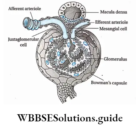
Juxtaglomerular Apparatus Functions:
When the blood pressure falls due to reduced blood flow to the kidneys, the sympathetic nervous system generates a stimulus. This stimulus causes the JGA to secrete renin.
It generates a decapeptide angiotensin-l by splitting angiotensinogen, a plasma protein. This under the influence of angiotensin-converting enzyme (ACE), forms angiotensin 2. This, in turn, stimulates the secretion of aldosterone from the adrenal cortex.
Aldosterone increases the reabsorption of water and Na+ ions in the DCT, thereby increasing the blood flow which raises the blood pressure. It finally increases the glomerular filtration rate (GFR).
During the shortage of oxygen in tissues, JG cells secrete a hormone called erythropoietin or renal erythropoietic factor (REF). This stimulates the production of RBCs in the bone marrow.
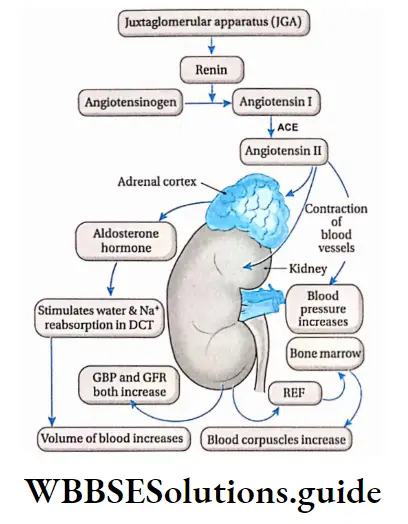
Atrial natriuretic peptide (ANP)
Atrial natriuretic peptide (ANP) Definition: The proteinaceous substance, secreted by the atrium of the heart, that acts as a vasodilator and maintains the balance between water and sodium ions in blood, is called an atrial natriuretic peptide (ANP)
Atrial natriuretic peptide Description: Earlier, it was considered that there are some factors that regulate the excretion of Na+ through urine. The process was termed natriuresis and the factors were called natriuretic factors.
Later, it was discovered, that cardiac atria secrete a factor with such a function. The first natriuretic hormone isolated from the heart was atrial natriuretic peptide (ANP).
The hormone ANP is also known as atrial natriuretic factor (ANF). ANP was subsequently isolated from other tissues, including the brain, where it exists in two forms that are smaller than circulating ANP.
Its secretion is stimulated by a rise in blood pressure and an increase in the effective i circulating volume.
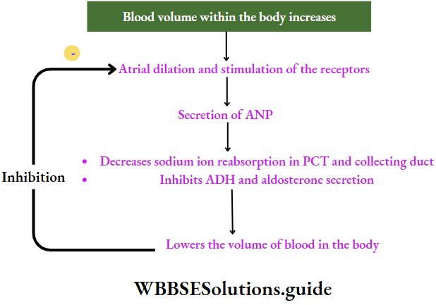
Functions: It has effects both on the kidney and the blood pressure. The effects are as follows—
On kidneys:
- It inhibits NaCI reabsorption by the medullary portion of the collecting duct.
- It inhibits ADH- ADH-stimulated water reabsorption across the collecting
duct.
On blood pressure:
It reduces blood pressure by dilating the walls of the heart and enhancing urinary NaCI and water excretion.
It inhibits the secretion of renin by the JGA which in turn inhibits the secretion of aldosterone by the adrenal cortex. This promotes the excretion of sodium along with water. This causes blood pressure and blood volume to decrease.
It reduces the secretion of ADH from posterior pituitary.
ANP secretion by the heart rises with hypertension and expansion of extracellular fluid volume, causing vasodilation of the afferent arteriole and vasoconstriction of the efferent arteriole.
The net effect of ANP is therefore to produce a mild increase in GFR with little change in RBF (renal blood flow).
Disorders due to sodium imbalance in the blood
Hypernatremia: If the sodium level in blood plasma is above normal (130-145 mmol/L), the condition is called hypernatremia.
Hyponatremia: If the sodium level in blood plasma is below normal, the condition is called hyponatremia.
Human excretory system: structure, function, and waste elimination process
Antidiuretic Hormone (ADH) And Diabetes Insipidus
The hormone, secreted by the pituitary gland, that acts on the kidneys to regulate the volume and osmolarity of the urine, is called antidiuretic hormone (ADH) or vasopressin.
Source of ADH: It is synthesized by neuroendocrine cells within the hypothalamus region of the brain. The synthesized hormone is packed in granules. These granules are stored at the nerve terminals in the posterior pituitary.
Functions of ADH:
- ADH increases the reabsorption of water in the DCT and the collecting duct.
- When plasma ADH levels are elevated, less volume of concentrated urine is excreted. This condition is called antidiuresis.
- When plasma ADH levels are low, more volume of urine is excreted. This condition is called diuresis. The urine, that is released, is dilute.
- It lowers the reabsorption of Cl” ions and thus large quantities of CP ions are excreted through the urine.

Diabetes insipidus Definition: The syndrome, that is characterized by the release of a large volume of hypotonic urine due to deficiency of ADH hormone in the blood or due to malfunctioning of kidneys, is called diabetes insipidus.
Causes: The causes of diabetes insipidus are—
Damage or infection of pituitary gland or associated neurons: If certain regions of the nervous system (the supraoptic and paraventricular nuclei, the hypothalamohypophysial tract), or the posterior pituitary gland gets damaged or infected, diabetes insipidus results.
The disease may develop after surgical removal of the posterior lobe of the pituitary. However, this condition may be temporary. The fibers may recover, make new vascular connections, and begin to secrete vasopressin again.
Failure of ADH to bind to receptors: If the ADH cannot bind to V-2 receptors (a type of vasopressin receptor) due to kidney malfunctioning, this disease may result. It mainly occurs as a genetic defect.
Mutation of water channel proteins aquaporin-2: It inhibits the reabsorption of water in the DCT and collecting duct. This situation may also develop this disease.
Types: Diabetes insipidus is generally of the following two types—
Vasopressin-sensitive or neurogenic or central diabetes insipidus: The type of diabetes insipidus caused by the damage of the hypothalamus or posterior pituitary gland, leading to less ADH secretion is called vasopressinsensitive or neurogenic or central diabetes insipidus.
Low vasopressin secretion decreases water and salt reabsorption in the DCT and collecting duct. This leads to the frequent discharge of dilute urine.
Nephrogenic diabetes insipidus: The type of diabetes insipidus that occurs due to malfunctioning of the kidneys, is called nephrogenic diabetes insipidus. Two forms of nephrogenic diabetes insipidus are generally seen.
In one form, the gene for the type 2 vasopressin receptor (V2) is mutated, making it unresponsive.
In the other case, mutations occur in the autosomal gene for aquaporin-2. This in turn, gives rise to mutated aquaporin-2 proteins.
These mutated aquaporins get trapped in intracellular locations, instead of reaching the collecting duct. This impairs the reabsorption of water and leads to frequent urination, releasing more volumes of urine.
Nephrogenic diabetes insipidus Symptoms:
- This disorder is marked by frequent urination. This condition is called diuresis.
- The symptoms of diabetes insipidus include the passing of large amounts of dilute urine. This condition is called polyuria.
- ADH stimulates thirst. Increased ADH secretion generates excessive thirst which leads to the intake of large quantities of water, which may lead to a medical condition called polydipsia. But, this is what keeps these patients healthy.
- If their sense of thirst is depressed for any reason and their intake of dilute fluid decreases, they develop dehydration that may be fatal.
- Excessive thirst causes dryness of the mouth.
Nephrogenic diabetes insipidus Treatment: Desamino D-Arginine Vasopressin (DDAVP) is a medication prescribed for this disorder. It has similar functions as ADH.
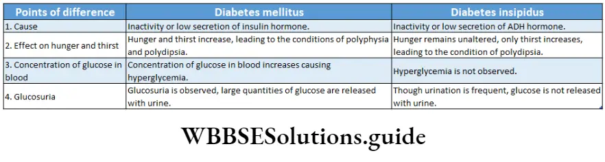
Role Of Other Organs In Ecreation
Besides the kidneys, there are certain other organs that remove waste products from the body. These include liver, skin, lungs, etc.
Role Of Skin
Skin is the largest organ in the human body. It is an accessory excretory organ. The skin contains sweat glands and sebaceous glands. The sweat glands secrete sweat while the sebaceous glands secrete sebum.
The different excretory functions of the skin have been discussed below.
Sweating:
There are numerous sweat glands present in the skin. They are microscopic, tubular structures that remain coiled within the dermis.
They are surrounded by a network of blood capillaries. They open up on the skin through minute pores.
These glands absorb several metabolic wastes such as urea, salts, and amino acids along with excess water from the blood and excrete them through the sweat.
The sweat glands remain very close to the blood capillaries. Thus, the metabolic wastes can diffuse out of the blood and enter the sweat glands. From here, these are excreted through the sweat.
In addition to excreting metabolic wastes, sweat also provides a cooling effect to the body.
About 1-1.5 L of sweat is excreted per day.
Types of sweating: Sweating is of two types.
Sensible sweating or perspiration: Sensible sweating or perspiration is a form of sweating or perspiration that can be sensed by the body. This perspiration absorbs the latent heat of the body, providing a cooling effect.
Insensible sweating or perspiration: Insensible sweating or perspiration is a kind of sweating or perspiration that occurs before the body can sense it. This can happen through the skin, mucous membranes of the lungs, and nasal cavity, in the form of vapor only. It is an obligatory process and has no connection with the sweat glands.
Other roles: A small amount of carbon dioxide gets excreted through the skin. It is about 1/100th of that excreted through the lungs. The skin of the external auditory meatus, in the external ear, contains ceruminous glands. They secrete a wax-like substance called cerumen. It contains several waste products.
Sebum, secreted by sebaceous glands, is an oily fluid. Different types of, sterols, fatty acids, etc., are excreted through the sebum.
Role Of Lungs
The lungs act as an accessory excretory organ besides being the principal respiratory organ.
The functions of; the lungs as excretory organs are as follows—
- About 18 L of C02 is produced every day by respiration. This is eliminated from our body through the air expired by the lungs.
- About 400 ml of water vapor is released from the body to the environment per day, through the expired air.
- Small amounts of acetone, ammonia, alcohol, etc., get excreted from the body by the lungs.
Role Of Liver
The liver is responsible for detoxifying and breaking down any toxin that enters our bodies.
Ammonia gas is generated within the body by deamination reactions. It is harmful to the body. The liver converts ammonia into urea, by the ornithine cycle. The liver filters the blood and recycles older and damaged RBCs.
It removes the following substances through the bile—heavy metals like copper, zinc, etc., certain drugs, bacteria, toxins from the blood (like alcohol), bile pigments and cholesterol, lecithin, etc.
Role Of Salivary Glands
The salivary gland produces saliva, which contains certain waste products, for example, inorganic salts, organic substances like urea), some drugs, and heavy metals such as lead, mercury, etc People who consume alcohol, excrete it through saliva. Large amounts of thiocyanates are present in the saliva of smokers. Even the microbes causing several diseases get released through the saliva.
Role Of Large Intestine
The feces, which are formed in the large intestine and eliminated from the body through the anus, contain heavy metals (e.g., bismuth, mercury, etc.), bile pigments, drugs, and many more waste products. Therefore, the large intestine also serves as an accessory excretory organ.
Disorders Related To Excretory System
Some of the different disorders related to the excretory System-
Uremia Definition: The disorder caused due to profound renal malfunction, that leads to the accumulation of urea and other nitrogenous wastes in the blood instead of being excreted with the urine is called uremia.
Excretory System Causes:
- It develops if urea concentrates in the blood instead of getting released through the urine due to renal malfunction.
- It develops when the byproducts of protein metabolism other than urea, such as creatinine, amino acids, etc., accumulate in the blood.
Excretory System Symptoms:
- Anorexia, Symptoms of vomiting, and uremia include mental deterioration, confusion, muscle twitching, convulsions, and coma.
- The blood urea nitrogen (BUN) and creatinine levels are high. The blood levels of these substances are used as an index of the severity of uremia.
- It includes the accumulation of urea and creatinine along with that of other toxic substances (organic acids or phenols)
Excretory System Treatment:
- Patients or the ones vulnerable to uremia are often haemodialysed by using a dialysis machine. This procedure has been described later in detail.
- Another method to treat uremia is haemofiltration. It involves filtering the blood under high pressure.
Renal failure
Renal failure Definition: The malfunctioning of the kidneys either acute or chronic, caused due to low glomerular filtration rate (GFR) resulting in the accumulation of nitrogenous substances in the blood, is called renal failure.
Renal failure Causes: Renal Failure may occur due to
- Bacterial infection
- Injuries
- Hemorrhage in the kidneys
- A disease called glomerulonephritis (inflammation of glomeruli),
- less electrolyte concentration in the body fluids.
Renal failure Types: It is generally of the two types—
Acute renal failure (ARF) —in this case, the kidneys lose their ability to excrete wastes and to regulate the homeostasis of blood volume, pH, and electrolytes. It deteriorates over a relatively short period of time (hours to days).
Chronic renal failure—it is the slow deterioration of the renal functions, that extend for a much longer period and finally lead to symptoms of uremia.
Symptoms: It results in failure to excrete different waste products from the body. Its symptoms include uremia, accumulation of wastes in the blood, disruption of water-salt balance in the body, and failure to secrete erythropoietin.
Renal failure Treatment:
- Avoiding medications that may cause renal failure as a side-effect.
- Haemodialysis may also be prescribed.
Nephritis
Nephritis Definition: Inflammation of the kidneys is called nephritis.
Nephritis Causes: It can be caused by infection, but is most commonly caused by autoimmune disorders that affect the major organiser
Nephritis Types: It may involve inflammation of the glomeruli, renal tubules, or interstitial tissue surrounding the glomeruli and tubules.
It is generally of two types—
- Glomerulonephritis and
- Pyelonephritis.
Nephritis Symptoms:
Symptoms of glomerulonephritis include proteinuria and haematuria.
Nephritis causes additional problems like water retention, as the kidneys cannot function properly to excrete water. Water retention or edema, can further cause swelling of the feet, ankles, legs, and hands.
In the case of pyelonephritis, the concentration of the urine decreases, leading to painful urination and severe body ache.
Nephritis Treatment: Primarily, nephritis tends to be treated with antibiotics. Occasionally it is treated with steroids, particularly in those cases when it is caused by lupus (an immune system disease). However personal hygiene is important as microbial contamination is one of the major causes of this disease.
Differences between ammonia, urea, and uric acid excretion
Renal Calculi
Renal Calculi Definition: The solid aggregations of the minerals in the urine, that do not get excreted, are called kidney stones or renal calculi.
Renal stone calculus or lithiasis is one of the most common diseases of the urinary tract. It occurs more frequently in men than in women. It is rare in children.
It shows a familial predisposition. Urinary calculus is a stone-like body, composed of urinary salts bound together by a colloid matrix of organic materials.
Renal Calculi Causes: The causes of renal calculi are—
Renal Calculi Dehydration: Due to dehydration, water reabsorption increases but reabsorption of the ions does not take place. This leads to their accumulation as calculi.
Changes in pH of the urine: If the buffering system in the kidneys gets affected, the urine that is released becomes alkaline. Substances like phosphates, etc., that form calculi, are released with such urine.
Microbial contamination: When crystalline substances form layers over pus, tissue fragments, etc., produced by microbial contamination, kidney stones are formed.
Metabolic conditions: Hyperparathyroidism, gout, etc., are also other causes of the formation of kidney stones.
Accumulation of calcium, uric acid, and oxalates leads to the formation of kidney stones.
Renal Calculi Types: Basically the renal stones can be divided into two major groups—
Primary stones: They appear in apparently healthy urinary tract without any preceding inflammation.
Secondary stones: They are usually formed as a result of inflammation.
Renal Calculi Symptoms: The symptoms include pain in the pelvic area and lower abdomen. This condition is called renal colic. It is associated with haematuria and painful urination. If the kidney stones descend and block the urethra, it may prevent the passing of urine through it. Retention of urine inside the bladder may result in urinary tract infection.
Renal Calculi Treatment: The kidney stones may be removed by surgery. Nowadays, laparoscopy is used to remove kidney stones.
In this process, the stones are collected from the kidney or the ureter through a small incision made in the respective organ. It is also called ‘keyhole surgery’.
The use of medicines like allopurinol (decrease uric acid production), diuretics, etc., may prevent the formation of stones in the kidney.
Another measure for treatment of the kidney stones is lithotripsy. It involves mechanical destruction of the kidney stones. Fragments of the stones are excreted through the urine.
Some other renal disorders body towards the dialyzer
Glomerulonephritis: This involves inflammation of the glomerulus.
Pyelonephritis: This involves inflammation of the renal pelvis due to infection in the ureters.
Some Important Procedures For Treatment Of Renal Disorders
There are some treatment procedures that are important for treating the above disorders. Some of them are discussed below.
Dialysis And Artificial Kidney
Dialysis and artificial kidney Definition: The clinical procedure by which the blood is purified artificially is called dialysis.
Dialysis uses the principle of diffusion of molecules, from high to low concentration.
In this process the diffusion occurs through a natural or synthetic semipermeable membrane, that allows only small molecules to pass through. This membrane is called a dialysis membrane. Dialysis is generally of two types.
Haemodialysis
The term dialysis refers to the separation of molecules on the basis of their ability to diffuse through an artificially selectively permeable membrane.
The principle of ultrafiltration is used in the “artificial kidney machine”, which is generally used for hemodialysis. This machine is also called the dialysis machine.
Haemodialysis Process:
In a dialysis machine, the patient’s blood with heparin at 0° C flows slowly through many small channels made of semipermeable membranes, through which only small molecules can diffuse.
A dialyzing solution or dialysate flows on the other side of these membranes.
The composition of this dialysis solution is crucial. It contains water, glucose, and electrolytes like sodium, potassium, calcium, magnesium, and chloride. This composition varies with the patient’s needs.
The total osmotic potential of the dialysis fluid must be equal to that of the plasma.
Molecules such as urea, uric acid, creatinine, etc., and ions that need to be removed from the blood are absent in the dialysis fluid.
These molecules and ions diffuse from the area of higher concentration (patient’s blood) to the area of lower concentration (dialyzing fluid). Thus, the blood in the patient’s body is purified by the removal of harmful products.
About 500 ml of the patient’s blood is in the dialysis machine at any one time, and the unit processes several hundred milliliters of blood per minute.
A patient with severe renal failure must be on the dialysis machine for 4 to 6 hours, three times a week.
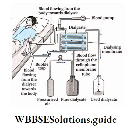
Importance of hemodialysis: Loss of kidney function or renal failure, results in uremia.
A person who suffers complete renal failure may die within 2 weeks, if not treated.
A drastic but highly successful treatment for such patients is a kidney transplant, but it needs a donor with proper compatibility with the patient. Therefore, artificial kidneys may be used as an immediate option.
Peritoneal dialysis
When the peritoneum inside the person’s body is used as the dialysing membrane, it is known as peritoneal dialysis.
Kidney Transplantation
Hemodialysis is not the permanent solution to complete renal failure. The damaged kidneys need to be transplanted by healthy kidneys from donors. This procedure is called kidney transplantation.
Excretory Products And Their Elimination Notes
- Catabolism: A series of metabolic reactions which break down complex molecules into simpler units.
- Chemical messengers: Metabolites that transport messages from one part of the body to the other, e.g., hormones, neurotransmitters, etc.
- Endocytosis: This is a form of active transport in which a cell engulfs foreign particles.
- Hemorrhage: Escape of blood from a ruptured blood vessel.
- Homeostasis: The tendency towards a relatively stable equilibrium between interdependent elements, especially as maintained by physiological processes.
- Peritoneum: The membrane that forms the lining of the abdominal cavity or coelom.
Points To Remember
- The organ system, that is responsible for the release of waste products from the body, is called the excretory system.
- Wastes generated from the body may be nitrogenous (urea, ammonia, uric acid, etc.) and non-nitrogenous (C02, oxalic acid, etc.).
- Organisms can be ammonotelic, ureotelic or uricotelic. Human beings are ureotelic.
- Protonephridia, metanephridia, and malpighian tubules are excretory organs found in different invertebrate organisms.
- The human excretory system consists of a pair of kidneys, a pair of ureters, a urinary bladder, and a urethra.
- According to the structure, the kidney can be pronephric (primitive structure), mesonephric (slightly modified), or metanephric (modified and differentiated).
- The structural and functional unit of the kidney is the nephron. It is where the urine is synthesised. 8- The different steps of urine formation, i.e., ultrafiltration, selective reabsorption, and tubular secretion, take place within the different parts of the nephron.
- The urine is a slightly yellowish, acidic fluid, that has a characteristic pungent odor and is involved in removing the nitrogenous and non-nitrogenous wastes from the body.
- The concentration of different constituents of urine varies according to different conditions of the body.
- The countercurrent mechanism regulates the proper concentration of water, several ions in blood and urine.
- The process of release of urine from the body is called micturition.
- Urine formation is also responsible for maintaining osmotic concentration of the blood and other body fluids. This process is called osmoregulation.
- A disease, named Bartter syndrome, is developed due to the absence of a channel protein called Barttin, present within the thick ascending loop of Henle. As a result, reabsorption of ions does not take place, which severely impairs water reabsorption in collecting ducts. Thus, liquid content of the body decreases.
- The functioning of the kidney is regulated by the juxtaglomerular apparatus (JGA), atrial natriuretic peptide (ANP), and antidiuretic hormone (ADH).
- About 2/3rd to 7/8th fraction of glomerular filtrate gets reabsorbed in the PCT, by passive absorption. This process is called obligatory water reabsorption Some amount of water reabsorption takes place in the DCT, due to the effect of ADH. This process takes place by active absorption. This process is called facultative water reabsorption.
- Hyposecretion of ADH causes diabetes insipidus.
- The accessory excretory organs include skin, lungs, liver, salivary glands, et
- Some of the disorders related to excretion include Uremia, nephritis, renal failure, etc.
- Renal calculi or kidney stones is another disorder related to the excretory system.
- Dialysis and kidney transplantation are important procedures involved in the treatment of these diseases.
- In the case of diseases like multiple myeloma, leukemia, lymphosarcoma, etc., a different globulin-type protein is found in the urine. These proteins are known as Bence-Jones proteins.
- The amount of any substance that is released through the urine per minute, is also present in the plasma. The amount of plasma in which this amount of substance is present is known as plasma clearance.