Living World Question And Answers

Class 11 Biology WBCHSE The Living World Some Important Questions And Answers

Question 1. Differentiate between catabolism and anabolism.
Answer: Catabolism involves breaking down larger molecules into smaller ones, thereby releasing energy, while anabolism involves the formation of larger molecules, using smaller molecules, thereby utilizing energy. Respiration is a catabolic process, while photosynthesis is an anabolic process.

The Living World

Question 2. What is intussusception?
Answer: Overall growth of a body due to an increase in the dry weight of the protoplasm, is called intussusception.

Question 3. Which are the coordinators within the body of plants and animals?
Answer: In case of animals, hormones act as chemical coordinators, while nerves act as physical coordinators. In case of plants, only hormones are present, which act as coordinators. Due to their presence, different cells, tissues, organs and organ systems function in a coordinated fashion.

Read and Learn More WBCHSE Solutions For Class 11 Biology

Question 4. What is homeostasis?
Answer: Homeostasis is the ability of maintaining a stable internal environment of the body irrespective of the changes in the external environment. For example, normal body temperature is 98.6°F. When the body temperature rises above this, hypothalamus sends signals all over the body, through the motor neurons. As a result, sweat is released which lowers the temperature to normal.

Question 5. What is taxonomy?
Answer: The branch of biology, that deals with the classification of organisms and provide required information for classification such as nomenclature; rules of nomenclature, identification, is known as taxonomy.

Question 6. What is systematics?
Answer: The branch of biology, that reveals relationship j among different organisms with the help of identification, nomenclature, description, and classification is known as systematics.

Class 11 Biology WBCHSE

Question 7. What is the difference between taxonomy and systematics?
Answer: The science which deals with identification, classification and nomenclature of organisms is known as taxonomy. On the other hand systematics is the science, that studies diversification of organisms — both past and present and the relationship among them, over the course of time.

Question 8. What is binomial nomenclature?
Answer: The system of naming, that is done by using only genus and species name is known as binomial | nomenclature. For example, Pisum sativum.

Question 9. What are taxonomic category and taxonomic hierarchy?
Answer: The different levels of classification are known as categories or taxonomic categories.
When these categories of classification are arranged in a definite order, then it is known as hierarchy or taxonomic hierarchy.

Question 10. What are monotypic and polytypic genus?
Answer: The genus which includes only one species is known as monotypic genus. For example, sapiens is the only species present in the genus Homo. Hence, Homo is a monotypic genus. The genus which includes more than one species is known as polytypic genus. For example, tigris and leo, both these species come under the genus Panthera. Hence, Panthera is a polytypic genus.

Class 11 Biology WBCHSE The Living World Multiple Choice Question and Answers

Question 1.  Joint Forest Management Concept was introduced in India during —

  1. 1970s
  2. 1980s
  3. 1990s
  4. 1960s

Answer: 2. 1980s

Question  2. Which is the National Aquatic animal of India?

  1. River Dolphin
  2. Blue whale
  3. Sea-horse
  4. Gangetic shark

Answer: 1. River Dolphin

Question 3. Red List contains data or information on— 

  1. All economically important plants
  2. Plants whose products are in international trade
  3. Threatened species
  4. Marine vertebrates only

Answer: 3. Threatened species

Question 4. Nomenclature is governed by certain universal rules. Which one of the following is contrary to the rules of nomenclature?

  1. The first word in a biological name represents the genus name and the second is a specific epithet
  2. The names are written in Latin and are italicised
  3. When written by hand, the names are to be underlined
  4. Biological names can be written in any language

Answer: 4. Biological names can be written in any language

Question 5. Following are the two statements regarding the origin of life —

The earliest organisms that appeared on the earth were non-green and presumably anaerobes

The first autotrophic organisms were the chemoautotrophs that never released oxygen Of the above statements which one of the following options is correct?

  1. 2 is correct but i is false
  2. Both 1 & 2 are correct
  3. Both 1 & 2 are false
  4. 1 is correct but 2 is false

Answer: 2. Both 1 & 2 are correct

Question 6. Taxonomic categories showing correct hierarchical arrangement in ascending order is—

  1. Kingdom→ Order → Division→ Class → Genus → Species
  2. Species→ Genus→ Division → Class → Order → Kingdom
  3. Kingdom → Division → Class → Order → Family → Genus → Species
  4. Species→ Genus → Family → Order → Class → Division → Kingdom

Answer: 4. Species→ Genus → Family → Order → Class → Division → Kingdom

Question 7. Which one of the following shows the hierarchical arrangement of taxonomic categories of plants in descending order?

The Living World Taxonomic Categories Of Plants In Descending Order

Answer:  4. Kingdom→Division→Class→Order→Family→Genus→Species

Question 8. A taxonomic group of any rank is –

  1. Taxon
  2. Tribe
  3. Race
  4. Variety

Answer: 1. Taxon

Question 9. Which of the following is correctly sequenced?

  1. Phylum, class, order, family
  2. Phylum, order, class, genus
  3. Phylum, class, family, order
  4. Phylum, order, family, class

Answer: 1. Phylum, class, order, family

Question 10. Family is placed between—

  1. Genus and species
  2. Order and class
  3. Class and genus
  4. Order and genus

Answer: 4. Order and genus

Question 11.  ICBN stands for

  1. Indian Congress of Biological Names
  2. International Code of Botanical Nomenclature
  3. International Congress of Biological Names
  4. Indian Code of Botanical Nomenclature

Answer: 2. International Code of Botanical Nomenclature

Class 11 Biology WBCHSE The Living World Very Short Question And Answers

Question 1. Name the bird whose beak structure and feeding habit were studied by Darwin, while studying evolution.
Answer:  Finch

Question 2. Name the book written by Charles Darwin.
Answer: Origin of Species

Question 3. What is taxon?
Answer: According to international rule of classification, each unit of classification is known as taxon.

Question 4. In which journal did Linnaeus publish his modified classification system?
Answer: Systema Naturae.

Question 5. What is hierarchy?
Answer: The arrangement of taxonomic categories one above the other in a definite logical way is known as hierarchy.

Question 6. Expand the abbreviation ICNCP?
Answer: ICNCP stands for International Code of Nomenclature for cultivated Plants.

Question 7. What is genus?
Answer: The genus is a unit of classification which is formed by collection of species with similar | characteristics.

Question 8. What is meant by trinomial nomenclature?
Answer: The nomenclature that includes genus name, species name, and subspecies name is called: trinomial nomenclature.

Question 9. What is species?
Answer: Species is a group of living organisms hearing similar characteristic features and they are. capable of interbreeding.

Question 10. What is subspecies?
Answer: There are several differences among organisms belonging to same species due to differences in geographical location. These species are thus divided into smaller groups known as subspecies.

Question 11. What do you mean by polytypic genus?
Answer: The genus which contains more than one species is known as a polytypic genus. Example- Plasmodium is a polytypic genus. It has GO j species, some of which are, Plasmodium vivax, P. falciperum, P. malariae, etc.

Question 12. What is meant by couplets in identification key?
Answer:  The pair of opposite characteristic features written in taxonomic key is known as couplet.

Question 13. What is identification key?
Answer: The identification key is a set of alternate characters arranged in such a fashion that by selection and elimination of them one can quickly identify the organism. It is an important tool for studying taxonomy It may be printed or computerised.

Class 11 Biology WBCHSE

Question 14. What is lead?
Answer: The different characteristics which are written as a couplet are known as lead.

Question 15. Name the popular key of taxonomy.
Answer: Indented key is the popular key of taxonomy.

Question 16. Given below is the scientific name of mango.

Identify the correctly written name.

  1. Mangifera Indica
  2. Mangifera indica

Answer: 2. Mangifera indica

Question 17. Linnaeus is considered as father of taxonomy. Name two other botanists known for their contribution to the field of taxonomy.
Answer: George Bentham and Sir J.D. Hooker.

Question 18. What does ICZN stand for?
Answer:  ICZN stands for International Code of Zoological Nomenclature.

Question 19. What is a museum?
Answer: A museum is an institution that preserves objects of scientific, artistic, cultural or historical importance.

Question 20. Can you identify the correct sequence of Itaxonomical categories?

  1. Species —> Order —> Phylum —> Kingdom
  2. Genus —> Species —> Order —> Kingdom
  3. Species→ Genus→ Order → Phylum

Answer: 3. Species→ Genus→ Order → Phylum

Question 21. Taxonomic key is one of the taxonomic tools in the identification and classification of plants and | animals. It is used in the preparation of—

  1. Monographs
  2. Flora
  3. Both 1 and 2
  4. None of these

Answer: 3. Both 1 and 2

Question 22. The term systematics refers to—

  1. Identification and classification of plants and j animals
  2. Nomenclature and identification of plants and ; animals
  3. Diversity of kinds of organisms and their ; relationships
  4. Different kinds of organism and their classification

Answer: Both 1 And 2

Question 23. Which is the largest botanical garden in the world? Name a few well known botanical gardens in India.

Ans. Royal Botanical garden, ICew (London). Some well known botanical gardens in India are

  1. Acharya Jagadish Chandra Bose Indian Botanic Garden, Shibpur, Howrah;
  2. Lloyd Botanical Garden, Lucknow.

Question 24. Genus represents—

  1. An individual plant or animal
  2. A collection of plants or animals
  3. A group of closely related species of plants or animals
  4. None of these

Answer: 3. A group of closely related species of plants or animals

Class 11 Biology WBCHSE

Question 25. How is key helpful in identification of living organisms?
Answer: Key helps to identify various taxa such as genus, species, etc. It is a systemic representation of different identifying characters belonging to different taxa.

Question 26. How can you relate metabolism with growth?
Answer: Metabolism involves both catabolism and anabolism. Growth occurs when the rate of anabolism is greater than the rate of catabolism.

Question 27. How can you distinguish between human beings and other living organisms?
Answer: Human beings have ability to think about higher concepts that other animals cannot. This feature distinguishes human beings from other animals.

Question 28. Amoeba multiplies by mitotic cell division. Is this phenomenon growth or reproduction? Explain.
Answer: In unicellular organisms like Amoeba, cell division is the means of multiplication. So, it helps in reproduction. But it also helps in the growth of their colony. Thus, in unicellular organisms, mitosis helps in both growth and reproduction.

Biological Classification Important Notes

Biological Classification Introduction

  • Every day we come across different kinds of organisms. We see different kinds of plants in the fields, gardens, etc. But why are the plants all so different from each other? Or why are the animals we see around us so different from each other? This is because different organisms have different characteristic features.Φ
  • Depending on these characteristics, they have been classified under different classes or groups. This is known as biological classification. In this chapter, we shall learn about the different types of classification of the organisms, that have been followed since ancient times to modern days.

Biological classification Definition: Biological classification is the scientific classification of living organisms based on their morphology, habitat, evolution similarities and differences with other organisms.

Read and Learn More: WBCHSE Notes for Class 11 Biology

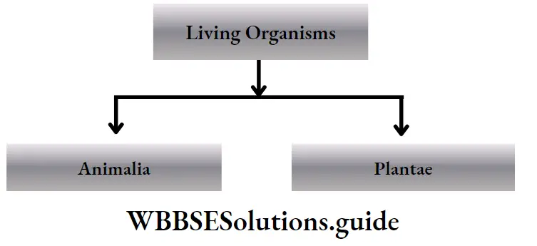
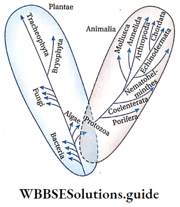
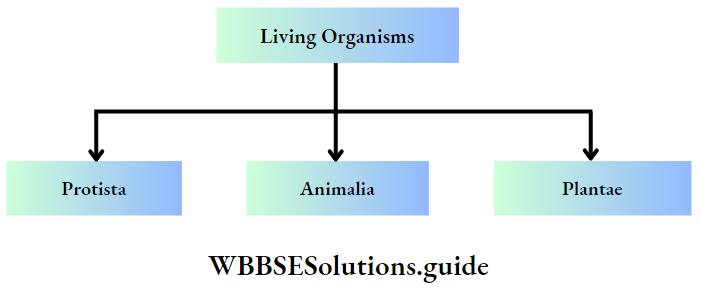
  • Classification of organisms has been carried out since the dawn of human civilisation. Aristotle (384-322 BC) had classified organisms into two large groups—plant kingdom and animal kingdom.
  • But this system failed to classify several organisms, such as unicellular green algae, fungi, etc. These organisms could not be placed in any of the above-mentioned two groups.
  • It even failed to distinguish between prokaryotes and eukaryotes. Later on, several other modified classification systems have been developed.

Importance of biological classification:

Biological classification is important in following ways—

  • Identification of organisms: Using a proper classification system, organisms can be identified easily.
  • Attaining knowledge about the living world: A classification system helps to categorise organisms on the basis of their morphological features, habitat, etc.
  • Correlating different groups of organisms: Classification helps us to correlate different groups of organisms, through their similarities and dissimilarities.
  • Discovery of new species: The system of classification makes it easier to classify newly discovered species. Since the dawn of civilisation, there have been many attempts to classify living organisms. But it was done without using scientific criteria.

Cyanobacteria or Blue-Green algae (BGA)

Cyanobacteria Definition: The gram negative, unicellular prokaryotes, containing photosynthetic pigments that can carry out photosynthesis, are called cyanobacteria or cyanophyta.

  • Cyanobacteria contain photosynthetic pigments like chlorophyll and various accessory pigments. They are also called oxygenic photoautotrophs.
  • Cyanobacteria are considered to be first organisms on earth that released oxygen. With evolution, several aerobic organisms developed from the cyanobacteria.
  • They were known as | blue-green algae, myxophyceae or cyanophyceae. In 1978, ICNB (International Code of Nomenclature of | Bacteria) named them as cyanobacteria.

Distribution: Cyanobacteria are usually free-living marshy regions. They are also found in sea, under the ice, desert region, lakes, etc.

General Features

General features Structure:

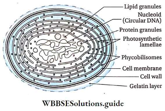
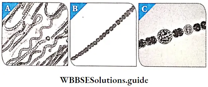
  1. Shape and size: Cyanobacteria are unicellular,  They may exist as unit (Spirulina sp.), colony (Nostoc sp.) or filamentous (Oscillatoria sp.). The cells may be large, spherical or oval shaped, with size ranging from l//m to 50 pm.
  2. Protoplasm: The protoplasm is generally divided into two parts. The pigment-containing part, called chromoplasm or chromatoplasm, is present towards the periphery. While the inner region contains a colourless part called centroplasm. Thylakoids are present in the chromoplasm.
  3. Cell wall: They possess a cell wall with an inner thick peptidoglycan layer, similar to gram positive eubacteria. The outer membrane contains proteins, lipids and carotenoids. There is a mucilage layer outside the cells.
  4. Cell membrane: The lipoprotein-containing cell membrane is present just below the cell wall.
  5. Lamellasome: Similar to mesosome in bacteria, cyanobacteria have a circular extracellular appendage, that helps in respiration. These are  known as lamellasomes.
  6. Thylakoids: All cyanobacteria contain photosynthetic pigments like chlorophyll a. They also contain blue phycobilin pigments, phycocyanin and allophycocyanin. Many members also contain another pigment, phycoerythrin, making the cells appear red, or sometimes black.
  7. These phycobilins are present in some special structures, on the thylakoid membranes. These structures are known as phycobilisomes. These are highly efficient channels that transfer captured solar energy (excitation energy) to the reaction centres of photosystems.
  8. Nucleoid: Cyanobacteria lack true nuclei and other organelles like mitochondria and chloroplastids. The naked, circular DNA (DNA without histone proteins) is arranged closely within the nucleoid. Plasmids may also be present.
  9. Ribosomes: 70S ribosomes may exist and form polyribosomes.
  10. Gas vacuoles: Cyanobacterial cells contain numerous gas vacuoles.Gas vacuoles provide buoyancy to the cells, allowing the cyanobacteria to float on the surface. This allows more exposure to sunlight for photosynthesis.

Biological Classification Cyanobacterial Cell

Stored materials: Cyanobacteria also contain various types of reserve food. They include—

  1. α-granules  which are cyanophycean starch and look similar to glycogen (polyglucose) granules, which store carbon
  2. β- granules(lipid granules),
  3. Protein granules, polyphosphate or volutin granules, etc., which allow cells to accumulate energy and nutrients.

Locomotion: Cyanobacteria move by gliding along the surfaces of the substratum.

Reproduction: It reproduces by vegetative and asexual modes of reproduction. Sexual reproduction is totally absent.

  1. Vegetative reproduction: It occurs by the following : methods—
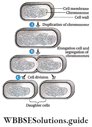
    1. Fission: Unicellular cyanobacterial cells usually divide and reproduce by fission. The cell divides into two daughter cells,
    2. Fragmentation: The filamentous members reproduce vegetatively through fragmentation. Each fragmented piece of  the filament, germinates into a new colony.
    3. Hormogonia: At the end of the growing season, filaments or trichomes break into multicellular pieces. These pieces secrete thick wall around the mass of cells, forming structures called hormogonia: These hormogonia can germinate into new filaments at the onset of favourable conditions.
  2. Asexual reproduction: Many non-motile cyanobacteria reproduce asexually by spore formation. The different types of spores formed are—
    1. Endospore: These are formed within the cell, Here the cells increase in size and their protoplasm divides to form endospores, e.g., Dermocarpa.
    2. Exospore: These are produced outside the cell.
    3. By the dissolution of the apical region of the cell surface, the protoplast gets exposed. From this exposed region of the protoplasm, round spores are formed in basipetal succession. E.g., Chaemosiphon.
    4. Akinetes: These are formed close to the heterocysts. The akinete mother cells increase in size and then develop a wall around it. Under favourable conditions, they give rise to new filaments. E.g., Cylindrospermum.
    5. Heterocyst: Some filamentous cyanobacteria form special types of thick walled cells, known as the heterocysts. The outer layer of the cell wall is made up of pectin or cellulose and the inner layer is of cellulose. They are usually yellow in colour due to the presence of carotene. They have rudimentary reproductive structures, as they can germinate into new filaments, under favourable conditions. E.g., Anobaena sp., Nostoc sp., Spirulina sp., Scytonema sp.

Biological Classification Different Types Of Cyanobacteria Nostoc Anabaena Heterocyst of Anabaena

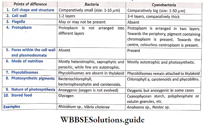
Biological Classification Differences Between Bacteria And CyanobacteriaMycoplasma

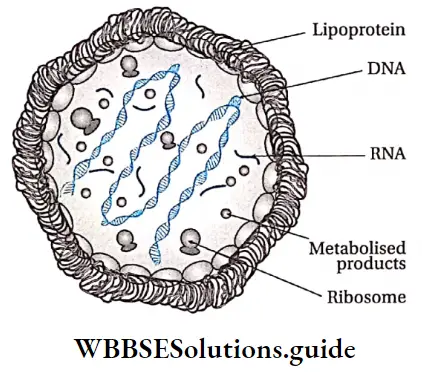
Mycoplasma Definition: Mycoplasmas are simple, small, gram-negative, prokaryotic organisms that lack a cell wall and can survive without oxygen.

Nocard and Roux discovered mycoplasma in 1898. Mycoplasma is also known as PPLO (Pleuro Pneumonia Like Organism) or MLO (Mycoplasma Like Organism). They are the smallest living cells known. Many mycoplasmas are pathogenic to both animals and plants.

Example: Mycoplasma gallisepticum, Mycoplasma genitalium, Mycoplasma pneumoniae, etc.

Biological Classification Mycoplasma Cell

General Features

Cell type and structure: They are tiny, prokaryotic microorganisms (0.1-0.15/ym). The shape of mycoplasma varies from being spherical to filamentous with no cell walls. Due to absence of cell wall, they can change their structure.

This phenomenon is known as polymorphism. They contain acetylglucosamine in the cell membrane. Mesosome is absent. Ribosomes are present. Their genome consists of a double-stranded linear but coiled DNA molecule.

  • Nutrition: They can be parasitic or saprophytic.
  • Respiration: They are obligate anaerobes, usually without any electron transport system (ETS). Even if present, ETS is inactive. ‘
  • Reproduction: Mycoplasma reproduce by binary fission. Sometimes, only the nucleoid shows binary fission, without cytokinesis.
  • Effect on animal world: Several species are pathogenic in humans. These include M. pneumoniae, which causes pneumonia and other respiratory disorders. Another is M. genitalium, which causes pelvic inflammatory diseases.
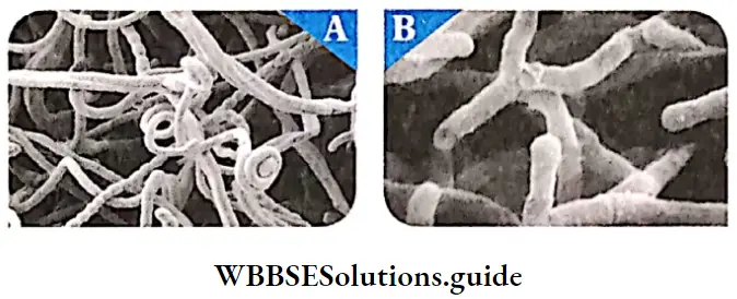
  • Distribution: Actinomycetes are primarily soil dwellers. But they are also widely distributed in a diverse range of aquatic ecosystem, including sediments | obtained from seabed.

General Features

General Features Structure: Actinomycetes are gram positive, prokaryotic microorganisms that have branched hyphae. The hyphae are usually aseptate but septa may be | formed under special conditions in some forms. The hyphae form mycelium like structure. The cell wall is made up of peptidoglycan, teichoic acid, teichuronic acid and polysaccharides. The chemical composition of their cell wall is similar to that of gram-positive bacteria.

The rigid cell wall maintains the cell shape. There are tiny chromatin granules present within the cell.

  • Nutrition: They are heterotrophic in nature. Some are saprophytic aerobes growing in soils and natural habitats.
  • Respiration: They are facultatively anaerobic.
  • Temperature sensitivity: They are generally mesophilic in nature. They are active at a temperature of 35°C.
  • Effects on animal world: Some of the actinomycetes species are used to prepare antibiotics. Some actinomycetes also cause disease (Actinomycosis) in humans.

Biological Classification Different actinomycetes Slreptomyces Actinomyces

Spirochaetes

Spirochaetes Definition: The heterotrophic prokaryotes, that are j present in water and muddy regions are called j spirochaetes.

General Features

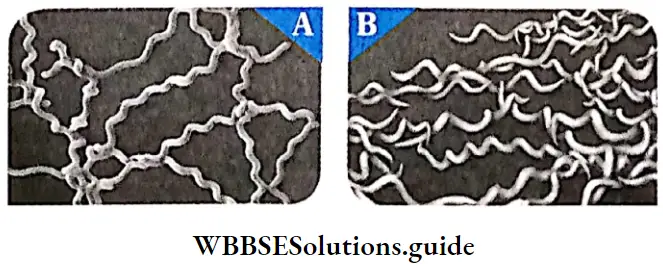
General Features Structure: Spirochaetes have long, helically coiled cells. The cells usually have diameter around 0.1 – 0.6pm and have cell wall. They are gram-negative bacteria. They are motile organisms. Spirochaetes are chemoheterotrophic in nature. They are sensitive to desiccation.

Effects on animal world: The majority of spirochaetes cause disease in man.

Examples: Leptospira sp., Borrelia sp., Treponema pallidum, etc.

Biological Classification Different Types Of Spirochaetes Leptosira Borelia

Rickettsiae

Rickettsiae Definition: Rickettsiae intracellular, gram negative coccobacillary forms that ‘ multiply within eukaryotic cells.

Rickettsiae General Features

Rickettsiae Cellular structure: Cells are 0.3 – 0.5 x 0.8 – 2.0pm in size. The cell wall is chemically similar to that of gram negative bacteria. It contains diaminopimelic acid and lacks teichoic acid.

  • Their outer membrane is composed mostly of lipopolysaccharides.
  • They lack a distinct nucleus and membrane bound organelles. Flagella, pili, mucilage capsule are absent.
  • They contain both RNA and DNA as nucleic acids. Mesosomes and ribosomes are also present in the cells.
  • Nutrition: They are parasitic in nature (obtain nutrition from host cell).
  • Growth and Reproduction: Rickettsia normally multiplies by transverse binary fission.
  • Effects on animal world: In their arthropod vectors (insects that can carry the organism), the rickettsia multiply within the epithelium of the intestinal tract. They are transmitted to man, via the arthropod saliva, through a bite. In their mammalian host (i.e., man), they are found mainly in the endothelium of the small blood vessels, particularly in those of the brain, skin and heart.
  • Rocky Mountain spotted fever is transmitted by the bite of a Dermacentor tick, which carries the rickettsia. This disease is common in warm-blooded animals, including humans.

Rickettsiae Example: Rickettsia typhi.

Kingdom Protista

  • This is the kingdom comprising unicellular, eukaryotic organisms that have features similar to those of fungi, plants or animals. The concept of kingdom Protista was given by taxonomist E. Haeckel in 1866.
  • All single-celled eukaryotes, other than green algae and red algae, have been placed under Protista. This kingdom forms a link between Kingdom Monera and the Kingdoms Fungi, Plantae and Animalia.
  • Distribution: Most of the members of this kingdom live in water or aquatic habitats. Majority of them are found as planktons within marine water, fresh water, etc.

Kingdom Protista General Features

Kingdom Protista General Features Cell type: The members of this kingdom are tiny, unicellular eukaryotes. Some of them may exist as colony or filaments.

  • Cell covering: Cell wall is usually absent; if present it may contain silica (e.g., diatoms). Cell membrane is covered by a pellicle, cuticle or shell. This forms a double-envelope system around the cells.
  • Protoplasm: The protoplasm contains a well defined nucleus and other membrane bound organelles. The cells contain 80S (60S + 40S) ribosome in the cytoplasm. On the other hand, 70S ribosome is present in mitochondria and plastids.
  • Chloroplast: The members may be photosynthetic or non-photosynthetic. Thylakoids, – containing photosynthetic pigments, are present within the j chloroplast of the photosynthetic members.
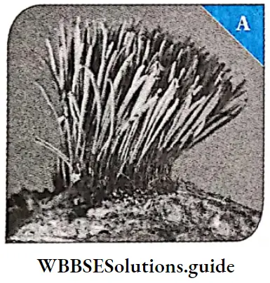
  • Locomotion: Most of them can show locomotion. The cells may contain cilia, flagella or pseudopodia. The cytoplasm, along with organelles, show a flowing j movement. This movement is called cytoplasmic j streaming or cyclosis.
  • Nutrition: Some are photoautotrophs, some heterotrophs, and some are mixotrophs (combining photosynthesis and heterotrophic nutrition). Some are holophytic, holozoic, parasitic, saphrophytic or symbiotic in nature. Some autotrophs are symbiotic in nature.
  • Respiration: Most protists are aerobic, but parasitic and ones living deep in the ocean, are anaerobic.
  • Reproduction: Protists reproduce asexually as well j as sexually.Asexual reproduction occurs by binary fission, j multiple fission, sporulation, cyst formation, etc. Sexual reproduction, on the other hand, occurs by syngamy and j conjugation. j
  • Cell division: Both mitosis and meiosis occur.
  • Life cycle patterns: The three basic life cycle patterns found in eukaryotes are represented by pr.otists. They are—Zygotic or haplontic, gametic or diplontic, sporic or haplodiplobiontic. At some point in the life cycle of many protists cysts (resistant cells) are formed.

Classification of Kingdom Protista

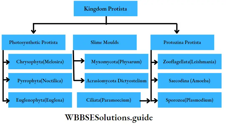
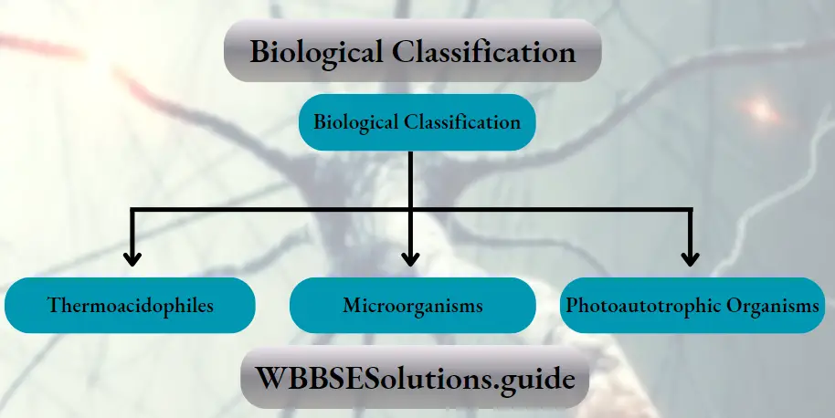
The members of Protista fall under the following three categories as shown in the chart below. These have been discussed under separate heads.

Biological Classification Kingdom Protista

Photosynthetic protista

These are unicellular, eukaryotic organisms, containing photosynthetic pigments. They are mainly found in aquatic ecosystems. They are mainly of three types—

Chrysophyta, Dinoflagellata and Euglenophyta.

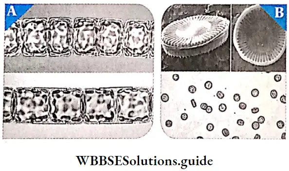
Chrysophyta: This group includes diatoms and golden algae. They are fresh water algae as well as marine dwellers. They are pollution indicators, i.e., they cannot grow in polluted regions, hence indicate the level of pollution.

Chrysophyta General Features

  1. Cellular structure: They are unicellular, microscopic and planktonic i.e., they float passively in water currents.The cells are enclosed within a shell made up of silica, called frustule.ln diatoms, the frustule divides to form two thin halves, which fit together as the lids in a soap-case.The cell wall is made up of silica along with cellulose, that makes it hard and porous. Chromatophores are present in the cells.
    • These contain pigments like chlorophyll, /2-carotene and a special type of xanthophyll (diatomin). Fucoxanthin, which is a characteristic pigment of brown algae, is present. Due to the presence of /3-carotene and diatomin, diatoms appear golden.
  2. Locomotion: Diatoms do not have flagella. Mucilage and oil globules help them to float on water.
  3. Symmetry: Both bilateral and radial symmetry is observed among the members.
  4. Nutrition: Most diatoms are photosynthetic (autotrophic) but some have evolved to become  heterotrophic.
  5. Reproduction: Diatoms reproduce by vegetative, asexual and sexual means. Vegetative reproduction takes place by cell division.Asexual reproduction occurs by binary fission. Sexual reproduction takes place by the formation of auxospores.

Chrysophyta Examples: Melosira sp., Cyclotella sp., Striatella sp., Hydrosira sp., etc.

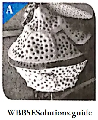
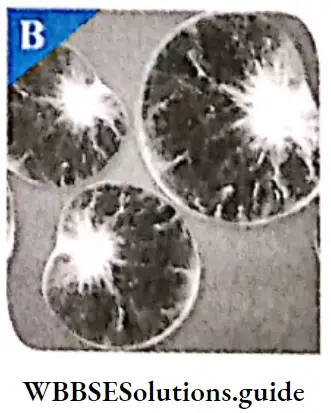
Biological Classification Photosynthetic Algal Protista Melosira Cyclotella

Dinoflagellata or Pyrrophyta: These are biflagellate, unicellular, marine algae.

Dinoflagellata General Features

Dinoflagellata Cellular structure: These organisms are mostly ; marine and photosynthetic. They appear yellow, green, brown, blue or red depending on the pigments present. Dinoflagellates may be covered by an outer covering outside their cells.

  • Depending on their; presence, they are of two types—unarmoured (without outer covering) and armoured (with outer covering).
  • Unarmoured dinoflagellates have a smooth pellicle or periplast. The cell wall has hard, cellulosic plates on the outer surface in case of armoured. These are known as the theca. The nucleus is large.
  • It has distinct nucleolus and nuclear envelope. Chromatin is present in chromosome form. Histone proteins are absent. Such a nucleus is known as mesokaryon.

Flagella: Most of them have two flagella. One lies longitudinally and the other transversely, in a furrow between the wall plates.

  • The flagella that is present in the longitudinal groove functions as a radar. The other flagella fits transversely in the furrow. It is used to create a rotational motion within the water.
  • Nutrition: Most of these organisms are autotrophic. They have chlorophyll a, chlorophyll c, fucoxanthin, etc., as photosynthetic pigments.
  • Stored food: Food is stored in the form of starch (polysaccharides) and oils (lipids).
  • Reproduction: Dinoflagellates show two types of reproduction— asexual and sexual. During normal condition, they reproduce asexually, by cell division.
  • But under certain stressful conditions, they undergo a different process of reproduction. Two dinoflagellates undergo fusion and form a diploid zygote.
  • This diploid zygote state remains throughout the period of stress. Once conditions become favourable, the diploid zygote breaks off into small pieces called cysts, that grow into new organisms.

Examples: Gonyaulax sp., Gymnodium sp., etc.

Biological Classification Gonyaulax

Biological Classification Noctiluca

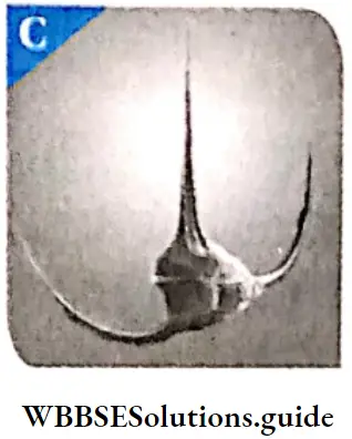
Biological Classification Ceratium

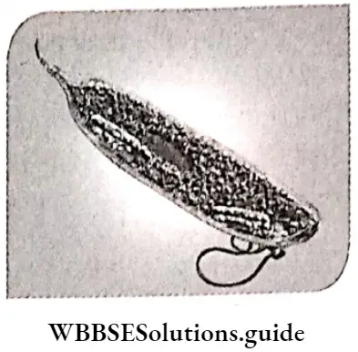
Euglenophyta: They are unicellular, photosynthetic, fresh water protists that have features similar to those of plants and animals.

Euglenophyta General Features

  1. General Features Distribution: Majority of them are fresh water organisms, living in stagnant water.
  2. Structure: The cells of most euglenoids are spindle-shaped. There is no cell wall or other rigid structure covering the plasma membrane.
  3. Cell covering: Instead of a cell wall, they have a protein rich layer, called pellicle, which makes their body flexible.
  4. Flagella: They have two flagella, a short and a long one.The flagella are tinsel shaped and have hairs on them.Both the flagella arise from their respective basal granules.Blepharoplasts or basal bodies are present at the basal end of the flagella.
  5. Eye-spot: Eye-spot is present at the end of the flagella. It contains a red-coloured astaxanthin pigment, that acts as photoreceptor (sensitive to light). The eyespot appears to be connected to the flagellum by special strands of cytoplasm that transmit signals from one organelle to the other.
  6. Nutrition: Euglenoids are mostly autotrophic in nature. Photosynthetic euglenoids contain chloroplast just like green algae. Chlorophyll a, chlorophyll b and xanthophyll pigments are present in the chloroplast. Non-photosynthetic euglenoids are saprophytic in nature. Some of the non-photosynthetic euglenoids are holotrophic while some are mixotrophic in nature.
  7. Stored food: Euglenoids have a carbohydrate food reserve, called paramylon. It is generally present in the form of small pyrenoid granules.
  8. Locomotion: They generally swim in water, using their flagella, but may also move over wet land by creeping.
  9. Reproduction: Reproduction in euglenoids takes place by asexual means, i.e., by mitotic cell division. Example: Euglena viridis, etc

Biological Classification Euglena

Euglenoids

The euglenoids were first defined by Otto Biitschli in 1884 as the flagellate order, Euglenida. They possess flexible cell coverings, that enable them to move about freely. They also ingest their food through a structure called gullet. So earlier they were considered as animals. However, they had certain characteristics similar to plants. Botanists subsequently created the algal division Euglenophyta. Presently they are classified as both animals and plants.

Slime moulds

Slime moulds Definition: Slime moulds are saprophytic protists that do not have a specific structure (amoeboid) and can grow in humid environment.

Slime moulds Distribution: Slime moulds were formerly classified as fungi. They are found mainly in wet, marshy regions. Some of the species of slime moulds are aquatic in nature.

Slime moulds General Features

General Features Structure: During favourable conditions, they live as independent, amoeba-like cells. These structures have single nucleus and are called myxamoeba. They feed on fungi and bacteria. However, during scarcity of food and unfavourable temperature, they form an aggregated structure. This structure is called plasmodium.These structures may grow and spread over a large distance. The outer part of the body is covered by mucilage, hence the name slime mould.

  • Locomotion: They move very slowly using pesudopodia. Such a motion is called amoeboid movement.
  • Nutrition: They are saprophytic and phagotrophic in nature. Some of them are parasites.
  • Reproduction: Both asexual and sexual types of reproduction are found. Asexual reproduction takes place by binary fission, plasmotomy (type of asexual reproduction in which a multinucleate protozoan cell divides into two or more multinucleate daughter cells, without mitosis), cysts or sclerotium formation, etc.
  • Life cycle: Zygotic and gametic meiosis take place within the life cycle.

Slime moulds General Features Types: There are two main groups of slime moulds in the Kingdom Protista. They are—Plasmodial slime moulds and Cellular slime moulds.

Plasmodial slime moulds or Myxomycota or true not have a specific structure. They are large single-celled mass, without the cell wall but contain thousands of nuclei (structure called plasmodium). A slimy secretion is released by the slime mould.

Examples: Physarum sp., Fuligo sp.

Cellular slime moulds or Acrasiomycota: They spend most of their lives as haploid, single nucleus containing, amoeboid protists. They show amoeboid motion, hence called myxamoeba. Upon the release of a chemical signal, the myxamoeba aggregate into a clustered structure, known as a pseudoplasmodia. This pseudoplasmodia eventually form multicellular slugs. They contain a layer of cellulose, outside the spores.

Example: Physarum sp., Stemonitis sp., Fuligo sp., Dictyostelium sp.

Biological Classification Stemonitis Sp

Protozoa Protista

Protozoa are unicellular, heterotrophic, eukaryotic organisms. They live as predators or parasites. They are considered to be the primitive relatives of animals.

Protozoa Protista General Features

  • General Features Symmetry: The body may be asymmetrical, or radially, bilaterally or spherically symmetrical.
  • Shape: They are of different shapes—spherical or oval shaped. Some of them do not have a definite shape or structure, such as Amoeba sp.
  • Body structure: They are eukaryotic, unicellular organisms. The cell may contain one or more nucleus, They have very thin cell membrane. Sometimes the cell  membrane may be covered by pellicle (Paramoecium  sp.) or calcareous or siliciferous shells (Elphidium sp.).
  • Locomotion: Generally, members of protozoa such as Plasmodium, show contractile motion by pseudopodia, flagella or cilia. On the other hand, members like Monocystis, do not have organs for locomotion. Such organisms move about by cellular contractions, using contractile fibrils called myoneme threads.
  • Osmoregulation: Protozoa living in fresh water are subjected to a hypotonic environment. Protozoa have contractile vacuoles, water vacuoles, etc., that collect and expel excess water. Some waste products and carbon dioxide are also excreted by this vacuole, along with the water. This is how protozoa perform osmoregulation.
  • Nutrition: The mode of nutrition in most of the free-living protozoa is holozoic. Other modes of nutrition found in many protozoa are saprophytic, symbiotic or parasitic.
  • Respiration: Aerobic respiration is seen in free-living organisms, while anaerobic respiration is seen in ectoparasites (parasites living on the body surface of the host).
  • Excretion: Waste products of protozoans are nitrogenous wastes like ammonia, C02, excess water, etc. These are expelled from the body through the contractile vacuole.
  • Reproduction: Both asexual and sexual reproduction are observed in these organisms. Asexual reproduction takes place by binary fission. Sometimes, multiple fission may also occur. Sexual reproduction may occur by syngamy and conjugation. Under unfavourable conditions, they form cysts, which become active during favourable conditions.
  • Reserve food: Reserve food is generally glycogen (example,. Entamoeba sp.).

Protozoa Protista General Features Types: There are four major groups of protozoans according to the organ of locomotion. They are—

Zooflagellata, Ciliata, Sarcodina, Sporozoans

Zooflagellata (Flagellated protozoans):

Zooflagellata General Features

  1. The members of this group are either free-living or parasitic.
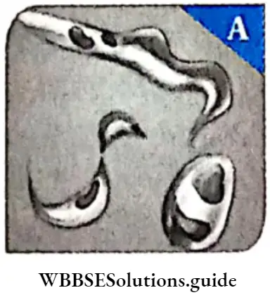
  2. They have one or more flagella. Some of them (parasitic forms) can cause diseases, e.g., Trypanosoma sp. causes sleeping sickness.
  3. The flagellated protozoans range from a simple oval cell with one or more flagella to collared flagellates (organisms with funnel-shaped collar of interconnected microvilli, at the base of the flagella).
  4. Most of the members have lost the ability to photosynthesise. They generally show parasitic nutrition, some may show saprophytic nutrition.
  5. Only asexual reproduction, by longitudinal binary fission, is observed. They may exist either as single cells (e.g., Paraphysomonas sp.) or as colonies (e.g., Codosiga sp.).

Biological Classification Trypanosoma

Biological Classification Leishmania

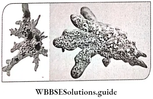
Biological Classification Amoeba

Sarcocfina (Amoeboid protozoans)

Sarcocfina General features

  1. These organisms live in fresh water, sea water or moist soil.
  2. The cells may contain more than one nucleus.
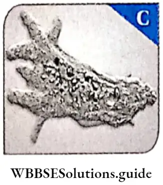
  3. The organisms move and capture their prey by stretching out their pseudopodia (false feet) as in Amoeba sp.
  4. The mode of nutrition is heterotrophic and holozoic. Some of them such as Entamoeba sp. are parasites.
  5. Marine forms have silica shells.
  6. Reproduction occurs both by asexual and sexual means. Asexual reproduction occurs by binary or multiple fission. Sexual reproduction occurs by syngamy.

Sarcocfina Example: Amoeba, etc.

Biological Classification Amoeba Proteus

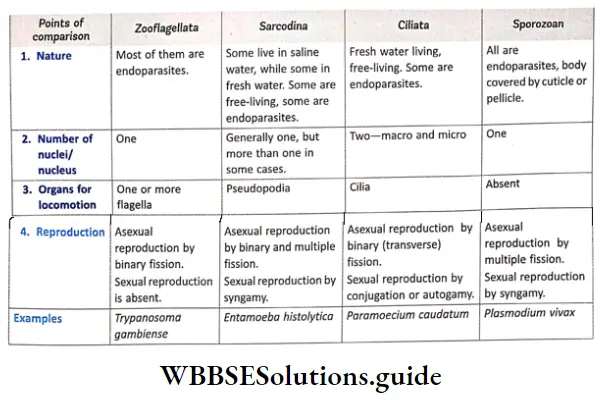
Comparison among the four classes of protozoa:

Biological Classification Comparison Among The Four Classes Of Protozoa.

General Features

  1. These are aquatic, actively motile organism.
  2. Most of them are free-living,however, some may form colonies. Some may also be endoparasitic  in nature.
  3. The cilia are the main organs of locomotion.
  4. Most of these are heterotrophic and holozoic. They have modified structures for absorbing their food, such as cytostome, cytopyge or cytoproct. Digestion takes place within the food vacuoles.
  5. They have a cavity (gullet) that opens to the outside of the cell surface. The coordinated movement of rows of cilia makes any food in the water to enter the gullet.
  6. There are two nuclei in the cell, the larger one called macronucleus and smaller one called micronucleus.
  7. Both asexual and sexual reproduction are seen among the members. Asexual reproduction takes place by transverse binary fission. Sexual reproduction takes place by conjugation.

Example: Paramoecium sp.

Sporozoans:

Sporozoans General features:

  1. This group includes organisms that have an infectious spore-like stage in their life cycle.
  2. Generally, they are unicellular in nature. © They remain as endoparasites (parasites living within the body of the host) within vertebrates and invertebrates.
  3. During most of their life cycle, they are unable to move by themselves, as they lack organs for locomotion.
  4. The body is covered by a permeable cuticle or pellicle layer.
  5. The mode of nutrition is heterotrophic and saprozoic.
  6. Both asexual and sexual types of reproduction are observed in the organisms. Asexual reproduction occurs by multiple fission or schizogony. Sexual reproductiontakes place by syngamy.
  7. Life cycle is haplontic.

Sporozoans Examples: Pneumocystis carinii, Plasmodium vivax, etc.

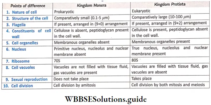
Differences between kingdom Monera and kingdom Protista:

Biological Classification Differences Between Kingdom Monera And kingdom Protista

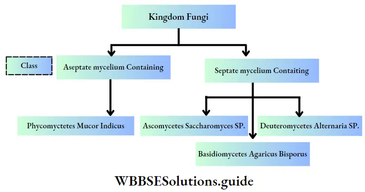
Kingdom Fungi

This kingdom of the biological world comprises of eukaryotic, multicellular, non-chlorophyllous, spore-forming organisms are called fungi.

Kingdom Fungi Distribution: Fungi are found in air, water, soil and, on animals and plants. They prefer to grow in warm j and humid places. Most of the fungi grow in the soil, Some of the fungi may also be aquatic. Some fungi remain as parasites, within host organisms, both intracellularly and intercellularly.

Kingdom Fungi General features

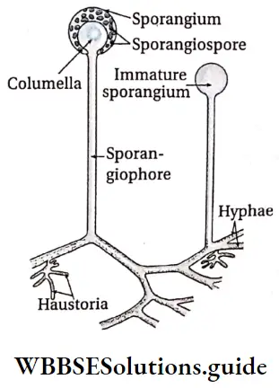
Kingdom Fungi Body Structure: Most of the fungi are simple, unicellular or multicellular and filamentous in nature, filamentous fungi consist of long, slender thread-like structures called hyphae. Due to extensive growth, the hyphae form a cottony network, known as mycelium. Some hyphae are long, slender, thread-like in nature, called hyaline hyphae. Some hyphae are filamentous in

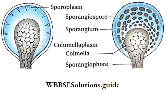
Biological Classification Structure Of Hyphae In Mucor

Nature, filled with multinucleated cytoplasm. These are called coenocytic hyphae. Some fungi have septa or cross walls in their hyphae and are called septate. Coenocytic hyphae do not have septa (aseptate), but septa develop within them during reproduction. There may or may not be pores across these septae, that allow transport of nutrients to other parts of the mycelium.

  • Cellular structure: The cells are eukaryotic. The cell wall is composed of chitin and polysaccharides. Except chloroplast, all other organelles are present in the cell.
  • Nutrition: Most fungi are heterotrophic. They absorb soluble organic matters from dead and decayed plant or animals. Hence, they are called saprophytes. They digest the extracellular substances and derive nutrition by absorbing them. Thus, fungi show an absorptive mode of nutrition. Some fungi can grow on other living organisms and draw nutrition from them (hosts). They are called parasites. The fungi can also live as symbionts—in association with algae as lichens and with roots of higher plants as mycorrhiza.
  • Reserve food: The reserve food materials are- glycogen, starch, sucrose, maltose oil, etc.
  • Reproduction: Reproduction in fungi can be performed vegetatively through fragmentation, fission and budding. Asexual reproduction is performed by the formation of spores called conidia, sporangiospores, oidia, sclerotia, chlamydospores, aplanospores or zoospores.
  • Sexual reproduction occurs, through isogamy, anisogamy and oogamy. The gametes unite to form different kinds of spores like oospores, ascospores and basidiospores. The various spores are produced in distinct structures called fruiting bodies or fructifications. Fertilisation involves the following three steps—
  1. Protoplasmic fusion of two motile or non-motile gametes, called plasmogamy.
  2. Subsequent fusion of two nuclei within the hyphae called, karyogamy.
  3. Meiosis within the zygote, resulting in the formation of haploid spores.

Sexual reproduction in fungi

  • During sexual reproduction, two haploid hyphae of compatible mating types come together and fuse. In certain fungi, the two nuclei of opposite nature fuse immediately resulting in the formation of diploid zygotes (2n).
  • However, in others (Ascomycetes and Basidiomycetes), an intervening dikaryotic stage (n + n, i.e., two nuclei per cell) occurs; such a condition is called a dikaryon and the phase is called dikaryophase of fungus. After dikaryophase ends, the parental nuclei fuse and the cells become diploid.

Classification Of Kingdom Fungi

Different scientists have classified fungi differently. Among them, the most widely accepted classification was propunded by Gwynne-Vaughan and Barnes (1927, 1937). This classification has been discussed below.

Biological Classification kingdom Fungi

Phycomycetes

This is the class of fungi having aseptate mycelium.

Phycomycetes Distribution: Members of phycomycetes are found mostly in aquatic habitats. They may also grow as saprophytes on dead and decaying organic matters in moist and damp places. They also grow as obligate parasites on plants.

Phycomycetes Body structure:

  1. The mycelium is aseptate and coenocytic.
  2. The cell wall is made up of chitin and other polysaccharides (such as glucan, cellulose).

Phycomycetes Reproduction:

  1. Asexual reproduction takes place by zoospores (motile) or by aplanospores (non-motile). Zoospores are produced endogenously within the zoosporangium.
  2. Sexual reproduction takes place by isogamy and oogamy. The gametes are non-motile. Oospores are produced by the union of two dissimilar gametes. Zoospores are produced by the union of two similar gametangia through zygospore formation.

Phycomycetes Examples: Mucor sp., Rhizopus sp. (the bread mould) and Albugo sp. (the parasitic fungi on mustard).

Biological Classification Columella In Mucor

Ascomycetes

This is one of the three classes of fungi having septate; mycelium.

Ascomycetes Distribution: Members of ascomycetes are generally terrestrial in nature. They may also exist as saprophytes or parasites (on plants).

Ascomycetes Body structure:

  1. Vegetative body is unicellular (yeasts) or multicellular.
  2. They commonly have a J r well-developed, branched septate mycelium.
  3. The cell wall is generally composed of chitin and glucans.

Ascomycetes Reproduction:

  1. Vegetative reproduction takes place by fragmentation (in filamentous form), fission and ; budding (in unicellular form).
  2. Asexual reproduction takes place by non-motile spores, such as conida, oidia and ; chlamydospores.
  3. The sexually reproducing units are called the ascospores. Ascospores are grown inside a small if specialised sac-like structure, called ascus (plural asci).
  4. The fruiting bodies (inside which asci are developed); are called the ascocarps.
  5. There are 8 ascospores within each ascus.
  6. The ascocarps may be cleistothecium (Penicillium sp.), apothecium (Ascobolus sp.), perithecium (Daldinio sp.) or ascostroma (Elsinoe veneta).

Ascomycetes Examples: Saccharomyces cerevisiae, Penicillium notatum E Aspergillus, Claviceps, Neurospora, etc.

Types of ascocarp :

In cleistothecium, the ascocarp is small or ovoid body that is closed from all sides. In apothecium, the ascocarp is cup-shaped or disc-shaped, while perithecium, it is flask-shaped with definite apical pore. In ascostroma (also known as pseudothecium), ascii are produced in cavities containing stroma.

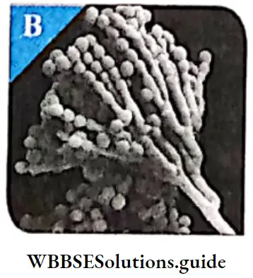
Biological Classification Different Types Of Fungi Aspergillus

Biological Classification Penicillium

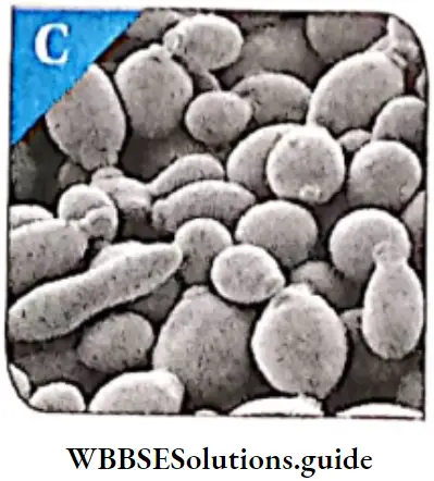
Biological Classification Yeast

Basidiomycetes

This is another class of fungi having septate mycelium.

Basidiomycetes Body structure:

  1. Presence of well-developed, branched and septate mycelium.
  2. The mycelium is of two types—primary and secondary.
  3. The mycelial cells may contain one nucleus, called monokaryotic (or uninucleate, common in primary mycelium) or two nuclei, called dikaryotic (or binucleate, common in secondary mycelium).
  4. The secondary mycelia may organise and form fruit body.© The cell wall is mainly composed of chitin and glucans.

Basidiomycetes Nutrition: They are mostly saprophytic and parasitic.

Basidiomycetes Reproduction:

  1. Primary mycelium may reproduce asexually by oidia, conidia, chlamydospores, etc.
  2. Vegetative reproduction takes place by budding and fragmentation.
  3. Sex organs are absent. During sexual reproduction, the dikaryotic cell is formed by somatogamy, plasmogamy (dikaryotization of the cell by somatogamy of monokaryotic and dikaryotic mycelium).
  4. The dikaryotic phase persists for long period of time. Karyogamy occurs in club-shaped structures, called basidia or basidium.
  5. 4-haploid basidiospores are formed by meiosis. They are oval, cylindrical or spherical in shape. They may be unicellular and uninucleate.
  6. Basidiospores are developed exogenously on the horn-shaped or tubular outgrowths of the basidium. These outgrowths are called sterigmata.

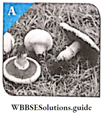
Basidiomycetes Examples: Agaricus campestris (mushroom), Puccinia graminis (rust fungus), Ustilago (Smut) etc.

Biological Classification Agaricus

Biological Classification Microsporum

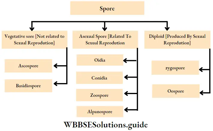
Different types of spores produced by the fungi have been depicted in the following flowchart.

Biological Classification Spore

Deuteromycetes

This is another class of fungi having septate mycelium.

Deuteromycetes Distribution: They are called the fungi imperfecti because only the asexual or vegetative phases of these j fungi are known. They are mostly terrestrial, with only a few being aquatic. They are generally decomposers and parasitic in nature, found as saprophytes in the soil.

Deuteromycetes Body structure: The vegetative body is mycelial and composed of profusely branched and septate hyphae. The sually multinucleated. In parasitic species, the hyphae grow intra or intercellularly.

Deuteromycetes Nutrition: Most of the fungi are saprophytic in nature. They act as decomposers of litter and help in mineral cycling. Some are parasites.

Deuteromycetes Reproduction:

  1. They reproduce mainly by asexual methods.
  2. The asexual reproduction takes place commonly by conidia, blastospores, chlamydospores and arthrospores.

Deuteromycetes Examples: Alternoria spv Colletotrichum sp. and Trichodermo sp.

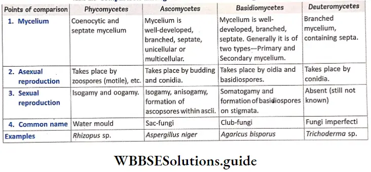
Compare among different classes of kingdom fungi:

Biological Classification Comparison Among Different Classes Of Kingdom Fungi

Importance Of Fungi:

Fungi play a very important role in our daily life, Some fungi are useful. These are used as food, natural sources of medicine, etc. Some fungi are j directly or indirectly harmful to mankind. They spoil food and other substances and also cause diseases in; both plants and animals, Some common fungal diseases of plants are early blight of potatoes, late blight of potato, etc.

Poisonous fungi :

Some fungi like Amanita phalloides produce toxins, like a-amanitin which causes lesions of stomach cells, It also produces phalloidin which affects the liver, Fungi, like Aspergillus flavus, A. fumigatus and Penicillium islandicum can infect groundnut and mustard cakes. They produce a poisonous substance, called aflatoxin, that binds with DNA and inhibits transcription in the host plant cell. This results in the inhibition of protein synthesis, ultimately leading to cell death.

kingdom Plante

Plants are multicellular organisms composed of eukaryotic cells. The cells are organised into tissues. The cells have cell wall that contains cellulose. The cells also contain plastids and large vacuoles in the cytoplasm. They obtain nutrition by photosynthesis. They are generally autotrophic and are called producers. Stored food is starch. Growth is not distinct. They continue to grow throughout their lifetime.

Examples: mosses, ferns, conifers, and flowering plants. We shall learn about this kingdom later.

kingdom Anmalia

Animals are multicellular organisms composed of eukaryotic cells. The cells are organised into tissues. The cells lack cell walls. They respond to stimuli very fast. Their growth is distinct. They grow only upto a certain period of their lifetime. They do not carry out photosynthesis and obtain nutrients from other organisms. Thus they are heterotrophic and are decomposers in nature.

Examples: sponges, worms, insects, and vertebrates. We j shall learn about this kingdom in details.

Virus Viroids And Lichens

These are also considered to be members of the living world.

Virus

Virus Definition: Viruses are microscopic, acellular organisms, that form an intermediate group between living and non-living organisms. The term ‘virus’ is derived from the same word in Latin, meaning venom or poisonous fluid. It was coined by M.W. Beijerinck in 1898. Viruses and viroids have not been placed in classification system as they do not have a cell structure and are not considered living.

Discovery of virus:

  1. D.J. Ivanowsky (1892) recognised certain microbes as causal organism of tobacco mosaic disease. These organisms were, found to be smaller than bacteria because they could pass through bacteria-proof filters.
  2. M.W. Beijerinck (1898) demonstrated that the extract of the virus infected tobacco plants could cause infection even in healthy plants. He named the fluid as contagium vivum fluidum (meaning ‘contagious living fluid’).
  3. W.M. Stanley (1935) showed that viruses could be crystallised.

However, once they infect a cell, they behave like living organisms. They take over the replication machinery of the host cell to replicate themselves, thereby killing the host cell.

Therefore, the question still remains whether the viruses are living or non-living.

Virus General features: The viruses are members of the j intermediate group of organisms, between the living and the non-living. They are inert outside their specific host cell.

As soon as they enter the host cell, they become living and start reproducing. Viruses are cannot complete their life cycle without the specific host.

Virus Shape and size:

  1. Viruses are very minute (microscopic) organisms, that have an average diameter of 20-30nm.
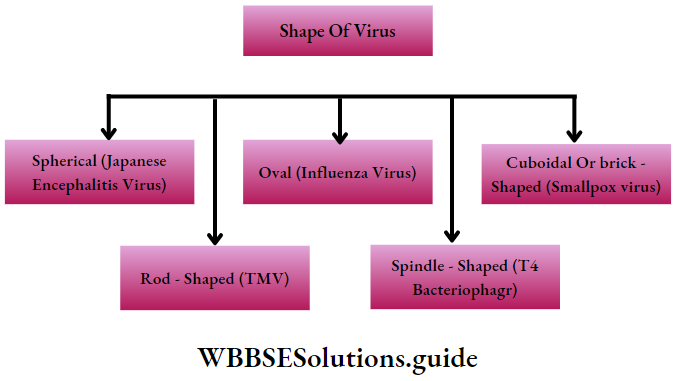
  2. They may be spherical (e.g., Poliovirus, Influenza virus), cylindrical (e.g., TMV), brick-shaped (e.g., Smallpox virus), or spindle-shaped (Phage virus).

Biological Classification Shape Of Virus

Virus Structure: Virus does not contain protoplasm or cell membrane. Virus consists of two parts—

  1. Nucleic acid (centrally placed) and
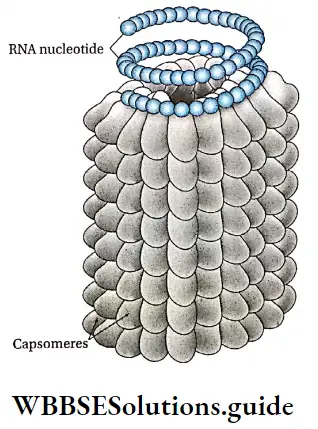
  2. Protein coat, sometimes with an additional envelope, made of lipoprotein. The protein coat, called capsid, is made up of small subunits, called capsomeres. These capsomeres are arranged in helical or polyhedral shapes. The capsid protects nucleic acid.
  3. Viruses contain either RNA or DNA as nucleic acid. A virus is a nucleoprotein (containing nucleic acid and protein) particle and the genetic material is infectious. In general, viruses that infect plants have single-stranded RNA.
  4. On the other hand, viruses that infect animals have either single or double-stranded RNA or double-stranded DNA. Bacteriophages (viruses that invade the bacteria) are usually double-stranded DNA viruses. Structural details is discussed under separate heads below.

Virus Living features: Presence of—

  1. Nucleic acid,
  2. Reproduction and
  3. Mutability.

Virus Non-living features:

  1. Lack of protoplast. The cell structure is not complete.
  2. Absence of respiration,
  3. High specific gravity like non-living things.

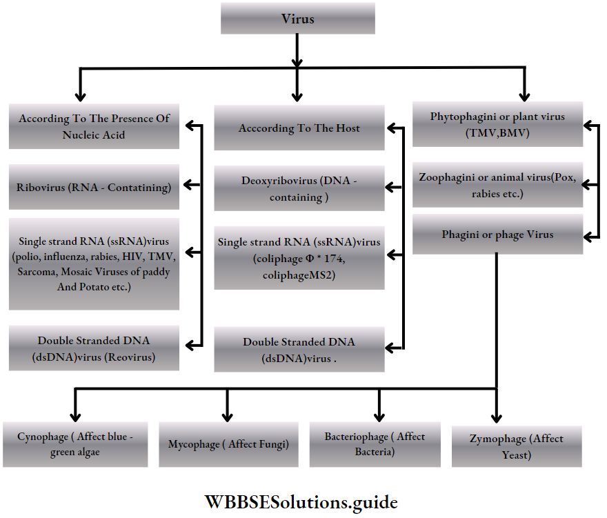
Virus General features Types: According to the presence of the nucleic acids and the host cell, the virus are classified into the following groups—

Biological Classification Virus

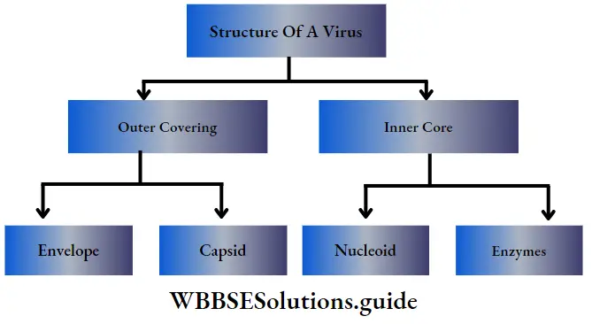
Structure of a virus

Biological Classification Structure Of A Virus

Outer covering

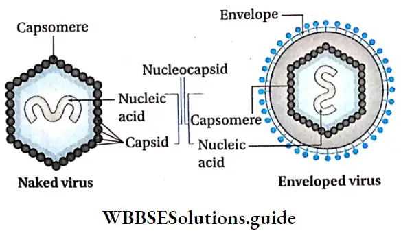
Outer covering Capsid: The protein coat surrounding the genome in a virus is called capsid. The capsid together, along with the enclosed nucleic acid, is called nucleocapsid. The capsid is made up of a number of protein subunits, called capsomeres.

Biological Classification Structure Of Virus

Outer covering Envelope: Many mammalian viruses have a protein, carbohydrate or lipid bilayer surrounding the j nucleocapsid. This loose covering is called envelope, The envelope is a part of the host cell membrane. It: is made up of several units called peplomers. The envelope may possess several outgrowths called spikes.

  • The virus which have lipid envelope outside the capsid are called lipoviruses. The viruses which do not have envelope are called naked virus (Example—TMV).

Internal or inner core

Nucleoid: The nucleic acids enclosed within the capsid region make up the nucleoid. Viruses contain only one type of nucleic acid, either DNA or RNA. DNA containing viruses are called deoxyviruses, whereas viruses having RNA are called riboviruses.

  • They vary in the structure of their nucleic acid. The nucleoid forms viral genome. The nucleoid consists of about 1000-250,000 nucleotide pairs. The nucleoid remains inactive as long as it does not come in contact with the host cell, The amount of nucleic acid of a virus usually depends on its size.

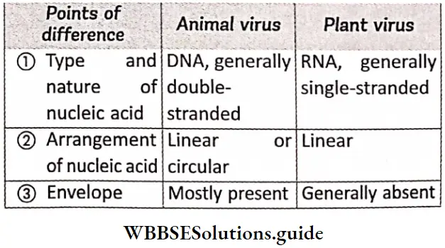
Genetic material of plant virus:

Most of the plant viruses have RNA either single (TMV) or double stranded (Rice ragged stunt viruses), except a few have DNA, either single (Gemini viruses) or double stranded (Dahlia mosaic virus). Animal viruses have mostly double stranded DNA or either single (Polio virus) or double stranded RNA (Reo virus) and bacteriophages contain mostly double stranded DNA, but they also have single stranded RNA or single stranded DNA.

Enzymes: Hemagglutinin esterase, integrase, reverse transcriptase, viral neuraminidase, polymerase, lysozyme, etc., are some of the enzymes found in viruses.

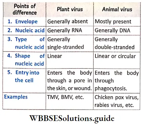
Differences between plant and animal virus:

Biological Classification Differences Between Plant And Animal Virus

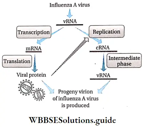
Viral Reproduction: Reproduction in virus takes place when its nucleic acid replicates within the host cell. The process of reproduction is slightly different in bacteriophage, from that of the other virus.

Reproduction in virus, other than bacteriophage: Principal events involved in viral replication, other than that in bacteriophage, are given below—

  1. Infection phase: It consists of two stages—
    1. Attachment: The first step in viral infection of a cell is attachment of the virus to the cell surface. Attachment is via temperature-independent ionic interactions. There are specific receptor molecules (generally protein, carbohydrate or lipid in nature), on the cell surface. Virus have a protein called viral attachment protein. This protein recognises the specific receptor on the cell and attaches the virus. Cells without the specific receptors are not susceptible to the virus,
    2. Penetration: After attachment, the virus has to enter or penetrate the host cell. The virus enters the cell in a variety of ways according to the nature of the virus. Plant viruses enter the host cell either via vectors (aphid, mites, etc.) or through damaged cell membrane. Animal viruses enter the host cell through processes like phagocytosis, endocytosis, etc.
  2. Eclipse phase: It occurs in two stages—
    1. Uncoating: The nucleic acid must be uncoated so that viral replication can begin. As soon as the nucleic acid becomes uncoated, the eclipse phase begins. This phase continues until new infectious virions are made.
    2. Synthesis of viral nucleic acid and protein: In this phase, the viral nucleic acid is replicated. Some 0 Assembly/maturation: New virus particles are assembled using the synthesised nucleic acids and coat proteins. Thus, the virus particles attain a maturation phase that follows the initial assembly process.
  3. Lysis (Release): Virus may be released due to cell lysis, or, if enveloped, may form bud on the cell.


Biological Classification Viral Replication

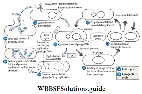
Reproduction in bacteriophage: Reproduction in j bacteriophage takes place by two cycles

  1. Lytic cycle
  2. Lysogenic cycle. The former type is seen in virulent | and the latter is found in avirulent or temperate phage. These cycles have been discussed under separate heads.

Lytic Cycle:

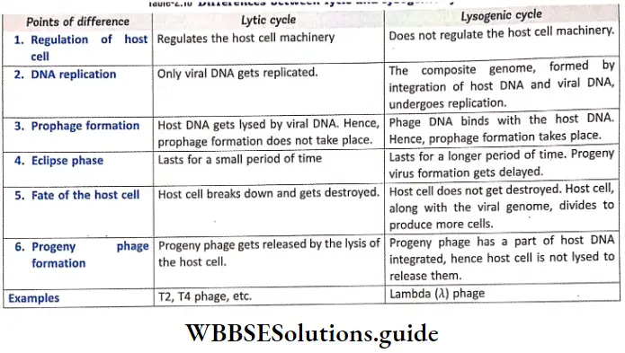
The cyclic reproduction mechanism, by which virulent | bacteriophage produces progeny virus by replication is called lytic cycle. It is common in T-even phages (T2, T4, etc.) which attack Escherichia coli. It takes place for about 20-30 minutes.

Lysogenic Cycle:

The cyclic reproduction mechanism by which phage virus introduces its nucleic acid into the host cell, divides to form progeny viruses is called lysogenic cycle. Lwoff (1953) discovered this cycle in Lambda (A) phages attacking £ coli. The phage involved in this cycle is called temperate phage, while the bacterium is of lysogenic strain.

Differences between lytic and lysogenic cycle:

Biological Classification Differences Between Lytic And Lysogenic Cycle

Biological Classification Reproduction Of A Typical Virus Bacteriophage

Characteristics of some important viruses

Characteristics of some of the viruses have been discussed below in details—

Bacteriophage

Bacteriophage Definition: Bacteriophages are viruses that infect bacteria, reproduce within them and later on lyse them to release the progeny phage.

Bacteriophage Example: T2 phage, lambda phage, phage MS2, ΦX 174 phage, T4 phage.

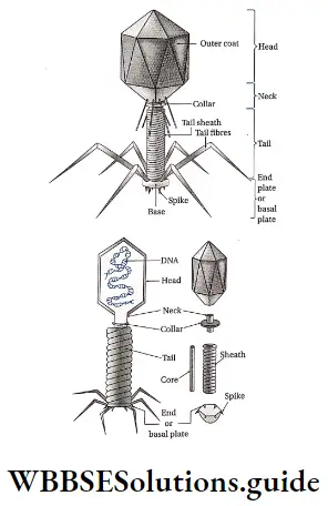
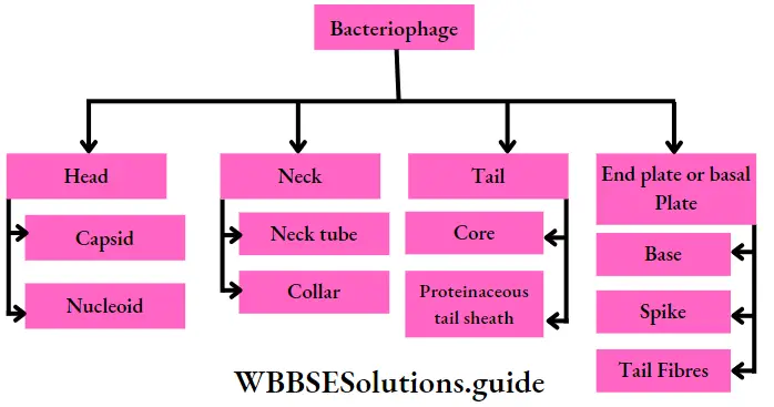
Bacteriophage Structure: T4 is one of the largest bacteriophages; it j is approximately ranging from 25nm-200nm long and 80-100nm wide. They are made up of four important parts—head, neck, tail and end plate or basal plate.

  • Head: All phages contain a structure called head, which varies in size and shape.
  1. Some are icosahedral (with 20 sides) and others are filamentous. The head is covered by a bilayered protein membrane called capsid.
  2. Capsid is made up of several units called capsomeres. It encloses nucleic acid and acts as the protective covering.
  3. The nucleic acid is about 0.05nm long double stranded DNA, containing hydroxymethylated cytosine.

Biological Classification Structure Of Bacteriophage

  • Neck: It is a small part that extends just after the head. It is made up of neck tube and collar.
  • Tail: Some phages have another structure called tail attached to the phage head. The tail has a hollow tube, called core. The nucleic acid is passed to the host cell during infection.
  • T4 tail is surrounded by a Y contractile sheath, called tail sheath, which contracts during infection of the bacterium. At the end of tail, phages like T4 have another structure called base plate. One or more tail fibres remain attached to it.
  • End plate or base plate: The tail has a plate-like structure at its end. This structure is called end plate or base plate. Six spikes are arranged, along with a long tail fibre, on this plate. The base plate and tail fibres are involved in the binding of the phage to the bacterial cell. All phages do not have base plates and tail fibres.

Biological Classification Bacteriophage

Tobacco mosaic virus

Tobacco mosaic virus Definition: The rod-shaped plant virus, that contains coiled RNA and causes mosaic diseases in tobacco plants are called tobacco mosaic virus (TMV).

Shape and size: TMV has a cylindrical hollow tube-like structure, with a length of 300nm and diameter 15-18 nm.

Tobacco mosaic virus Structure:

  1. The virus is composed of single strand of RNA, wrapped inside a capsid.
  2. The virus particle that the virus can attain the cylindrical structure.
  3. The capsid is composed of about 2130 capsomeres (70 j angstrom x 20 angstrom).
  4. The capsomeres are arranged like bricks in a cylindrical chimney-like structure. This gives the capsid a shape of spiral staircase.
  5. Each capsomere contains 158 amino acids.
  6. The single-stranded RNA is arranged spirally (as a spring), having 6400-7300 ; nucleotide units.
  7. The RNA strand encodes four : proteins. These include two proteins that replicate the ; viral RNA, a protein that transports the RNA from cell to cell, spreading the infection, and the capsid protein.

Biological Classification Structure Of Tobacco Mosaic Virus

HIV, AIDS and AIDS wasting syndrome

HIV or Human Immunodeficiency Virus is a type of j retrovirus, that causes the disease AIDS (Acquired j Immunodeficiency Syndrome) in humans. AIDS wasting syndrome is a condition in which a person suffering; from AIDS loses at least 10% of his body weight, especially that of the muscles.

Tobacco mosaic virus Importance of virus

Viruses can be both useful and harmful and hence, have both advantages as well as disadvantages. Some of these are as follows—

Tobacco mosaic virus Importance of virus Advantages:

  • Use as bactericide: Bacteriophages are sometimes j used in ‘polluted water as bactericides. They act as scavengers that eradicate the bacterial population j present in the water. Similarly, they can also kill the disease-causing bacteria.
  • Use in scientific experiments: In space research,  lysogenic phage cultures are used as radiation detector, Avirulent or temperate phages are useful in studying genetic recombination (transduction) and are used ; widely in biotechnological research.
  • Treatment and prevention of diseases: Sometimes bacteriophages are used in therapy and prophylaxis of some bacterial diseases. Viruses are utilised in the production of vaccines used to develop immunity against viral infection.

Importance of virus Disadvantages:

Causal agents of various diseases: Viruses are responsible for causing various diseases of both plants and animals. Some of the viral diseases in plants are tobacco mosaic, yellow vein mosaic of lady’s finger, leaf roll of potato, leaf curl of papaya, etc. The plant viruses cause damage to different parts like root, leaf, fruit, seed, etc. These incur economic losses by hampering the quality and quantity of the plant products. Some viral diseases in animals include small pox, meningitis, pneumonia, mumps, bronchitis, etc. In case of animals, the diseases may even prove fatal.

Oncovirus: Some of the viruses are responsible for causing cancer. These viruses are called oncoviruses.

Viroids

Viroids Definition: The tiny, non-coiled, infectious, ssRNA containing particles that can cause diseases in plants are called viroids.

Viroids General features

Viroids Structure: The viroids do not have capsid (i.e., protein coat) around the RNA molecule.Their genome contains a circular RNA strand extensively base paired within itself. So, they can resist RNase attack.

Viroids Genome:

  1. Viroids have small, low-molecular weight, circular, single stranded RNA molecule. The ssRNA is made up of about 250-390 bases. This number of bases is insufficient to code even for one enzyme, required for replication. Some of the nucleotides even lack AUG(initiation codon).
  2. The molecular weight is about 75,000 to 1,20,000 Da.
  3. There are five domains in the viroid genome—TL (Terminal Left), P domain (Pathogenicity Domain), CCC
  4. Viruses can be both useful and harmful and hence, have both advantages as well as disadvantages. Some of these are as follows— leaf curl of papaya, etc. The plant viruses (Central Conserved Region), TR (Terminal Rigid), V Domain.

Viroids Nature:

  1. The viroids are smaller and simpler than virus particles.
  2. The viroids lack any protein coat and remain as free RNA.
  3. The size of RNA is 25-370 bases in viroids as compared to 3.2-20 kb in viruses.
  4. The ability to mutate is more in case of viroids, than in viruses.
  5. They generally reproduce under high temperature. They reproduce after attaching to the host cell DNA.
  6. Its RNA cannot be degraded by RNase enzyme.

Viroids that cause diseases in plants: Nearly twenty plant diseases are known to have caused by viroids. Some of such viroids are —Potato Spindle Tuber Viroid (PSTVd), Coconut Cadang-Cadang Viroid (CCVd), Avocado Sunblotch Viroid, etc.

Only two diseases such as Scrapie disease of sheep and Kuru disease of human are confirmed to be caused by viroids.

Biological Classification Differences Between Virus And Viroids

Lichens

The term ‘Lichen’ was given by Theophrastus. Its symbiotic characteristic was however, stated by, Schwander in 1867.

Lichens Definition: Lichens are symbiotic associations of fungi and algae.

Lichens Distribution: Lichens can grow over a range of habitats, such as stones, bark of trees, soil, even in water.

Lichens General features

  1. General features Structure: Lichens are composite thalloid, generally leaf-like or cylindrical in shape. The thalloid may be of different colours—grey, green, yellow, pink or brown. There are three layers in the structure of the thalloid— upper and lower layers are made up of fungal hyphae, with the middle cortex layer made up of algae. Algal layer is also known as gonidial layer.
  2. Nutrition: They depend on atmospheric source of nutrition. The lichens that grow on soil or rock, absorb nutrients from their substrate.
  3. Reproduction: Lichen reproduces by all the three means—vegetative, asexual, and sexual.Vegetative reproduction takes place by fragmentation, decaying of older parts, by soredia and isidia. Asexual reproduction takes place by the formation of oidia. Sexual reproduction takes place by the formation of ascospores or basidiospores. Only fungal component is involved in sexual reproduction.
  4. Components: The algal partner (known as photobiont or phycobiont) may be cyanobacteria (Nostoc sp.) or green algae (Trebouxia sp.).
    • Generally the fungal partner (known as mycobiont) occupies most of the thallus and produces its own reproductive structures. It absorbs water and mineral salts and transports them to the algae.
    • In about 98% lichens, fungal component is a member of ascomycetes, while the rest are those from basidiomycetes and deuteromycetes.
    • The algal component, on the other hand, manufactures the food ; through photosynthesis. This food probably diffuses out from the algal body and is absorbed by the fungal component.
  5. Classification of lichens: Lichens are classified on the basis of mutualism and morphological structure.

Types of lichen according to mutualism:

Biological Classification Types Of Lichen According To Mutualism

Types of lichen according to morphology:

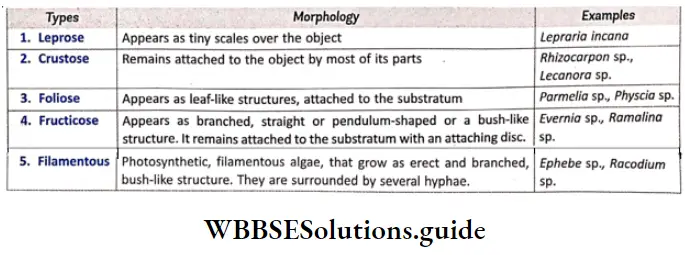
Biological Classification Types Of Lichen According To Morphology

Importance of lichens: The lichens are beneficial as well as harmful to mankind. They are used as food and fodder, and medicines. They also have various kinds of industrial uses.

  • As food: Some species of Parmelia sp. are used as curry powder in India, while various others are used as food in other countries. Lichens like Lecanora saxicola and Aspicilia calcarea, etc., are used as food by several other organisms.
  • As fodder: Ramalina fraxinea, R. fastigiato, Evernia prunastri, etc., are used as fodder for animals.
  • As medicine: Lichens are medicinally important due to the presence of lichenin and some bitter alkaloids or astringent substances. They are used in the treatment of diseases like jaundice, diarrhoea, epilepsy, etc.
  • Industrial uses: Lichens are also used in different kinds of industries—
  1. Tanning industry: Some lichens like Lobaria pulmonaria and Cetraria islandica are used in tanning leather.
  2. Brewery and distillation: Lichens like Lobaria \ pulmonaria are used in brewing of beer. In Russia and Sweden, Usnea florida, Cladonia rangiferina and Ramalina fraxinea are used in production of alcohol due to their rich lichenin content.
  3. Preparation of dye: Dyes obtained from some lichens have been used since ancient times for colouring fabrics, etc. Litmus, an acid-base indicator dye (substance that shows colour change in the presence of an acid or base), is extracted from Roccella tinctoria, R. montagnei and also from Lasallia pustulata.
  4. Cosmetics and perfumery: Certain aromatic compounds (organic compounds with characteristic odour) are available in lichen and are extracted.
    These are used in the preparation of cosmetics and perfumes. Essential oils (oils present in cosmetics) extracted from Ramalina sp. and Evernia sp. are used in the manufacture of cosmetic soaps.
  5. Harmful activities:
    1. Lichens like Amphiloma sp. and Cladonia sp. grow on mosses and cause total destruction of moss colonies.
    2. Different crustose lichens cause severe damage to window glasses and marble stones of old buildings,
    3. Lichens like Letharia vulpina (wolf moss) are highly poisonous due to the presence of vulpinic acid.

Ecological importance of lichens

  1. Pioneer of rock vegetation: Due to their ability to grow using minimum nutrients and water, crustose lichens can colonise fast.
  2. Accumulation of radioactive substances: Lichens can absorb different radioactive substances from the environment through bioremediation.
  3. Sensitivity to air pollutants: Lichens are very sensitive to air pollutants like S02, CO, C02, etc. Thus lichens are important bioindicators of these gaseous pollutants.

Notes

  • Amitosis: A type of cell division, which involves simple
  • Bioremediation: Using ‘ plants or microorganisms cleavage of the nucleus, followed by division of the j (naturally growing or artificially introduced) to consume cytoplasm. j or break down pollutants in the environment.
  • Biota: Total collection of organisms of a particular region, at a specific time period.
  • Chemosynthetic: Organisms which obtain energy by oxidising inorganic molecules into organic ones.
  • Equatorial plane: Plane present between the poles of a cell, that remains perpendicular to the spindle apparatus in the dividing cell.
  • Febrile: Feverish
  • Heterotrophic: Organisms which cannot produce their own food.
  • Histone: A type of protein that is wrapped around by DNA molecule to form chromatin fibre.
  • Holozoic: A type of nutrition involving ingestion, digestion, absorption and assimilation of the food. Osmolability: Unstable towards changes in osmotic pressure.
  • Peptidoglycan: A polymer made of sugar and amino ! acids, that forms the basic component of the cell wall.
  • Pseudopodia: A type of transient organ of locomotion j produced by protoplasmic extension, present in some organisms.
  • Ruminant animals: Animals that acquire nutrients from ! plant-based food, by fermenting it in a special stomach before digestion. This fermented ingesta is regurgitated I and chewed by these animals.
  • Syngamy: Fusion of two cells, or their nuclei, during ; reproduction.

Points To Remember:

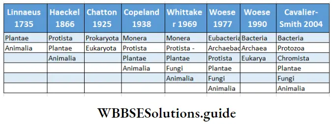
  1. Carolus Linnaeus (1753) classified living organisms into two kingdoms—Plant kingdom and Animal kingdom.
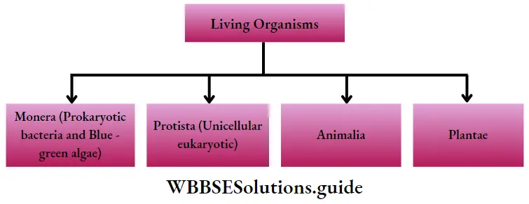
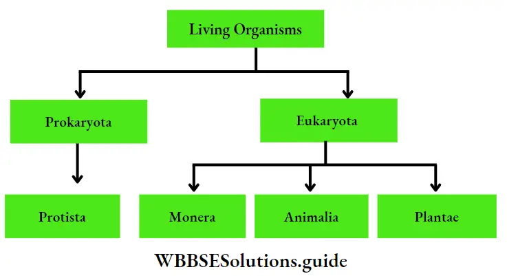
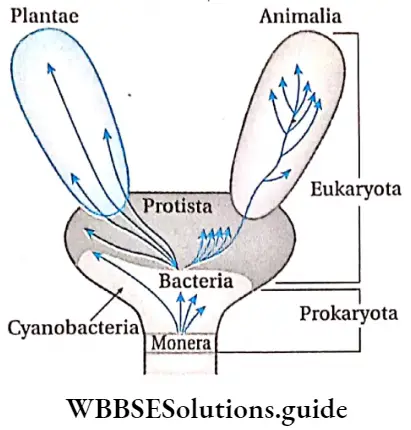
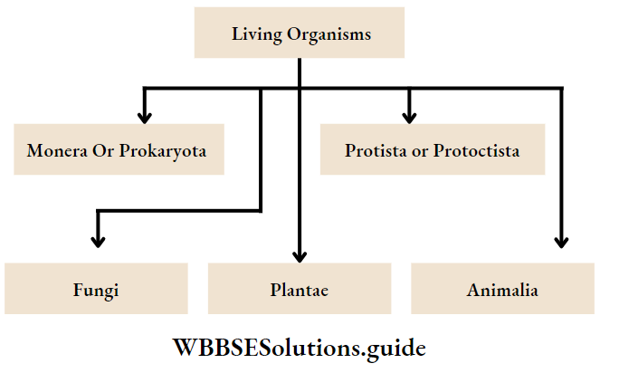
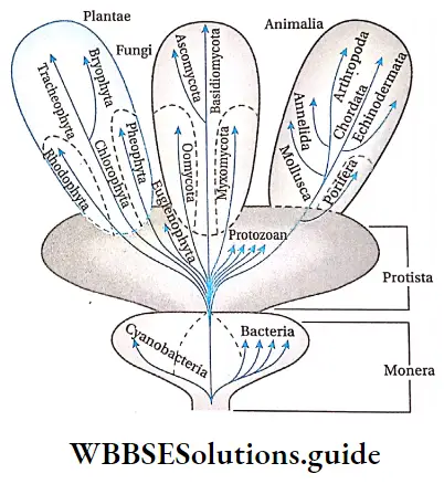
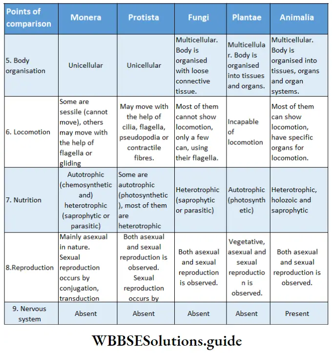
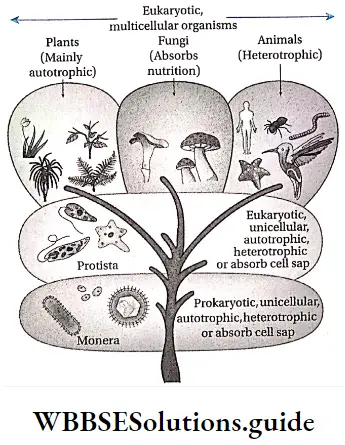
  2. R. H. Whittaker (1969) classified living organisms into five kingdoms. These kingdoms are Monera, Protista, Fungi, Plantae and Animalia.
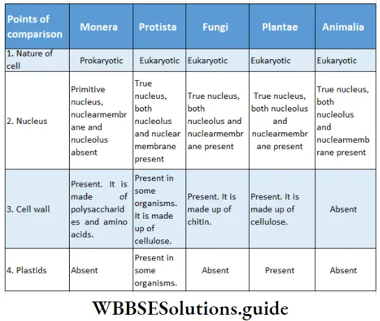
  3. Kingdom Monera consists of only prokaryotes. Kingdom Protista, . Fungi, Plantae and Animalia consist of eukaryotes.
  4. Bacteria under the kingdom Monera was first discovered by Leeuwenhoek (1676).
  5. Cell wall of bacteria is composed of peptidoglycan or \ murein.
  6. The coiled structure formed by the invagination of j plasma membrane of Gram positive and Gram \ negative bacteria is called mesosome.
  7. The circular DNA molecule present in bacterial cell as I a substitute of nucleus is called nucleoid or genophore.
  8. Some bacteria (like Chlorobium) possess the j photosynthetic pigment bacteriochlorophyll. These bacteria are capable of photosynthesis.
  9. The protist which has characteristics of both animal j and fungi is called slime mould (Myxomycetes).
  10. Ascomycetes and basidiomycetes of kingdom Fungi i are called sac fungi and club fungi respectively.
  11. Edible mushroom is Agaricus bisporus.
  12. Each basidiocarp (mushrooms) has two parts—stalk like structure is called stipe and umbrella like structure is called pileus.
  13. The causative fungus of Black stem rust disease of wheat is Puccinia graminis tritici.
  14. Late blight of potato is caused by the fungus Phytopthora infestans.
  15. Lichen is a symbiotic organism formed by the association of algae and fungi. Its fungal part is mycobiont and algal part is phycobiont.
  16. Mycoplasma are prokaryotes that lack cell wall.
  17. Silica can be found in the cell wall of diatoms.
  18. The protozoa Plasmodium has four species. Each of them is a carrier of malaria. Such as
    1. Plasmodium vivax— Benign malaria
    2. Plasmodium falciparum—Malignant malaria
    3. Plasmodium malariae—Quartan malaria
    4. Plasmodium ovale—Mild tertian malaria
  19. The term virus was coined by the Dutch microbiologist Martinus Beijerinck. In the early 20th century. Frederick Twort discovered that bacteria could be attacked by viruses.
  20. Viruses are intermediate between living and non-living organisms.
  21. The viruses which invade the body of harmful bacteria and kill them are called bacteriophages.


Archaebacteria Characteristics and Types Notes

Domain Archaea

Domain Archaea Characteristics:

  1. They are the most primitive organisms.
  2. Members of this domain have prokaryotic cells.
  3. Cell membranes have branched hydrocarbon chains attached to glycerol by a type of linkage called ester linkage.
  4. Certain chemical constituents of cell membrane help them to withstand extreme temperature and highly acidic environment.
  5. Peptidoglycan is absent in cell wall.
  6. Extreme halophiles (organisms thriving in highly saline environment) and hyperthermophiles (organisms thriving in extremely hot environment) are best examples of Archaea.

Domain Archaea

Read and Learn More: WBCHSE Notes for Class 11 Biology

Kingdoms included: Domain Archaea contains only one kingdom called Archaebacteria. This kingdom is further divided into three groups—methanogens, halophiles and thermoacidophiles. These groups have been discussed later in the chapter.

Domain Bacteria

Domain Bacteria Characteristics:

  1. This group includes prokaryotic bacteria.
  2. Cell membranes have unbranched fatty acid chains attached to glycerol by a type of linkage called ester linkage.
  3. Peptidoglycan is present in cell wall.
  4. Most of them are decomposers, some are autotrophic in nature.
  5. Most of them cause diseases in higher organisms but some may be beneficial.

Kingdoms included: This group also has a single kingdom called Eubacteria. This kingdom is again subdivided into four groups— Proteobacteria (Rhizobium 1 sp.), Cyanobacteria (Nostoc sp.), Spirochaetes (Treponema sp.), Firmidicutes (Clostridium sp.) and Mycoplasmas. These groups too have been discussed later in the chapter.

Domain Eukarya

Domain Eukarya Characteristics:

  1. The members of Eukarya have eukaryotic cells, which have cell membranes nearly similar to that of bacteria.
  2. True nucleus and membranous cell organelles are present.
  3. Some eukaryotes may have cell wall, but peptidoglycan is absent.

Kingdoms included: Domain Eukarya is further divided into Kingdom Protista (algae, protozoans, etc.), Kingdom Fungi (yeast, mould, etc.), Kingdom Plantae (flowering plants, ferns, etc.) and Kingdom Animalia (insects, vertebrates, etc.).

Biological Classification Comparison Among The Three Domains

Kingdom Monera

The Kingdom comprising of the simplest unicellular, prokaryotic living organisms, is known as Kingdom Monera.

Kingdom Monera Distribution: The members of kingdom Monera are present in all types of environment. Some of them are even present in extremes of environment (such as high temperature, saline, alkaline or acidic environment, etc.).

Kingdom Monera General features:

  • Cell nature: These organisms are prokaryotic. They are simple, unicellular and microscopic. They may form filament or thread-like structure.
  • Cell shape and size: Cell shape is generally characteristic of a given bacterial species. They are microscopic, with a volume of about 0.2-10.0pm3, length being 0.1-0.5pm.
  • Cell wall: Cell wall is present in some organisms. It is generally made up of peptidoglycans. In case of archaebacteria, the cell wall is made up of proteins. The cell wall is absent in case of mycoplasma.
  • Cytoplasm: The cytoplasm appears as colourless jelly. It contains nuclear material, non-membranous cell organelle and gas vacuoles. True cytoskeleton is absent but may contain primitive cytoskeleton.
  • Nuclear material: They do not possess a definite nucleus or a nuclear membrane. The nuclear material without the nuclear membrane and nucleus, is known as nucleoid.
  • Genetic material: The genetic material is constituted of naked double-stranded DNA, that remains without histone proteins. It is present within the cytoplasm. This type of DNA is known as prochromosomes.In some cases, plasmids (small DNA molecules that replicate independently) and episomes (small DNA molecules that can replicate independently as well as integrated within the chromosome) are present.
  • Cell organelles: Double membrane-bound cell organelles are absent.Chloroplast is absent. The pigment-containing, membranous thylakoids remain freely suspended in the cell. These are known as chromatophores. Mesosomes are cell organelles that carry respiratory enzymes. These organelles are present in the inner lining of the cell membrane.
  • Vacuoles: Gas vacuoles may be present.
  • Cellular appendages: Sometimes single-stranded flagella may be present. Some of the members may also contain pili and fimbrae.
  • Nutrition: Some of the members of this kingdom can synthesise their food (autotrophs), while some derive nutrition from others (heterotrophs).
  • Respiration: They perform both aerobic and anaerobic types of respiration.
  • Cell division: Cell division is mainly amitosis.
  • Reproduction: Reproduction is asexual in nature. This occurs by budding and binary fission.Genetic recombination (exchange of genetic material between chromosomes) is observed.
  • Nitrogen fixation: Some bacteria and blue-green algae have the capacity to fix atmospheric nitrogen as ammonia. They are called nitrogen fixing bacteria. Examples: Bacteria, mycoplasma, and blue-green algae.

Classification of kingdom Monera

The microorganisms in Kingdom Monera are considered the most ancient living forms on earth. The kingdom is divided into two groups—Archaebacteria and Eubacteria. These have been discussed under separate heads.

Biological Classification Monera

Subkingdom—Archaebacteria

Archaebacteria are small, primitive microbes that can survive in extreme and harsh environmental conditions. Hence, they are also known as extremophiles.

Subkingdom General features:

  • Cell wall: Cell wall is made up of polysaccharides (other than cellulose) and proteins. In particular, their cell wall lacks peptidoglycan. Thus they may be resistant to antimicrobial agents, interfering with peptidoglycan biosynthesis.
  • Cell membrane: Single-layered lipids are present in the cell membrane. Fatty acids are attached to the glycerol units by ether bond instead of ester bond.
  • Nucleotides: 16S rRNA nucleotide is different from that in other organisms. RNA polymerase is also structurally dissimilar in different organisms.
  • Respiration: Most of them are anaerobes in nature.
  • Effect of antibiotics: They do not get affected by These have bacterial antibiotics.
  • Classification of archaebacteria: Based on their habitats, the archaebacteria are classified as methanogens, halophiles and thermoacidophiles. They have been discussed under separate heads.

Methanogens

Methanogens Characteristics:

  1. The members of this group are anaerobic organisms. They cannot survive under aerobic conditions.
  2. They produce methane as a metabolic byproduct in anoxic conditions, using carbon dioxide and energy from decaying organisms.
  3. They live in marshy areas, such as lakes, muddy puddles, etc; They also live within the guts of the ruminant animals.
  4. They are responsible for the production of biogas from the dung of the above-mentioned animals.
  5. They are autotrophic in nature.

Examples: Methanobacterium ruminantium, Methanobacterium bryantii, Methanobacterium formicicum and Methanococcus sp., etc.

Halophiles

Halophiles Characteristics:

  1. The term halophile means salt loving in Greek. Therefore, as the name suggests, the members of this group live in extremely saline regions.
  2. Halophiles include all microorganisms that are found in acid lakes, Dead Sea and oceans.
  3. Their bodies contain a photoreceptor (light-sensitive) pigment called bacteriorhodopsin. This pigment helps to synthesise ATP, on absorbing light of a specific wavelength.
  4. The osmotic concentration of fluids within their body is high. This, in turn, helps them to survive in saline environment.
  5. They are aerobic in nature.

Example: Halobacterium sp., Halococcus sp., Nitzschia, diatoms, etc.

Biological Classification Sulpholobus And Halococcus

Thermoacidophiles

Thermoacidophiles Characteristics:

  1. They live in highly acidic, sulphur-rich, extremely hot environments.
  2. They prefer temperatures ranging between 70-80°C and pH between 2-3.
  3. They live mostly in hot springs and/or within deep ocean hydrothermal vents (fissures on the floor of the sea, from which mineral-rich, hot water flows).
  4. Thermoacidophiles are anaerobic in nature.
  5. They show autotrophic mode of nutrition and synthesise food by chemosynthesis.

Examples: Thermoplasma sp., Sulpholobus sp., Picrophilus sp., and Thermococci sp., etc.

Subkingdom—Eubacteria

Eubacteria, known as ‘true bacteria’, are unicellular, prokaryotic organisms that have a rigid cell wall.

Classification of eubacteria: Eubacteria is further divided into 6 groups—bacteria, cyanobacteria, mycoplasma, actinomycetes, spirochaetes and rickettsia. All of them have been discussed under separate heads.

Bacteria

Bacteria Definition: Unicellular prokaryotic organisms, containing primitive nucleus, that can survive under all kinds of environment are known as bacteria.

Bacteria Distribution: Bacteria are the most abundant living organisms in nature. They are found in the land, water, air, i.e., everywhere around us.

Bacteria General features:

  1. Bacteria are microscopic organisms containing non-organised nucleus.
  2. Their cell wall contains peptidoglycan.
  3. They contain a single, highly coiled, double-stranded DNA, that is not bound to histone proteins.
  4. Some amount of plasmid DNA may be present.
  5. Sometimes bacteria may contain cellular appendages such as flagella, pili or fimbrae.
  6. Membranous cell organelles are absent. However, mesosome and 70S ribosomes are present.
  7. Cellular respiration is carried out using mesosomes.
  8. These bacteria may be aerobic or anaerobic.
  9. They show autotrophic or heterotrophic mode of nutrition. Pigments like bacteriochlorophyll, bacterioviridin, etc., are present in autotrophic bacteria.
  10. Cell division is amitosis.
  11. Bacteria reproduce mainly by fission. Sometimes, under unfavourable conditions, they produce structures called spores. These spores germinate when conditions are favourable. They reproduce by sexual reproduction, which involves DNA transfer from one bacterium to the other.
  12. The majority of bacteria are decomposers in nature.
  13. Many of them have a significant impact on human life. They are helpful in making curd from milk, production of antibiotics, nitrogen fixation in legumes, etc. Some, however, are pathogens, causing damage to human beings, crops, farm animals and pets.

Biological Classification Bacterial Cell

Classification of Bacteria

According to shape: Bacteria may be classified into different types on the basis of their shape—cocci (spherical shaped), bacilli (rod-shaped), spirochaetes (spiral), etc. The table below shows the different types of bacteria according to their shapes.

Biological Classification Different types Of Bacteria Their Features Example And Shapes

Biological Classification Different types Of Bacteria Their Features Example And Shapes.

According to the staining behaviour: Bacteria may also be classified on the basis of their staining behaviour. A Based on Gram staining, bacteria may be classified into two types—Gram positive and gram negative.

Biological Classification Bacteria

  • Gram positive bacteria have a thicker peptidoglycan layer in their cell wall. This layer retains the crystal violet stain during the alcohol wash.
  • Hence, the bacteria show violet colouration. Gram negative bacteria, on the other hand, have a thinner peptidoglycan layer in the cell wall.
  • This layer does not retain the crystal violet stain but retains safranine (red-coloured counterstain). Hence after alcohol wash, the bacteria appears red instead of violet.

According to the nature of respiration: On the basis of the need of oxygen during respiration, bacteria are classified into two types— aerobes and anaerobes. Those bacteria that need oxygen are called aerobes.

  • Those that do not need oxygen are called anaerobes. Both aerobes and anaerobes can be obligate or facultative. The bacteria, that cannot grow in the absence of oxygen, are called obligate aerobes (E.g.,Nitrobacter spv Thiobacillus sp.).
  • The bacteria, that cannot grow in the presence of oxygen, are called obligate anaerobes (E.g., Clostridium acetobutylicum).
  • In some bacteria, respiration generally takes place in the presence of oxygen, but may also take place in its absence.
  • These are called facultative aerobes (E.g., Escherichia coli, Clostridium tetani). In some bacteria, respiration usually takes place in the absence of oxygen, but may also take place in its presence.
  • These are called facultative anaerobes (E.g.,Rhodobacter sp.). In some bacteria, anaerobic respiration takes place, even in presence of oxygen. These are called aerotolerant anaerobes (E.g.,Clostridium intestinale).

According to thermal sensitivity: On the basis of the temperature required for growth, bacteria can be classified into the following groups—

  1. Psychrophiles (cold-loving) can grow at 0°C but optimum temperature for their growth is about 15°C.
  2. Psychrotrophs can grow at 0°C also but optimum temperature for j their growth is 20 – 30°C. 0 Mesophiles (moderate temperature-loving) grow best at moderate temperature around 37°C.
  3. Thermophiles (heat-loving) have an optimum growth at around 60°C.
  4. Hyperthermophiles have optimum growth at 80°C or even higher temperature (as in case of archaea).

According to number and position of flagella: Flagella are fine hair-like, proteinaceous structures present in motile bacteria.There are also some bacteria like Corynebacterium diphtheriae that do not have flagella. Such bacteria are called atrichous. On the basis of the number of flagella, the bacteria are classified into four groups.These are—

  1. Monotrichous bacteria having one flagellum at one end.E.g., Vibrio cholerae.
  2. Lophotrichous bacteria having more than one flagella at one end. E.g., Pseudomonas sp.
  3. Amphitrichous bacteria having one flagellum present on either end of the cell. E.g., Spirillum sp.
  4. Peritrichous bacteria having flagella distributed all over the body. E.g., Salmonella typhi.

Biological Classification Classification Of Bacteria According To Number And Arrangement Of Flagella

According to the mode of nutrition: On the basis of the mode of nutrition, bacteria may be classified into different types. These have been depicted through a flowchart on the next page.

Reproduction in bacteria: Bacteria reproduce by vegetative, asexual and sexual means.

Vegetative reproduction

  1. Binary fission: In this method, a single cell divides into two equal halves. Cells undergo division after every 20-30 minutes.
  2. Budding: In case of budding, the bacterial cell develops a small swelling. This swelling is called the bud. It gradually increases in size. DNA in the mother cell produces daughter DNA. The nucleus undergoes division first, forming two daughter nuclei. One such daughter nucleus remains with the mother cell and the other, along with some cytoplasm, moves to the bud. The pinched off bud gets separated from the mother cell by the formation of partition wall in between them. Thus another bacterial cell is formed. Examples are hyphomicrobium vulgare, Rhodomicrobium vannielia, etc.

Biological Classification Binary Fission Of Bacteria

Asexual reproduction

  1. Cyst formation: Cysts are formed by the deposition of additional wall or layer surrounding the mother cell wall. They survive the unfavourable conditions and germinate at the onset of favourable conditions. Example, many members of Azotobacter sp.
  2. Endospore formation: Endospores are formed during unfavourable environmental conditions like desiccation and starvation. The nucleoid replicates to form endospores with cytoplasmic material.
  3. On germination during favourable conditions, a spore gives rise to a normal bacterial cell. Example, Bacillus subtilis, Clostridium tetani, Escherichia coli, etc.

Sexual reproduction

  1. Conjugation: It is the transfer of genetic material between two bacterial cells, through plasmid. This occurs by direct cell-to-cell contact or by a tubular connection between two cells. It was discovered in 1946 by Joshua Lederberg and Edward Tatum.
  2. Transduction: It is the transfer of genetic material between bacterial cells by a virus (also known as vector). This virus is usually a bacteriophage. This was studied by Lederberg and Zinder in Salmonella sp. bacteria.

Classification of bacteria on the basis of mode of nutrition:

Biological Classification Classification Of Bacteria On The Basis Of Mode Of Nutrition

Transformation: It is the transfer of genetic material between bacterial cells by extraction or liberation of genes. Bacterial transformation was discovered by Griffith in Pneumococcus sp. (bacterial species causing pneumonia).

Classification System Of Living Organisms Notes

Sequential Development Of Classification System Of Living Organisms

  • Initially, biologists divided all living things into two large groups or kingdoms—the plant and the animal. However there were certain limitations, so this classification system was discarded. Gradually, three and four-kingdom classification systems were proposed.
  • But they too were discarded due to some reason. With time, five and six-kingdom classification systems were proposed. Today, most biologists favor the five-kingdom system of classification.

Two kingdom classification:

The two-kingdom system of classification was proposed by Carolus Linnaeus. As mentioned before, it has two large groups— Plant Kingdom and Animal Kingdom. This classification system was developed based on locomotion and food habits seen in organisms. A.L. de Jussieu gave the major subdivisions of Kingdom Plantae. The major phyla of Kingdom Animalia were given by G.L.Cuvier.

Read and Learn More: WBCHSE Notes for Class 11 Biology

Classification System Of Living Organisms Notes

Drawbacks: This system included all plants and animals, without discriminating the eukaryotes and prokaryotes, unicellular and multicellular, autotrophic and heterotrophic organisms.

This system also failed to classify organisms that did not fall under either of these two groups. However, this two-kingdom system of classification was used for a long time but was eventually replaced by a three kingdom system.

Biological Classification Two kingdom classification

Three kingdom classification:

With the progressive development of the microscope, several microscopic organisms were discovered. These microorganisms were neither plants nor animals and so needed to be placed in another group. Hence, German; Scientist Ernst Haeckel (1866) introduced another kingdom called Protista. Haeckel included bacteria and cyanobacteria in this group. This modified classification system came to be known as the kingdom classification system.

Biological Classification Living Organisms 2

Biological Classification Three kingdom classification

Drawbacks:  This system failed to distinguish between prokaryotic and eukaryotic organisms. Unicellular and multicellular organisms were still placed in the same group under this system.

Four kingdom classification:

With the discovery of electron microscope, the microorganisms could be observed better. As a result, prokaryotic and eukaryotic cells could be distinguished clearly. Based on this, Herbert F. Copeland proposed the four-kingdom classification system in 1938. He separated prokaryotic bacteria and blue-green algae from kingdom Protista and placed them under another kingdom called Monera.

Biological-Classification-Living-Organisms-3-1

Towards the beginning of 1960s, Stanier and Van Niel introduced another category (also called rank) above the Kingdom in the classification system. This category or rank came to be known as the Superkingdom or Empire. The living world was divided into two Superkingdoms— prokaryota and eukaryota.

Biological Classification Living Organisms 4

Biological Classification Four kingdom classification

Five kingdom classification

  • In 1969, R.H. Whittaker introduced the five-kingdom system of classification. According to Whittaker, all protists must be unicellular. He divided this kingdom Protista into two parts called subkingdoms.
  • One subkingdom included all uninucleated (eukaryotic) organisms—protozoans, diatoms, euglenoids, and many other microscopic organisms. Non-nucleated (prokaryotic) organisms, on the other hand, were included in the other subkingdom.
  • This group included both the true bacteria and the cyanobacteria. Cyanobacteria is an important group of prokaryotes that carry out oxygenic photosynthesis. They are often referred to as blue-green algae.
  • Whittaker excluded all of the fungi, marine algae, and other multicellular organisms from kingdom Protista and placed them in separate groups. This system of classification is called the Five Kingdom classification.

Basis of five kingdom classification: The previous classification systems (two, three and four kingdoms) had certain drawbacks. Many of such issues were resolved through Five kingdom classification system. The basis of classification under this system is as follows—

  1. Complexity in body structure,
  2. Complexity in cellular organization,
  3. Mode of nutrition,
  4. Variation in ecosystem,
  5. Phylogenetic relationship.

Biological Classification Living Organisms 5

Biological Classification Five kingdom classification

On the basis of the above-mentioned features, living organisms were arranged hierarchically into three levels—

  1. Prokaryotic organisms (placed under kingdom IVlonera);
  2. Eukaryotic, unicellular organisms (placed under kingdom Protista); and
  3. Eukaryotic, multicellular organisms (placed under kingdoms Fungi, Plantae and Animalia).

Comparison between the Five Kingdoms:

Biological Classification Comparison Between The Five Kingdoms.

Biological Classification Comparison Between The Five Kingdoms..

Merits and demerits of five kingdom classification

The five kingdom system of classification system has both merits and demerits.

Merits:

  1. Monera has been placed as a separate kingdom. This is correct, as the members of Monera have characteristic features different from other unicellular organisms.
  2. Bacteria, fungi and some algae were eliminated from Kingdom Plantae. This has led to proper study of the evolution of plants, in particular.
  3. The group protozoa has been separated from Kingdom Animalia, which makes kingdom Animalia more homogeneous. Now the kingdom contains more organisms with similar features.
  4. The groups of fungi and slime mould have been placed in a Kingdom. This is justified because they are totally different from other primitive prokaryotes like algae and protozoans.
  5. The kingdom Plantae and Animalia are now more homogeneous groups than they were in the previous classification systems. This is due to the fact that now they contain only plants and animals, respectively.
  6. The five kingdom classification puts stress on cellular organisation and modes of nutrition, both of which are basic characteristics of life.
  7. It is the most accepted system of classification. This is mainly because the different groups of organisms have been placed according to their phylogenetic nature.

Demerits: The five kingdom classification has certain demerits as well, particularly with reference to j the lower forms of life. These are—

Biological Classification Primitive Microorganism

  1. Different types of algae have been introduced under three separate; kingdoms, which is wrong. For example, cyanobacteria has been included under Monera, while bacillariophyceae and dinophyceae under Protista, and red algae, brown algae and green algae under Plantae. This is not acceptable as all of these belong to the same group—algae. Hence, these should not be placed in different kingdoms.
  2. Kingdom Protista is made up of different types of organisms. This makes the kingdom more heterogeneous, hence complicates the relation between the organisms.
  3. Chlamydomonas sp., inspite of being unicellular, has not been included under Protista as it closely resembles other members of green algae.
  4. Red algae and brown algae, though placed under Kingdom Plantae, are quite different from the other members of the kingdom.
  5. Viruses have not been placed under any kingdom in this system of classification.

Six kingdom classification

Cavalier Smith was first to propound the six kingdom classification in 1998. This system includes six kingdoms—bacteria, protozoa, chromista, plants, fungi and animals. The table below depicts different systems of classification as propounded by different scientists.

Biological Classification Classification Of Organisms Based On Evolution

  • Three domain classification: Towards the mid 1970s, scientists began classification of living organisms according to their genetic constitution. They placed organisms, that originated from the same ancestor, within the same group.
  • This led to the introduction of three domain classification system. It was put forth by American microbiologist, Carl Woese in 1990. In this system, the living world is divided into three groups or domains— Archaea, Bacteria and Eukarya.
  • Basis of classification: While searching for new unicellular organisms, Carl Woese discovered certain microorganisms. These organisms could survive under extreme conditions such as acid lakes, hot water springs, etc.
  • He named these as archaebacteria. Analysis of their DNA structure showed similarities with that of the eukaryotes but dissimilarities with prokaryotes. Later he named this domain as Archaea.
  • Classification: Basically, it is a system of classification of living organisms into three domains based on the differences in their 16S rRNA genes. According to Carl Woese, all organisms have evolved from a primitive organism or a progenote.
  • According to the genetic differences, he classified the kingdom Monera into eubacteria (mainly bacteria) and archaebacteria (presently archaea). The rest of the eukaryotic organisms were placed under the domain Eukarya. Each domain is further divided into several kingdoms.

Biological Classification Three Domain Classification

Biological Classification Question And Answers

Biological Classification Questions And Answers

Question 1. What is a domain?
Answer. The category above kingdom is the domain. In 1977, Carl Woese first used the term domain.

Biological Classification

Question 2. In how many domains is the living world classified and what are they?
Answer. Carl Woese classified the living world into three domains. These are—archaea, eukarya and bacteria.

Question 3. What are thermoacidophiles?
Answer. The obligate anaerobic bacteria or archaebacteria that thrive at places with high temperatures (110°C) and low pH (pH 2.0) are called thermoacidophiles.

Question 4. What are aerotolerant microorganisms?
Answer. The bacteria that can carry out anaerobic respiration at low concentrations of oxygen are called aerotolerant microorganisms. E.g., Lactic acid fermenting bacteria.

Question 5. What are the dissimilarities found between bacteria and archaebacteria?
Answer. Archaebacteria and bacteria differ in genetic constitution and biochemical constituents. The cell wall of bacteria possesses peptidoglycan but the cell wall of archaebacteria lacks peptidoglycan.

Read and Learn More WBCHSE Solutions For Class 11 Biology

Question 6. What are photoautotrophic organisms?
Answer. The microorganisms (bacteria) that contain photosynthetic pigment (such as bacteriochlorophyll, bacteriophaeophytin) and can prepare their food in the presence of sunlight, are called photoautotrophic organisms: E.g., Chlorobium sp.

Question 7. What are myxamoeba, plasmodium and pseudoplasmodium of slime moulds?
Answer. The unicellular vegetative body of slime mould is called myxamoeba. Plasmodium is its naked protoplasm containing many nuclei. When myxamoeba fuse, the structure formed is called pseudoplasmodium.

Question 8. What is the basis of the five-kingdom classification?
Answer. The basis of the five kingdom classification is—

  1. Complexity in the body system,
  2. Complexity in cellular organisation,
  3. Mode of nutrition,
  4. Variations in the ecosystem,
  5. Phylogenetic relationship.

Question 9. What are lichenology and mycology?
Answer. The study of lichens is called lichenology and the study of fungi is called mycology.

Question 10. Why are lichens called indicators of air pollution?
Answer. Lichens cannot survive in increased concentrations of air pollutants, mainly in high sulphur dioxide concentrations. So, the death rate of lichens is considered an indicator of air pollution.

Question 11. What is known as virion?
Answer. The nucleic acid containing and protein encapsulated pathogenic virus or its single unit is called a virion.

Question 12. What are prions?
Answer. The smallest infectious pathogen that lacks nucleic acid and is composed of only proteins is called prions. It is responsible for some diseases of the nervous system.

Question 13. What are dinoflagellates?
Answer. The unicellular, photosynthetic, golden brown protists, that possess two flagella are called dinoflagellates. E.g., Gonyaulaxs p., Gymnodinium sp.

Question 14. Write two differences between animal viruses and plant viruses.
Answer. Differences between animal and plant viruses are as follows—

Biological Classification Differences Between Animal And Plant Viruses

Biological Classification Very Short Answer Type Questions

Question 1. Who propounded the five-kingdom classification?
Answer: R.H. Whittaker propounded the five-kingdom classification of organisms.

Question 2. Name the five kingdoms of organisms.
Answer: The five kingdoms are—Monera, Protista, Fungi, Plantae and Animalia.

Question 3. Name the kingdoms whose organisms possess cell walls.
Answer: The cells of organisms of the kingdom Monera, Fungi and Plantae possess cell walls.

Question 4. Name the protein present in the flagella of bacteria.
Answer: Flagellin

Question 5. What is the full form of PPLO? What is its other name?
Answer: The full form of PPLO is Pleuro Pneumonia Organisms. It is also called mycoplasma.

Question 6. Which component is found in the cell wall of fungus?
Answer: Fungal cell walls have fungal cellulose or chitin. Besides, it also has polysaccharides, proteins, lipids, etc.

Question 7. Name the smallest free-living microorganism.
Answer: The smallest free-living microorganism is Mycoplasma.

Question 8. Which class of fungi is called club fungi?
Answer: Fungi of class Basidiomycetes are called club fungi.

Question 9. Which class of fungi is called sac fungi?
Answer: Fungi of the class Ascomycetes are called sac fungi.

Question 10. What type of stored food is found in fungi?
Answer:  Glycogen is present as stored food in fungi.

Question 11. Name two symbiotic relations of fungi.
Answer: Two symbiotic relations of fungi are—lichen (fungi and algae) and mycorrhiza (fungi and roots of advanced plants).

Question 12. Which group of bacteria is known as ray fungi?
Answer: Bacteria belonging to the group Actinomycetes are called ray fungi.

Question 13. Name the chemical component present in the cell wall of bacteria.
Answer: The chemical component present in the cell wall of bacteria is peptidoglycan or murein.

Question 14. Name the causative agent of ‘black rust disease1 of wheat.
Answer: The causative agent of black rust disease of wheat is Puccinia graminis vartritici.

Question 15. Name the protozoa that cause malaria.
Answer: The causative agent of malaria is Plasmodium.

Question 16. Which phylum of Protista shows a resemblance with animals and fungi.
Answer: The phylum euglenophyta of the kingdom Protista shows a resemblance with animals and fungi.

Question 17. Why are some fungi classified as fungi imperfect?
Answer:  Some fungi are classified as fungi imperfecti because they lack a sexual reproductive stage or perfect stage in their life cycle.

Question 18. Name one organism having a eukaryotic nucleus and lacking chlorophyll.
Answer: Fungi have eukaryotic nuclei but lack chlorophyll.

Question 19. Who first crystallised the virus?
Answer: The virus was first crystallised by W. M. Stanley.

Question 20. Who coined the term virus?
Answer:  The name virus was coined by M.Beijernick.

Question 21. What genetic component is present in TMV?
Answer: The genetic component of TMV is RNA.

Question 22. Name one RNA molecule that shows features of a virus.
Answer:  Viroids are RNA molecules showing features like viruses.

Question 23. Name the causative agent of AIDS.
Answer: The causative agent of AIDS is HIV (Human Immunodeficiency Virus).

Biological Classification Multiple Choice Question and Answers

Biological Classification Multiple Choice Questions

Question 1. Viroids differ from viruses in having—

  1. DNA molecules without protein coat
  2. RNA molecules with protein coat
  3. RNA molecules without protein coat
  4. DNA molecules with protein coat

Answer: 3. RNA molecules without protein coat

Question 2. Which of the following are found in extreme saline conditions?

  1. Eubacteria
  2. Cyanobacteria
  3. Mycobacteria
  4. Archaebacteria

Answer: 4. Archaebacteria

Read and Learn More WBCHSE Multiple Choice Question and Answers for Class 11 Biology

Question 3. Which among the following are the smallest living cells, known without a definite cell wall, pathogenic to plants as well as animals and can survive without oxygen?

  1. Pseudomonas
  2. Mycoplasma
  3. Nostoc
  4. Bacillus

Answer: 2. Nostoc

Question 4. The primary producers of the deep-sea hydrothermal vent ecosystem are—

  1. Blue-green algae
  2. Coral reefs
  3. Green algae
  4. Chemosynthetic bacteria

Answer: 4. Chemosynthetic bacteria

Question 5. The primary prokaryotes responsible for the production of biogas from the dung of ruminant animals, include the—

  1. Thermoacidophiles
  2. Methanogens
  3. Eubacteria
  4. Halophiles

Answer: 2. Methanogens

Question 6. Which one of the following is wrong for fungi?

  1. They are heterotrophic
  2. They are both unicellular and multicellular
  3. They are eukaryotic
  4. All fungi possess a purely cellulosic cell wall

Answer: 4. All fungi possess a purely cellulosic cell wall

Question 7. Methanogens belong to—

  1. Dinoflagellates
  2. Slime moulds
  3. Eubacteria
  4. Archaebacteria

Answer: 4. Archaebacteria

Question 8. Select the wrong statement.

  1. Diatoms are chief producers in the oceans
  2. Diatoms are microscopic and float passively in water
  3. The walls of diatoms are easily destructible
  4. Diatomaceous earth is formed by the cell walls of diatoms.

Answer: 3. The walls of diatoms are easily destructible

Question 9. Select the mismatch.

  1. Protists- Eukaryotes
  2. Methanogens- Prokaryotes
  3. Gas vacuoles- Green bacteria
  4. Large central vacuoles- Animal cells

Answer: 4. Large central vacuoles- Animal cells

Question 10. Select the wrong statement.

  1. Cyanobacteria lack flagellated cells
  2. Mycoplasma is a wall-less microorganism
  3. Bacterial cell wall is made up of peptidoglycan
  4. Pili and fimbriae are mainly involved in motility of bacterial cells

Answer: 4. Pili and fimbriae are mainly involved in motility of bacterial cells

Question 11. Which of the following statements is wrong for Viroids?

  1. They are smaller than viruses
  2. They cause infections
  3. Their RNA is of high molecular weight
  4. They lack a protein coat

Answer: 1. They are smaller than viruses

Question 12. One of the major components of cell wall of most fungi is-

  1. Chitin
  2. Peptidoglycan
  3. Cellulose
  4. Hemicellulose

Answer: 1. Chitin

Question 13. Which of the following is not a feature of the plasmids?

  1. Circular structure
  2. Transferable
  3. Single-stranded
  4. Independent replication

Answer: 4. Independent replication

Question 14. Which of the following would appear as the pioneer organisms on bare rocks?

  1. Lichens
  2. Liverworts
  3. Mosses
  4. Green algae

Answer: 1. Lichens

Question 15. Chrysophytes, Euglenoids, Dinoflagellates and Slime moulds are included in the kingdom —

  1. Monera
  2. Protista
  3. Fungi
  4. Animalia

Answer: 2. Protista

Question 16. Choose the wrong statement—

  1. Yeast is unicellular and useful in fermentation
  2. Penicillium is multicellular and produces antibiotics
  3. Neurospora is used in the study of biochemical genetics
  4. Morels and truffles are poisonous mushrooms

Answer: 4. Morels and truffles are poisonous mushrooms

Question 17. Cell wall is absent in—

  1. Nostoc
  2. Aspergillus
  3. Fun aria
  4. Mycoplasma

Answer: 4. Mycoplasma

Question 18. In which group of organisms the cells walls form two thin overlapping shells which fit together?

  1. Slime moulds
  2. Chrysophytes
  3. Euglenoids
  4. Dinoflagellates

Answer: 2. Chrysophytes

Question 19. Select the wrong statement—

  1. Mosaic disease in tobacco and AIDS in human being are caused by viruses
  2. The viroids we discovered by D.J. Ivanowski
  3. W.M. Stanley showed that viruses could be crystallized
  4. The term contagium vivum fluidum was coined by M.W. Beijerinek

Answer: 2. The viroids we discovered by D.J. Ivanowski

Question 20. The structures that help some bacteria to attach to rocks and/or host tissues are

  1. Holdfast
  2. Rhizoids
  3. Fimbriae
  4. Mesosomes

Answer: 3. Fimbriae

Question 21. The imperfect fungi which are decomposers of litter and help in mineral cycling belong to—

  1. Ascomycetes
  2. Deuteromycetes
  3. Basidiomycetes
  4. Phycomycetes

Answer: 2. Deuteromycetes

Question 22. Which the following are most suitable indicator of S02 pollution in the environment?

  1. Fungi
  2. Lichens
  3. Conifers
  4. Algae

Answer: 2. Lichens

Question 23. Pick up the wrong statement—

  1. Nuclear membrane is present in Monera
  2. Cell wall is absent in Animalia
  3. Protista have photosynthetic an heterotrophic modes of nutrition
  4. Some fungi are edible

Answer: 1. Nuclear membrane is present in Monera

Question 24. Five kingdom system of classification suggested by RH Whittaker is not based on

  1. Presence or absence of a well defined nucleus
  2. Mode of reproduction
  3. Mode of nutrition
  4. Complexity of body organisation

Answer: 1. Presence or absence of a well defined nucleus

Question 25. Archaebacteria differ from eubacteria in—

  1. Cell membrane structure
  2. Mode of nutrition
  3. Cell shape
  4. Mode of reproduction

Answer: 1. Cell membrane structure

Question 26. Which of the following shows coiled RNA strand and capsomeres?

  1. Polio virus
  2. Tobacco mosaic virus
  3. Measles virus
  4. Retrovirus

Answer: 2. Tobacco mosaic virus

Question 27. Viruses have—

  1. DNA enclosed in a protein coat
  2. Prokaryotic nucleus
  3. Single chromosome
  4. Both DNA and RNA

Answer: Both DNA and RNA

Question 28. Identify the correct sequence of events in the viral replication process.

  1. Eclipse
  2. Maturation
  3. Adsorption
  4. Assembly
  5. Penetration
  6. Lysis

(1), (2), (3), (4), (5), (6)

(2), (1), (3), (4), (5), (6)

(3), (5),(1), (2), (4), (6)

(3), (5), (6), (4), (2), (1)

Answer: 3. Adsorption

Question 29. As per Whittaker’s classification, an organism possessing eukaryotic cell structure, multicellular organisation, with a cell wall and nuclear membrane showing heterotrophic nutrition can be placed under the kingdom—

  1. Monera
  2. Protista
  3. Plantae
  4. Fungi
  5. Animalia

Answer: 4. Animalia

Question 30. Which of the following does not apply to Ascomycetes?

  1. Mycelium coenocytic and aseptate
  2. Commonly known as sac fungi
  3. Asexual spores called conidia are produced exogenously
  4. Sexual spores called ascospores are produced endogenously
  5. They are saprophytic, decomposers, parasitic or coprophilous

Answer: 1. Mycelium coenocytic and aseptate

Question 31. Which of the following groups of organisms have a protein rich layer called pellicle?

  1. Chrysophytes
  2. Euglenoids
  3. Dinoflagellates
  4. Slime moulds
  5. Protozoans

Answer: 2. Euglenoids

Question 32. Identify the correct pairs of combination

Viroid — Bovine spongiform encephalitis.

Prion — Creutzfeldt-Jakob disease.

Measles virus — Glycoprotein projections.

Rabies virus Polyhedral symmetry.

  1. (3) and (4)
  2. (1) and (3)
  3. (1) and (2)
  4. (2) and (3)

Answer: 4. (2) and (3)

Question 33. The protozoan with heliopodia as locomotory structures

  1. Actinophryx
  2. Entamoeba
  3. Entamoeba
  4. Euglypha

Answer: 1. Actinophryx

Question 34. The type of syngamy seen in Trichonympha is—

  1. Hologamy
  2. Anisogamy
  3. Isogamy
  4. Conjugation

Answer: 1. Hologamy

Question 35. Protists obtain their food as—

  1. Photosynthesisers
  2. Chemosynthesisers
  3. Heterotrophs
  4. Both 1 and 2

Answer: 4. Both 1 and 2

Question 36. Which of the following does not belong to kingdom—Protista?

  1. Chrysophytes
  2. Euglenoids
  3. Ascomycetes
  4. Dinoflagellates
  5. Protozoans

Answer: 3. Dinoflagellates

Question 37. Bacteria differ from plants in that they do not have—

  1. DNA
  2. RNA
  3. Cell wall
  4. A well defined nucleus

Answer: 4. A well defined nucleus

Plant Kingdom – Classification and Characteristics Notes

Plant Kingdom Introduction To Play Kingdom

The kingdom Plantae or plant kingdom includes all eukaryotic chlorophyll-containing autotrophic organisms which are commonly called plants. Some of the general features of the plants are as follows

Habitat: This kingdom comprises the following groups—Algae, Bryophyta, Pteridophyta, Gymnosperms and Angiosperms. The algae thrive in aquatic habitats. Members of the Bryophyta and Pteridophyta include both aquatic and terrestrial members.

The gymnosperms and angiosperms are mostly terrestrial. However, they also include several aquatic species as well, for example, Hydrilla sp., Vallisneria sp., etc.

The aquatic species are found in freshwater, brackish water or seawater. The terrestrial members may live in different places such as mountains, deserts, marshlands, river basins, etc.

Read and Learn More: WBCHSE Notes for Class 11 Biology

Structural organisation: All the members of this kingdom are eukaryotic in nature. Many members of primitive groups, i.e., algae and bryophytes, have a thallus-like body structure. Such a body is not differentiated into roots, stems and leaves.

The body of the higher plants, i.e., pteridophytes, gymnosperms and angiosperms, are generally differentiated into true root, stem and leaves.

Nutrition: Most plants are autotrophic in nature, they can synthesise their own food. Some may be partially heterotrophic such as insectivorous plants or parasitic plants. Bladderwort and Venus flytrap are examples of insectivorous plants. Cuscuta sp. is an example of a parasitic plant.

Cell covering and vacuoles: The plant cells have a eukaryotic structure with a prominent cell wall. The cell wall is mainly made up of cellulose. Each mature cell may have a large central vacuole.

Plastids: All plants have plastids in their cells. The green parts of the plant have chlorophyll-containing chloroplastids. The coloured parts have chromoplastids, that contain pigments other than chlorophyll. The parts of the plant that store food, contain leucoplastids, which are without any pigment.

Pigments: Chlorophyll a is present in the chloroplasts of almost all plants. Chlorophyll b, c, and d are present in addition to Chlorophyll a, in different plant groups. Other than chlorophyll, carotenoids (orange), xanthophyll (yellow), phycocyanin (blue-green), phycoerythrin (red), etc., are also present.

Reserve food: Starch is the major reserve food in plants. Other than this, fat or oil droplets, some proteins and crystals of different salts are also found in plant cells as reserve food.

Growth: Except in some unicellular plants, all other plants show unregulated growth. Meristematic tissues are the tissues that form various plant parts and keep the plant growing. Some higher plants also show secondary growth besides primary growth.

Movement and locomotion: Only some unicellular green algae are able to show locomotion. The rest of the plants are static in one place and spread their branches from there. However, some plants (such as algae, bryophytes, pteridophytes and some gymnosperms) produce motile gametes (reproductive units or cells).

Response to stimuli: Despite the absence of a nervous system, plants respond to external stimuli. They produce several growth-regulating chemicals to respond to environmental stimuli. Movement or growth toward stimuli like gravity, light, water, etc., is known as tropism. Shoots tend to grow or move toward light source (phototropism) and roots grow towards gravity (geotropism).

The male gametes of bryophytes show chemotropism (stimulus for such movement is a specific biochemical substance) towards female gametes.

If the response to stimuli is independent of the direction of stimuli, then such response is called nastic movement. Insectivorous plant Venus flytrap rapidly closes its laminar halves when an insect sits on its leaf.

Presence of vascular tissues: Except for algae and bryophytes, all plants have vascular tissues to transport water and nutrients across the cells. In some bryophytes, there are special tissues, called hydroid and leptoid, that are similar to vascular tissues.

Reproduction: Reproduction in plants is of three types—vegetative, asexual and sexual. Plants reproduce asexually by different types of asexual reproductive units or spores. In angiosperms, vegetative reproduction occurs through underground stems, roots, leaves, etc.

During sexual reproduction, plants produce male and female gametes. These gametes may or may not be morphologically similar Union of gametes leads to the formation of zygotes which develop into new plants.

Alternation of generations and life cycle: Life cycle patterns of the members of the plant kingdom show two distinct phases—the diploid (2n) or sporophytic phase and the haploid (n) or gametophytic phase—that alternate with each other throughout the life cycle.

This phenomenon is called the alternation of generations. The duration of each phase, and their dependence on each other, vary, among different groups of plants.

Classification Of Plant Kingdom Into Major Groups

The history of plant classification is fascinating. To understand the concerns that affected the classification system let us consider classification within Angiosperms.

Classification is a logical system of organisation of several categories or taxa, each containing one or more organisms. Plants are also classified into different taxas by different authors. These classifications are categorised into three types—artificial, natural and phylogenetic.

Artificial system: It is the earliest system of classification of plants, which was based on one or a few morphological features such as habit, colour, number ar|d shape of leaves, floral characters, etc.

Theophrastus (370-285 B.C.), the father of botany,c|assjfjed a|| p|ants on the basis of form and texture. John Ray (1628-1705) broadly divided plants into two groups -Herbae, i.e., herbaceous and Arborae, i.e. woody. The Herbae were divided into two groups Imperfectae (non-flowering plants or cryptogams) and Perfectae (flowering herbaceous plants).

The group Arborae contains the flowering trees. He classified the group perfectly and airborne into monocots and dicots. Carolus Linnaeus (1936), also known as the father of taxonomy classified the then existing plants based on number, union and length of stamens.

Natural System: In this system, the plants are grouped on the basis of both external and internal features, like ultrastructure, anatomy, and embryology. Classification propounded by George Bentham and sePh Dalton Hooker is one of the most popular natural systems of classification.

This was published in the book Genera Plantarum (1862-1883). In Bentham and  Hooker’s system of classification, plants are grouped into two sub-kingdoms, Cryptogamia (non-flowing plants) and Phanerogamia (flowering plants).

Phylogenetic system: In this system, plants are classified on the basis of their course of evolution, and genetic and phylogenetic relationships. Some examples of a phylogenetic system of classifications are those propounded by A. W. Eichler (1875), A. Engler and Karl A. E. Prantl (1936), John Hutchinson (1959), etc.

In some modern phylogenetic systems of classification information available from various fields of study, such as palaeobotany, biochemistry, cytology, molecular biology, anatomy, etc., are used along with morphological features of the plants.

Classifications propounded by A. Takhtajan (1980), Arthur Cronquist (1981) and Thorne (1983) are examples of modern phylogenetic systems.

The outline classification of plants which is widely accepted among the biologists is shown below in tabular form.
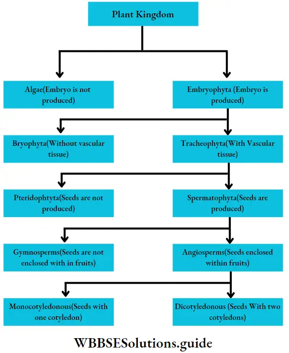
Plant Kingdom Plant kingdom

Each group mentioned in this classification has been discussed separately, later in this chapter.

Phaeophyceae (Brown algae)

Phaeophyceae Definition: Phaeophyceae (Greek: phaios= brown; phyton = plant) are the multicellular, mainly marine, brown-coloured group of algae.

This group of algae is commonly known as brown algae, as they appear golden brown in colour due to the presence of a xanthophyll pigment—fucoxanthin. The fucoxanthin partially masks the chlorophylls and carotenes. Thus gives the characteristic brown colouration.

It is a large group of algae consisting of 240 genera and over 1500 species. Among these 32 genera and 93 species have been reported from India.

Phaeophyceae Distribution: They are mostly marine in habitat and a few are freshwater. The freshwater members are Pleurocladia spv Heribaudiella sp., Lithoderma sp. and Sphacelaria sp. Pleurocladia lacustris grow both in freshwater and marine habitats. They are generally found attached to the marine substratum, in the colder regions of the sea.

Phaeophyceae General features:

  1. They range from simple microscopic (1 mm) heterotrichous filament (Ectocarpus sp.) to the largest alga (60-90 metres) (Macrocystis pyrifera).
  2. Giant brown algae are called kelps. The plant body is non-motile, multicellular and highly differentiated, both externally and internally. Their shapes may range from ribbon-like, filamentous, leaf-like, bush-like, fern-like or some may be branched like trees. Unicellular, colonial and unbranched filamentous forms are completely absent.
  3. The plant body is differentiated into holdfast, short or elongated stipe and an expanded leaf-blade or lamina. The lamina performs photosynthesis and bears reproductive structures. Many species can float due to the presence of air bladders.
  4. The photosynthetic plastids present in Phaeophyceae are called phaeoplasts. The phaeoplasts contain three thylakoid-containing lamellae. The photosynthetic pigments include chlorophyll a, chlorophyll c, beta-carotene, luteins and xanthophylls. The pigment fucoxanthin gives it a distinct brown colour
  5. The growth pattern may be apical (Fucales, Dictyotales), intercalary (Laminariales) or trichothallic (Ectocarpales).
  6. The cell wall is differentiated into outer mucilaginous and inner cellulose layers. The outer layer contains succinic and alginic acids. The cell wall is covered by a layer of gelatinous hydrocolloid called algin. This protects the cell during unfavourable conditions.
  7. The cells usually have many small vesicles and white granules. The granules are called fucosan granules.
  8. Single, stalked pyrenoides are present.
  9. Motile zoospores and gametes have two laterally inserted unequal flagella. The larger one is tinsel or pantonematic and the smaller one is whiplash or acronematic type.
  10. The reserve foods are commonly laminarin and mannitol. Sucrose and glycerol are also present in some members.
  11. Reproduction takes place vegetatively (mainly by fragmentation and special propagules), asexually (asymmetrical flagella-containing zoospores or non-flagellate tetraspores) and sexually (isogamy, anisogamy and oogamy).
  12. In most of the members, fertilisation is external. The zygote does not undergo meiotic division and on germination, it develops into a diploid thallus.
  13. The life cycle is diplohaplontic.
  14. The alternation of generations is isomorphic or heteromorphic in nature.
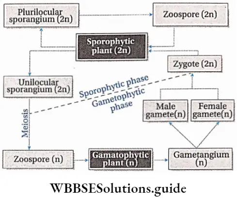
Plant Kingdom Life cycle ofa brown alga

Embryophyta

Embryophyta Definition: The embryophytes are the most familiar sub-kingdom of green plants that form embryos from the zygote after fertilisation.

Embryophyta Definition General features:

  1. Embryophyta is mainly characterised by the presence of multicellular sporophytes, cuticles, archegonia, antheridia and sporangia, as well as the presence of sporopollenin in spore walls.
  2. They are a terrestrial species of plants. They produce embryos but do not produce accessory spores.
  3. The gametangia are covered by a jacket of sterile cells.

Embryophyta Definition Classification: According to the presence of conducting tissue, embryophyta can be divided into two groups—Bryophyta (without vascular tissue), and Tracheophyta (with vascular tissue). These two groups have been discussed under separate heads.

Tracheophyta Tracheobionta

The term tracheophyta came from two Greek words ‘trachea’ (vascular strand) and ‘phyton’ (plants).

Tracheophyta Definition: Tracheophyta are the major terrestrial plant group, with conducting tissues.

The members of this group are also called vascular plants. Vascular plants include the club mosses, ferns, gymnosperms and angiosperms.

Tracheophyta General features:

  1. The tracheophyta is the largest and most advanced group of plants.
  2. The sporophyte (2n) is the dominant generation in the life cycle of tracheophytes.
  3. The sporophyte is independent and autotrophic and possesses specialised vascular tissues.
  4. The plant body is differentiated into true roots, stems and leaves.
  5. Leaves are broad, containing a large amount of chlorophyllous tissues. Stomata are present on the leaves, for the exchange of gases.
  6. Reproduction is mostly vegetative and sexual. Formation of the embryo takes place.
  7. They contain vascular or conductive tissues—xylem and phloem. The xylem transports water and minerals from soil to leaves. The phloem transfers photosynthetic products from leaves to all other parts of the plant.
  8. Their size may range from a few millimetres to several hundred metres.
  9. Their life cycle extends from about some weeks to several years.

Classification of Tracheophyta:

Tracheophytes are classified into two groups—

  1. Pteridophyta (seeds are not produced) and
  2. Spermatophyta (seeds are produced). These have been discussed below under separate heads.

Pteridophyta

Pteridophytes (Greek: pteron-feather, phyton-plants) are non-flowering vascular plants. Hence they are also known as ‘vascular cryptogams’.

Pteridophytes Definition: Pteridophyta is the large group of primitive land plants, with true body differentiation and conducting tissues but does not produce seeds.

They are represented by about 400 genera and about 10,500 species including both living and fossil plants.

Pteridophytes Distribution: Most of the pteridophytes are terrestrial in nature. They are mainly found in damp, shady parts of mountains, damp walls, tree trunks, etc. Aquatic plants like Azolla sp. and parasitic plants like Polypodium sp. are also included in this group.

Pteridophytes Life cycle: Pteridophytes show mainly two types of life cycle—sporophytic and gametophytic.

Characteristics of sporophytes:

  1. Pteridophytes are the first true land plants. The predominant generation is the sporophytic plant body, differentiated into true root, stem and leaves. The sporophyte is independent and autotrophic.
  2. The stem is aerial or rhizomatous. The stem is generally branched—either dichotomous or monopodial. Stem sometimes is densely covered with brown scales.
  3. The primary root is short-living. The primary root is replaced by a dichotomously branched, adventitious root.
  4. Conducting tissues (xylem and phloem) are present. But tracheids and companion cells are absent in the xylem and phloem respectively.
  5. Leaves are of two types— brown-coloured scale leaves and green-coloured sporophylls bearing sporangia, Sporophylls may be small microphyllous and large megaphyllous. The young leaf tips of ferns show circinate ptyxis (arrangement of immature leaf coiled form, which unfolds with maturity).
  6. Haploid (n) spores are produced within sporangia. In some species (for example Dryopteris) all the spores are similar in structure. They are called homosporous pteridophytes. In some species (for example Marsilea) spores are dimorphic (i.e., larger female spores or megaspores and smaller male spores or microspores). Such plants are known as heterosporous pteridophytes.
  7. In the case of heterosporous plants, megasporangia are borne on megasporophyll, while microsporangia are borne on microsporophyll.
  8. Some pteridophytes bear a cluster of sporangia called sori (singular: sorus) on either the lower surface of leaves (example Dryopteris sp.) or inside special structures called sporocarps (example Marsilea sp.) or in spike(for example Ophioglossum sp.).
  9. The sori are covered by a bilayered, smooth membrane, called indusium.
  10. These spores germinate to form gametophytic plants.
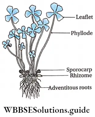
Plant Kingdom Pteridophyte—Marsileasp

Plant Kingdom Sori ofDryopteris sp

Characteristics of gametophytes:

  1. The gametophytic plant is haploid, autotropic, independently living, dorsiventral, heart-shaped thallus. It is called prothallus.
  2. Prothalli are rather simple structures and do not have vascular tissues. They may be attached to the substrate by fine multicellular rhizoids. They resemble thallose liverworts.
  3. In the case of homosporous plants, the spores germinate to produce monoecious prothallus, which produces both antheridia and archegonia.
  4. In the case of heterosporous plants, antheridia are produced on male prothallus developed from microspores and archegonia are produced on female prothallus developed from megaspores.
  5.  Antheridia are small club-shaped structures, that produce motile sperms or antherozoids. They show chemotactic movement towards archegonia. Archegonia are flusk-shaped structures, with a cup-shaped venter and a long neck. Inside the venter an egg cell is present.
  6. The zygote is produced after fertilisation and quickly undergoes repeated mitotic divisions to form an embryo. The embryo develops the sporophytic plant.
  7. Flowers, fruits and seeds are not produced.

Some common examples of Pteridophytes are— Psilotum sp., Lycopodium sp., Selaginella sp., Isoetes sp., Pellia sp., Equisetum sp., Ophioglossum sp., Pteris sp., etc.

Alternation of generations: All pteridophytes show a diplohaplontic life cycle, heteromorphic alternation of generations.
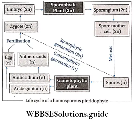
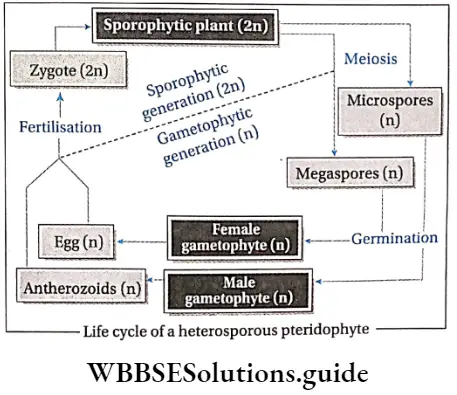
Plant Kingdom Life cycle ofhomosporousfern

Plant Kingdom Life cycle ofheterosporousfern

The alternation of generations in the life cycle of a typical pteridophyta (Dryopteris sp.) is shown in the figure below.
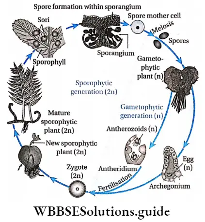
Plant Kingdom Alteration ofgenerations in life cycle ofDryopteris sp.

Commercial importance of pteridophyta: Many species of pteridophytes are used for various purposes.

Source of Food: The young leaves or fronds of Ampelopteris proliferate, leaf tips of Matteuccia struthiopteris and leaves of Diplazium esculentum, Marsiiea sp., are also used as vegetables.

Source of fodder: Dry fronds of many ferns are used as the livestock feed for cattle. The leaves of Marsiiea sp. are used as fodder for animals. The rhizome of many ferns such as Pteris sp., which is rich in starch, is used as animal food. The corm (modified stem) of Isoetes sp. is consumed as food by pigs, ducks and other animals.

Source of medicines: Lycopodium sp., Equisetum sp., Selaginella sp., Marsiiea sp., etc., are some pteridophytes that are used to treat neural disorders.

The spores of Lycopodium sp. have been widely used in pharmacies as a protective powder for tender skin and also as water-repellants. The foliages of Lycopodium sp. are used as tincture, powder, ointment and cream and used as a diuretic too.

Equisetum sp. (Horsetail) is rich in silicic acid and silicates. Potassium, aluminium and manganese, along with fifteen types of flavonoid compounds, have been reported from Equisetum.

The flavonoids and saponins are assumed to cause the diuretic effect. The silicon is believed to exert connective tissue-strengthening and anti-arthritic action.

Several ferns are used as herbal medicine. Oil extracted from Aspidium rhizome is used as a vermifuge (medicine against worms). The leaf extract of Polypodium is used to cure stomach and liver problems.

The root extract of Osmunda regatis is used for the treatment of jaundice. The root extract of Osmanda vulgaris is used to heal wounds.

Aesthetic uses: Many species of pteridophytes are cultivated for their aesthetic value. Some epiphytic species of Lycopodium sp. are grown on hanging baskets.

Several species of Selaginella are used in decoration during festive occasions due to their decent foliage and colour. Several ferns such as Angiopteris sp., Aspienium sp., Marattia sp., etc., have beautiful soral arrangements, hence used for ornamentation.

Polishing purpose: Accumulation of silica in the stem cells of Equisetum sp. gives it rough structures. Thus, it can be used as a scrubber for polishing purposes.

Preparation of fire-crackers: Since the spores of Lycopodium sp., are highly inflammable, hence they can be used to prepare fire-crackers.

Biofertilisers: Azolla sp., is a free-floating water fern which can grow very quickly through vegetative propagation. They live symbiotically with a nitrogen-fixing cyanobacteria—Anabaena sp., etc.

The alga provides nitrogen to the plant and to the growing aquatic medium. Thus, Azolla sp., growing in rice fields serves as a green manure for better crop yield.

Metal indicators: Equisetum sp., accumulates minerals, especially gold, in their stem. The rate of accumulation even reaches up to about 4.5 ounces per ton.

They may be referred to as gold indicator plants, which help in identifying a region with gold ore deposits. Similarly, Aspienium adulterinum is an indicator of nickel and Actinopteris australis is a cobalt indicator plant.

Fossil fuels (coal): During the carboniferous period, the flora of the earth was dominated by huge pteridophytes (Lycopods), ferns and other large leafy plants. The coal formed after their death had become sources of different fossil fuels.

Classification of pteridophyta: Pteridophytes have been classified into four major classes by Doyle (1971)—Psilopsida, Lycopsida, Sphenopsida and Pteropsida.
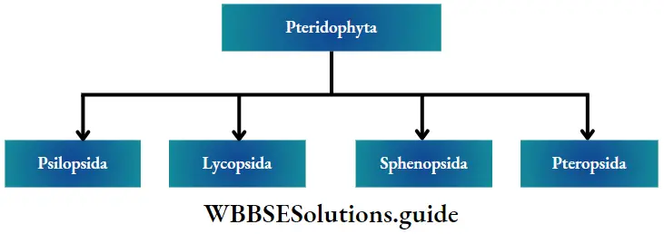
Plant Kingdom Pteridophyta

Fossil fuels General features:

  1. They are mainly terrestrial pteridophytes, found in warm climates.
  2. The plant body is a rootless sporophyte that differentiates into a subterranean (just below the earth’s surface) rhizome and an aerial erect shoot.
  3. Branching is dichotomous in both rhizome and shoot.
  4. The large rhizoids borne on the rhizome, absorb water and nutrients from the soil.
  5. The aerial shoots bear spirally arranged scale-like (for example Psilotum sp.) or leaf-like appendages (for example Tmesipteris sp.).
  6. Stele is protostelic or siphonostelic with sclerenchymatous pith.
  7. Cambium is absent Hence, secondary growth is absent.
  8. Bi or trilocular sporangia are borne in the axils of leaf-like appendages.
  9. The mode of sporangial development is of eusporangiate (more than one cell initiates sporangial development)type.
  10. They are homosporous.
  11. The gametophytes or prothalli are independent, colourless, cylindrical, branched and subterranean. They grow as saprophytes with an associated endophytic fungus.
  12. Antherozoids are spirally coiled and multi-flagellated. Examples: Psilotum sp. (living); fossil members are- Rhynia sp., Cooksonia sp., Zosterophyllum sp etc.

Lycopsida (Lycopods)

Lycopsida General features:

  1. Thin and small structured pteridophyta, found mostly in forests and mountains.
  2. The sporophytic plant body is differentiated into definite root, stem and leaves.
  3. The sporophytes are dichotomously branched.
  4. The leaves are usually small and microphyllous. The leaves are either isophyllous or dimorphic. In some species, ligules (tongue-shaped membranous structures) may present towards the leaf apex.
  5. The xylem arrangement in the stem is exarch.
  6. Stele may be protostele, siphonostele or polystele in nature
  7. Sporangia are borne singly on the adaxial (upper) surface of the sporophylls. In most cases, sporophylls join to form cones or strobili.
  8. The spores are homosporous (for example Lycopodium sp.) or heterosporous (for example Selaginella sp.).
  9. The spores develop into independent gametophytes.
  10. Antherozoids are flagellated.

Examples: Lycopodium sp. and Selaginella sp. are living genera and there are 14 extinct genera—Asteroxylon sp., Baragwanathia sp., etc.

Sphenopsida (Horsetails)

Sphenopsida General features:

  1. The living members of this group are found in damp and shady regions of mountains, forests and marshlands.
  2. The stems and branches are differentiated into nodes and internodes. Internodes are with longitudinal ridges and furrows.
  3. Branches arise in whorls.
  4. The leaves are extremely reduced and borne in whorls at the nodes of aerial branches and stems.
  5. Stele is Protostele or siphonostele in nature.
  6. The sporangia develop on an elliptical pallet appendage called sporangiophore. Sprorangiophores are arranged into a cone shaPed strobilus (P|ural: strobila)-
  7. Most of the members are homosporous including Equisetum sp. However, some extinct forms were heterosporous (for example Catamites Hashana).
  8. The gametophytes are exosporic (develop outside the spore) and green.
  9. Antherozoids are multiflagellate.

Examples: This class is represented by a single living genus and 18 extinct genera. The living genus is Equisetum sp. and the fossil genera are Catamites sp. Annularia sp., etc.

Pteropsida

This group is commonly known as ferns. The pteropsida differs from other classes in having large leaves (fronds). This is the largest and highly evolved group of pteridophytes and is represented by about 9,000 species which show a wide range of distribution. Pteropsida are known from as far back as the Devonian period of the Paleozoic Era.

Pteropsida General features:

  1. Members of Pteropsida are found in shady and damp forests, high altitudes of mountains, marshlands and moist soil of plains. Some members are aquatic and some are epiphytic.
  2. The sporophytes are usually perennial in nature and differentiated into roots, stems and spirally arranged leaves.
  3. The stem is rhizomatous, mostly short and stout. Adventitious roots grow from it.
  4. The rhizome is covered with brown scales.
  5. The leaves are large, simple (Ophioglossum sp.) or pinnately compound (Dryopteris sp.) and are called fronds. In Adiantum sp., the rachis is dichotomously branched and bears a fan-shaped leaflet.
  6. The rachis is covered with tiny brown hairs called ramenta.
  7. Young fronds show deracinate ptyxis, except Ophioglossum sp.
  8. The stele can be of different types, for example protostele, siphonostele, etc.
  9. Vegetative reproduction fragmentation, adventitious buds and bulbils (fleshy globose structures). In Adiantum sp., new plants, are produced from leaf tips when they come in contact with the soil.
  10. Matured vegetative leaves bear sori on their lower surface. They are then known as sporophylls. Sori bear a number of sporangia. Sporangial clusters are sometimes enclosed by a shield-like structure called indusium.
  11. These plants are homosporous in nature. Haploid spores are formed through meiosis from diploid spore mother cells.
  12. Monoecious gametophytes or prothalli are produced by the germination of spores. Rhizoids present on prothallus.
  13. Antherozoids are ciliated and coiled.

Examples: Ophioglossum sp., Pteris sp., Dryopteris sp., etc.

Similarities between bryophyte and Pteridophyta

  1. Both are non-flowering and terrestrial.
  2. Both have multicellular and multiflagellate reproductive organs.
  3. Both their antheridia and archegonia are similar in structure, with a layer of sterile cells covering them. Antheridia are club-shaped and archegonia are flask-shaped in both.
  4. Antherozoids are coiled and motile in both.
  5. Both have a definite alternation of generations.
  6. Asexual reproduction takes place by spores.
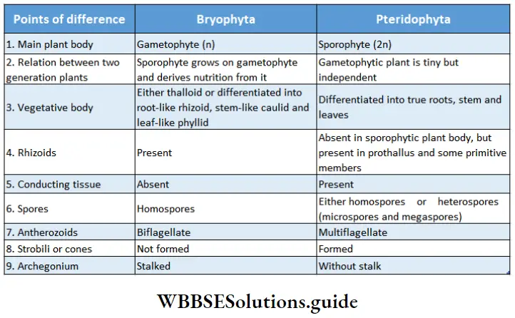
Plant Kingdom Differences between bryophyta and pteridphyta

Spermatophyta

Spermatophyta Definition: Spermatophyta is the higher group of flowering land plants where seeds are produced and embryo development takes place.

Spermatophyta Distribution: They are found all over the land and sometimes in water as well.

Spermatophyta General features:

  1. The body is differentiated into roots, stems and leaves. Vascular tissues (xylem and phloem) and mechanical tissues, both are present.
  2. The plants are heterosporous with microspores and megaspores.
  3. Gametophytic plants are dependent on sporophytic plants.
  4. The reproductive organs are multicellular. Instead of being released, the megaspores remain within the ovules where one or more female gametes are formed.

Classification of the Spermatophyta: The spermatophytes are further classified into two groups
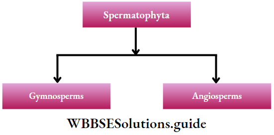
Plant Kingdom Classification of the Spermatophyta

These topics are discussed under separate heads.

Cycadopsida

Members of the class Cycadopsida produced the most dominating flora during the mid-Mesozoic Era.

Cycadopsida General features:

The general features of Cycadopsida are as follows—

  1. Plants generally with stout trunks having manoxylic wood. They have large pith regions but have woody cortexes.
  2. Leaves are large and frond-like. They are basically pinnately compound in form or in venation.
  3. Annual rings are absent.
  4. Presence of ciliated sperms.
  5. These plants are usually dioecious with the reproductive organs borne on specialised leaves (sporophylls).
  6. The pollen grains (microspores) form within the microsporangia on the sporophylls of the male cones.
  7. The ovules (megaspores) develop without protective coverings on the sporophylls of the female cones.
  8. The pollination is carried out by wind or beetles.
  9. After fertilisation, the seed develops without a protective pericarp. The seed is radially symmetrical. The seed has an outer fleshy layer and the embryo has two cotyledons.
  10. The life cycle is diplohaplontic, alternation of generations is heteromorphic. The life cycle of cycads has two distinct phases. The gametophyte stage is microscopic and enclosed within the microspores and megaspores.

Examples: Cycas sp., Ceratozamia sp., Zamia, etc.

Ginkgopsida

The class Ginkgopsida includes a single order of Ginkgoales—now represented by a single living member Ginkgo biloba. The members of this group first appeared in the Permian period, but gradually depleted from the earth.

The leaf impressions of G. biloba have been identified from the Permian rocks and probably from the Carboniferous rocks. G. biloba has escaped extinction and still exists today being the oldest living seed plant Thus, Ginkgo biloba is referred to as a ‘living fossil’.

Ginkgopsida General features:

The general features of Ginkgopsida are as follows—

  1. The members of this group are mostly tall, woody, deciduous and branched trees. They have both long and dwarf shoots.
  2. Secondary wood is pyroxylin i.e., has a large number of thick-walled tracheids and fibres.
  3. Leaves are leathery, strap-shaped or fan-shaped, often with dichotomous venation.
  4. These plants are dioecious, i.e., both male and female gametophytes are present on the same plant. Produce both male and female cones.
  5. Male fructifications are axillary, unbranched catkin-like bearing microsporangiophores.
  6. Each microsporangiophore bears 2-12 pendulous microsporangia.
  7. Ciliated sperms are present.
  8. Ovules are 2-10 in number, terminal on axillary branching or almost unbranched axes.
  9. Seeds are large showing radial symmetry.
  10. Seeds have with fleshy outer layer and a stony middle layer.

Examples: Ginkgo biloba

Coniferopsida

The Coniferopsida includes four orders—Cordaitales, Voltziales, Coniferales and Taxales. They probably appeared during the Upper Devonian and were the most dominating taxa during the Upper Carboniferous to Triassic. At present, this class is represented by 57 living genera.

Coniferopsida General features:

The general features of Coniferopsida are as follows—

  1. Mostly tall, woody, evergreen plants which are profusely branched.
  2. Secondary wood is pyroxylin.
  3. They have needle-shaped, paddle-shaped or fan-shaped foliage leaves. The arrangement of leaves is whorled or a|ternate. The )eaves are covered with a thick cuticle.
  4. Long and dwarf shoots are present.
  5. Stele in the stem are endarch, pith and cortex are small, with large conducting tissues. The woody part is more in these plants.
  6. Resin canals are often present in leaves and stems.
  7. The plants are usually dioecious. The reproductive organs are borne on specialised leaves (sporophylls), which are compactly arranged in cones or strobili.
  8. Male cones or microcstrobili are smaller and survive for less number of days. Female cones or megastrobili are large, and long and survive for longer periods of time.
  9. The gametophyte stage is represented by microspores or pollen.
  10. Male gametes are represented by the male nucleus only.
  11. The pollen forms within the microsporangia present on the scales (microsporophylls) of the male cones. They are often winged.
  12. The ovules are borne on the sporophylls of the female cones and the pollen is usually transferred to the ovules by wind.
  13. After fertilisation, the seed develops directly on the female sporophylls.
  14. Seeds are bilaterally symmetrical. The embryo has two too many cotyledons.

Examples: Pinus sp., Picea sp., Abies sp., Cedrus sp., etc.

Gnetopsida

The class Gnetopsida is considered to be the highly evolved group of gymnosperms. The class comprises three distinct orders—Ephedrales, Welwitschiales and Gnetales, with a monogeneric family in each. However, some scientists prefer to retain them within a single order, Gnetales, with a monogeneric family in each.

Gnetopsida General features:

The general features of Coniferopsida are as follows—

  1. The members of this group are small trees or shrubs or lianes in nature.
  2. The plants bear opposite or whorled leaves, with reticulate venation.
  3. Vessels are found in the secondary xylem. For that reason, they are considered to be ancestral to angiosperms.
  4. Resin canals are absent.
  5. Flowers are monoecious. Male and female cones are borne separately.
  6. Compound male and female strobili resemble the inflorescence of angiosperms.
  7. Male flowers consist of a stalk bearing two or more sporangia. They may bear bract and perianth.
  8. Female flowers consist of ovules only.
  9. They show unique fertilisation features i.e., a pollen tube grows in order to fertilize the egg. Thus, the sperms themselves are not motile, like other gymnosperms.
  10. The embryo contains two cotyledons.

Examples: Gnetum ula, Ephedra sp., Welwitschia sp., etc
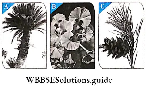
Plant Kingdom Different types ofgymnosperm

Similarities between Pteridophyta and gymnosperms

  1. Both have a major sporophytic phase and a less important gametophytic phase.
  2. Both have sporophytic plant bodies, differentiated into roots, stems and leaves.
  3. Both have vascular tissues, without vessels in the xylem (exception: Gnetopsida) and companion cells in the phloem.
  4. Pteridophytes are heterosporous and have motile male gametes (reproductive cells) like many gymnosperms (for example Cycas sp.).
  5. Both show definite alternation of generation.
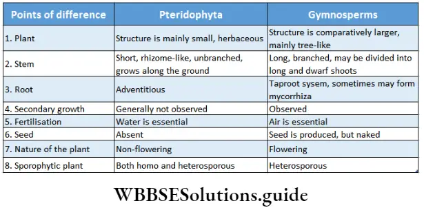
Plant Kingdom Differences between pteridophyta and gymnosperms

Angiosperms

Angiosperms Definition: Angiosperms are flowering plants that produce fruits, that enclose the seeds.

Angiosperms are the highest-order plants in the plant kingdom. They are the largest group of plants, that are flowering and contain seeds in special structures, called fruits.

Angiosperms Distribution: Angiosperms originated quite late on earth, yet they occupy the majority of it. Most of them are terrestrial, and only a few are aquatic. They are found under the ocean, as well as at 6000m above sea level.

Angiosperms General features:

  1. The main plant body is sporophytic in nature. The sporophytic stage is independent and diploid (2n). The gametophytic phase is haploid (n) and dependent on the sporophytic phase.
  2. Angiosperms are trees, shrubs or herbs in nature. The trees and shrubs are generally perennials, while the herbs are annuals, biennials or perennials.
  3. An angiospermic plant consists of different organs—root, stem, branches, leaves, flowers, fruits and seeds.
  4. The body of an angiosperm plant consists of two systems— the root system and the shoot system.
  5. The roots may be taproot or adventitious.
  6. The stem may be branched or unbranched. In some cases, they may be climbers or lianes.
  7. The leaves are simple or compound, dorsiventral or isobilateral.
  8. The conducting tissues are the xylem and phloem. The vascular bundle of roots are radial and exarch, while that of the stem is collateral or collateral, free or enclosed and endarch. Secondary growth is observed in dicots.
  9. Angiospermic plants contain chlorophyll in their leaves and thus are able to photosynthesise.
  10. Flowers may be complete (all four floral whorls are present), incomplete, bisexual, unisexual (male or female), or sterile in nature. They have accessory whorls (sepals and petals), along with reproductive whorls (androecium and gynoecium).
  11. Androecium has an anther and filament. The anther produces pollen grains.
  12. The gynoecium has an ovary, style and stigma. One or more ovules are produced within the ovary.
  13. The male gametophytes form pollen tubes. They contain two nuclei.
  14. The archegonia are not produced within female gametophytes. Instead, the female gametophytes get reduced to four megaspores.
  15. Only one of the megaspores develops into an embryo sac which contains an egg nucleus. Double fertilisation, occurs in an egg angiosperms. Apart from a zygote, a triploid endosperm is also produced from this.
  16. After fertilisation, the ovule transforms into seed and the ovary into fruit.
  17. The zygote develops into an embryo by repeated cell division. After germination, it grows into an adult plant.

Sexual Reproduction in Flowering Plants: Various steps of the sexual reproductive procedure of flowering plants are discussed below.

Microsporogenesis: Microspore i.e., the pollen grain is the first cell of the male gametophyte, which contains only one haploid (n) nucleus. During the early stage of development, four microspores or pollens (n) are formed from each microsporocyte or pollen mother cell through meiosis.

Pollination: Transfer of pollen grains to the stigma takes place by several agents, such as wind, water, insects, etc. This process is called pollination. It takes place by two means—self-pollination (within the same flower or two different flowers of the same plant) and cross-pollination (between two different flowers of two plants).

Development of male gametophyte: Firstly, the pollen cell (n) undergoes mitotic division and forms a small generative cell and a large vegetative or tube cell. The generative cell further divides and gives rise to two male gametes. This is known as the 3-celled stage containing a vegetative cell and two male gametes.

The division of the generative cell may take place either in the pollen grain or in the pollen tube. The pollen tube comes out through germ pores after pollination. The nucleus of the vegetative cell, commonly known as the tube nucleus, usually degenerates with the maturation of the generative cell.
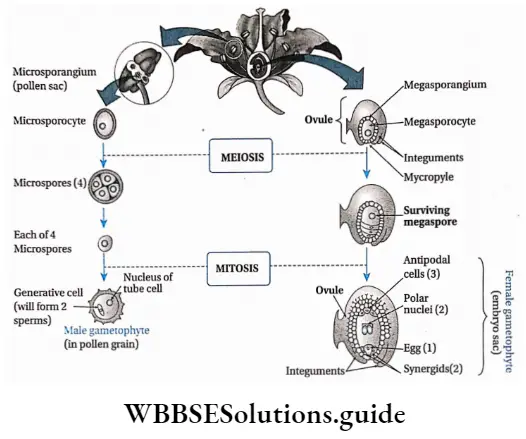
Plant Kingdom Development ofmale andfemale gametophyte in angiosperms

Development of megaspore mother cell: The ovule, inside the ovary, develops as a multicellular placental outgrowth including the epidermal and numerous hypodermal cells. This structure develops into a mass of tissue called nucellus, with one or two integuments.

One of the hypodermal (just below the outermost layer) cells of the nucellus enlarges in size and has a dense cytoplasm and a conspicuous nucleus. This is called an archesporial cell, which subsequently divides transversely and forms an inner primary sporogenous cell and an outer primary parietal cell.

The inner primary sporogenous cell functions as a megasporocyte or megaspore mother cell and the primary parietal cell through repeated vertical divisions forms layers of parietal cells. Sometimes, the archesporial cell does not divide and directly functions as a megaspore mother cell.

Megasporogenesis: The diploid megaspore mother cell meiotically divides to form four haploid (n) megaspores, within the ovule. Two subsequent transverse divisions form four, linearly arranged, haploid megaspores (linear tetrad).

Out of four megaspores, only one moves towards the chalazal end (mostly towards the stalk) and becomes the functional megaspore.

The other three, remain at the micropylar end (the small opening of the integument), where they gradually disintegrate. The functional megaspore enlarges to form the female gametophyte i.e., the embryo sac.

Development of female gametophyte: Megaspore (n) is the first cell of the female gametophyte. The functional megaspore enlarges and forms the embryo sac. The nucleus of this embryo sac through three successive divisions forms eight nuclei.

Only one of these eight nuclei forms the egg cell (n). Four of these eight nuclei reside towards the micropylar end and the remaining four towards the chalazal end. A single nucleus from each pole then moves towards the centre and forms a pair of polar nuclei, which then fuse to form a diploid nucleus, called the definitive nucleus (2n).

This is also called a polar fusion nucleus or secondary nucleus. The three nuclei at the micropylar end (one egg cell and two synergids) form the egg apparatus and the rest three at the chalazal end, are called antipodal cells.

In the egg apparatus, each nucleus remains surrounded by a viscous mass of cytoplasm without any wall. The m‘dd*e largest one is called egg or ovum or oosphere and the rest two on each side of the egg are the synergids. The antipodal cells have a viscous mass of cytoplasm, each covered by a cellulosic wall.

Fertilisation: Fertilisation is a process where male and female gametes fuse with each other to form a zygote, which later on forms the multicellular embryo. In angiosperms, the embryo sac remains embedded into the ovarian cavity.

The pollen grains reach the stigma through pollination. The pollen grain germinates on the stigma and forms a pollen tube which grows through the style and reaches the ovule. Here it releases the male gametes near the egg.

One of these male gametes fuses with the egg, to form the diploid zygote (2n). The other male gamete fuses with the definitive nucleus (2n) to form the endosperm nucleus (3n). This gives it the name, double fertilisation.
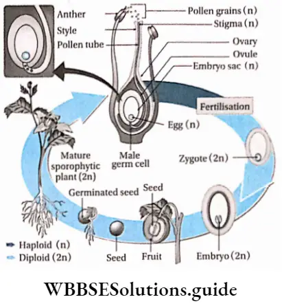
Plant Kingdom Life cycle ofangiosperms

Germination of pollen grains

The pollen grain usually develops a single pollen tube (monosiphonous), but occasionally in some cases, it may; develop more than one tube (polysiphonous). Sometimes the single pollen tube may be branched.

The commercial importance of angiosperms:

Human civilisation is dependent on plants. The three major necessities of human beings— food, clothing and shelter, are fulfilled mainly by plants.

Food source:

Plants yield various types of food such as—

  1. Cereals, i.e., rice, wheat, maize, etc.
  2. Millets, i.e., small-grained food such example, jowar, bajra, ragi, etc.
  3. Pulses, like, gram, pea, pigeon pea, lentil, mung, etc.
  4. Vegetables, like, radish, carrots, sweet potatoes, etc.
  5. Fruits, like, orange, banana, apple, guava, mango, etc.

Food adjuncts: Plants yield spices, flavouring materials, beverages, etc.

  1. Spices and other flavouring materials like ginger, turmeric, cinnamomum, cloves saffron, pepper, etc.
  2. Beverages like tea, coffee, etc.

Production of medicines and drugs:

  1. Medicinal plants such as Cinchona sp., Ipecac sp„ Rauvolfia sp. etc., are sources of various alkaloids. These alkaloids are used to produce medicines.
  2. Many angiosperms are sources of fumitories and masticatories, for example, tobacco, °P‘um’ marijuana (ganja), etc.

Industrial use of plants and plant products: Plants yield fibres (for example jute, cotton, hemp, etc.), timber (for example teak, sal etc.), and essential oils (for example Eucalyptus, Citronella, etc. sugars (for example sugarcane, palm), cellulose products etc. Each of these has different uses in our daily lives.

Fuel: Dead remains of plants and woody plant parts are used as fuel.

Similarities between gymnosperms and angiosperms

  1. Both have major sporophytic phases. The gametophytic phase was dependent on the sporophytic phase.
  2. Both have their body differentiated into roots, stem and leaves. Both produce flowers.
  3. Both have open or closed collateral vascular bundles.
  4. Both show the growth of pollen tubes.
  5. Both are heterosporous plants in nature.
  6. In both of them, female spores remain within sporangia or ovules.
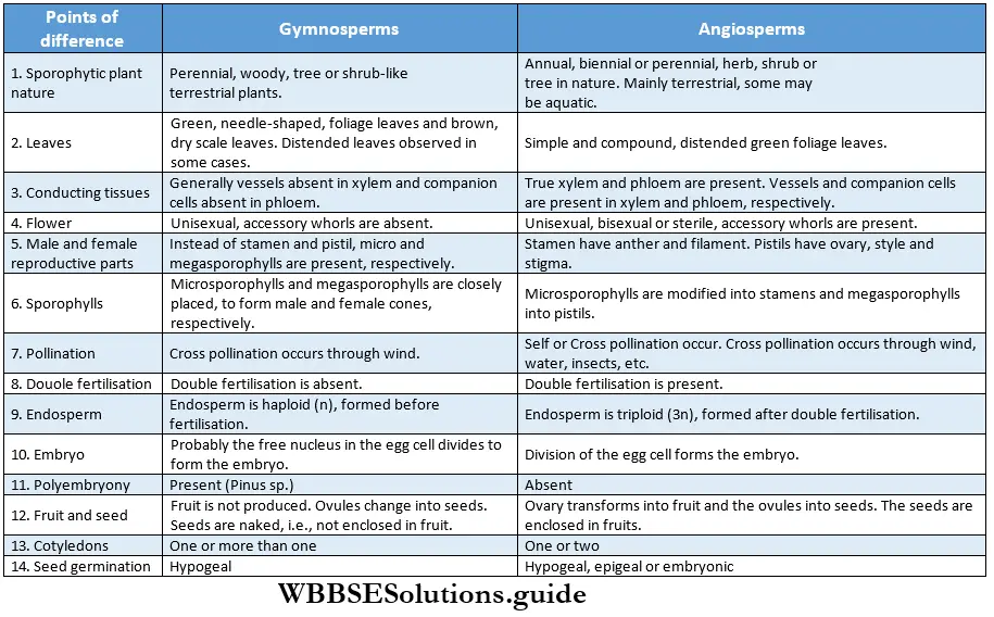
Plant Kingdom Differences between gymnosperms and angiosperms

Classification Of Angiosperms

Bentham and Hooker’s System: Bentham and Hooker worked together for about 25 years and jointly published their work in the book Genera Plantarum (1862-1883) in three volumes.

They divided the Angiospermae into two major classes—

  1. Monocotyledonae (comprises plants having single cotyledon in seed, i.e., monocotyledonous plants) and it was divided into seven series. The Series were further divided into cohorts (= orders), and cohorts into natural orders (= families). Natural orders include genera and finally, each genus includes species.
  2. Dicotyledonae (comprises plants having two cotyledons in seed, i.e., dicotyledonous plants). It was further divided into three sub-classes—Polypetalae, and Monochlamydeae. Polypetalae was divided into three series; Gamopetalae was divided into three series and Monochlamydeae into eight series. These groups are discussed under separate heads.

Monocotyledonous Plants

Definition: Monocotyledonous plants are those angiosperms that have a single cotyledon in their seeds.

Monocotyledonous plants General features:

  1. Generally, they are annual plants, but the trees are perennial.
  2. Plants are usually herbaceous, with weak stems (for example rice, wheat, grass, etc.). Some may be shrubs and trees (palm, date palm, coconut), while some may be climbers (orchids).
  3. The embryo contains one cotyledon. The endosperm is well-developed and store food for future use.
  4. The leaves may be simple or compound. They show parallel venation and sheathing leaf bases.
  5. Leaves are isobilateral, mesophyll tissue is not differentiated into spongy and palisade parenchyma.
  6. Stems are generally unbranched, with nodes and internodes. In paddy and bamboo, the internodes are hollow, while in maize and sugarcane, those are solid. In some plants like ginger, onion, etc., stems are underground.
  7. At the early stage of development, plants lose their tap roots and fibrous root systems develop.
  8. The flowers are trimerous (the number of whorls present is 3 or in multiples of 3).
  9. Calyx and corolla fuse to form the perianth.
  10. Stem vascular bundles are conjoint, collateral and closed. The bundles remain scattered throughout the cortex. No stele is formed.
  11. The number of vascular bundles in the root is usually six or more.
  12. Due to the lack of cambium in vascular bundles, no secondary growth is observed in the stem and roots.
  13. The plumule is present near the embryo axis. Coleoptile and coleorhiza are present in seed.
  14. The seeds show hypogeal germination.

Examples: Pisum sativum, Mangifera indica, Hibiscus rosa-sinensis, etc.

Dicotyledonous Plants

Dicotyledonous plants Definition: Dicotyledonous plants are those angiosperms that have two cotyledons in their seed.
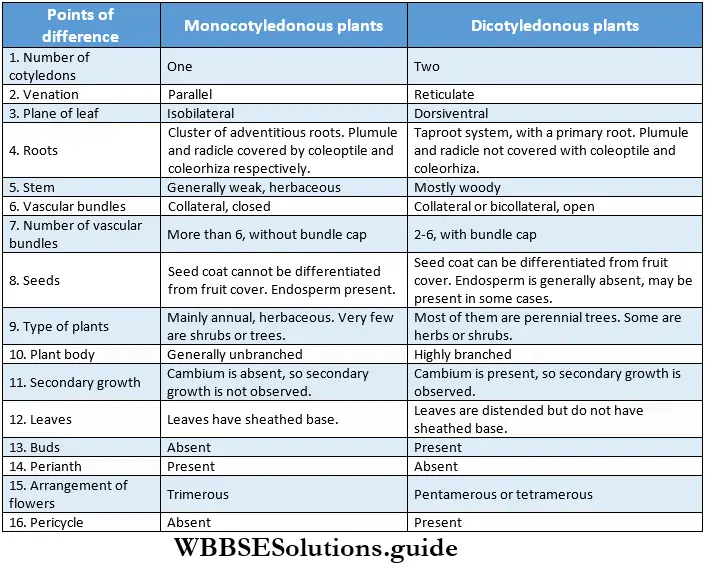
Plant Kingdom Differences between monocotyledonous and dicotyledonous plants

Plant Life Cycles And Alternation Of Generations

A life cycle is a sequence of events that occur from birth to death of an organism. Generation is any entity with distinct morphological features that represent a particular event or phase in the life cycle.

Based on chromosome number an organism can be haploid or diploid. In plants, haploid gametes are produced by gametophytes while the diploid structures that produce spores by meiosis are called sporophytes.

In their life cycles, plants have the gametophytic or sporophytic phase as the dominant one, in which they pass most of their life span. Depending upon these dominant phases, life cycles are mainly of three types.

Those types are discussed below—

Haplontic or haplobiontic life cycle:

  1. In this type of life cycle, the gametophyte (haploid) stage is dominant throughout the life cycle and the sporophyte (diploid) stage is represented only by the zygote.
  2. The gametophytic plant develops haploid gametes inside the gametangia.
  3. The fusion between these two gametes forms the zygote.
  4. The zygote immediately undergoes meiotic division and forms four haploid meiospores.
  5. These meiospores develop into haploid plants. The alternation of generations can be interpreted by changes in the chromosome number.

Example: This type of life cycle is observed in Volvox sp., Chlamydomonus sp., Spirogyra sp., etc.
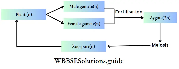
Plant Kingdom Plant

Diplontic or diplobiontic life cycle:

  1. In this type of life cycle, the plant body is sporophytic and diploid and the haploid gametophytic phase is represented only by the gametes.
  2. Sporophytic plants bear gametangia inside which gametes are produced by meiosis.
  3. The gametes upon fertilisation form diploid zygote.
  4. The zygote gives rise to a new sporophytic plant body.

Example: This type of life cycle is observed in Cladophora sp., Fucus sp., etc., algae and in all seed-bearing plants i.e., gymnosperms and angiosperms.

Plant Kingdom Fertilisation

Haplo-diplobiontic life cycle:

  1. In this type of life cycle, both the haploid and diploid phases are prominent.
  2. They differ in chromosome number and reproductive function. The haploid gametophytic plant reproduces sexually, i.e., by producing gametes.
  3. The gametes produce diploid zygotes through fertilisation. The zygote gives rise to the diploid sporophyte.
  4. The sporophytic plant reproduces by asexual processes, i.e., by producing haploid spores.
  5. In this life cycle, alternation of two generations occurs by sporogenic meiosis and fusion of gametes.
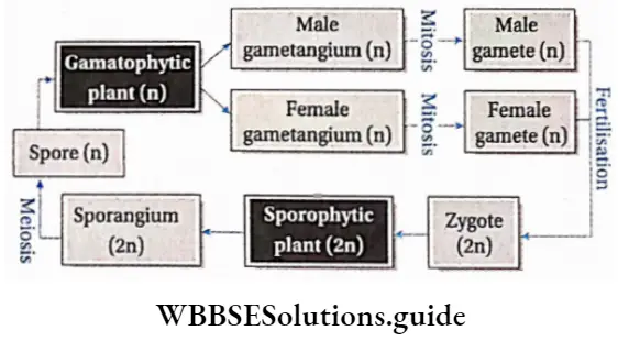
Plant Kingdom Gamatophytic plant

  1. Isomorphic or homologous types, i.e., gametophyte and sporophyte are morphologically similar. Example: This type of life cycle is observed in Ectocarpus sp., Polysiphonia sp., etc.
  2. Heteromorphic or heterologous type, i.e., the gametophyte and sporophyte are morphologically dissimilar. Example: This type of life cycle is observed in bryophytes and pteridophytes.

Plant Kingdom Notes

  • Alkaloid: A class of nitrogenous organic compounds of plant origin having physiological actions on humans.
  • Cilium (plural—cilia): An organelle found in eukaryotic cells. In some organisms, cilia are the means of motility.
  • Coenocytic: A multinucleate thallus/body with a continuous mass of protoplasm enclosed by one cell wall.
  • Coleoptile: A sheath protecting the tip of the plumule in monocots.
  • Coleorhiza: A sheath protecting the tip of a radicle in monocots.
  • Dichotomous branching: A pattern of branching where an equal division of an apical bud or apical growth point forms two equal branches.
  • Dorsiventral: An organ or body of an organism having dissimilar upper (dorsal) and lower (ventral) surfaces.
  • Epigeal: Germination of seeds with one or more seed leaves or cotyledons appearing above the ground.
  • Hypogeal: Germination of seeds with cotyledons appearing above the ground. Plumule: Part of the embryo which will grow into the shoot.
  • Radicle: Part of the embryo which will grow into root.
  • Sporopollenin: It is an inert biopolymer found in the tough outer wall of spores and pollen grains.
  • Tinsel flagellum: A type of flagellum which has fine minute hairs along the axis.
  • Whiplash flagellum: A type of flagellum which has a smooth surface.
  • Xylan: A type of polysaccharide found in the cell walls of plants and algae.

Points To Remember

  1. Linnaeus first used the term algae.
  2. The branch of science that deals with the study of algae is called phycology.
  3. Body of algae is a simple thallus, that consists of parenchymatous cells.
  4. Large marine algae are known as seaweeds or kelp.
  5. The longest phase in the life cycle of algae is its gametophytic phase.
  6. Stored food in green algae are starch and oil.
  7. Red algae appear reddish due to the presence of the pigment phycoerythrin.
  8. The pigment responsible for the brownish colour of brown algae is fucoxanthin.
  9. The main stored food of Phaeophycean algae is laminarin.
  10. Algae mostly exist as haplobiontic, diplobiontic and triplobiontic.
  11. The cell wall of Chlamydomonas sp. lacks cellulose.
  12. Volvox sp. is a freshwater green algae that forms colonies.
  13. Volvox sp. was first observed by Antonie van Leeuwenhoek in 1700.
  14. Chlorella sp. is a unicellular green algae that is used as a food.
  15. Chlorella sp. is a source of antibiotic chlorellin.
  16. The common name of Spirogyra sp. is pond silk.
  17. The algae Spirogyra sp. has three types of reproduction—vegetative, asexual and sexual.
  18. Algae are consumed by various organisms in the aquatic ecosystem.
  19. lodin is obtained from some phaeophycean algae such as Laminaria digitatia, Ecklonia sp., etc.
  20. Bryophytes are known as amphibians of the plant kingdom.
  21. Alternation of generations is observed in mosses.
  22. Although bryophytes are terrestrial, they require water during the process of fertilisation.
  23. The sporophytic phase of the life cycle in bryophytes depends on the gametophytic plant body for food.
  24. The common name of bryophytes of the Class Anthocerotopsida is hornworts.
  25. Apospory can be seen in some species of Anthoceros.
  26. Seedless, vascular plant fern has approximately 10,000 species.
  27. Salvinia sp., Azolla sp., and Ceratopteris sp., are aquatic ferns.
  28. Pteridophytes have a distinct vascular system made up of xylem and phloem tissues. They are called vascular cryptogams.
  29. Generally, four types of ferns are seen—Psilopsida, Lycopsida (clubmoss or ground pine), Sphenopsida (horsetail) and Filicopsida or Pteropsida (ferns).
  30. Lycopsids are of two types-(a) homosporous (like Lycopod/t/m sp.) and (b) heterosporous (like Selaginella sp.).
  31. The aquatic fern Azolla is used as organic manure.
  32. Lycopodium sp. is used to cure skin diseases.
  33. Gymnosperms are intermediates between ferns and angiosperms.
  34. Psilotum sp. is known as a primitive fern. It lacks true roots. Leaves are small and lack veins.
  35. Sequoia sempervirens is the largest gymnosperm (366 ft), Zamia sp. is the smallest gymnosperm (25 cm), and Sequoiadendron gigantia is the oldest gymnosperm (3,500 years based on the annual ring count).
  36. The vascular bundle of gymnosperms is conjoint, collateral and open.
  37. Gnetum sp. is the intermediate between gymnosperm and angiosperm.
  38. The wood obtained from gymnosperms is called softwood.
  39. The endosperm of gymnosperms are produced before fertilisation, so the endosperms are haploid (n).
  40. Polyembryony is observed in gymnosperms, like, Pine.
  41. The largest ovum, ovule and antherozoa are present in Cycas sp.
  42. The gymnosperm Ginkgo biloba (maidenhair tree) is known as a living fossil.
  43. Cycas revoluta is the scientific name of the plant sago palm.
  44. Pine tree bears numerous pollens which are yellow in colour. These pollens have wing-like structures and are dispersed by air. When these are carried in the air, it appears like a yellow cloud. This is known as a ‘shower of sulphur’ or a ‘shower of golden dust’.
  45. Fertilisation in pine trees may occur one year after pollination. Polyembryony is seen in pine trees. However, they produce one embryo after maturation.
  46. Angiosperms are vascular plants, that form fruits, containing seeds.
  47. Angiosperms are classified into two divisions— monocotyledons and dicotyledons
  48. Angiosperms are of three types—herbs, shrubs or trees. They can also be annual, biennial or perennial.
  49. Some species of P|ants on|Y grow other plants but do not obtain food from the host. These are known as epiphytes. Aerial roots of epiphytes absorb water from air through the spongy cells, called velamen, present at the tips.
  50. Endosperms of angiosperms are produced by double fertilisation (fusion of a male gamete to a diploid secondary nucleus). Thus, endosperm cells have a triploid (3n) set of chromosomes.
  51. The vascular bundle in the roots of angiosperms is radial and exarch. Number of the vascular bundles is more than 6 in monocots and 2-6 in dicots.
  52. The vascular bundle in the stem of angiosperms is conjoint, collateral or collateral, open or closed and endarch.
  53. Secondary growth is seen in dicot plants but it is not seen in monocot plants.
  54. The largest flower is Rafflesia Arnoldi, whose diameter is 1 meter and weighs approximately 15 kg. The smallest flower and smallest angiosperm is Wolffia microscopic (duck weed).
  55. Largest angiosperm in Eucalyptus regnance.
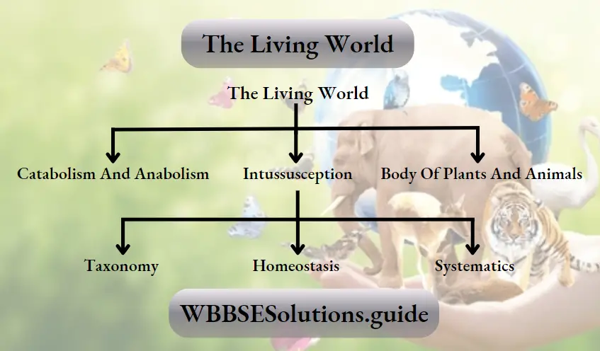
Characteristics And Classification Of Gymnosperms

Gymnosperms

Gymnosperms Definition: Gymnosperms are plants in which seeds are not enclosed in an ovule.

The gymnosperms are seed-forming plants that include conifers, cycads, Ginkgo, and members of the order Gnetales. The term ‘gymnosperm’ comes from the Greek words gymnos (=naked) and sperma (=seed), meaning ‘naked seeds’. The naked condition of their seeds is due to the fact that the carpels do not fuse, enclosing the ovules, to form an ovary.
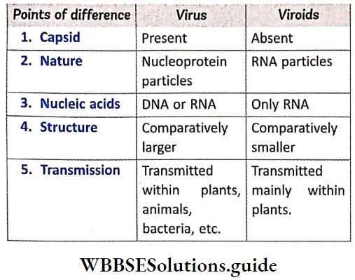
It is considered that gymnosperms have developed from a type of plant species, called progymnosperms They are intermediate to the pteridophyta and angiosperms. About 900 species of gymnosperms have been discovered so far.
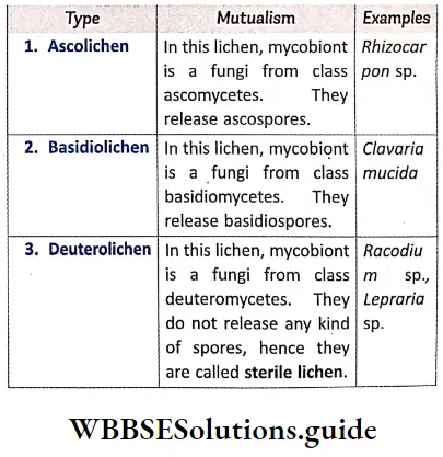
Gymnosperms Distribution: Gymnosperms are present all over the world, but they are found more in the temperate zone, than the equatorial zone.

Read and Learn More: WBCHSE Notes for Class 11 Biology

Gymnosperms General features:

1. Most of the gymnosperms are perennial and evergreen in nature, with long, hard and woody stems. Some of them are shrub-like.

2. The main plant body is sporophytic (2n). It is independent, with the body being differentiated into true roots, stems, leaves, flowers and seeds.

3. Plants have a tap root system, that generally persists for a long time. Some adventitious roots also develop along with the primary root. In some cases, blue-green algae and fungi are found in roots in symbiotic association with the roots.

4. The stem is erect and woody. It may be branched (Pinus sp.) or unbranched (Cycas sp., Zamia sp.). Stem branches are of two types—long shoots and dwarf shoots.

5. Long shoots have unlimited growth while dwarf shoots show regulated and limited growth.

6. Vascular bundles in stems are of two types—open and collateral.

7. In roots, radially arranged xylem is made up of tracheids, parenchyma and xylem fibre. Vessels or trachea are only present in the xylem of Gnetum sp. and Ephedra sp. Phloem consists of sieve cells, phloem parenchyma and phloem fibre. The companion cells are absent.
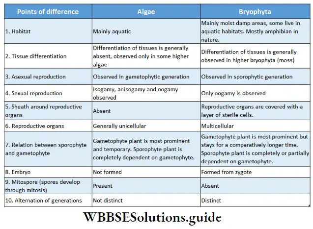
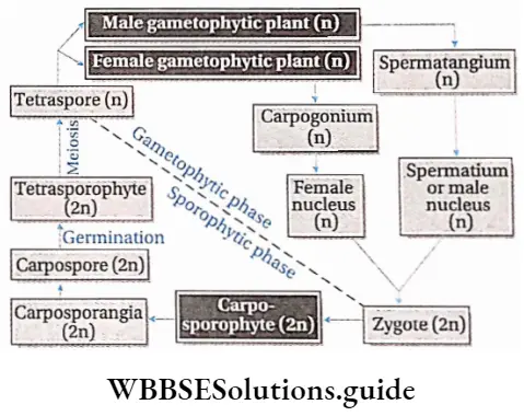
8. Leaves are of two types—brown scale leaves and green foliage leaves. Long shoots bear scale leaves, while dwarf shoots bear both scale and foliage leaves

9. Venation in leaves may be reticulate (Gnetum sp.) parallel (Pinus wallichiana) or dichotomous (Gingko sp.).

10. The leaves are covered with thick cuticles, sometimes with a wax layer. Stomata are present on the lower surface of the leaves. Mesophyll may (Cycas sp.) or may not be (Pinus sp.) divided into palisade and spongy parenchyma.

11. Secondary growth is present in all members, where the mature metaxylem shows bordered pits (In Stangeria sp. and Zamia sp., the metaxylem shows scalariform thickening).

12. Flowers are monoecious, naked and devoid of accessories whor|s.

13. Male flowers are formed of microsporophylls, and female flowers are formed of megasporophylls, called carpels.

14. Microsporophylls and megasporophylls remain arranged and condensed on an axis to produce male and female cones respectively.

Plant Kingdom A male cone and a microsporophyll bearing tivo microsporangia

15. Microsporangia are present on the ventral side of the microsporophylls and megasporangia are present on the dorsal side of the megasporophylls.

16. Ovules or megaspores are exposed on the megasporophylls. Each megasporophyll may bear two or more ovules.

17. Inside microsporangia, numerous microspores (pollen grains) are produced.

18. Microspores produce male gametophytes. Male gametes are either ciliated or non-ciliated.

19. Megaspores in megasporangia produce female gametophytes. Egg cells are produced in one or more archegonia within female gametophytes.

20. Pollination occurs mainly by wind. Male gametes come in direct contact with the ovule (siphonogamy).

21. Sometimes more than one embryo may develop from a zygote (polyembryony).

22. Endosperm is haploid (n) and produced within female gametophytes before fertilisation.

23. Ovule (seed) consists of a single jacket or integument (unitegmic). Cotyledon may be one or two (Cycas sp.) or many (Pinus sp.).

24. The alternation of generations observed in gymnosperms is heteromorphic.

Plant Kingdom Alteration ofgenerations in the life cycle ofa typical gymnosperm

Plant Kingdom Cones ofgymnosperm

Some common examples of gymnosperms are— Cycas sp., Pinus sp., Ginkgo sp., Gnetum sp., etc.

Commercial importance of gymnosperms: Gymnosperms are of great economic importance. The commercial uses of gymnosperms are as follows—

Timber: The coniferous wood may be very light or hard, strong or soft, coarse or grained and durable with distinct annual rings.

Pinus sp., Cedrus sp., Abies sp., etc., yield good quality timber that is used for making furniture, plywood, matchsticks, pencils, etc.

Resin source: Resin exudates from coniferous plants. Resin is a nitrogenous waste produced by plants which is soluble only in organic solvents. The resin is used in varnishes, plasters, enamels, medicine, printing ink, laundry soap, oil, etc.

Resins are of different types like—rosin (solid, obtaining Pinus sp. mainly), copals (hard, with essential oils, in Agathis sp.), sandarac (translucent, pale yellow or orange-coloured, in Tetraclinis articulate), Canada balsam (transparent, non-crystalline, in Abies balsamea)

Source of essential oils and gums: Almost all conifers yield volatile essential oils along with resins. Spruce oil is obtained from Picea abies which is used in room sprays, bath salts and deodorant.

The Siberian fir needle oil, obtained from Abies sibirica is also used in deodorant, toilet and shaving soaps, etc. Cedar oil is obtained from Cedrus deodara and Juniperus virginiana and is used in perfumery as well as in microscopic studies.

Source of paper: Conifers yield good quality timbers which are used for making paper pulp, used in the newsprint industry. Writing and printing papers are manufactured from the wood of Picea sp., Abies sp., Larix sp. and Tsuga sp., whereas craft papers are produced from Pinus sp. and Cryptomeria sp.

Production of medicines: Ephedra sp. is an important source of alkaloid, ephedrine. This is used as a bronchodilator in cough mixture used for the treatment of bronchial asthma. The leaves of Ginkgo biloba are an important source of the ginkgolide compound.

This compound is used to antagonize platelet-activating factor (PAF) in vertebrate blood. Another compound bilobalide obtained from Ginkgo leaves used in the treatment of neural disorders like Alzheimer’s disease. Extracts of these leaves are also used in the treatment of muscular disorders, cerebral insufficiency, vertigo, etc.

Taxus brevifolia is an important source of an anticancer drug, taxol. The leaves of Taxus baccata are useful in the treatment of asthma, bronchitis, hiccups, epilepsy and indigestion. The seeds of Cycas sp., are used as an herbal drug for curing boils, wounds and sores.

Source of tannins: The bark of Tsuga canadensis, Picea alba, Sequoia, etc., yields tannins which are used for different purposes.

As ornamental plants: Several varieties of Cycas sp., Pinussp., Cedrus sp., etc., are widely planted as ornamental plants in gardens, parks temples etc., for their beautiful appearance, symmetrical growth and evergreen nature Their branches and leaves are used as decorations.

Source of food: Spongy pith of Cycas, Zamia, and Macrozamia contain large amounts of starch which is used to prepare sago. It is used as a food. The seeds of Pinus sp., are used in cooking and are eaten raw.

Seeds of Juniperus sp., are used as condiments in cooking. The young unfolded succulent leaves and tender shoots of many species of Cycas sp., are taken as cooked food.

Classification of gymnosperms: The living gymnosperms plants are grouped into four main types—

  1. Cycadopsida,
  2. Ginkgopsida,
  3. Coniferopsida
  4. Gnetopsida. These groups have been discussed under separate heads.

Bryophyta -Definition and Characteristics Notes

Bryophyta

The term ‘bryophyta’ came from two Greek words bryon (moss) and phyton (plant).

Bryophyta Definition: Bryophytes are the simplest and most primitive, non-flowering, embryophytes, that do not contain vascular tissues.

Bryophyta Distribution: Bryophyta is the first group of plants to inhabit the land. They generally occur in moist and shady places. They are found at higher altitudes, on moist soil, stones, trunks and branches of trees. Though they are terrestrial, they require water to complete their life cycle, specifically during fertilisation. Therefore, they are considered as ‘the amphibians of plant kingdom’.

Bryophyta Size and shape: The size of bryophytes may range from very small (1-3 mm) to large (60 cm). Generally, they grow within a height of 15 cm and are herbaceous in nature. They grow closely packed as mats or cushions on rocks, and soil, as epiphytes on the trunks and leaves or as free-floating in water. Most of the bryophytes show dichotomous branching. They are either thalloid or foliose.

Read and Learn More: WBCHSE Notes for Class 11 Biology

Bryophyta General features:

The important characteristics of bryophytes are as follows—

  1. The life cycle of bryophytes is divided into gametophytic and sporophytic phases (i.e., heteromorphic).
  2. The gametophytic phase is predominant and ecologically persistent, i.e., green, independent and long-lived.
  3. The sporophytic phase is very short-lived and completely dependent upon the gametophyte for nutrition and growth.

Plant Kingdom Riccia thallus

Characteristics of gametophytes: The characteristics of gametophytes have been discussed below.

Structure:

Gametophytes show the following structural features—

  1. The gametophytic plant body is either thalloid or differentiated into the root-like rhizoid, stem-like erect caulid and leaf-like phyllid.
  2. Roots are absent in bryophytes. Instead, structures called rhizoids, are present, and those perform the function of roots. They may be branched or unbranched, unicellular or multicellular. They are of two types smooth-walled (inner wall smooth) or tuberculated (inner wall with peg-like projections).
  3. The thread-like body structure, produced during the germination of spores to produce immature gametophytes is called protonema.
  4. The stem-like structure of bryophytes, above the ground is called caulid. These bear leaf-like structures are called phyllids.
  5. Multicellular, coloured scales, are present at the ventral side of the thallus. They protect the cells from friction with the substratum.
  6. Xylem and phloem are absent in these plants. They may have special lignin-containing, conducting tissue in sporophytes. Otherwise, the plant body is made up of parenchyma cells only.
  7. Air pores present in the thallus and stomata present in the capsule in moss, allow gaseous exchange with the environment.

Bryophyta Reproduction: They reproduce by vegetative and sexual means. Asexual reproduction is completely absent in bryophytes.

  1. Vegetative propagation takes place by fragmentation or some special, structures, like gemmae, tubers, protonema, adventitious branches, persistent apices, etc.
  2. Sexual reproduction is of monogamous type. They have large, multicellular gametangia (sex organs), covered with a jacket of sterile cells.
  3. Bryophytes are unisexual, either homothallic (monoecious) or heterothallic (dioecious),
  4. The male sex organs, called antheridia, are multicellular, stalked, globose or ovoid structures. They have a single-layered thick jacket surrounding androgenic mother cells.
  5. Biflagellate sperms or antherozoids (male gametes) are produced from these cells,
  6. The female sex organs, called archegonia, are multicellular, vase-shaped or round-bottom flask-shaped structures,
  7. They have a swollen base, called a venter, containing a ventral canal cell and an egg, and the upper elongated neck containing neck canal cells.
  8. Both the venter and neck are surrounded by the single-layered jacket, made of sterile cells.
  9. The sperms are motile and swim through the water to reach the archegonia. Hence, water is required for sperm dispersal and subsequent fertilisation.
  10. The zygote does not pass through any resting phase. The diploid zygote or oospore develops into the embryo.
  11. Embryonic growth of the sporophyte begins within the venter of the archegonium, soon after fertilisation.

Plant Kingdom Differences between rhizoids and scales

Exoscopic mode of development

The embryo follows an endoscopic mode of development. In this development, the zygote first divides transversely to form an outer epibasal cell and an inner hypobasal cell. The embryo develops from the epibasal cell. Thus, the shoot-forming apical cell is directed outwards, i.e., towards the neck of the archegonium.

Characteristics of Sporophytes:

  1. The sporophytes are diploid.they remain attached to the gametophytic plants for nutrition.
  2. They have bulbous feet, an unbranched stalk-like seta and a single terminal sporangium called a capsule. Sometimes the sporophyte is represented only by a capsule (Riccia sp.).
  3. The capsule has a protective covering called calyptra, which is a part of the gametophyte.
  4. Bryophytes are homosporous (spores are morphologically and physiologically identical). Spores are produced from the sporogenous cells of the capsule through meiotic division.
  5. The haploid (n) spore after germination either produces a filamentous germ tube that gives rise to a young gametophyte (for example Riccia spv Marchantia sp.) or produces a juvenile plant or protonema that ultimately gives rise to an adult gametophytic plant.

Sporophytes Alternation of generations: The alternation of generations is of a heteromorphic nature. The main plant body is gametophytic and bears haploid germ cells. After fertilisation, a diploid zygote is produced.

This undergoes division and differentiation to form a diploid sporophytic (2n) plant. Haploid spores are produced within the spore mother cells of the sporangia in sporophytic plants by the process of meiosis.

These spores germinate to produce haploid gametophytic(n) generation. The alternation of generations In a typical bryophyte has been depicted In.

Some common examples of Bryophytes are—Riccia sp., Marchantia sp., Anthoceros sp., Pellia sp., Pogonatum sp., etc.

The commercial importance of Bryophyta: There are various commercial applications of bryophytes These have been discussed as follows—

Indicator Species: Both liverworts and mosses are often good indicators of environmental conditions. In Finland, terrestrial bryophytes are used to characterise forest types. Bryophytes are used as indicators for mineral concentration. Copper mosses grow almost exclusively in areas with high concentrations of copper, particularly copper sulphate. Thus they serve as good metal indicators.

Soil erosion control: Cultivation of bryophytes on bare land by spreading spores or vegetative fragments or propagules of bryophytes could help to prevent soil erosion. The protonemata, which develop from both fragments and spores, form mat-like structures that cover and bind exposed soil particles.

Sphagnum sp. has a high water holding capacity. They control the water content of the soil during spring runoff in the Arctic. also responsible for soil formation and storing water It is during floods. They are used as nursery beds, for proper conservation of the plants.

Barometer

In Japan, H. Taoda (1976) developed a bryometer, a bag of mosses that respond in predictable ways to various levels of air pollution. By exposing a variety of mosses to various levels of SO2, he determined that most species are injured by 10-40 hours of exposure to 0.8 ppm SO2. Since that time, the use of the barometer has spread around the world.

Nitrogen fixation: Nitrogen is often a limiting nutrient for plant growth, especially in agriculture. Bryophyte crusts, endowed with nitrogen-fixing cyanobacteria, can contribute considerable nitrogen, particularly to dry soils.

Nostoc sp., a cyanobacterium living symbiotically with Anthoceros sp., is able to fix atmospheric nitrogen as an ammonium salt. Hence, it can be used as fertiliser.

pH regulation of soil: Sphagnum sp., helps to maintain the acidity of the soil. Hence, acid-loving plants or oxylophytes are known to grow in such soil.

Food sources: Bryophytes are very rarely consumed by the animals. They may be the source of specific needs of animals at a time when fresh food is scarce. These are used as food in snow-capped mountain regions.

Medicines: Bryophytes (especially liverworts) are used as herbal medicine. Numerous compounds, including oligosaccharides, polysaccharides, sugar alcohols, amino acids, fatty acids, and aliphatic compounds occur in bryophytes.

Bryophytes such as Sphagnum sp. are used to treat skin diseases, and Marchantia polymorpha is used to treat liver ailments. Rhodobryum giganteum and R. roseum are used to treat cardiovascular diseases and nervous prostration.

Polytrichum commune reduces inflammation and fever. It is also used as a diuretic laxative and hemostatic agent. Haplocladium microphyllum is used to treat cystitis, bronchitis, etc.

Plant Kingdom Alteration ofgenerations in the life cycle ofa typical Bryophyte

Classification of Bryophyta: Rothmaler (1951) classified bryophytes into three classes—Hepaticopsida, Anthoceropsida and Bryopsida. In accordance with the International Code of Botanical Nomenclature. Proskauer (1957) modified the classification of Rothmaler by changing the class name Anthoceropsida to Anthocerotopsida.

Plant Kingdom Bryophyta

Class: Liverworts or Hepaticopsida

Liverworts  General features:

  1. Gametophytes are dorsoventrally flattened, and thalloid in nature. Thallus dichotomously branched (apical growing point divided into two growing parts producing two branches in a forked manner). Each lobe of the thallus has a dorsal groove and an apical notch.
  2. The thallus structure has multicellular scales and unicellular rhizoids.
  3. The thallus has chloroplasts but is devoid of pyrenoids.
  4. Multicellular sex organs, i.e., antheridia and archegonia, are present on the dorsal side of the thallus. In some species (Marchantia sp.) sex organs are bore on specified structures called antheridiophores and archegoniophores.
  5. Male antheridia are stalked, globose or ovoid. Each antheridium has several antherozoid mother cells which produce biflagellate,comma-shaped antherozoids or sperms.
  6. Female archegonia is flask-shaped. Its lower part is called the venter and its upper slender part is called the neck. Inside the venter single egg is present.
  7. A sporophyte is devoid of foot and seta. Capsules are globose or ellipsoid, brown or black in colour. Setae are small delicate stalks. [Exception: Foot and seta are absent in the sporophyte of Marchantia sp.]
  8. The capsule wall is made up of a single-cell layer. The capsule contains haploid spores developed from spore mother cells through meiosis. Some species (for example Marchantia sp.) have haploid, needle-like elaters. These elaters help in the dehiscence of the capsule.

Some examples of Hepaticopsida are: Riccia sp., Marchantia sp.

Plant Kingdom Some common members ofHepaticopsida

Class: Hornworts or Anthocerotopsida

Hornworts General features:

  1. Gametophytes are thalloid, with the structure being dorsoventrally flattened and irregularly lobed.
  2. The thallus is not differentiated into stems and leaves and is always homogeneous. Each cell has a chloroplast with centrally placed pyrenoids.
  3. Air cavities and scales may be absent, but smooth and septate rhizoids are present.
  4. The reproductive organs are present on the dorsal side of the thalamus.
  5. In some species, the thallus sometimes contains mucilage cavities which are inhabited by Nostoc, a cyanobacterium.
  6. The mature sporophyte shows a bulbous foot and a slender cylindrical capsule standing out like a bristle from the thallus. The meristematic zone is present at the basal part of the capsule. Seta is absent.
  7. Capsules have a central column of sterile cells called columella (Exception: In Notothylas, columella is absent). The spore mother cells remain surrounding the columella.
  8. The spore mother cells generate haploid spores through meiosis. The capsule also contains numerous unicellular structures resembling elaters, called pseudoelaters. Some examples of Anthocerotopsida are— Anthoceros sp., Notothylus sp., Ceratophyllum sp., etc.

Plant Kingdom Some members ofAnthocerotopsida

Class: Moss or Bryopsida

Moss or Bryopsida General features:

  1. Matured gametophytes, originating from branched protonema, are erect plants with a stem-like axis (caulid).
  2. The axis is covered with spirally arranged leaf-like phyllids.
  3. Phyllids are flattened leaf lamina-like structures, light green in colour with serrated margins. The midrib is prominent.
  4. Multicellular and branched rhizoids are present instead of roots. Caulids possess a cylinder of primitive conducting tissue, made up of thick-walled cells. A central, parenchymatous pith-like structure is also seen.
  5. Multicellular sex organs are present at the top of the branches of caulids. The sex organs develop at the axils of leafy structures called perichaetial leaves.
  6. Antheridia are small, stalked and dumbbell-shaped, while the archegonia are distinct and flask-shaped.
  7. Sporophyte is dependent on gametophytic plants. It is differentiated into foot, seta and capsule.
  8. The foot is bulbous and acts like a haustorium. Capsules may be green, brown, yellow or red in colour, with a short green, yellow or translucent seta.
  9. The capsule contains a dome-shaped spore sac containing sporogenous tissue, overarching a centrally located columella and single cell-layered thick ribbon-like trabeculae.
  10. At the top of the capsule, there is a cap-like structure called an operculum. Below the operculum one or more ring or teeth-like structures (peristome teeth) are present. These help in the opening of the capsule and the release of spores.
  11. The mature capsule is protected by a covering called calyptra. The diploid sporogenous tissues in the spore sac can produce haploid spores through meiosis. These spores germinate to form haploid protonema.

Some examples of Bryopsida are: Sphagnum sp., Funaria hygrometrica, Pogonatum sp., etc.

Plant Kingdom Some members ofMoss

Similarities between algae and Bryophyta

  1. Both are non-flowering, autotrophic, without any vascular tissue.
  2. Both do not have roots. Generally, both of them have a thallus body structure.
  3. Both require water for their fertilisation. The male gametes are flagellate and hence, they are motile in nature.
  4. Both have cellulose in the cell wall.
  5. Protonema of mosses resembles the thread-like algae.
  6. Both have similar photosynthetic pigments.
  7. Gametophyte is the main plant body in both.

Plant Kingdom Comparison among Hepaticopsida, Anthocerotopsida and Bryopsida

Plant Kingdom Differences between algae and bryophyta

Red Algae General Characteristics Notes

Rhodophyceae (Red Algae)

Rhodophyceae Definition: Rhodophyceae (Greek: ~rhodo= red; phyton=plant) are a non-flagellate, bright red-coloured group of marine algae.

The Rhodophyceae or red algae are red due to the presence of a water-soluble red pigment,r-phycoerythrin. This pigment masks the green colour of chlorophyll a, giving the characteristic red appearance They also contain a blue pigment, r-phycocyanin.

Rhodophyceae Distribution: Most of the members of Rhodophyceae are marine. Generally, they are found in the warm regions of the sea. They are attached to the substratum (lithophytes), at the bottom of the sea (even below 600 metres). Several red algae are found in freshwater (for example Batrachospermum sp.,), and wet soil (i.e., Porphyridium sp.). Some of these members are also found as parasites or epiphytes.

Read and Learn More: WBCHSE Notes for Class 11 Biology

Rhodophyceae General Features

  1. The plant body may be unicellular (Porphyridium sp.) or multicellular. The multicellular form may be filamentous (Goniotrichum sp.), parenchymatous (Porphyra sp.)pseudoparenchymatous (Helminthodadia sp.), feathery (Polysiphonia sp.) or ribbon-like (Chondrus sp.). They may attain a size up to 2 metres as in Schizymenia sp
  2. The flagellated motile stages are totally absent
  3. The cell wall consists of the outer pectin and an inner cellulose layer. The mucilaginous material of the outer layer mainly consists of agar-agar and carrageenans. These constitute a major portion of the dry weight of the cell wall.
  4. In multicellular members of this group, the cell walls have pits, through which cytoplasmic connections are maintained. These cytoplasmic threads are called plasmodesmata.
  5. The members of Rhodophyceae show variation in the number of nuclei in a cell. In the members of the subclass Bangioideae, cells are uninucleate, but in the members of the subclass Florideae, most of the members are multinucleate. The number of nuclei is 3,000-4,000 in Griffithsia sp.
  6. The cells may have one chromatophore with a central pyrenoid (Bangioideae) or many discoid and parietal chromatophores with pyrenoids (Florideae).
  7. The photosynthetic pigments are chlorophyll a, chlorophyll b, chlorophyll d, a and -carotenes, and xanthophylls. Along with these pigments, some phycobiliproteins are also present, such as r-phycoerythrin, r-phycocyanin and alpha phycocyanin.
  8. The reserve food is Floridian alpha phycocyanin. starch. Other than this, galactosides, floridiside and mannoglycerate are also present in the cytoplasm.
  9. Reproduction takes place by all three means vegetative, asexual and sexual. Vegetative reproduction takes place only in unicellular forms. Asexual reproduction takes place by monospores, neutral spores, carpospores, bispores, and tetraspores.
    Sexual reproduction is of advanced oogamous type. The male and female sex organs are called spermatangium and carpogonium respectively. The latter are flask-shaped with a long neck, called trichogyne. Non-motile, male gametes are produced within antheridia, called spermatia. The spermatia are carried away by the water current to the extended part of the carpogonium.
  10. The sporophytic plants may develop certain structures called tetrasporangia within which tetraspores (any of the spores produced in a group of four) are produced.
  11. The life cycle is haplobiontic or diplobiontic in nature.
  12. The alternation of generations varies from isomorphic to heteromorphic (where two generations are morphologically dissimilar).

Plant Kingdom Life cycle ofa red alga

Plant Kingdom Comparative study of Chlorophyceae, Phaeophyceae and Rhodophyceae

Similarities between algae and fungi

  1. Both are eukaryotic and unicellular or multicellular.
  2. Both are thallophytic, i.e., the body is not differentiated into roots, shoots and leaves.
  3. Both have cell walls.
  4. Both do not have vascular tissue.
  5. Both reproduce by vegetative, asexual or sexual means. The reproductive organs are unicellular. Asexual reproduction occurs by spores.

Plant Kingdom Differences between algae and fungi