
Haemocyte
The nucleated cells present in the plasma of hemolymph are called hemocytes. Haemocytes are of seven types, among which a few are discussed.
Haemocyte Functions
Haemocyte Transportation: Plasma transports soluble substances and digested food to the tissues. It also transports nitrogenous waste products out of the body during excretion. It helps in transporting the secreted substances to the target organs.
Haemocyte Storage of water: Stores water in the body (as 70% of hemolymph is made up of water).
Haemocyte Killing of germs: The haemocytes kill germs and purify the blood.
Read and Learn More: WBCHSE Notes for Class 11 Biology
Haemocyte Heart: The main organ which is responsible for I circulation of blood throughout the body is called the heart.
- It is made up of connective tissue and muscular tissue.
- It lies within the pericardial sinuses.
Haemocyte Position: The heart is present along the mid-dorsal line of the thorax and abdomen, below the terga.
Haemocytes in insects notes PDF
Haemocyte Structure:
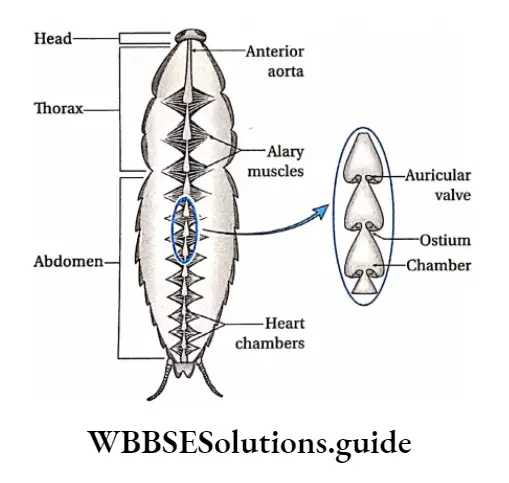
- The heart is an elongated, muscular organ, comprising 13 segmentally arranged, funnel-shaped chambers.
- The cavity or lumen of the heart is lined by the sarcolemma of muscle cells.
- Each chamber of the heart has a pair of minute lateral openings called ostia. These are guarded by valves that allow the blood to flow into the heart from the pericardial sinus.
- These valves also prevent the flow of blood in the reverse direction. The first chamber of the heart leads into the anterior aorta.
- Each segment of the heart gives rise to a pair of arteries called excurrent segmental arteries.
- The anterior aorta and the segmental arteries finally open into the hemocoel.
- The wall of the heart is supported by a bundle of 12 pairs of triangular alary muscles attached to the dorsal membranous diaphragm.
- The pointed ends of these muscles remain inserted into the terga. They help in the contraction and relaxation of the heart.
- The chambers of the heart present between mesothorax and metathorax are called booster hearts. From these regions, hemolymph is transported to the wings of the cockroach.
hemocytes
Function: It is a contractile organ that pumps the hemolymph in one direction i.e., from the anterior aorta to the body cavity.
Blood sinuses or lacunae: In cockroaches, the body cavity extends from the anterior to the posterior end of the body.
It is filled with hemolymph and is known as hemocoel (haem: blood, coel: cavity).
| Class 11 Biology | Class 11 Chemistry |
| Class 11 Chemistry | Class 11 Physics |
| Class 11 Biology MCQs | Class 11 Physics MCQs |
| Class 11 Biology | Class 11 Physics Notes |
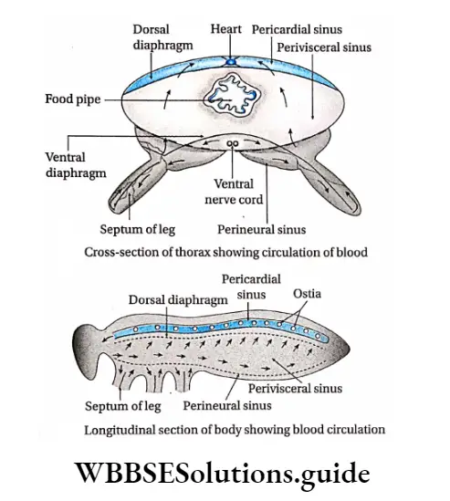
The hemocoel lacks the epithelium of a true coelom. It is divided into three chambers by two parallel membranes at the thorax and abdomen. These three chambers filled with blood or hemolymph are called blood sinuses.

Blood sinuses Structure: The three blood sinuses are formed by two horizontal muscular membranous septa or the diaphragms. These are—the dorsal diaphragm and ventral diaphragm.
Both the diaphragms are perforated so that the three sinuses remain connected with each other.
Types of haemocytes in insects and their functions
Blood sinuses Types: The three blood sinuses are as follows—
Blood sinuses Dorsal pericardial sinus: It lies above the dorsal diaphragm surrounding the heart, aorta, and alary muscles.
Blood sinuses Middle perivisceral sinus: It lies between the two diaphragms and contains the other visceral organs.
Blood sinuses Ventral sternal or perineural sinus: It lies below the ventral diaphragm and encloses the nerve cord. It also extends into the legs.
Blood sinus Valves present in the heart of cockroaches
Different valves found in the heart of the cockroach are—
Auricular valves: Valves present between the pericardial sinus and the heart chambers at the opening of the ostia are called auricular valves. Blood flows from the heart to the pericardial sinus through this valve.
Ventricular valves: Valves located between the heart chambers are called ventricular valves. Due to their action, hemolymph always flows from the heart to different organs.
Pulsatile vesicle: There is an accessory pulsatile vesicle at the base of each antenna. It pumps hemolymph from the head sinus into the antenna.
Pulsatile vesicle Mechanism of circulation:
- When the alary muscles contract, the pericardial sinus expands in volume.
- Hence, hemolymph flows through the perforations of the dorsal diaphragm into the pericardial sinus.
- Again when alary muscles relax, hemolymph enters the heart from the pericardial sinus through the Ostia.
After hemolymph enters the heart, two phases occur respectively. They are—
Systole phase:
- During systole, the valves of the ostia close. This prevents the backflow of hemolymph into the pericardial sinus. Therefore, some of the hemolymph is pumped into segmental arteries, while most of it is poured into the head sinus through the anterior aorta.
- From the head sinus, the hemolymph flows towards the thorax and abdomen, thereby filling the perineural sinus.
- From the perineural sinus, the hemolymph flows into the perivisceral sinus through the pores of the ventral diaphragm.
- The contraction of the alary muscles flattens the dorsal diaphragm and the pericardial sinus is enlarged.
- This causes the hemolymph to again flow into the pericardial sinus from the underlying perivisceral sinus through the pores of the dorsal diaphragm.
Diastole phase:
- During the diastole of the heart, relaxation of the Respiratory system of the alary muscles occurs.
- This narrows the pericardial sinus. As a result, the hemolymph is forced into the heart through the ostia, passing through the auricular valve.
Haemocytes in insect blood: structure and function
Diastasis: The next systole after the diastole follows after a short interval. This interval is called diastasis.
haemocyte
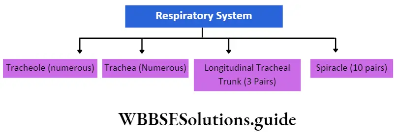
Respiratory System Definition: The physiological system involved in the exchange of CO2 and O2 between the tissues and the atmosphere, with the help of respiratory organs is known as the respiratory system.
The components of the respiratory system.
” hemocyte”
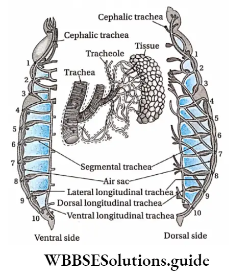
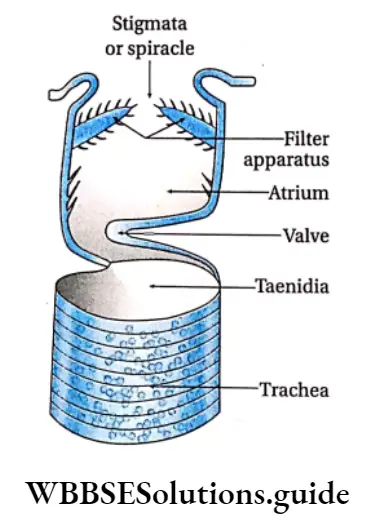
Stigmata Or Spiracle Definition: The openings or pores that line the lateral surface of the body of cockroaches enabling gaseous exchange between the body and the atmosphere are called spiracles.
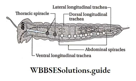
Stigmata Numbers and Stigmata types: There are ten pairs of spiracles. Two pairs of spiracles are in the thoracic region, known as eight pairs of spiracles are in the abdominal region, known as abdominal spiracles.
Stigmata Position: The ten pairs of spiracles are present on the lateral surface of each segment, one on each side of the segment.
Stigmata Structure:
- Each spiracle is a slit-like oval aperture, guarded by bristles on the inner margin. These bristles act as filtering apparatus by preventing dust particles from entering.
- The spiracles open and close by valves that are regulated by the sphincter muscles.
- Each of the spiracles of the mesothorax is guarded by two valves—anterior and posterior valves. Its anterior valve is firm but the posterior valve is movable.
- Spiracles of metathorax are guarded only by the anterior valve. Spiracles of the abdomen lack valves.
- Each spiracle is surrounded by a ring-shaped hard structure (sclerite) called peritreme.
- Some of the spiracles lead into an internal small chamber called atrium which leads into a tube-like structure called tracheal trunk.
- Thoracic spiracles directly open into the trachea, but abdominal spiracles open first into the atria and then into the trachea.
- Functions: The main function of spiracles (or stigmata) is an exchange of gases between the body and the atmosphere.
- The walls of spiracles are made up of chitinous bristles which act as filters.
- They prevent water and other substances from entering into the respiratory system.
Circum-oesophageal connective: It is a pair of commissures (a bundle of nerve fibers that join two parts of the nervous system) that connects supra-oesophageal ganglia with sub-oesophageal ganglia.
The supra-oesophageal ganglia, sub-oesophageal ganglia, and circumoesophageal connectives, together form a structure around the oesophagus. This structure is called the nerve ring.
Role of haemocytes in insect immunity and defense
Circum-oesophageal connective Position: It is present encircling the esophagus.
Circum-oesophageal connective Structure: It comprises comparatively thin and broad nerves. Among the nerves in a pair, one is short and the other is long. These are connected through transverse commissures.
Ventral nerve cord: It is the nerve cord that arises from the sub-oesophageal ganglion. It is formed of two ventral cords.
Circum-oesophageal connective Position: It lies mid-ventrally below the alimentary canal, extending up to the 7th abdominal segment.
Circum-oesophageal connective Structure: There are two ventral nerve cords that run very close and parallel to each other.
They pass along the thorax and abdomen. They are made up of nine segmental ganglia—three thoracic and six abdominal.
Thoracic ganglion: They are three in number. Each thoracic segment bears a large ganglion. They are named prothoracic, mesothoracic, and metathoracic ganglia.
Abdominal ganglion: There are six abdominal ganglia located in the 1st, 2nd, 3rd, 4th, 5th and 7th abdominal segments.
Each of the above-mentioned ganglion, except the 6th abdominal ganglion, is formed by the fusion of paired ganglia.
The last or 6th abdominal ganglion is the largest among them. Unlike others, it is formed by the fusion of several ganglia.
Peripheral nervous system: Nerves that originate from the central nervous system and are distributed to the organs located on the lateral part of the body, form the peripheral nervous system.
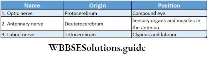
Three pairs of nerves originate from the supra-oesophageal ganglion—optic, antennary, and labrofrontal nerves. The first two innervate the eyes and antennae.
The third one is divided into two parts—the labral nerve which innervates the labrum and the frontal nerve which joins the sympathetic nervous system.
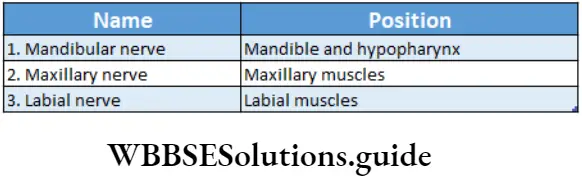
Three pairs of nerves arise from the sub-oesophageal ganglion—mandibular, maxillary, and labial nerves. As the names suggest, the nerves supply to the mandibles, maxillae, and labium, respectively.
Nerves originating from the three thoracic ganglia spread over the different muscles (wings and legs) of the respective segment.
Nerves originating from the first five abdominal ganglia spread over the heart, spiracles, and muscles of the dorsal and ventral body wall of the 2nd, 3rd, 4th, 5th, and 6th abdominal segments.
haemocytes
Three pairs of nerves arise from the 6th abdominal ganglion, which spread over the body wall and other parts of the abdomen.
These parts include reproductive organs, copulatory appendages, anal cerci, and anal styles in males of the 7th, 8th, and 9th abdominal segments of the cockroach.
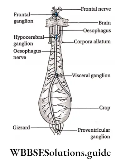
Autonomic nervous system or sympathetic or stomatogastric nervous system: The stomatogastric nervous system consists of nerves, ganglia, and their connectives, that spread to the alimentary canal, the heart, and other visceral organs.
This division of the nervous system regulates the functions of those organs; hence, it is also called the visceral nervous system.
It consists of four nerve ganglia and an intracerebral complex.
Ganglia: The four nerve ganglia that make up the autonomic nervous system are—the frontal ganglion, occipital or hypocerebral ganglion, visceral or ingluvial ganglion, and proventricular ganglion.
Functions of the esophagus, crop, and gizzard are controlled by these ganglia.
Retrocerebral complex:
- It is a neuroendocrine complex.
- It consists of a pair of corpora cardiaca, a pair of corpora allata, and connectives.
Sense organs Definition: Organs that are responsible for perceiving the major senses like touch, smell, hearing, sight, etc., are called sense organs.
In cockroaches, sense organs are compound eyes, antennae, ocelli, and sensillae.
Compound eye Definition: Compound eyes are a pair of large, black, bean-shaped sense organs, present on the head, that help in vision.
Compound eye Position: It is situated dorsolaterally on the head, one on each side.
Compound eye Structure:
- The external surface of the compound eye is covered with a convex transparent cuticle called a cornea.
- The compound eye has several polygonal facets or structures. Each of these polygonal structures denotes a single visual unit, called the ommatidium (plural: ommatidia).
- Each compound eye of a cockroach has about 2,000 ommatidia.
- Each ommatidium is made up of two parts—diopteric and receptive region.
- The dioptric region focuses on the light rays coming from an object, while the receptive region converts the image into an impulse.
- The dioptric region consists of one corneal lens, two corneagen cells, a crystalline cone, and four cone cells (also known as vitrellae).
- The corneal lens is a cuticular secretion of two epidermal cells, called corneagen cells, lying below. On the other hand, the crystalline cone is a conical secretion of the cone cells.
- The receptive part is made up of a spindle-shaped structure, secreted by eight elongated cells. These cells are called retinulae. The structure formed is called a rhabdome, which is made up of eight parts called rhabdomeres.
- The rhabdomeres contain the visual pigment called retinene.
- The retinulae continue as nerve fibers, that join to form the optic nerve.
Functions: Several images are formed in each ommatidium when light rays fall on it.
This kind of image is called a mosaic image. This type of vision, known as mosaic vision, is adapted for detecting movement more efficiently.
Other sense organs: Besides compound eyes, other sense organs in cockroaches include—
Antennae: Antennae are a pair of long, segmented, thread-like structures present on the head.
They arise from the membranous antennal sockets in front of each eye. They can be moved freely in all directions.
They possess small sensory bristles and act as organs of touch and smell. Each antenna has three parts—
Scape: It is the basal part of the antenna which articulates in the antennal socket.
Pedicel: It is the mid-region of the antennae.
Flagellum: It is a long, many-jointed structure that is located next to the pedicel. This part is covered with numerous bristles. The antennae act mainly as sense organs of touch and help to search for food.
Ocelli or fenestrae: A pair of small, circular, whitish degenerated structures on each side of the compound eye represent the simple eyes or ocelli.
They are located on the dorsal side of the head, near the base of the antennal.
They comprise of single corneal facet and are sensitive to light eye representing the simple eyes or ocelli.
They are located on the dorsal side of the head, near the base of the antennal. They comprise of single corneal facet and are sensitive to light.

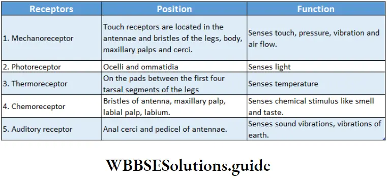
Receptors Within The Sense Organs
Groups of specialized cells that can detect any stimuli are called receptors. They are generally present within the sense organs.
They can be of different types. The following table describes some of these receptors—
Excretory system Definition: The excretory system is an organ system concerned with the removal of metabolic waste products from the body through specific organs.
Organelles: Malpighian tubules are the main excretory organs of cockroaches.
- In addition, fat bodies, amoeboid cells, nephrocytes, cuticles, rectal glands, and uricose glands serve as accessory excretory organs.
- In cockroaches other than the Periplaneto species, another accessory gland called the mushroom gland may be present that helps in excretion.
Class 11 zoology haemocytes in insects notes
Malpighian Tubule Definition: The thin, unbranched, thread-like tubes, which are the main excretory organs of cockroaches are known as malpighian tubules.
Malpighian Tubule Position: Malpighian tubules are found at the junction of the midgut and hindgut.
Malpighian Tubule Number: There are 60-150 malpighian tubules arranged in 6-8 bundles. Each bundle consists of 15-20 tubules.
Malpighian Tubule Structure:
- They are numerous, thin, long, filamentous, unbranched, thread-like yellow-colored tubules,
- Each tubule is 16 mm long and 0.5 mm in diameter,
- The proximal end of each tubule opens to the lumen of the gut and the distal blind end floats in the hemolymph of the hemocyte.
- The tubules are lined by a single layer; of glandular ciliated epithelial cells.
Malpighian Tubule Types: On the basis of staining by silver nitrate, the tubules are classified into two types
Deeply stained malpighian tubules, lightly stained malpighian tubules.
Malpighian Tubule Functions:
- Malpighian tubules reabsorb the nitrogenous metabolic waste products from the hemolymph of the hemocoel. These wastes are drained out of the body through the hindgut.
- It also acts as an osmoregulatory organ.
- Accessory excretory organs Besides Malpighian tubules, there are some accessory excretory organs.
They are as follows—
Amoeboid cell: In the nymphal (very young) stage, some amoeboid cells store excretory products within the cuticle. These cells get removed along with the cuticle during metamorphosis.
Cuticle: Nitrogenous wastes and metabolic salts are deposited in the cuticle of cockroaches. The waste products are shed off along with the cuticle during the process of molting.
Fat bodies: Numerous white lobule-like bodies are present below the body surface. These bodies comprise fat-storing cells. The fat cells of cockroaches are analogous to the liver of human beings.
Some fat cells collect excretory substances from the blood such as—urate, uric acid, etc. However, the way these stored substances are eliminated is not clear.
They contain two types of cells—
- Trophocyte cells (stores food),
- Urate cells (stores uric acid).
Mushroom gland: The mushroom gland is present in male cockroaches. In some species, it possesses long, blind tubules on its periphery known as uricase glands.
These glands synthesize uric acid. The mushroom gland is also known as utriculi majores. It stores uric acid and releases it during copulation over the spermatophore.
Nephrocytes or Pericardial cells: Large, aggregated cells, located along the heart, absorb excretory substances (uric acid, calcium salts, etc.,) from the hemolymph and store them in the cytoplasm.
These cells are called nephrocytes. They release these excretory substances in the gut.
Rectal gland: They are present in the papillae of the internal wall of the rectum. They help in osmoregulation by facilitating the reabsorption of water and minerals from the rectum.
Short notes on haemocytes in insects for quick revision
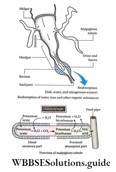
Mechanism of excretion by malpighian tubules: The process of excretion by malpighian tubules is as follows—
- In the distal blind region of the tubule, nitrogenous waste is absorbed from the hemolymph by glandular cells. It should be noted that peristalsis within malpighian tubules (5-15 times at 20-25°C) helps in the reabsorption of excretory substances.
- Reabsorption takes place by diffusion. The excretory substances are salts of uric acid (potassium urate, sodium urate, etc.), and water. These substances react with water and CO2 to produce potassium bicarbonate and uric acid.
- Potassium bicarbonate and water are reabsorbed into the hemolymph through the proximal convoluted region of the tubule.
- Uric acid, which is left in the tubule, is released into the hindgut, by peristalsis.
- The colon and rectum again reabsorb water from uric acid, converting it into solid crystals. This is released by the anus from the body. Hence, the cockroach is a uricotelic animal.
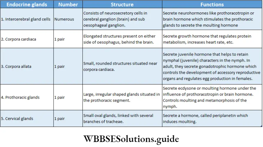
Endocrine system Definition: The system formed by the glandular neurosecretory cells that produce secretions that control various physiological functions is known as the endocrine system.
The various components of the endocrine system are discussed under the following heads—
Neurohormones: The endocrine system comprises five types of bundles of cells. Out of these five, three bundles are located at the front and two, at the back.
These cells are known as neurosecretory cells. The secretions of these neurosecretory cells which control the various physiological functions are called neurohormones.
Types of glands: The different types of endocrine glands are discussed in the following table—
Reproductive System Definition: The system that is involved with the production of offspring (reproduction) is known as the reproductive system.
The cockroaches show sexual dimorphism, i.e., males and females are separate.
The male and the female reproductive systems are discussed as follows—
Male reproductive system: The male reproductive system comprises a pair of testes, vasa deferentia, seminal vesicles, an ejaculatory duct, a utricular or mushroom gland, conglobate or phallic gland, and the external genitalia or male gonapophyses.
Testes Definition: The Testis (Plural: Testes) is the primary reproductive organ of males which produces the male gamete, sperm.
Testes Position: Each testis is located in the dorsolateral side of the abdominal cavity, which extends from the 4th – 6th abdominal segments.
Difference between haemocytes and blood cells in insects
Testes Structure:
- Testes are paired, three-lobed structures, that are about 1 cm in length,
- They are more prominent, well well-developed in the young ones,
- Each lobe is made up of 30-40 small follicles that are arranged cylindrical along the vas deferens.
Testes Function: The primary function of testes is sperm production. Testes become non-functional in older cockroaches.
Vas Deferens Definition: The ducts or tubes that originate from the testes of cockroach and extend towards the posterior end to fuse with the ejaculatory duct is called vas deferens.
Vas Deferens Position: Vas deferens arise from each testis. They run posteriorly and then curve anteriorly from the lateral sides of the abdominal cavity towards the center.
Vas Deferens Structure: Vas deferens are paired, thin, slender white ducts. They open into the ejaculatory duct.
Vas Deferens Function: They help in the transport of sperm from the testes to the next structure called the ejaculatory duct.
Seminal vesicle Definition: Seminal vesicles are sac-like accessory reproductive structures into which the vas deferens open and release the sperms.
Seminal vesicle Position: They are present at the junction of the vas deferens and the ejaculatory duct, in the 6th-7th abdominal segments.
Seminal vesicle Structure: They are opaque, whitish, bulbous, sac-like structures. Generally, there are two seminal vesicles in a cockroach.
Seminal vesicle Function: They store mature sperm temporarily.
Mushroom gland Definition: Mushroom gland is a large accessory reproductive gland arising from the outer wall of the seminal vesicle.
Mushroom gland Position: They are situated at the junction of the vas deferens and ejaculatory duct in the 6th-7th abdominal segments.
Mushroom gland Structure: Mushroom gland is made of numerous blind, slender, thread-like tubular structures. The arrangement of these tubules gives the gland a mushroom-like appearance.
One end of the gland is blind but the other end is open and it connects with the ejaculatory duct. The mass of glandular tubules exists in two forms—
Utriculi majores: It consists of long peripheral tubules.
Utriculi breviores: It is made of short central tubules.
Utriculi breviaries Function: The two parts of the mushroom gland act in two ways.
Utriculi majores form the inner layer of the wall of the spermatophore (bundle of sperms). Utriculi breviaries provide nourishment to the sperm.
Ejaculatory Duct Definition: The Ejaculatory duct is an elongated, wide duct that carries the sperms from the seminal vesicles to the genital pouch.
Ejaculatory Duct Position: It is a median duct that lies between the seminal vesicle and the male genital pore (gonopore) in the abdomen.
Ejaculatory Duct Structure: It is a long, wide, muscular tubule with glandular lining. It gradually enters the genital pouch and opens outside through the male genital pore. Only one ejaculatory duct is seen in the body of a cockroach.
Ejaculatory Duct Function: The ejaculatory duct facilitates the ejaculation of sperm, stored in the seminal vesicles, during mating.
Conglobate or phallic gland Definition: Conglobate gland is a male accessory reproductive gland that opens separately through an aperture that lies beside the male gonopore.
Conglobate or phallic gland Position: It lies below the ejaculatory duct slightly to the right of the nerve cord. It extends upto the genital pouch.
Function of insect haemocytes in wound healing and phagocytosis
Conglobate or phallic gland Structure: The conglobate gland is an elongated, flat, club-shaped structure. Its one end is blunt but its other end narrows posteriorly into a tubular structure.
This tubular structure opens into the male genital pouch. The cockroach has only one conglobate gland.
Conglobate or phallic gland Function: It secretes a substance that forms the outermost layer of the spermatophore.
Genital pouch Definition: Genital or reproductive pouch is the cavity at the hind end of the abdomen of male cockroaches that comprises the anus, male genital pore, and gonapophyses.
Genital pouch Position: It is located on the ventral side of the 9th sternum and on the dorsal side of the 10th abdominal segment.
Genital pouch Structure: It is a pouch-like structure. The organs present within it are the dorsally placed anus, ventrally placed male gonopore, and gonapophysis. A cockroach possesses only one genital pouch.
Genital pouch Function: It ejects sperm outside the body.
Gonapophysis or phylloxera Definition: The chitinous, asymmetrical appendages that surround the male gonopore within the genital pouch are called the male gonapophysis.
Gonapophysis or phylloxera Position: Gonapophysis is present surrounding the male genital pore.
Gonapophysis or phylloxera Structure: Each of the three chitinous, sclerite-containing appendages is called phylloxera. The phallomeres together are called gonapophyses (sing. gonapophysis).
There are two phallomeres present laterally and one present ventrally.
Three gonapophyses are seen in cockroaches. These are—
Ventral phylloxera: It is located beneath the right phylloxera. It is also known as phallus or deugus. The ejaculatory duct opens at the base of
this flat, plate-like structure.
Right phallomere: The right phallomere is mid-dorsal in position. It bears two chitinous opposing plates, a hook, and a serrate lobe.
Left phylloxera: The left phylloxera has a broad base. It bears—
- The titillation, which is a long, thin, hook-like part present towards the left of the pseudopenis,
- Pseudopenis, which has a hammer-like head,
- Accutobolus, which is the innermost part and contains the hook,
- Asperate, which is the groove-like outer part of the accutobolus.
Left phylloxera Function: It acts as a copulatory organ and helps in copulation.
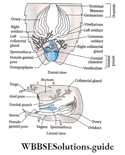
Female reproductive system: The female reproductive system comprises ovaries, oviducts, vagina, spermatheca, collateral gland, genital pouch or brood pouch, female gonopore, and gonapophyses.
Ovary Definition: Ovary is the primary female reproductive organ that produces the female gamete, egg.
Ovary Position: Each ovary is located on the lateral and posterior end of the abdominal cavity, which extends from the 2nd to 6th abdominal segments.
Ovary Structure: Ovaries are paired, yellow-colored organs. Each ovary is made up of 8 beaded, tube-like structures called ovarioles or ovarian tubules.
One end of these tubules is closed. Each ovariole is divided into many segments. These are—
Germarium: The anterior narrow part of the ovariole, containing immature germ cells.
Vitellarium: The posterior region of the ovariole which contains mature ova.
Terminal filament: The long, thread-like filaments That lie Within The fat Bodies.
Therminal Filaments Arise from all the ovarioles. all the terminal filaments fuse within the ovary to form a structure called the suspensory ligament. Ovary Fuses With Fat Bodies Through The Ligament.
Pedicel: The egg chamber continues posteriorly into a thin-walled hollow stalk which is called a pedicel.
It opens into the lateral oviduct. The secretion from the epithelium of the ovariole nourishes the ova.
Function: Ovaries produce ova.

Oviduct Definition: The Oviduct is a part of the female reproductive system that is formed by the union of eight ovarioles at the posterior end of the ovary.
Oviduct Position: Each oviduct arises from the posterior part of each ovary. Therefore, a pair of oviducts are found in cockroaches.
Oviduct Structure: Each oviduct arising from each ovary is called a lateral oviduct. The two lateral oviducts fuse to form another wider, median duct called a common oviduct.
Oviduct Function: They transport the eggs to the vagina.
Vagina Definition: The posterior part of the common oviduct of a female cockroach that opens into the female genital pore is called the vagina.
Vagina Position: The vagina lies near the female gonopore I on the 8th sternum. A female cockroach bears only one vagina.
Vagina Structure: The vagina is a broad, short sac-like structure. It remains fused with the genital pouch. The slit-like opening of the vagina is called the female gonopore.
It opens into the genital chamber.
Vagina Function: It releases the mature eggs through the female gonopore into the genital chamber.
NEET haemocytes in insects important points and MCQs
Spermatheca Definition: The sac-like structure that is joined with the genital pouch of a female cockroach and receives sperm during copulation is called spermatheca.
Spermatheca Position: They are located at the posterior part of the genital pouch.
Spermatheca Structure: Spermathecae are paired, club-shaped sac-like structures. Both the spermathecae are not of equal size.
They unite to form a small common duct. The duct opens within the genital chamber on a small spermathecal papilla.
Spermatheca Function: They receive spermatozoa during copulation and store it for fertilization.
Collateral Gland Definition: (Material glands are accessory reproductive glands that are present on both sides of the genital pouch in a female cockroach.
Collateral Gland Position: These glands lie behind and above the ovaries and open on the dorsal face of the female gonopore.
Structure: Collateral glands are paired, branched tubular structures. The left collateral gland is larger and more well-developed than the right gland. They unite to form a collateral duct that opens outside.
Function: The secretion of this gland forms a structure called ootheca.
Female gonopore Definition: The slit-like opening of the vagina is called female gonopore.
Female gonopore Position: It lies at the posterior part of the vagina in the 8th sternum inside the genital pouch.
Female gonopore Structure: It is a slit-like aperture of the vagina.
Female gonopore Function: It allows mature eggs to pass from the; vagina into the genital pouch.