Nervous Tissue
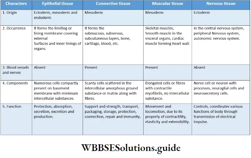
Nervous Tissue Definition: The tissue, derived from the embryonic ectoderm, that is responsible for coordination of the physiological functions with the external as well as internal environment is known as nervous tissue.
Nervous Tissue Origin: Nervous Tissues Have Originated From Ectoderm.
Nervous Tissue Characteristics: Nerve cells or neurons are the structural and functional unit of the nervous system, They form nerves, that serve as the mode of
communication within the body.
These cells have three main properties that enable them to communicate with other cells of a nerve—
Excitability (irritability): Neurons exhibit the property of irritability. Neurons are excitable—i.e., they get excited and respond to environmental changes (stimuli).
Excitability Conductivity: Neurons respond to stimuli by producing electrical signals. These signals are conducted to other distant cells within the body by the nerves.
Read and Learn More: WBCHSE Notes for Class 11 Biology
Excitability Secretion: When the electrical signal reaches the end of a nerve fiber, the neuron secretes a chemical called neurotransmitter. This neurotransmitter crosses the gap between two neurons.
On reaching the next neuron, it stimulates it, thereby transmitting the impulse.
Excitability Functions
Excitability Coordination: It coordinates and controls various organs of the body.
Excitability Sensations: It perceives various sensations of the body like touch, pain, pressure, heat, cold, vision, hearing, smell, and taste.
Excitability Homeostasis: It maintains the body’s equilibrium with respect to the changes in the external environment.
Response to stimuli: It brings about an appropriate response to various stimuli.
Consciousness: It helps in carrying out conscious activities.
Components: Nervous tissue consists of nerve cells (neurons) and associated supporting cells (neuroglial cells)
A neuron or nerve cell
A neuron or nerve cell Definition: The structural and functional unit of nervous tissue, which transmits nerve impulses, throughout the body, is called a neuron.
Parts of a neuron: A typical neuron consists of two parts—cell body and cellular processes. These have been discussed under separate heads.
Cell body (neurocytoma or soma) Definition: The nucleated cell body of a neuron is known As Neurocyton or Soma.
Cell body (neurocytoma or soma) Structure
Shape and volume: The cell body of a neuron is polymorphic in shape, i.e., it may be star-shaped, pyramidal, oval, round, or flask-shaped.
Some cell bodies are so large that they can be seen through the naked eye. Very small cell bodies are also found in some neurons.
Covering: The outer covering of the cell body is called neurilemma.
Nucleus: Each cell has a single, large, and oval nucleus with a nucleolus.
Cytoplasm: Cytoplasm is granular in nature. It is also called neuroplasm.
Nissl’s granules: The neuroplasm contains numerous, minute basophilic granules called Nissl granules.
They are composed of rough endoplasmic reticulum along with ribosomes. Nissl granules act as the sites of protein synthesis.
On the basis of the structure and function of the neuron, the number of Nissl granules may vary. For example, more Nissl granules are present in motor neurons than in sensory neurons.
Neurofilament: Bundles of fine protein filaments, called the neurofilaments, form neurofibrils.
These are found scattered throughout the neuroplasm. They take part in the transmission of nerve impulses.
Centrosome: Earlier it was believed that neuron lacks centrosome. This was considered to be the reason why the neurons did not undergo cell division.
However, in recent years, centrosome has been observed in a neuron under an electron microscope.
However, the process of cell division is still absent. Hence, it is considered that the centrosome of neurons may be inactive in nature.
Other organelles: In addition, the neuroplasm contains mitochondria, Golgi bodies, and ribosomes.
Liposuction: It is a brown pigment present in the cytoplasm. It contains lipid residues formed result of lysosomal digestion.
Its abnormal accumulation induces degenerative disorders (disorders in which the structure as well as the functions of the tissues degrade).
Functions
Receives impulse: The cell body of a neuron remains associated with other neurons through its short branching processes (dendrites). These dendrites help to receive impulses from the neighboring neurons.
Secretion: Secretes various structural and functional proteins.
Processes of the neuron Definition: The fiber-like protoplasmic elongations arising from the cyton are known as processes of the neuron.
Processes of the neuron include the axon and the dendron. These have been discussed under separate heads.
Axon: A single, long (generally unbranched) cytoplasmic process arising from the cyton is known as an axon.
Structure
Shape and origin: It is a long, thin appendage with a uniform diameter throughout its length. Axon arises from a conical elevation, called axon hillock, on the cell body.
Axoplasm: The cytoplasm within the axon is known as axoplasm. It is comprised of neurofibrils, mitochondria, and smooth endoplasmic reticulum. But Nissl’s granules are absent.
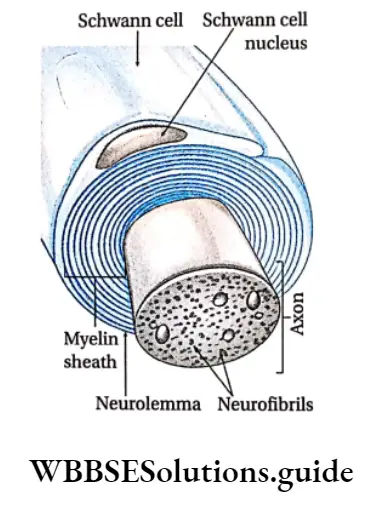
Covering: The cell membrane of the axon is called axolemma. The axon may be surrounded by another covering over the axolemma, called the myelin sheath.
The outermost thin neurilemma consists of tubular cells, called Schwann cells, that are arranged along the length of the axon.
Each of these cells is wrapped around the axon so that it is covered by a number of concentric layers of Schwann cell plasma membrane.
These membranes fuse to produce the myelin sheath, which is composed of a shining, fatty substance called myelin. The outermost layer of the Schwann cell plasma membrane is called the neurilemma.
Nodes of Ranvier: The myelin sheath is not continuous along the entire length of the axon.
It is interrupted at regular intervals to form certain gaps known as the Nodes of Ranvier.
They are points of branching of the axon. They also contain channels that transport ions, required during the conduction of nerve impulses.
Collateral branches: Generally, the axon lacks branches. However, sometimes the axon may show branching. These branches are called collateral branches.
End brush: Axon ends in numerous fine branches known as axon terminals. These axon terminals form a tuft-like structure called end brush.
Synaptic knob: Each axon terminal swells up into a knob-like structure known as synaptic knob. It contains abundant mitochondria and secretory vesicles, called the synaptic vesicles.
Functions: Axons conduct nerve impulses from the cell body to the next neuron. Hence, it is efferent in nature.
Dendron: The shorter branching processes of the i-cell body of a neuron are called dendrons.
Structure: It is of shorter length than the axon. The diameter of the dendron is not uniform throughout its length. The outer covering of a dendron is 4 neurilemma.
- The thin terminal branches of dendrons are known as dendrites.
- The cytoplasm of dendron ( contains Nissl granules, neurofibrils, and mitochondria.
Functions: Dendrons receive stimulations and conduct them towards the cyton. Hence, they are afferent in nature.

Classification of neuron
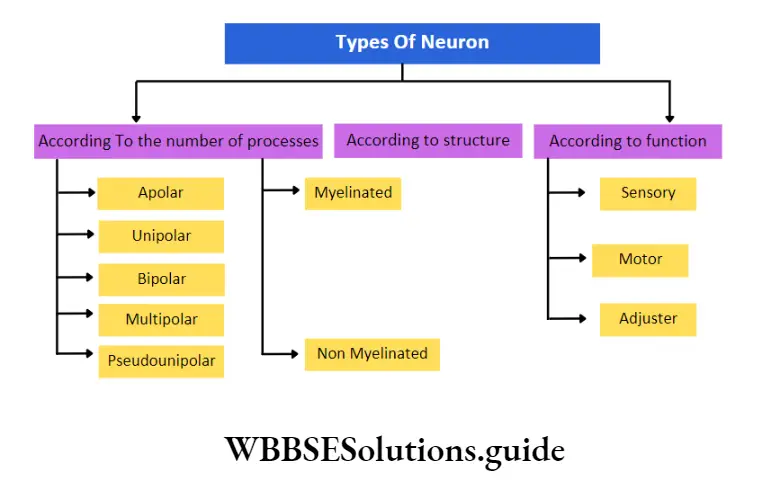
On the basis of processes arising from the cell body: According to the number of processes arising from the cell body, neurons may be classified into—
Apolar neuron: The neuron without any processes arising from its cell body is known as apolar neuron. Such neurons are found in the CNS of the vertebrates.
Unipolar neuron: A neuron with only one process arising from the cell body is known as a unipolar neuron. Such neurons are found in the early stages of embryonic development.
Bipolar neuron: The neuron with two processes (one axon and one dendron) arising from the cell body is known as bipolar neuron.
Such neurons are found in the rod and cone cells of the retina and in the olfactory epithelium.
Multipolar neuron: A neuron with more than two processes arising from the cell body is known as a multipolar neuron.
One of these processes acts as an axon and the rest as dendrons. Such neurons are found in the central nervous system and in the ganglia of the autonomic nervous system.
Pseudounipolar neuron: The neuron with a single process formed by the union of the dendron and axon known as a pseudounipolar neuron.
It soon bifurcates and separates into dendrons and axons after emerging from the cell body.
Such neurons are found in the dorsal root ganglia of the spinal nerve in adult vertebrates.
On the basis of structure: According to structure, neurons are classified into two types.
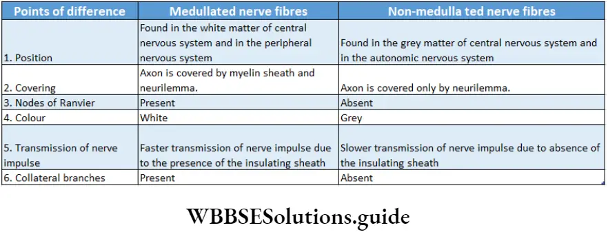
Medullated or myelinated neurons: Medullated neurons are the neurons in which the axon is surrounded by an inner thick medullary sheath or myelin sheath and an outer thin neurilemma or neurolemma.
Medullated or myelinated neurons are found in the white matter of the central nervous system (CNS) and in the peripheral nervous system.
Non-medullated or non-my/inated neuron: Non-medullated neurons are the neurons in which the axon lacks myelin sheath i.e., it is covered only by neurilemma. These neurons also lack the Node of Ranvier.
Non-medullated or non-myelinated neurons are found in the grey matter of the central nervous system (CNS) and in the autonomic nervous system.
On the basis of function: According to the function, neurons are of three types—sensory, motor, and relay neurons.
Sensory or afferent neurons: They transmit impulses from sense organs that receive the stimuli to the central nervous system.
Motor or efferent neurons: They transmit electrical signals from the central nervous system to the effector organs in the body.
Relay neurons: They are found within the central nervous system between the sensory and the motor neurons. These neurons transmit the electrical impulses from sensory to motor neurons. They are also called adjuster neurons interneurons or connector neurons.
Neuroglial cells Definition: The specialized, non-neural, accessory cells of the nervous system that provide support to the nerve cells are known as neuroglial cells.
These are essential for the function and survival of nerve cells.
Neuroglial cells Position: These cells are found both in the central nervous system and peripheral nervous system.
Neuroglial cells Types: The different types of neuroglial cells are as follows—
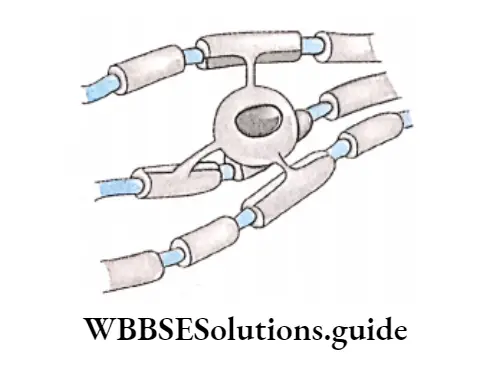
Oligodendrocytes: These are myelin-secreting cells of the CNS. Their main function is to provide support and insulation to the axons within the CNS
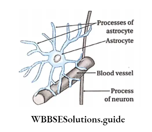
Astroglia or Astrocytes: They are star-shaped cells having several processes extending from their cell body into the surrounding network of nerve fibers.
They provide physical support as well as essential nutrients to nerve cells of the brain and spinal cord.
They help in the transport of ions, taking place between the nerve cell and the extracellular fluid. They also help to form the blood-brain barrier (thin, semipermeable membrane that separates the blood from the cerebrospinal fluid in the brain).
Astrocytes differ from oligodendrocytes in having thicker and more number of processes.
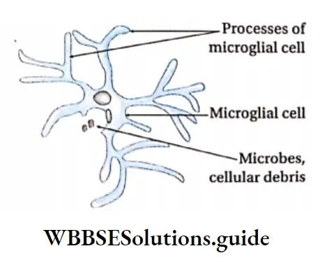
Microglia or microglial cells: These cells are small cells that are similar to macrophages (large scavenger cells) present within the immune system, with respect to functions. They are the phagocytes of the central nervous system.
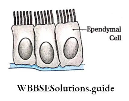
Ependymal cells or ependymocytes: These cells are found in the central nervous system.
They form an extremely thin epithelial-like lining in the ventricles of the brain and central canal of the spinal cord.
This lining is called ependyma. The apical surface is covered by cilia, which keep the cerebrospinal fluid in circulation. They also form cerebrospinal fluid (CSF).

Satellite glial cells and Schwann cells
Besides the above-mentioned cells, there are certain other accessory cells, that help the nerve cells in different ways. They are called satellite glial cells and Schwann cells.
- Satellite glial cells: They surround cell bodies of neurons in ganglia (ganglion cells). They provide electrical insulation and nutrients to neurons.
- Schwann cells: They are responsible for the myelination of axons in the peripheral nervous system.
Synapse Structure: Synapse is the junction between the dendrites of one neuron and the axon of another neuron.
Parts: The terminal swollen part of the axon called the synaptic knob, is covered by a membrane called the presynaptic membrane.
The membrane of the dendrite of the other neuron is called the postsynaptic membrane.
The minute space between the presynaptic and postsynaptic membrane is called the synaptic cleft.
Functions of neuroglia: Various functions of neuroglia are—
- They (oligodendrocytes) synthesize myelin sheaths.
- They (ependymal cells) coordinate the flow of CSF.
- They (microglia) have phagocytic properties.
- They (astrocytes) form the blood-brain barrier.
- They (astrocytes) maintain an appropriate balance of Ca2+ and K+ ions needed for synaptic transmission.
Nerve or Nerve trunk
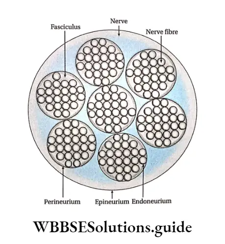
A nerve is formed of several bundles of nerve fibers. Such bundles are called the fasciculi. Each nerve fiber in a bundle is covered by a thin sheath of connective tissue, called the endoneurium.
Each fasciculus is enclosed by another sheath of white fibrous tissue, called the perineurium.
All the fasciculi are surrounded by a thick coat of white fibrous tissue, called the epineurium.
