Nervous Tissue – Characteristics, Structure, Function Notes

Nervous Tissue

Nervous Tissue Definition: The tissue, derived from the embryonic ectoderm, that is responsible for coordination of the physiological functions with the external as well as internal environment is known as nervous tissue.

Nervous Tissue Origin: Nervous Tissues Have Originated From Ectoderm.

Nervous Tissue Characteristics: Nerve cells or neurons are the structural and functional unit of the nervous system, They form nerves, that serve as the mode of
communication within the body.

These cells have three main properties that enable them to communicate with other cells of a nerve—

Excitability (irritability): Neurons exhibit the property of irritability. Neurons are excitable—i.e., they get excited and respond to environmental changes (stimuli).

Excitability Conductivity: Neurons respond to stimuli by producing electrical signals. These signals are conducted to other distant cells within the body by the nerves.

Read and Learn More: WBCHSE Notes for Class 11 Biology

Excitability Secretion: When the electrical signal reaches the end of a nerve fiber, the neuron secretes a chemical called neurotransmitter. This neurotransmitter crosses the gap between two neurons.

On reaching the next neuron, it stimulates it, thereby transmitting the impulse.

Excitability Functions

Excitability Coordination: It coordinates and controls various organs of the body.

Excitability Sensations: It perceives various sensations of the body like touch, pain, pressure, heat, cold, vision, hearing, smell, and taste.

Excitability Homeostasis: It maintains the body’s equilibrium with respect to the changes in the external environment.

” define nervous tissue”

Response to stimuli: It brings about an appropriate response to various stimuli.

Consciousness: It helps in carrying out conscious activities.

Components: Nervous tissue consists of nerve cells (neurons) and associated supporting cells (neuroglial cells)

A neuron or nerve cell

Class 11 Biology Class 11 Chemistry
Class 11 Chemistry Class 11 Physics
Class 11 Biology MCQs Class 11 Physics MCQs
Class 11 Biology Class 11 Physics Notes

A neuron or nerve cell Definition: The structural and functional unit of nervous tissue, which transmits nerve impulses, throughout the body, is called a neuron.

Parts of a neuron: A typical neuron consists of two parts—cell body and cellular processes. These have been discussed under separate heads.

Cell body (neurocytoma or soma) Definition: The nucleated cell body of a neuron is known As Neurocyton or Soma.

Cell body (neurocytoma or soma) Structure

Shape and volume: The cell body of a neuron is polymorphic in shape, i.e., it may be star-shaped, pyramidal, oval, round, or flask-shaped.

Some cell bodies are so large that they can be seen through the naked eye. Very small cell bodies are also found in some neurons.

Nervous Tissue – Characteristics, Structure, Function Notes

Covering: The outer covering of the cell body is called neurilemma.

Nucleus: Each cell has a single, large, and oval nucleus with a nucleolus.

Cytoplasm: Cytoplasm is granular in nature. It is also called neuroplasm.

” neuron tissue function”

Nissl’s granules: The neuroplasm contains numerous, minute basophilic granules called Nissl granules.

They are composed of rough endoplasmic reticulum along with ribosomes. Nissl granules act as the sites of protein synthesis.

On the basis of the structure and function of the neuron, the number of Nissl granules may vary. For example, more Nissl granules are present in motor neurons than in sensory neurons.

Neurofilament: Bundles of fine protein filaments, called the neurofilaments, form neurofibrils.

These are found scattered throughout the neuroplasm. They take part in the transmission of nerve impulses.

Centrosome: Earlier it was believed that neuron lacks centrosome. This was considered to be the reason why the neurons did not undergo cell division.

However, in recent years, centrosome has been observed in a neuron under an electron microscope.

However, the process of cell division is still absent. Hence, it is considered that the centrosome of neurons may be inactive in nature.

Other organelles: In addition, the neuroplasm contains mitochondria, Golgi bodies, and ribosomes.

Liposuction: It is a brown pigment present in the cytoplasm. It contains lipid residues formed result of lysosomal digestion.

Its abnormal accumulation induces degenerative disorders (disorders in which the structure as well as the functions of the tissues degrade).

Functions

Receives impulse: The cell body of a neuron remains associated with other neurons through its short branching processes (dendrites). These dendrites help to receive impulses from the neighboring neurons.

Secretion: Secretes various structural and functional proteins.

Processes of the neuron Definition: The fiber-like protoplasmic elongations arising from the cyton are known as processes of the neuron.

Processes of the neuron include the axon and the dendron. These have been discussed under separate heads.

Axon: A single, long (generally unbranched) cytoplasmic process arising from the cyton is known as an axon.

Structure

Shape and origin: It is a long, thin appendage with a uniform diameter throughout its length. Axon arises from a conical elevation, called axon hillock, on the cell body.

Axoplasm: The cytoplasm within the axon is known as axoplasm. It is comprised of neurofibrils, mitochondria, and smooth endoplasmic reticulum. But Nissl’s granules are absent.

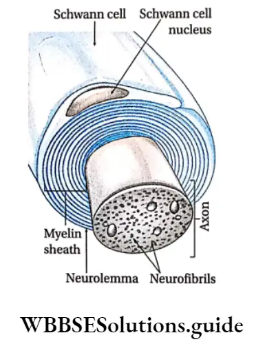
Covering: The cell membrane of the axon is called axolemma. The axon may be surrounded by another covering over the axolemma, called the myelin sheath.

The outermost thin neurilemma consists of tubular cells, called Schwann cells, that are arranged along the length of the axon.

Each of these cells is wrapped around the axon so that it is covered by a number of concentric layers of Schwann cell plasma membrane.

These membranes fuse to produce the myelin sheath, which is composed of a shining, fatty substance called myelin. The outermost layer of the Schwann cell plasma membrane is called the neurilemma.

nervous tissue function and structure

Nodes of Ranvier: The myelin sheath is not continuous along the entire length of the axon.

It is interrupted at regular intervals to form certain gaps known as the Nodes of Ranvier.

They are points of branching of the axon. They also contain channels that transport ions, required during the conduction of nerve impulses.

Collateral branches: Generally, the axon lacks branches. However, sometimes the axon may show branching. These branches are called collateral branches.

End brush: Axon ends in numerous fine branches known as axon terminals. These axon terminals form a tuft-like structure called end brush.

Synaptic knob: Each axon terminal swells up into a knob-like structure known as synaptic knob. It contains abundant mitochondria and secretory vesicles, called the synaptic vesicles.

Functions: Axons conduct nerve impulses from the cell body to the next neuron. Hence, it is efferent in nature.

Dendron: The shorter branching processes of the i-cell body of a neuron are called dendrons.

Structure: It is of shorter length than the axon. The diameter of the dendron is not uniform throughout its length. The outer covering of a dendron is 4 neurilemma.

  • The thin terminal branches of dendrons are known as dendrites.
  • The cytoplasm of dendron ( contains Nissl granules, neurofibrils, and mitochondria.

Functions: Dendrons receive stimulations and conduct them towards the cyton. Hence, they are afferent in nature.

Biology Class 11 Chapter 7 Structural Organisation In Animals Differences Between Areolar And Adipose Connective Tissue

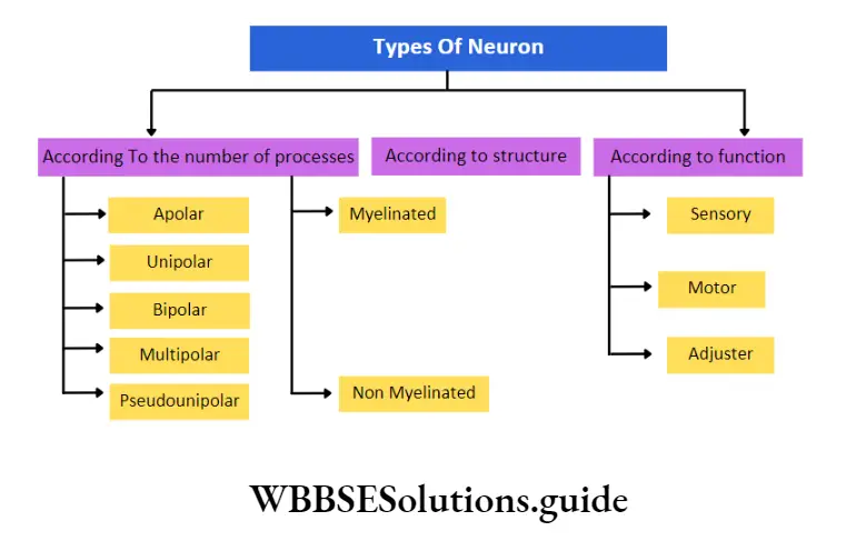
Classification of neuron

Biology Class 11 Chapter 7 Structural Organisation In Animals Types of neuron

structure and function of nervous tissue

On the basis of processes arising from the cell body: According to the number of processes arising from the cell body, neurons may be classified into—

Apolar neuron: The neuron without any processes arising from its cell body is known as apolar neuron. Such neurons are found in the CNS of the vertebrates.

Unipolar neuron: A neuron with only one process arising from the cell body is known as a unipolar neuron. Such neurons are found in the early stages of embryonic development.

Bipolar neuron: The neuron with two processes (one axon and one dendron) arising from the cell body is known as bipolar neuron.

Such neurons are found in the rod and cone cells of the retina and in the olfactory epithelium.

Multipolar neuron: A neuron with more than two processes arising from the cell body is known as a multipolar neuron.

One of these processes acts as an axon and the rest as dendrons. Such neurons are found in the central nervous system and in the ganglia of the autonomic nervous system.

Pseudounipolar neuron: The neuron with a single process formed by the union of the dendron and axon known as a pseudounipolar neuron.

It soon bifurcates and separates into dendrons and axons after emerging from the cell body.

Such neurons are found in the dorsal root ganglia of the spinal nerve in adult vertebrates.

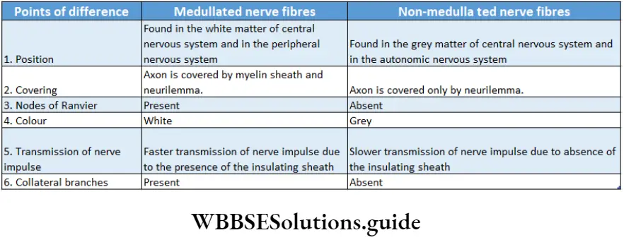
On the basis of structure: According to structure, neurons are classified into two types.

” characteristics of nervous tissue”

Medullated or myelinated neurons: Medullated neurons are the neurons in which the axon is surrounded by an inner thick medullary sheath or myelin sheath and an outer thin neurilemma or neurolemma.

Medullated or myelinated neurons are found in the white matter of the central nervous system (CNS) and in the peripheral nervous system.

Non-medullated or non-my/inated neuron: Non-medullated neurons are the neurons in which the axon lacks myelin sheath i.e., it is covered only by neurilemma. These neurons also lack the Node of Ranvier.

Non-medullated or non-myelinated neurons are found in the grey matter of the central nervous system (CNS) and in the autonomic nervous system.

On the basis of function: According to the function, neurons are of three types—sensory, motor, and relay neurons.

Sensory or afferent neurons: They transmit impulses from sense organs that receive the stimuli to the central nervous system.

Motor or efferent neurons: They transmit electrical signals from the central nervous system to the effector organs in the body.

Relay neurons: They are found within the central nervous system between the sensory and the motor neurons. These neurons transmit the electrical impulses from sensory to motor neurons. They are also called adjuster neurons interneurons or connector neurons.

Biology Class 11 Chapter 7 Structural Organisation In Animals Difference Between Medullated And Non- Medullated Nerve Fibers

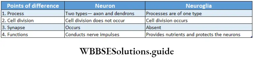
Neuroglial cells Definition: The specialized, non-neural, accessory cells of the nervous system that provide support to the nerve cells are known as neuroglial cells.

These are essential for the function and survival of nerve cells.

Neuroglial cells Position: These cells are found both in the central nervous system and peripheral nervous system.

Neuroglial cells Types: The different types of neuroglial cells are as follows—

Oligodendrocytes: These are myelin-secreting cells of the CNS. Their main function is to provide support and insulation to the axons within the CNS

Biology Class 11 Chapter 7 Structural Organisation In Animals Oligodendrocyte

“diagram of nervous system “

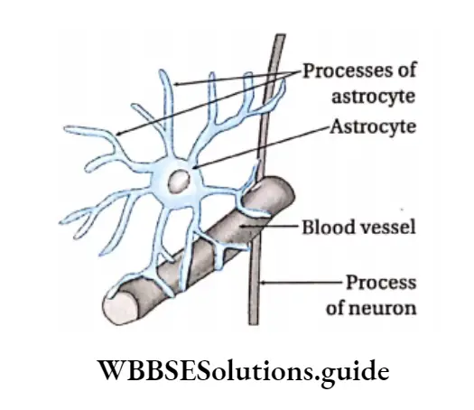
Astroglia or Astrocytes: They are star-shaped cells having several processes extending from their cell body into the surrounding network of nerve fibers.

They provide physical support as well as essential nutrients to nerve cells of the brain and spinal cord.

They help in the transport of ions, taking place between the nerve cell and the extracellular fluid. They also help to form the blood-brain barrier (thin, semipermeable membrane that separates the blood from the cerebrospinal fluid in the brain).

Astrocytes differ from oligodendrocytes in having thicker and more number of processes.

Biology Class 11 Chapter 7 Structural Organisation In Animals Astrocyte

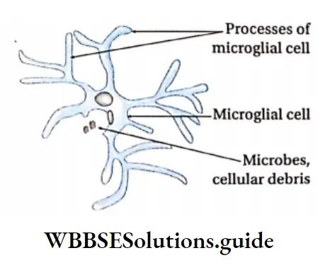
Microglia or microglial cells: These cells are small cells that are similar to macrophages (large scavenger cells) present within the immune system, with respect to functions. They are the phagocytes of the central nervous system.

Biology Class 11 Chapter 7 Structural Organisation In Animals Microglia

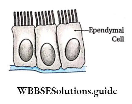
Ependymal cells or ependymocytes: These cells are found in the central nervous system.

They form an extremely thin epithelial-like lining in the ventricles of the brain and central canal of the spinal cord.

This lining is called ependyma. The apical surface is covered by cilia, which keep the cerebrospinal fluid in circulation. They also form cerebrospinal fluid (CSF).

Biology Class 11 Chapter 7 Structural Organisation In Animals Ependymal cell

Biology Class 11 Chapter 7 Structural Organisation In Animals Differences Between Neuron And Neurogila

Satellite glial cells and Schwann cells

Besides the above-mentioned cells, there are certain other accessory cells, that help the nerve cells in different ways. They are called satellite glial cells and Schwann cells.

  1. Satellite glial cells: They surround cell bodies of neurons in ganglia (ganglion cells). They provide electrical insulation and nutrients to neurons.
  2. Schwann cells: They are responsible for the myelination of axons in the peripheral nervous system.

Biology Class 11 Chapter 7 Structural Organisation In Animals Schwann cell

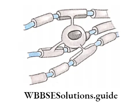
Synapse Structure: Synapse is the junction between the dendrites of one neuron and the axon of another neuron.

Parts: The terminal swollen part of the axon called the synaptic knob, is covered by a membrane called the presynaptic membrane.

The membrane of the dendrite of the other neuron is called the postsynaptic membrane.

The minute space between the presynaptic and postsynaptic membrane is called the synaptic cleft.

Functions of neuroglia: Various functions of neuroglia are—

  1. They (oligodendrocytes) synthesize myelin sheaths.
  2. They (ependymal cells) coordinate the flow of CSF.
  3. They (microglia) have phagocytic properties.
  4. They (astrocytes) form the blood-brain barrier.
  5. They (astrocytes) maintain an appropriate balance of Ca2+ and K+ ions needed for synaptic transmission.

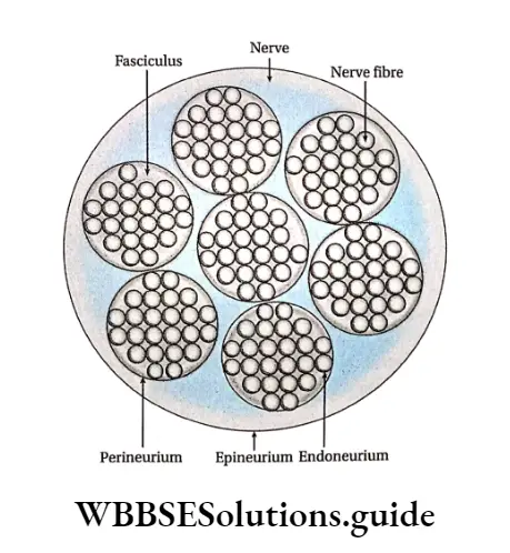
Nerve or Nerve trunk

A nerve is formed of several bundles of nerve fibers. Such bundles are called the fasciculi. Each nerve fiber in a bundle is covered by a thin sheath of connective tissue, called the endoneurium.

Each fasciculus is enclosed by another sheath of white fibrous tissue, called the perineurium.

All the fasciculi are surrounded by a thick coat of white fibrous tissue, called the epineurium.

Biology Class 11 Chapter 7 Structural Organisation In Animals Cross-section of nerve

Biology Class 11 Chapter 7 Structura Organisation In Animals Comparison Among Major Animal Tissues

Muscular Tissue – Structure, Functions and Types Notes

Muscular Tissue

Muscular tissue Definition: The tissue, derived from embryonic mesoderm, and involved in movement and locomotion by virtue of its property of contraction and relaxation is known as muscular tissue.

Muscular tissue Position: Some muscular tissues or muscles are associated with bones and so known as skeletal muscles, Some are associated with visceral organs and are called smooth muscles. Some muscles are associated with the heart, hence known as cardiac muscles.

Muscular tissue Origin: Muscular tissues have originated from mesoderm.

Muscular tissue Components: Muscular tissue is made up of muscle cells or fibers. The main components of the cell include 70% water, 20% proteins like actin, myosin, tropomyosin A, tropomyosin B, myogen, myoalbumin, myoglobulin, etc. 10% glycogen, 0.2% lipids like phospholipid and cholesterol, 1.5% inorganic salts like potassium phosphate, salts of calcium, sodium, magnesium, etc.

Read and Learn More: WBCHSE Notes for Class 11 Biology

Muscular tissue Structure

Muscular tissue Shape: Muscule Tissue Is Made of Bundles Of Narrow, Elongated Contractile Muscle Cell, called my myocytes. Due to their elongated nature, they are also known as muscle fibers.

muscles and muscle tissue

Muscular tissue Arrangement: They are contractile in nature. The contractile unit is made up of proteins such as mainly myosin, actin, troponin, tropomyosin, etc. The muscle fibers are arranged like a mesh.

Muscular Tissue – Structure, Functions And Types Notes

Muscular tissue Matrix: Intercellular matrix is absent but muscle fibers are surrounded by connective tissue.

Muscular tissue Covering: Myofibril or muscle fiber is covered with a plasma membrane known as sarcolemma. They also have mitochondria, known as sarcosomes, and endoplasmic reticulum, known as sarcoplasmic reticulum.

Muscular tissue Innervation: Sensory nerves are present in muscles.

Class 11 Biology Class 11 Chemistry
Class 11 Chemistry Class 11 Physics
Class 11 Biology MCQs Class 11 Physics MCQs
Class 11 Biology Class 11 Physics Notes

Muscular tissue Circulation of blood: Blood vessels are present in the muscular tissue. So, blood circulation takes place within muscle fibers.

Some Special Contractile Cells

Besides muscle cells, some other cells also have the capacity to contract and expand. Some of these cells are described below.

Muscular tissue Myoepithelial cells: The contractile cells which originate from the ectoderm are called myoepithelial cells. Their contraction is slow and involuntary. They are found in the sweat glands, mammary glands, lacrimal glands, and salivary glands.

Muscular tissue Pericytes: These are contractile cells that wrap around the endothelial cells of capillaries and venules throughout the body. They are also known as mural cells.

Myofibroblasts: The contractile cells that contain actin and myosin are called myofibroblasts. They help in wound repair.

Myofibroblasts Functions

Coordination of organs: Muscles (especially involuntary muscles) control coordination between various organs, movement and locomotion, body posture, speech, facial expression, etc.

” types of muscle tissue”

Coordination of visceral organs: Involuntary muscles present within the visceral organs, help in the movement of the organs.

Some examples of these movements are—the contraction of the muscles of the GL tract during peristalsis, uterine wall contraction during childbirth, urinary bladder contraction during micturition, contraction, and relaxation of the walls of the blood vessels for regulating blood pressure, etc.

Circulation of blood: The circulation of blood throughout the body as well as the beating of the heart, both are coordinated by the contraction and relaxation of cardiac muscle.

Response to stimuli: The contraction of muscles helps us to respond to external stimuli.

Coordination of respiration: Respiration is regulated by the contraction and relaxation of respiratory muscles (skeletal muscles). It is also regulated by the smooth muscles lining the trachea and airways of the lungs.

Generation of heat: The movement of muscles during vigorous physical activities such as exercise etc., helps to generate heat in the body.

Types: There are several types of muscular tissue or muscles based on position, structure, mode of action, etc.

On the basis of structure, location, and function, the types of muscles are—striated or skeletal or voluntary muscles, non-striated or smooth visceral or involuntary muscles, and cardiac muscles. The structure and function of each of these muscular tissues are described below

Skeletal Muscle

Skeletal Muscle Definition: The muscles with alternate light and dark bands, which remain associated with the skeleton and can contract on will, are called striated skeletal or voluntary muscles.

Skeletal Muscle Position: These muscles remain attached to the bones or the skeleton and are controlled by the somatic nerve.

Characteristic features: Skeletal muscle is called striated because of its appearance.

It contains alternate light and dark bands, that are visible through a phase contrast microscope.

Biology Class 11 Chapter 7 Structural Organisation In Animals Skeletal muscle

Components:

  1. The skeletal muscles (e.g. biceps, triceps) are spindle-shaped, i.e., swollen in the middle region and tapering at the ends.
  2. The muscles are attached to the bones by means of a hard, collagenous connective tissue, called tendon. One end of the tendon is bound to the perichondrium of bone and the other end is bound to the sarcolemma of the muscle fiber.
  3. Each muscle fiber is enclosed by a connective tissue membrane, known as an endomysium.
  4. A bundle of muscle fibers is known as a fasciculus (pi. fasciculi). This is enclosed by another connective tissue layer called the perimysium.
  5. Bundles of fasciculi are surrounded by another connective tissue layer called the epimysium.
  6. The main structural component of striated muscles or skeletal muscles is muscle cells or muscle fiber.
  7. The structural features of muscle fibre as seen under an electron microscope are described below.

Shape: Each muscle cell is elongated, cylindrical, unbranched fiber, with blunt ends. Each muscle fiber has a diameter of 10-100 pm and is 1-40 mm long.

“muscle tissue structure “

Sarcolemma: The muscle fiber is enclosed within a thin transparent membrane called sarcolemma. This membrane is semi-permeable in nature.

Sarcoplasm: The cytoplasm of muscle cells is known as sarcoplasm. The striated muscle fiber is multi-nucleated. During the formation of striated muscle fiber, its nucleus divides a number of times by mitosis.

This division is not followed by the division of cytoplasm. Due to this reason, muscle fibers are multi-nucleated.

Organelles: It contains sarcoplasmic reticulum (ER), sarcosomes (mitochondria), glycogen, and lipid droplets.

The most important organelle among them is

A-band sarcoplasmic reticulum. The terminal end of the sarcoplasmic reticulum is swollen and is called terminal cisternae. This part stores a huge amount of calcium ions.

These Ca2+ ions are used for muscle contraction.

T-tubule: The sarcolemma of muscle fibers invaginates and extends towards the cytoplasm, at some points.

These extended tube-like structures are called T-tubules or transverse tubules. Each T-tubule remains associated with two terminal cisternae of the sarcoplasmic reticulum.

This structure formed by the T-tubule and the two associated terminal cisternae is called a triad.

Biology Class 11 Chapter 7 Structural Organisation In Animals Three dimensional structure of a muscle finre

Myofibril: The sarcoplasm of skeletal muscle fiber consists of numerous longitudinal bundles of protein fibrils.

These fibrils are called myofibrils. Using an electron microscope, it can be seen that the myofibrils are made up of two types of thin, thread-like contractile protein filaments called myofilaments. The two myofilaments are thicker myosin filaments (100A in diameter) and thinner actin filaments (50A in diameter).

Each myofibril contains approximately 1500 myosin and 3000 actin filaments.

“function of muscle tissue “

The actin filament comprises actin, tropomyosin, and troponin proteins while the myosin filament is made up of myosin protein.

Alternate light and dark bands, called striations, appear throughout a myofibril. The structural details of these striations are as follows-

A Band And I Band: The Light Bands Of Myofilaments Which Are Composed Of Strands Of Only Actin known as l-bands or isotropic bands.

These bands are called ‘isotropic’ as they have the same refractive index in all planes.

The dark bands of myofilaments contain strands of myosin protein along with a few strands of actin protein scattered within. These are known as the A-bands or anisotropic bands.

These bands are called ‘anisotropic1 as they refract light differently in different planes.

Z-line or Z-disc: The thick line present at the center of the l-band is called the Z-line or Krause’s membrane. It is also known as Dobie’s line.

[The letter Z in ‘Z-line’ has been derived from the German word ‘Zwischenschiebe’ which means, Zwischen = between and Scheibe = disc.]

Sarcomere: The segment of the myofibril between two adjacent Z lines is known as sarcomere.

  1. It is the contractile unit of the myofibril and hence, that of the muscle.
  2. The ends of actin strands in adjacent sarcomeres are connected by the Z-line.
  3. Sarcomere = Half of l-band + One complete A-band + Another half of the next l-band

H-zone: Along the midline of each A-band is a lighter zone. This is known as the H-zone or Hensen’s zone.

M-line: A thick line is present vertically in the middle of the H-zone. This is known as the M-line.

[The letter ‘M’ in the M-line has been taken from the German word ‘Mittlescheibe’ which means ‘central disc’]

Biology Class 11 Chapter 7 Structural Organisation In Animals Structure of myofibril

Biology Class 11 Chapter 7 Structural Organisation In Animals Structure of myofibril

Function: The movement of the skeleton is under the conscious control of the body, hence controlled by striated muscles.

These include movement of limbs, fingers, toes, neck, etc. Changes during facial expressions Example ability to smile or to frown, are also controlled by voluntary muscles.

Smooth Muscle Definition: The muscles that are non-striated, involuntary in nature, and found in the visceral organs are known as visceral muscles or non-striated or involuntary muscles.

3 types of muscle tissue

Smooth Muscle Position: These muscles are found in the wall of the alimentary canal, urinary tract, blood vessels, uterus, fallopian tube, the dermis of the skin, iris, and ciliary bodies of the eyes.

Characteristic features:

  1. These muscles do not have any cross-striation and so these are known as smooth muscles.
  2. Due to their presence in the visceral organs, these are also known as visceral muscles.
  3. These muscles are controlled by the autonomic nervous system and hence, are involuntary in nature.
  4. The cells are capable of mitosis.
  5. These muscles can remain contracted for a long time. These muscles never get fatigued.

Components

Muscle fibers: Each muscle fiber is spindle-shaped. These are generally unbranched but, in some fibers, terminal branches can be seen.

Length and diameter: The average length of a muscle fiber is 20 pm with a maximum width of about 6 pm.

Outer covering: The outer covering of muscle fiber is called sarcolemma. It is indistinct in nature.

Cavioli and sarcoplasmic reticulum: Certain structures formed by sarcolemma within the smooth muscle cells are called cavioli. The inner portion of the ravioli contains the sarcoplasmic reticulum.

Nucleus: Cells are uni-nucleated with a centrally placed oval or elongated nucleus.

Mitochondria: Cells contain fewer mitochondria.

“muscular tissue function “

Golgi bodies: Cells contain smaller Golgi bodies.

Contractile protein: The sarcoplasm contains numerous fine contractile myofibrils. Unlike skeletal muscles, the myofibrils are not arranged in a distinct pattern in a smooth muscle cell.

The myofilaments, actin, and myosin, present within the myofibrils, are irregularly arranged.

They are not organized to form sarcomeres (structures formed by two adjacent l-bands with an A-band between), hence striations are not formed

Cellular connections: Adjacent smooth muscle cells are separated by a basal lamina.

However, this basal lamina is not continuous. It is absent in the regions where adjacent muscle cells are connected.

These regions that appear like band-shaped tight junctions are called fascia occludes or nexus.

Cellular connections Types

Single-unit smooth muscle: The muscles formed by the muscle fibers that contract together at the same time are called single-unit smooth muscles.

In such cases, the muscle cells are connected by tight junctions and hence, these muscles form a covering.

These tight junctions are known as cytoplasmic bridges. Due to this property of forming tight junctions, single-unit smooth muscle bundles form a syncytium.

This structure contracts in a coordinated manner, leading to muscle contraction.

Examples are the muscles of the uterus, gastrointestinal tract, and urinary bladder.

Multi-unit smooth muscle: The muscles formed by the muscle fibers that contract separately, each as a single unit are called multi-unit smooth muscles.

Examples are the muscles of walls of blood vessels, ciliary bodies, iris, esophagus, dermis, hair root, etc.

Cellular connections Functions

Peristalsis: The action of smooth muscles helps food to be carried along the gastrointestinal tract.

Regulation of opening and closing of orifice: Smooth muscle forms the structures called sphincters, which are present at the beginning of the orifices.

The sphincters control the opening and closing of a number of orifices in the body. This in turn regulates several physiological processes like the movement of food from the stomach into the intestine or evacuation of the urinary bladder and rectum.

“structure of skeletal muscle “

Controlling vasodilation: Vasodilation is controlled by the contraction of smooth muscles, thereby regulating blood pressure.

Contraction of the urinary bladder and uterus: Contraction of the urinary bladder during micturition and forceful contraction of the uterus during childbirth occurs with the help of smooth muscles.

Other functions: The smooth muscles of the stomach also help in the churning of food.

Cardiac Muscle Definition: The striated involuntary muscle present in the myocardium (wall) of the heart is known as cardiac muscle.

Cellular connections Position: These muscles are found in the wall of the heart.

Characteristic features:

  1. Cardiac muscles are associated with the heart.
  2. They are capable of involuntary and rhythmic movements.
  3. Cross striations are observed, hence they are a type of striated muscles.

Biology Class 11 Chapter 7 Structural Organisation In Animals Visceral and cardiac muscle

Components Muscle fibers: Cardiac muscle fibers are striated, and branched. The cells that form the cardiac muscle are called cardiomyocytes or myocardiocytes. They have a single, large, oval, central nucleus.

Sarcolemma: The outer covering of cardiac muscle (sarcolemma) is indistinct.

Syncytium: Cardiomyocytes are connected with one another forming a syncytium.

Intercalated disc: Sarcolemma of the cardiac muscle fiber is thickened at intervals to form plate or disc-like structures called intercalated discs.

They separate the cardiac muscle cells from one another. At each intercalated disc, the cell membranes of two adjacent cells fuse with one another, forming permeable communicating junctions (gap junctions). These junctions help in the transmission of nerve impulses.

Other types of cell junctions formed through intercalated discs include fascia adherens (binding site for actin, connect sarcomeres) and desmosomes (binding site for intermediate filaments, join the cells).

Sarcoplasm: Sarcoplasm is granular in nature. It has several mitochondria, scattered throughout. A small Golgi body, with few lipid droplets and sarcoplasmic reticulum (endoplasmic reticulum), are also present.

Myofilament: Numerous distinct myofibrils with alternate dark and light cross bands, forming prominent sarcomeres, are found within the sarcoplasm.

As stated earlier, myofibrils are made up of myofilaments, that are composed of actin and myosin protein strands. They are present as bundles.

T-tubules: Sarcolemma penetrates inside muscle fibers at specific regions. These structures formed by the sarcolemma are called transverse tubules or T-tubules. Together these tubules are known as T-system.

This network stores calcium ions, required for muscle contraction.

Functions: Pumping of blood through the heart takes place by alternate contraction and relaxation of cardiac muscles.

Besides the above-mentioned types, there are several types of muscular tissue, such as—

Types of muscles on the basis of function and pigments: Details regarding the classification (Locomotion and movement).

Types of muscles based on the nature of their activity: Muscles are classified on the basis of the nature of their activity into two groups.

White muscle or fast muscle: The skeletal or voluntary muscles which appear pale due to less blood vessels and the absence of myoglobin are called white muscles.

Such muscles show prominent striations. They react fast to impulses but cannot hold the contraction for a long time. Example muscles of the lip, tongue, etc.

Red muscle or stow muscle: Skeletal muscles that appear red due to more blood vessels and myoglobin are called red muscles.

Such muscles show fewer striations. They react slowly to impulses but can hold the contraction for a long time. Example muscles controlling body posture.

Few Concepts About Muscular Tissue

Refractory period: The brief period during which the muscle cannot get excited to a second stimulus after receiving the first stimulation is called the refractory period.

Heart muscles have a long refractory period. Hence, they do not fatigue easily.

Tetanus: In terms of physiology, if a series of I stimuli from the external environment are applied successively during a single contraction, then they fuse together leading to sustained contraction.

This phenomenon is called tetanus, Summation: A weak stimulation may fail to produce a contraction in the muscles.

But when this weak stimulation is applied repeatedly, it may produce considerable contraction in the muscles. This phenomenon is called summation.

Rigor mortis: The rigidity of muscles after death is called rigor mortis.

Biology Class 11 Chapter 7 Structural Organisation In Animals Comparisons Among Atriated, Unstriated And Cardiac Muscles

Glandular Epithelium – Definition, Structure and Functions Notes

Synthesis and secretion: The glands, formed by these glandular cells, synthesize and secrete mucus, hormones, enzymes, etc.

Storage: Some chemical substances are stored in these glandular cells, such as—protein in the stomach, lipids in the adrenal and sebaceous glands, and sugar and protein in the salivary gland.

Excretion: When present in sweat glands and sebaceous glands, glandular epithelium helps to excrete sweat and sebum, respectively.

Classification of glands: On the basis of the number of cells present in the epithelium, glands are divided into two types—

  1. Unicellular glands and
  2. Multicellular glands.

Unicellular gland: Unicellular gland may be defined as an isolated single secretory cell that itself functions as a gland.

Read and Learn More: WBCHSE Notes for Class 11 Biology

Unicellular gland Position: It is found in the mucous membrane of the stomach, intestine, rectum, and trachea.

Structure They are elongated and flask-shaped. The cell consists of a nucleus, surrounded by cytoplasm containing numerous Golgi bodies.

Class 11 Biology Class 11 Chemistry
Class 11 Chemistry Class 11 Physics
Class 11 Biology MCQs Class 11 Physics MCQs
Class 11 Biology Class 11 Physics Notes

The apical part of the cell contains granules which contain the glycoprotein mucin. The distal part of the cell becomes swollen and bursts. This causes secretion of mucin.

Glandular epithelium definition, structure, and functions notes PDF

Unicellular gland Functions: Some of them (goblet cells) secrete mucus which forms a protective layer on the intestinal mucous membrane,

  • They neutralize acids and alkalis, thereby maintaining pH balance.
  • They provide protection by engulfing bacteria and foreign particles that enter the trachea or alimentary canal.
  • They lubricate the alimentary canal and trachea.

Example: Goblet cells, calciform cells.

what is glandular epithelium

Multicellular gland: Multicellular glands may be defined as glands comprising aggregates of secretory cells.

On the basis of types of secretion, there are three types of multicellular glands—

Endocrine glands are ductless glands. They pour their secretions (hormones) directly into the blood. Example pituitary, thyroid or adrenal glands etc.

Exocrine glands, these glands are provided with ducts. They pour their secretions (enzymes) through ducts into the target organs. Example salivary, gastric-OPlntestjnal glands,

Mixed glands, glands comprise both endocrine and exocrine parts. Example pancreas— whose exocrine secretory part is called ‘acinus’ and endocrine secretory part is made up of cells called ‘islets of Langerhans’.

Glandular Epithelium – Definition, Structure and Functions Notes

On the basis of the mode of secretion, glands are of three types. They are—

Holocrine gland: Secretion is discharged along with the entire cytoplasm of the cell. This leads to the disintegration of the cell. Example sebaceous gland.

Apocrine gland: The apical surface of the secretory cell is discharged along with the accumulated secretions. Examples mammary glands, and apocrine sweat glands.

Merocrine gland: The secretion is released to the external environment through the cell membrane slowly by diffusion. The whole cell remains intact. Examples salivary gland, and pancreatic gland.

Biology Class 11 Chapter 7 Structural Organisation In Animals Secretions of holocrine,apocrine and merocrine gland

Types of multicellular glands on the basis of the nature of ducts

Simple gland: Simple glands are multicellular glands with unbranched ducts. These are two Simple tubular glands, which are unbranched, tube-like simple glands. Example intestinal gland, sweat gland,

Simple saccular or alveolar glands, which are unbranched, sac-like simple glands. Example mucous glands in frog’s skin. It is absent in mammals.

Glandular epithelium structure and function with diagram

Compound gland: Compound glands are multicellular glands with branched ducts. These are of two types—

Compound tubular gland, the distal end of these glands is branched and tube-like. Example Brunner’s gland of duodenum.

Compound saccular or racemose gland, a distal end of these glands is branched and sac-like.

Examples exocrine pancreas, salivary gland, and sebaceous gland.

Tubulo-saccular/alveolar gland, distal ends of these glands comprise both tubule and sac-like structures. Examples are parts of the salivary gland, mammary gland, and glands of respiratory passage.

Functions of glands

Hormone secretion: The endocrine glands are involved in the secretion of hormones. For example, thyroxine is secreted from the thyroid gland, insulin is secreted from the endocrine part of the pancreas, etc.

Enzyme secretion: The exocrine glands secrete various enzymes. For example, ptyalin is secreted from the salivary gland, pepsin from the gastric gland, etc.

Compound or stratified epithelial tissue

Functions of glands

Hormone secretion: The endocrine glands are involved in the secretion of hormones. For example, thyroxine is secreted from the thyroid gland, insulin is secreted from the endocrine part of the pancreas, etc.

Enzyme secretion: The exocrine glands secrete various enzymes. For example, ptyalin is secreted from the salivary gland, pepsin from the gastric gland, etc. Compound orstratified epithelial tissue.

Biology Class 11 Chapter 7 Structural Organisation In Animals Stratified epithelial tissue

Transitional epithelial tissue Definition: The epithelial tissue that has multiple (3-4) layers of cells and shows properties of both simple and compound epithelium, is called transitional epithelial tissue.

Transitional epithelial tissue Position: It is found in the renal pelvis, broader parts of the ureter, upper part of the urinary bladder, urethra, and urinary duct.

Transitional epithelial tissue Structure:

  1. The cells of the superficial layer are large, flat, irregular-shaped (generally quadrilateral), and may be binucleate.
  2. The next layer contains pyriform cells (flame-shaped cells). One end of these cells is round and the other end is narrow.
  3. Cells in the third layer are polyhedral. These cells are located in between the narrow ends of pyriform cells.
  4. The cells of the basal (bottom) layer are round or cuboidal in shape. © The nuclei are oval or spherical in shape and the cytoplasm is granular.
  5. This epithelium forms walls of the organs that undergo continuous expansion and contraction.

Transitional epithelial tissue Function:

  • It prevents the reabsorption of ions during excretion.
  • It regulates the distension of organs like the urinary bladder.

Biology Class 11 Chapter 7 Structural Organisation In Animals Transitional epithelial tissue

Stratified squamous epithelial tissue

Stratified squamous epithelial tissue Definition: Epithelial tissue in which the uppermost layer has squamous cells, that may or may not be keratinized, is called stratified squamous epithelial tissue.

Stratified squamous epithelial tissue Position: It is located in the epidermis of the skin, oral cavity, pharynx, esophagus, vag*na, cervix, cornea, etc.

Stratified squamous epithelial tissue Types: It is of two types—stratified keratinized squamous epithelium and non-keratinized stratified squamous epithelium. These are discussed under separate heads.

Stratified keratinized squamous epithelial tissue Definition: The epithelial tissue, consisting of multiple cellular layers, whose uppermost cellular layer contains keratin, is called stratified keratinized squamous epithelial tissue.

Stratified squamous epithelial tissue Position: It is found in the epidermis of the skin (nails, horns, the enamel of teeth, etc).

Stratified squamous epithelial tissue Structure:

  • Cells of the superficial layer are dead. However, these cells store keratin and are thus cornified.
  • Hence, this layer is called stratum corneum (Latin term, meaning ‘horny layer’).
  • Cells of the middle layer are polygonal and squamous in nature.
  • The lower layer has columnar cells which are attached to the basement membrane.
  • The cells of the superficial layer are constantly sloughed off and replaced by new cells from the inner layers.

Biology Class 11 Chapter 7 Structural Organisation In Animals Stratified keratinised squamous epithelial tissue

Function: Its major role is to protect the underlying soft tissues against mechanical injury and friction. It is water-resistant and hence, prevents water loss from the body.

Non-keratinized stratified squamous epithelial tissue Definition: The epithelial tissue, consisting of multiple cellular layers, in which the uppermost layer does not contain keratin is called non-keratinized stratified squamous epithelial tissue.

Types of glandular epithelium and their functions

Non-keratinized stratified squamous epithelial tissue Position: It is found in the lining of oral and nasal cavities, urethra, cervix, cornea, vag*na, anal canal, etc.

Non-keratinized stratified squamous epithelial tissue Structure:

  • This tissue has multiple layers of cells. These cells lack keratin.
  • The outer layer consists of living cells and the middle layer contains polyhedral cells.
  • Cells attached to the basement membrane are columnar.

glandular

Non-keratinized stratified squamous epithelial tissue Function: Its major role is to protect the underlying soft tissues against mechanical injury and friction. It is water-resistant and hence, prevents water loss from the body.

Prickle cell: The cells at the basal surface of stratified keratinized squamous epithelial tissue are connected with each other by intercellular fibers and protoplasmic appendages.

These fibers and appendages are called prickles and the associated cells are called prickle cells due to their thorny appearance. These cells can withstand pressure.

Stratified Cubodial Epithelial Tissue

Stratified Cubodial Epithelial Tissue Definition: The epithelial tissue, consisting of multiple cellular layers, in which the uppermost layer does not contain keratin is called non-keratinised stratified squamous epithelial tissue.

Stratified Cubodial Epithelial Tissue Position: It is found in the larger duct of sweat glands, salivary glands, female urethra, and pancreas.

Structure:

The tissue is usually multi-layered.

Cuboidal cells are present in the uppermost layer and polyhedral cells are found in the middle layer.

Function: It provides strength to the walls of the lumen and also helps in the secretion of sweat, saliva, etc.

Biology Class 11 Chapter 7 Structural Organisation In Animals Stratified cuboidal epithelial tissue

Class 11 biology glandular epithelium notes with examples

Stratified Columnar Epithelial Tissue Definition: The epithelial tissue, made up of multiple layers of cells, whose uppermost cellular layer has columnar cells is called stratified columnar epithelial tissue.

Stratified Columnar Epithelial Tissue Position: It is found in the pharynx, epiglottis, a mucous layer of the anus, and urethra.

Stratified Columnar Epithelial Tissue Structure:

The uppermost layer has columnar cells with an oval nucleus.

The lowermost layer has cuboidal cells.

Function: Its main function is to give protection to the underlying organ.

Biology Class 11 Chapter 7 Structural Organisation In Animals Stratified columnar epithelial tissue

Endothelium Definition and Function Notes

Endothelium

The squamous epithelial tissue present in the inner wall of blood vessels and lymphatic vessels is called endothelium.

Endocardium: The squamous epithelial tissue present in the inner wall of the heart is called the endocardium.

Endocardium Functions

  • Protection: It protects internal organs from mechanical injury and infection.
  • Diffusion: It helps in the diffusion of gases and nutrients, for example, lung alveoli and capillary endothelium.
  • Filtration: It helps in ultrafiltration in the Bowman’s capsule of nephron, epithelium, and endothelium.

Read and Learn More: WBCHSE Notes for Class 11 Biology

Biology Class 11 Chapter 7 Structural Differences Between Epithelium And Endothelium

Biology Class 11 Chapter 7 Structural Organisation In Animals Squamous epithelial tissue

Cuboidal Epithelial Tissue

Cuboidal Epithelial Tissue Definition: The epithelial tissue that consists of cube-shaped cells that rest on the basement membrane is called cuboidal epithelial tissue.

Cuboidal Epithelial Tissue Position: It is found in the thyroid gland, salivary gland, bronchioles in the lungs, proximal and distal convoluted tubules of the nephron, covering of the ovary (germinal epithelium), etc.

Class 11 Biology Class 11 Chemistry
Class 11 Chemistry Class 11 Physics
Class 11 Biology MCQs Class 11 Physics MCQs
Class 11 Biology Class 11 Physics Notes

Endothelium meaning

Cuboidal Epithelial Tissue Structure:

  1. Cells of this tissue are cuboidal in shape and similar in size i.e., they have almost equal length, breadth, and height.
  2. The nucleus is spherical and centrally located.
  3. The cytoplasm is granular.
  4. The free surfaces of the cells are sometimes lined by microvilli.
  5. Such a surface containing microvilli is called a brush border.

Endothelium Definition and Function Notes

Cuboidal Epithelial Tissue Function

Protection: It protects the organs to which it is associated.

Secretion: It secretes various secretory products when present in secretory glands such as the thyroid, pancreas, mucous gland, salivary gland, and sweat gland.

Endothelium definition and function notes PDF 

Reabsorption: Under special conditions (within convoluted tubules in nephrons), cuboidal epithelium reabsorbs molecules and ions from the urine. Thus, this tissue takes part in urine formation.

Cuboidal epithelial tissue Function Types: Different types of cuboidal epithelium, their definition, positions, and functions are given in the following table

Biology Class 11 Chapter 7 Structural Different Types Of Cuboidal Epithelium-definition,postion And Functions

Biology Class 11 Chapter 7 Structural Organisation In Animals Cuboidal epithelial tissue

Columnar epithelial tissue

Columnar epithelial tissue Definition: The epithelial tissue in which cells are column-shaped (i.e., height is more than their breadth), is called columnar epithelial tissue.

Columnar epithelial tissue Position: It is found in the lining of the digestive tract, gall bladder, bile duct, ducts of gastric gland, intestinal gland, sweat, and sebaceous gland, and pancreatic lobules.

Columnar epithelial tissue Structure: Cells of this tissue are tall and elongated.

The nucleus is oval-shaped and is situated near the base of the cell, in case the cell is secretory in nature. Otherwise, the nucleus lies at the center of the cell.

Endothelium function in blood vessels and cardiovascular system

“endothelial meaning “

A brush border is seen in the free surface of the epithelium lining the inner cavity of convoluted tubules of the small intestine.

Columnar epithelial tissue Functions

Absorption: It helps in the absorption of digested food into blood by the intestinal epithelium.

Secretion: Columnar epithelium of the gastric and intestinal glands transform into pitcher-shaped cells, known as goblet cells. They secrete mucus.

Protection: The mucus, secreted by the goblet cells, protects the lining of the stomach from the effect of the HCI acid secreted in it. In this way, they protect the organs to which the epithelium is associated.

Types: Types of columnar epithelium, their definition, position, and functions are given in the table below.

Biology Class 11 Chapter 7 Structural Different Types Of Columnar Epithelium Defination, Postion And Functions

Biology Class 11 Chapter 7 Structural Differences Between Squamous,CUboidal And Columanr Epithelium

Biology Class 11 Chapter 7 Structural Organisation In Animals Columnar epithelial tissue

Ciliated epithelial tissue Definition: The epithelial tissue that is made up of generally columnar, sometimes cuboidal cells, with fine JiaÿBce processes or cilia on the free surface is known as ciliated epithelial tissue.

Position: It is found in the fallopian tube, most of the uterus, the efferent tubule of testes, the lining of the ventricles of the brain, and the central canal of the spinal cord.

Structure:

  1. Cells of this tissue are either columnar or cuboidal.
  2. The free surface of the cells has 20-30 cilia.
  3. The root of the cilia has a row of particles or corpuscles at its base, called basal granules. These are considered to be the residue of centriole.
  4. Thread-like structures extend inward, from each basal granule, into the protoplasm of the cell.

Biology Class 11 Chapter 7 Structural Organisation In Animals Ciliated epithelial tissue

Types of cilia on the free surface of epithelial tissue

Kinocilia: These are motile in nature. These cilia originate from basal granules of the cytoplasm. These are present on the inner walls of the trachea.

Stereocilia: These are non-motile in nature. These cilia are formed by the foldings of the plasma membrane. These are seen in epididymis and vas deferens.

Types of cilia on the free surface of epithelial tissue Function

Protection: Wave-like unidirectional movement of cilia of the columnar epithelial cells helps to sweep out mucus and dust particles in organs such as the trachea, nasal passages, etc.

Transport of ovum: The fallopian tube, helps in the transport of the ovum towards the uterus.

“blood vessel lining “

Transport of cerebrospinal fluid (CSF): It helps in the circulation of CSF into cerebrospinal cavities of the central nervous system.

Glandular epithelial tissue Definition: The epithelial tissue that is made up of cells specialized mainly in secretory functions is known as glandular epithelial tissue.

The secretory organs formed by the glandular epithelia are called glands. The secreted substances can be enzymes, hormones, or chemicals. The secretions are transported to their target organs or cells either by duct (in case of enzymes, chemicals, etc.) or without duct (in case of hormones).

Class 11 biology endothelium notes with diagrams

Position: It is found in mammary glands, sweat glands, sebaceous glands, salivary glands, part of Intestinal glands, and thyroid gland.

“endothelial cells “

Glandular epithelial tissue Structure:

  1. Cells of this tissue are generally cuboidal, columnar, or polygonal in shape.
  2. The cells are usually arranged in one layer but in the salivary gland, a second layer may be found.
  3. Sometimes, the secretory cells may be present as bundles. These cells are specialized for the synthesis and secretion of enzymes, hormones, or certain chemicals required by the body.
  4. The cytoplasm is granular in nature. It contains membrane-bound secretory vesicles.
  5. Each cell bears a large nucleus.

Types And Functions Of Epithelial Tissues Notes

Epithelial Tissue

Types of tissues: The bodies of all vertebrates and most of the invertebrates are made of a variety of tissues.

However, all the tissues may be grouped into four main types. These are epithelial tissue, connective tissue, muscular tissue, and nervous tissue.

Epithelial Tissue Location: Epithelial tissue is present mainly in two regions—the external covering of the body (skin) and the walls (both inner and outer) of all internal organs.

Epithelial Tissue Components: Epithelial cells are compactly arranged with minimum intercellular spaces.

The tissue is made up of three major components. They are—the non-cellular basement membrane, epithelial cells, and intercellular cementing material. This cementing material is a mucoprotein complex containing hyaluronic acid and calcium salt.

Read and Learn More: WBCHSE Notes for Class 11 Biology

Epithelial Tissue Structure: An epithelium consists of one or more layers of cells. The outer layer of cells, when present in the skin, is exposed to the external environment outside the body.

“types and functions of epithelial tissues notes for class 11”

But, this layer is also present on the outer surface of internal organs. Here, it is exposed to a lumen or cavity within the body. The deep inner layer of cells is bound by a basement membrane.

Class 11 Biology Class 11 Chemistry
Class 11 Chemistry Class 11 Physics
Class 11 Biology MCQs Class 11 Physics MCQs
Class 11 Biology Class 11 Physics Notes

simple squamous epithelial tissue diagram

Basement membrane: Basement membrane is not actually a membrane. Rather, it is an extracellular matrix present between the epithelial tissue and the loose connective tissue below it.

It is composed of a network of collagenous fibers which are formed from secretions of the underlying cells of the connective tissue. It consists of two layers—the outer thin basal lamina and the inner thick, fibrous, reticular lamina (also known as lamina reticularis).

“short notes on epithelial tissues for NEET and exams”

The basal lamina is further made up of two layers—lamina lucida and lamina densa. The lamina lucida is closer to the epitheium while the lamina densa is closer to the connective tissue.

The lamina reticularis is attached to the basal lamina with collagen fibrils. This layer is also attached together by microfibrils made of fibrillin protein.

Types And Functions Of Epithelial Tissues Notes

Functions of the basement membrane of epithelium

  1. Anchorage: It binds the epithelium to the loose connective tissue underneath and holds the layers -c6
  2. Growth: Controls the growth and differentiation of cells within the epithelial tissue.
  3. Exchange of molecules: Acts as a filter during an exchange of large molecules between epithelium and the underlying connective tissue.
  4. Signaling: Helps in cell-to-cell signaling.

Lamina propria: It is a layer of loose vascular connective tissue, present at the base of the basement membrane. It provides mechanical support to the epithelium.

It also contains several blood vessels and nerves. These blood vessels and nerves provide nutrition and innervation to the epithelium.

“detailed notes on epithelial tissues with diagrams”

Papilla: The hair-like appendages that lie at the junction of epithelium and the underlying connective; tissue are called papillae (sing, papilla). They hold the epithelium in position.

Biology Class 11 Chapter 7 Structural Organisation In Animals Basic structure of epithelial tissue

Surface layers of cells: The epithelial cells have three surfaces. These are as follows—

Basal surface: It is the lower surface of the cells, adjacent to the basement membrane.

Apical surface: It is the upper surface of the cells, that remains free. It increases the surface area for absorption.

This property is achieved by modification of the surface into structures like microvilli, stereocilia, etc.

Lateral surfaces: These are surfaces of the cells, on both sides, facing adjacent epithelial cells.

“classification of epithelial tissues and their functions”

Biology Class 11 Chapter 7 Structural Organisation In Animals Free surface,brush border,cilia of tissue

Intercellular junctions: These are specialized regions between the plasma membrane of adjacent cells. These are also known as cell junctions. They provide mechanical support and help in cell-to-cell communication.

However, sometimes they may act as impermeable barriers, preventing the transport of certain molecules between the cells.

“difference between simple and stratified epithelial tissues”

Intercellular junctions are of the following types—

Tight junctions: These are specialized regions between adjacent epithelial cells where the cell membranes are fused together by means of sealing strands.

They bind the epithelial cells together and check the passage of molecules and ions between them.

Gap junctions: These are specialized junctions directly connecting the cytoplasm of cells. They allow the passage of ions and small molecules from one cell to the adjacent one.

“epithelial tissue characteristics and examples notes”

Adherens junctions: Junctions present in heart muscles and skin epithelium. They join the actin filaments present in the muscles to form a continuous belt.

Desmosomes: Intercellular junctions of epithelia and cardiac muscle. They are of two types—Belt and Spot desmosomes. The belt desmosomes are belt-like and the spot desmosomes are spot-like or circular in appearance.

Biology Class 11 Chapter 7 Structural Organisation In Animals Intercelluar junction

“squamous epithelial tissue “

Cilia and Flagella: These are microtubular structures that arise from the plasma membrane of the epithelial cells. Cilia are found in animal cells whereas flagella are primarily found in prokaryotes and unicellular eukaryotes.

They help in locomotion as well as to propel away harmful, unwanted particles.

Cilia and Flagella Functions: The functions of epithelial tissue are as follows—

Cilia and Flagella Protection: Epithelial tissue forms the skin of animals. It also forms the wall of internal organs, thus protecting them from injuries.

Terrestrial vertebrates have keratin in their skin cells. This makes them resistant to water loss from their skin. Ciliated epithelium, lining the respiratory tract, sweeps away impurities with the help of cilia.

“types and functions of epithelial tissues PDF notes download”

Cilia and Flagella Absorption: The gut is lined with epithelial tissue that absorbs nutrients from food. The lungs are also lined with epithelial tissue which helps them to absorb oxygen.

Cilia and Flagella Secretion: Glandular epithelium forms the exocrine and endocrine glands. Endocrine glands secrete hormones into the circulation. Exocrine glands secrete mucus, saliva, wax, milk, etc., through ducts.

“importance of epithelial tissue in protection and absorption”

Contractile property: Myoepithelium is the contractile epithelium, present in sweat glands, mammary glands etc. It helps in the flow of secreted fluids from the glands.

Reproduction: Gametes (such as sperm and ova) are produced from the germinal epithelium lining the testis and ovary.

“functions of epithelial tissue in different organs”

Sensation: It is perceived with the help of sensory epithelium present in the skin, taste buds in the tongue, nasal epithelium, etc.

Excretion: Ultrafiltration by Bowman’s capsule and tubular reabsorption during urine formation is carried out by renal epithelium in the kidney.

Transport: The beating of cilia in ciliated epithelium present in food pipe, respiratory tract, reproductive tract, etc., helps to transport food, mucus, gametes, etc.

Respiration: The epithelium of alveoli in the lungs helps in gaseous exchange.

Classification: Based on the shape of the cells that lie above the basement membrane and on the number of layers of cells, the different types of epithelial tissues are shown in the given flowchart.

Biology Class 11 Chapter 7 Structural Organisation In Animals Epithelial tissue

Simple epithelial tissue Definition: The epithelium that is made up of a single layer of cells, is called simple epithelial tissue or epithelium.

Based on the structure of the cells, the simple epithelium is classified as—squamous epithelium, cuboidal epithelium, columnar epithelium, ciliated epithelium, and glandular epithelium.

“structure and types of epithelial tissues explained”

Squamous Epithelial Tissue Definition: The epithelial tissue that consists of a single layer of large, flattened, polygonal cells is called squamous epithelial tissue.

Squamous Epithelial Tissue Position: It is found in the alveoli of lungs, endothelium of capillaries, Bowman’s capsule and Henle’s loop of the nephron, pericardium and the inner lining of the heart, and the peritoneal lining of the coelom.

Squamous Epithelial Tissue Structure:

  1. Cells of this tissue are flat, polygonal in shape with irregular margins. Their shapes resemble the scales of fish. It is also known as pavement epithelium as it forms a pavement-like structure.
  2. Cells are compactly arranged without any intercellular spaces.
  3. The nuclei are oval or disc-shaped and centrally located.
  4. Cytoplasm may be clear or granular.

What Is Aestivation ? Describe Various Types Of Aestivation

Aestivation

Aestivation Definition: The pattern of arrangement of calyx, corolla or perianth in a flower bud is known as aestivation.

Aestivation Types:

Different types of aestivation are as follows—

Morphology Of Flowering Plants Aestivation

what is aestivation

Open: The margins of the sepals or petals do not touch or overlap each other. example Magnolia champaca, Allaria sp., etc.

Morphology Of Flowering Plants Open aestivation

Valvate: The margins of sepals or petals just touch side by side closely, without overlapping each other. example, calyx of Hibiscus rosa-sinensis, both calyx and corolla of Mimosa pudica and Acacia nilotica, etc.

Read and Learn More: WBCHSE Notes for Class 11 Biology

Morphology Of Flowering Plants Valvate aestivation

“definition and types of aestivation in flowers”

Twisted or contorted: One margin of each sepal or petal overlaps the margin of the neighboring sepal or petal. example corolla of Hibiscus rosa-sinensis, Nerium odoratum, etc.

Morphology Of Flowering Plants Twisted aestivation

Imbricate: One member of the whorl remains completely outside without any overlapping both the edges of another member remain overlapped by the neighboring members and the rest are arranged in a twisted manner. example corolla of Solanum torvum, Nicotiana tabacum, Clerodendrum viscosum, etc.

Morphology Of Flowering Plants Imbricate aestivation

” aestivation in biology”

Quincuncial: Two members of the whorl remain completely external and two remain completely internal. The rest are present in a twisted manner. example petals of Lantana camara, sepals of Holarrhena antidysenterica, etc.

Morphology Of Flowering Plants Quincuncial aestivation

Vexillary: Out of five members the posterior single petal is the largest and outermost (vexillum), which overlaps the two lateral petals (wings). The two wings partially overlap the two smallest and innermost, partially fused petals (keel). example corolla of Pisum sativum, Clitoria ternatea, Crotolaria sp., etc.

Morphology Of Flowering Plants Vexillary aestivation

Variations Infloral Members

The floral parts, i.e., calyx, corolla, androecium, and gynoecium, show different variations in their shapes, position, arrangement, and association with other floral parts. These variations or forms of different floral members are described below.

Variation in calyx

Different types of sepals are discussed below.

Class 11 Biology Class 11 Chemistry
Class 11 Chemistry Class 11 Physics
Class 11 Biology MCQs Class 11 Physics MCQs
Class 11 Biology Class 11 Physics Notes

“importance of aestivation in plant classification”

Based on the color of the calyx: According to the color of sepals, calyx are of two types. The green-coloured calyx is known as sepaloid. example, Cocos nucifera. Sometimes the calyx becomes colored like the petals and is known as a petaloid or tepal. example Mussaenda sp.

What Is Aestivation Describe Various Types Of Aestivation

aestivation definition biology

Based on whether sepals are united or not: According to the union of sepals, calyx are of two types.

Polysepalous: In this type, the sepals remain free and separate. example Brassica nigra.

Gamosepalous: In this type, the sepals remain partially or completely united. example Datura metel, Hibiscus rosa-sinensis.

Morphology Of Flowering Plants Polysepalous(Rose)

Morphology Of Flowering Plants Gamosepalous(Datura)

Based on the duration of attachment of calyx to the flower: According to the duration of attachment of calyx to the flowers, calyx are of two types.

Caducous or fagacious: It sheds off just before the complete blooming of the flower. example Papaver somniferum.

Deciduous: It remains attached to the flower until fertilization. It sheds off with corolla. example Brassica nigra.

“difference between aestivation and phyllotaxy”

Persistent: It remains attached till fruit maturation and never sheds off. It is of two types—

Marcescent: This type of persistent sepals cease further growth after fertilization, get shrunk, and become wrinkled. example Solarium melongena.

Accrescent: This type of persistent sepals continues to grow along the growth of the fruit. example Cocos nucifera, and Dillenia indica.

Morphology Of Flowering Plants Different modifications ofcalyx

Based on the structure of calyx: The structure of calyx varies with species. Types of calyx based on their structures have been mentioned.

Morphology Of Flowering Plants Types of calyx

Variation In Corolla

On the basis of different characters corolla can be of various types. Sometimes the petals can be green in color, known as sepaloid or sepaline. example Annona squamosa, Pergularia daemia, etc.

Based on whether petals are united or not: According to the union of petals, corolla are of two types—

Polypetalous: The petals remain free. example Brassica nigra.

“aestivation in flower buds and its significance”

Gamopetalous or sympetalous: The petals are completely or partially united. example Datura metel, Catharanthus roseus, etc.

Morphology Of Flowering Plants Polypetalous (Prunus sp.)

Morphology Of Flowering Plants Gamopetalous(Brugmansia sp.)

Appendages of Corolla

Sometimes various types of appendages may develop on the corolla.

These are as follows—

  1. Saccate or gibbous: This is a sac-like or pouch-like structure formed by the dilation of the lower region of the corolla tube on one side. example Antirrhinum majus (snapdragon), Calceolaria mexicana.
  2. Corona: These are hair-like or scaly outgrowths of various kinds that develop from the inner wall of the corolla tube. example Passiflora suberosa, Nerium indicum, Scoparia dulcis, etc.
  3. Spurred or calcarate: These are tubular structures formed by the elongation of a single petal or petals of the corolla tube. example Delphinum majus and Aquilegia sp.

“twisted aestivation in flowers with diagram”

Based on the structure of corolla:

According to the structure, corollas are of the following types—

Polypetalous and regular type:

This type of Corolla is divided into three types—

  1. Cruciform: In this type, four clawed free petals are arranged in the form of a cross. example Brassica nigra (mustard) and Raphanus sativa (radish), etc.
  2. Caryophyllaceous: In this type, five petals have claws, arranged at right angles to their limbs. example Dianthus chinensis and Saponaria vaccaria, etc.
  3. Rosaceous: In this type, five or more almost sessile petals are widely spread outside. example Rosa lucida (wild rose), and Camellia sinensis (tea).

Morphology Of Flowering Plants Different types ofpolypetalous and regular corolla

Polypetalous and irregular type: Out of five irregular free petals, the posterior, odd petal is the largest and outermost. This is known as vexillum or standard. The vexillum overlaps two lateral petals, known as wings or alae.

The wings partially overlap the two smallest and innermost petals known as keel or carina. These two interior petals remain more or less united. It is also known as a papilionaceous type of corolla. Examples are Pisum sativum (pea), Sesbania grandriflora, Clitoria ternatea, etc.

Morphology Of Flowering Plants Papilionaceous (Clitoria sp.)

Gamopetalous and irregular type:

This type of Corolla is of three types—

  1. Ligulate: Five petals unite to form a flat tongue-shaped upper part of the corolla gradually forms the short tubular lower part. example, Ray florets of Helianthus annuus (sunflower).
  2. Bilabiate or two-lipped: The upper part of the corolla tube is divided into two lip-like structures to form a wide open mouth above the throat. example Ocimum sanctum, Anisomeles indica, Leucas linifolia.
  3. Personate or masked: The flower contains, a bilabiate corolla limb, but here the mouth is closed by a projection (palate) of the lower lip. example Antirrhinum majus (snapdragon), Lindenbergia indica, etc.

Morphology Of Flowering Plants Different types ofgamopetalous and irregular corolla

Gamopetafous and regular type:

This type of corolla is divided into the following types—

  1. Tubular: Here, the petals unite to form a cylindrical corolla tube. example Disc florets of Helianthus annuus (sunflower).
  2. Campanulate or bell-shaped: The petals unite to form a bell-shaped structure. example Cucurbita maxima (pumpkin), Thevetia peruviana (yellow oleander), Physalis peruviana, etc.
  3. Rotate or wheel-shaped: The short corolla tube spreads its flat limbs at right angles to the tube axis example Scoparia dulcis, Solanum melongena (brinjal), Calotropis procera, etc.
  4. Hypocrateriform or salver-shaped: The long and narrow corolla tube spreads its limbs at right angles to the tube axis. The limbs arrange themselves in a wheel-like structure. example. Ixora coccinea, Ipomoea quammoclit,etc.
  5. Infundibuliform or funnel-shaped: The lower tubular corolla gradually widens at the top like a funnel. example, Ipomoea pulchella (morning glory), and Datura sp. (thorn apple).
  6. Urceolate or urn-shaped: The corolla tube becomes inflated in the middle and comparatively narrower at both ends. example Rhododendron sp., Bryophyllum calycinum.

Morphology Of Flowering Plants Different types ofgamopetalous and regular corolla

Variation in androecium

On the basis of the number, location, structure, etc. of stamen, androecium is of different types.

“vexillary aestivation in Fabaceae family plants”

Based on the number of other lobes:

Based on the number of enter lobes, androecia are of the following types—

Monothecous: Anthers have a single lobe. Example Brassica nigra.

Dithecous: Anthers have two lobes. Example Hibiscus rosa-sinensis.

The number of stamens varies from one to many. On the basis of the number of stamens,

The flowers are grouped as—

  1. Monandrous: This type of flower contains single or solitary stamen. Example Curcuma longa (turmeric).
  2. Diandrous: This type of flower contains two stamens. Example Adhatoda vasica (vasaka).
  3. Triandrous: This type of flower contains three stamens. Example Triticum aestivum (wheat).
  4. Tetrandrous: This type of flower contains four stamens. Example Ocimum sanctum (tulsi).
  5. Pentandrous: This type of flower contains five stamens. Example Solanum nigrum.
  6. Hexandrous: This type of flower contains six stamens. Example Oryza sativa.
  7. Polyandrous: This type of flower contains numerous stamens. Example Rosa centifolia.

Based on the insertion of stamens:

Based on the insertion of stamens, androecia are of the following types—

Isostemonous: In this type, one whorl of stamens is arranged alternately with the sepals i.e., antisepalous (Example Pisum sativum) or petals i.e., antipetalous (Example Primrose)

Diplostemonous: In this type, the stamens are arranged in two whorls and the number of stamens is double the number of petals. Usually, the inner whorl remains opposite to the petals. Example Cassia’s fistula.

Obdiplostemonous: In this type, the stamens are arranged in two whorls such that the members of the outer whorl are opposite to petals and those in the inner whorl are opposite to sepals. Example Geranium sp.

Polystemonous: In this type, stamens are arranged in more than two whorls. Example Delphinium sp.

“classification of aestivation with examples for class 11”

Alternipetalous: In this type, the stamens remain opposite to the perianth. Example Diosporous embryopteris.

Based on the length of the stamen:

Based on the length of stamens, androecia are of the following types—

Didynamous: There is a solitary whorl of four stamens, among which two stamens are longer than the other two. Example Leonurus sibiricus.

Tetradynamous: Six stamens are arranged in two whorls. In the outer whorl, two stamens are there which are shorter than the four stamens of the inner whorl. For example, Brassica nigra.

Heterodynamous: In this type, stamens of different lengths occur in one whorl. Example Cassia Tora.

Based on the attachment of anther with the filament:

According to the attachment of anther with filament, stamens are of the following types—

Adnate: The filament remains attached throughout the length of the anther. Example Magnolia sp.

Basifixed or innate: The apex of the filament remains firmly attached to the base of the anther. Example Brassica sp.

Dorsifixed: The tip of the filament remains firmly attached to the dorsal side of the anther. Examples are Sesbania sp., Passiflora sp.

“role of aestivation in floral symmetry and taxonomy”

Versatile: The tip of the filament remains attached at a point near the middle of the connective and thus the anthers can move freely. Example Triticum aestivum.

Morphology Of Flowering Plants Different types ofattachments ofanther andfilament

Based on the shape of the anther: Different types of anthers, based on their shapes are given below.

Morphology Of Flowering Plants Types of anther

Morphology Of Flowering Plants Different shapes ofanther

Based on the dehiscence of anthers: The pollen grains inside the mature anther develop and exert some pressure on the outer wall. The anther wall bursts open due to the pressure and the pollen grains are set free in the air.

The dehiscence pattern of anthers can be of the following types—

Longitudinal: The anther lobes burst longitudinally. This occurs along the line of suture from base to apex. Example Datura sp.

Transverse: The anther splits up transversely along the suture. Example Hibiscus rosa-sinensis.

Apical or porous: The pollen grains are discharged through the apical pores of the anther. Example Solarium tuberosum.

Valvular: The walls of the anther open up like valves to discharge the pollens. Examples are Berberis vulgaris, Cinnamomum zeylanicum, etc.

Morphology Of Flowering Plants Dehiscence ofanther

Based on the union of stamens: The stamens may be fused with each other or with the gynoecium or perianth.

The union of stamens found in different flowers is of the following types—

Cohesion of stamen: Stamens may remain fused with each other or with filament or another. This is known as the cohesion of stamen.

This can be of the following types—

1. Adelphous: In this type, the stamens are united by the fusion of their filaments.

The adelphous condition may further be divided into the following types—

  1. Monadelphous: In this type, filaments of all the stamens are fused to form a single tubular structure. Example Hibiscus rosa-sinensis.
  2. Diadelphous: In this type, the stamens are fused at their filaments and form two bundles. Example Pisum sativum.
  3. Polyadelphous: In this type, the stamens are fused by their filaments and form more than two groups. Example Bombax ceiba.

2. Syngenesious: In this type, all the anthers remain fused but the filaments are free. Example Helianthus annuus (sunflower).

3. Synandrous: In this type, the anthers and filaments are fused throughout their length to form a compact structure. Example Cucurbita maxima (pumpkin).

Morphology Of Flowering Plants Cohesion ofanther

Adhesion of stamens: When the stamens are attached with other floral whorls or perianth, then the condition is known as adhesion of stamen.

it is classified into the following types—

  1. Epipetalous stamen: The stamens remain attached to the petals by their filaments. Example Solanum nigram.
  2. Epiphyllous stamen: The stamens are attached to the perianth by their filaments. Example Polyanthes tuberosa (tuberose).
  3. Gynandrous stamen: The stamens remain attached to the gynoecium, either throughout their length or only by their anthers. Example Calotropis sp.
  4. Episepalous stamen: The stamens remain attached to the calyx. Example Quinsqualis sp.

Morphology Of Flowering Plants Adhesion ofstamen

Variation in Gynoecium

Based on the number and position of the thalamus, the gynoecium is of various types.

“diagram-based explanation of aestivation in plants”

Based on the number of carpels in the gynoecium:

According number of carpels, gynoecium are of the following types—

Simple or monocarpellary gynoecium: The gynoecium is composed of solitary carpel. Example Pisum sativum.

Bicarpellary: The gynoecium is composed of two carpels. Example Justicia sp.

Tricarpellary: The gynoecium is composed of three carpels. Example Allium cepa.

Tetracarpellary: The gynoecium is composed of four carpels. Example Datura metel.

Pentacarpeliary: The gynoecium is composed of five carpels. Example Melia sp.

Compound or polycarpellary: The gynoecium is composed of more than five carpels. Example Papaver sp. It is of the following two types, on the basis of their union—

Apocarpous: In this type, the carpels remain completely free from one another to form multiple ovaries. Example Michelia Champaca.

Syncarpous: In this type, the carpels are fused with each other to form a single ovary. Example Mangifera indica.

Morphology Of Flowering Plants Cohesion ofcarpel

Based on the number of locules in the ovary: According to the number of locules or chambers in the ovary,

Gynoecium can be of the following types—

Morphology Of Flowering Plants Types of ovary

Based on the position of the ovary on the thalamus: The position of the ovary on the thalamus varies in plants.

“MCQs on aestivation and floral arrangements in plants”

Superior: In this type, the ovary is situated on the top of the thalamus and the other floral whorls reside just below the ovary. The flowers which bear this type of ovary are known as hypogynous flowers. Example Hibiscus rosa-sinensis.

Morphology Of Flowering Plants Superior ovary

Inferior: In this type, the ovary resides at a level that is lower than the other floral whorls on the thalamus. The thalamus becomes cup-shaped due to fusion with the ovary wall. The flowers which bear this type of ovary are known as epigynous flowers. Example Cucurbita maxima.

Morphology Of Flowering Plants Inferior ovary

Morphology Of Flowering Plants Semi-inferior ovary

Semi-inferior: In the type, the ovary is situated in such a position, which is intermediate to that of the superior and inferior types. the flowers that bear this type of ovary are known as perigynous flowers example pisum sotivum.

Phyllotaxy Notes

Phyllotaxy

The leaves remain arranged on the stem in such a manner that they can get the maximum amount of sunlight for their physiological functions.

Phyllotaxy Definition: The arrangement of leaves on stems or branches in a definite manner is known as phyllotaxy.

Phyllotaxy Types:

Phyllotaxy is mainly of three types—

  1. Alternate or acrylic or spiral,
  2. Opposite and
  3. Whorled. The last two types are known as cyclic type.

Morphology Of Flowering Plants Phyllotaxy

Alternate or acyclic or spiral: In this type, one leaf develops from each node and remains spirally arranged around the stem. Examples are china rose, and mango. Here, phyllotaxy is determined by passing an imaginary line through the bases of the leaves.

Read and Learn More: WBCHSE Notes for Class 11 Biology

This shows a spiral path is known as a genetic spiral. if this spiral path is placed on a flat surface then it is known as a flat spiral. The distance between the bases of two consecutive leaves is known as divergence.

“phyllotaxy notes for class 11 biology”

The angle subtended at the center of the shoot by two consecutive leaves is known as angular divergence. The imaginary vertical line that connects the leaves vertically is known as orthostichies.

Morphology Of Flowering Plants Alternate phyllotaxy

On the basis of the number of orthostichies or the number of rows in which the leaves are arranged,

Phyllotaxy Notes

Alternate phyllotaxy is divided into the following types—

  1. Distichous or 2-ranked phyllotaxy: Leaves are arranged in two rows, i.e., two orthostichies can be drawn. example, sugarcane, maize
  2. Tristichous or 3-ranked phyllotaxy: Leaves are arranged in three rows, i.e., three orthostichies can be drawn. exampleCyperus sp., orange.
  3. Pentastichous or 5-ranked phyllotaxy: Leaves are arranged in five rows, i.e., five orthostichies can be drawn. example Hibiscus rosa-sinensis.
  4. Octastichous or 8-ranked phyllotaxy: Leaves are arranged in eight rows, i.e., eight orthostichies can be drawn. example papaya.

Morphology Of Flowering Plants Different types ofalternate phyllotaxy

whorled leaf arrangement examples

Opposite: In this type, two leaves develop from each i node opposite to each other.

It may be of two types— Opposite decussate and opposite superposed.

  1. Opposite decussate: In this type, consecutive pairs of opposite leaves are arranged at a right angle to each other. example Ocimum sanctum, Calotropis procera, etc.
  2. Opposite superposed: In this type, successive pairs of opposite leaves are arranged in one plane. example Quisqualis sp., Hiptage madablota, etc.

Morphology Of Flowering Plants Opposite phyllotaxy

Whorled or verticillate phyllotaxy: In this type, more than two leaves develop from each node forming a whorl. example, Alstonia scholars, etc.

Morphology Of Flowering Plants Whorled phyllotaxy

phyllotaxy of leaves

Morphology Of Flowering Plants Differences between spiral and opposite phyllotaxy

Modifications Of Leaves

Other than photosynthesis and transpiration leaves perform other functions. For that purpose, they modify themselves through different adaptations.

Different types of modifications are discussed below—

Leaf tendrils: In weak plants, the lamina becomes modified either partially or wholly into a coiled thread-like structure called a tendril. This tendril helps the plant to climb up support.

Class 11 Biology Class 11 Chemistry
Class 11 Chemistry Class 11 Physics
Class 11 Biology MCQs Class 11 Physics MCQs
Class 11 Biology Class 11 Physics Notes

These can be of the following types—

Whole leaf tendril: In this type, the entire leaf gets modified into a tendril. example Lathyrus aphasia.

Leaflet tendril: In this type, the terminal leaflets are modified into tendrils. example Pisum sativum.

Morphology Of Flowering Plants Different types ofleaftendril

“phyllotaxy “

Petiolar tendril: In this type, the petiole is modified into tendrils. example Clematis gouriana.

Leaf apex tendril: In this type, the leaf apex is modified into a tendril. example Gloriosa Superba.

Stipular tendril: In this type, the stipules are modified into tendrils. example Smilax macrophylla.

Leaf spines: The leaf may be completely or partially modified into a spine. This protects the plants from predators and reduces the rate of transpiration in plants.

“types of phyllotaxy with examples notes”

These are of the following types-

  1. The apex of the lamina can be modified into spine as found in Phoenix sylvestris.
  2. The margin of the lamina can be modified into the spine as found in Argemone mexicana.
  3. The apex and margin of lamina both can be modified into spines as found in Aloe perfoliata.
  4. Leaves can be modified into spines. But the leaves that emerge from axillary buds remain normal, as found in Berberis vulgaris.

Morphology Of Flowering Plants Leafspine

whorled phyllotaxy

Fleshy or succulent leaves: In some plants, the leaves become fleshy due to the storage of water, mucilage, and food. This is mostly found in xerophytes and halophytes. example Bryophyllum sp., Basella rubra, etc.

Morphology Of Flowering Plants Succulent leaf (Bryophyllum sp.)

Morphology Of Flowering Plants Leafhook(Bignonia sp.)

Leaf or leaflet hooks: In some plants, three-terminal leaflets of a compound leaf get modified into sharp, curved, and anchored on the support for climbing example biononia angiocath.

Root-like structure: In some aquatic plants, the submerged leaves are modified into narrow root-like structures. These adventitious roots absorb water and help to float the plant on water. example Myriophyllum indicum.

Leaf-pitcher: In Nepenthes khasiana, an insectivorous plant, the leaf lamina is modified into a pitcher with a lid developed from the apex. The pitcher helps the plant to fulfill its nitrogen requirement by trapping and digesting insects.

“phyllotaxy in plants detailed notes”

Bladder: A rootless aquatic herb, the bladderwort {utricuiaria stellaris), has segmented leaves. Some of the segments of the leaves are modified to form bladder-like structures. These bladders help them to trap insects and digest them.

Water reservoir leaf: In Dischidia rafflesiana, a non-insectivorous plant, the leaf lamina is modified to form a pitcher-like structure. The main function of this pitcher is to store rainwater for future use.

This plant is an epiphytic climber. Certain adventitious roots develop from the node, from where the stalk of the pitcher develops. These roots absorb water from the pitcher by entering into the cavity of the pitcher.

Morphology Of Flowering Plants Bladderwort

Morphology Of Flowering Plants Water reservoir leaf

Scale leaves: These are membranous leaves that mainly provide protection to the plants. example zingiber officinale.

Morphology Of Flowering Plants Differences between stem tendril and leaf tendril

Morphology Of Flowering Plants Differences between phylloclade and phyllode

Functions Of Leaf

Leaves serve two types of functions—primary and special functions.

Primary functions: The basic functions of leaves are—

Photosynthesis: Leaves produce carbohydrates through photosynthesis, which is the main source of food.

Gaseous exchange: The gaseous exchange in plants occurs through the stomata present on the leaves.

Transpiration: The stomata also help in transpiration and thus maintain the water balance in the plant body.

Protects bud: Leaves protect the apical and axillary buds during their development.

Transportation: The vascular bundles of the leaves help in the conduction of food and water within the plant body.

“difference between alternate, opposite, and whorled phyllotaxy”

Special functions: Leaves of many plants perform some special functions. Those are—

Food and water storage: The fleshy leaves of xerophytes and some other plants store mucilage and food for future use. example Allium cepa, Aloe vera.

Protection: Some leaves are modified into thorns or spines. These structures protect the plants from different external factors. example Argemone spv Cacti sp.

Climbing: In some plants, leaves get wholly or partially modified into tendrils. These tendrils help the weak stem to climb up on supports. example Pisum sativum.

“phyllotaxy diagrams and explanation notes”

Reproduction: Adventitious roots and vegetative buds emerge from the leaf margin of Bryophyllum sp. These give rise to new plants.

Catching insects: Some plants have specialized leaves through which they can catch insects. Such plants are called insectivorous plants. These plants grow on nitrogen-deficient soil. To overcome this they feed on insects, from which they get nitrogen.

Helps in floating: In aquatic plants, the leaves contain aerenchyma cells, which help the plants to float on water.

The Inflorescence

As the plants mature, flowers grow on the floral axis either singly or in clusters. Flowers are arranged in various ways on the floral axis in different plants.

The Inflorescence Definition: The arrangement of flowers on the floral axis either singly or in clusters, is known as inflorescence.

The stalk of a solitary inflorescence is known as the floral axis or peduncle. Sometimes, it branches out and bears flowers at the branched apices. The stalk of the individual flowers is called pedicel.

“short notes on phyllotaxy for NEET”

A long, simple, or branched peduncle is known as a rachis. The small main axis of a spikelet present in grass-like plants is called rachilla.

The unbranched naked peduncle that develops from the underground stem, is termed as scape. The stalked flowers are called pedicellate flowers, whereas the flowers without stalks are known as sessile flowers.

“phyllotaxy and its significance in plants”

Some plants bear flattened peduncles. This is known as a receptacle. The part on which the floral parts grow is called the thalamus. The conical receptacle is known as the torus.

Bracts

Sometimes, the flowers grow at axils of expanded leafy structure, known as a bract. The flowers having bract are known as bracteate flowers and those without bract are known as ebracteate flowers.

Sometimes, very small thin bract-like, leafy, or scaly structures develop on the flower stalk in between the flower and bracts. These structures are known as bracteoles or secondary bracts.

Bracts are of various types—

  1. Leafy or foliaceous,
  2. Scaly,
  3. Spathy,
  4. Petaloid,
  5. Epicalyx,
  6. Involucre,
  7. Glume, and
  8. Cupule.

Morphology Of Flowering Plants Bract

Types of Inflorescence: Inflorescence is mainly of three types.

They are as follows—

Morphology Of Flowering Plants Inflorescence

Morphology Of Flowering Plants Question And Answers

Biology Class 11 WBCHSE Morphology Of Flowering Plants Some Important Questions And Answers

1. What is the difference between a root cap and a multiple root cap?
Answer: The root cap is single-layered and regenerates again after its disintegration. On the other hand, multiple root caps contain multiple layers of cells but can not be produced again.

Morphology Of Flowering Plants

2. Is it possible for a plant to absorb minerals and water from the soil, if the root hairs of its primary root get degenerated?
Answer: Yes. This is because root hairs are also present in the branched roots (secondary and tertiary roots). So, after the degeneration of the root hairs of the primary root, the plant can absorb water and minerals through the root hairs of the branched roots.

3. Why is the root hair region of the main root temporary?
Answer: The root hair region of the main root has a definite lifespan. After that, the root hairs degenerate and secondary roots grow from that region.

Read and Learn More WBCHSE Solutions For Class 11 Biology

4. Write about the distribution of roots in a dome-shaped tree.
Answer: The roots of these trees move deep into the soil, Besides these roots, some branch roots are also found near the lower surface of the trunk. The branch roots remain scattered at the base of the trunk to provide mechanical support to the tree.

Class 11 Biology Class 11 Chemistry
Class 11 Chemistry Class 11 Physics
Class 11 Biology MCQs Class 11 Physics MCQs
Class 11 Biology Class 11 Physics Notes

Important questions on morphology of flowering plants for NEET

5. Name the two functions of the plants that are performed only by the adventitious roots but not by the true roots.
Answer: Reproduction and respiration, are the two functions performed only by the adventitious roots.

Morphology Of Flowering Plants Question And Answers

6. What type of root is a beetroot? Name the pigment present in this root. Name the substance in which the pigment is soluble.
Answer: Beetroot is a napiform root. The pigment is betacyanin. It is soluble in water.

7. Name the plant, in which photosynthesis occurs through the roots.
Answer: Tinospora sp., and Trapa sp., are the two plants, in which photosynthesis occurs through the roots.

8. Name the plants in which buds are produced by adventitious roots.
Answer: The adventitious roots of Trichosanthes sp., and Dalbergia sp., can produce buds.

9. Write down the characteristics of roots in Orchid-like plants.
Answer:

Orchid-like plants bear two types of roots. They are—

  1. Clinging root and
  2. Aerial roots. The clinging root helps the plant to remain attached to the support. Aerial roots absorb water vapor from the air. They provide the required amount of water for photosynthesis to the plants.

10. What kind of root is found in sweet potato? Write down a special feature of this root.
Answer: Tuber-like roots are found in sweet potatoes which are known as tuberous roots. Special feature: These roots do not have any definite shape.

11. What do you mean by vegetative and reproductive organs of plants?
Answer: Vegetative organs perform functions other than reproduction. Whereas, reproductive organs take part in sexual reproduction to form new individuals.

Class 11 Biology Solutions

12. Give two examples of rootless plants.
Answer: Wolffia sp., and Utricularia sp.

13. Why do potatoes store a large amount of starch in them?
Answer: Vegetative reproduction in potatoes occurs by the axillary buds. These buds require glucose for their development. This glucose is provided by the starch stored in the potatoes. So, potatoes store a large amount of starch.

14. Why should plants be described morphologically?
Answer: The description of morphological features helps in identifying and assigning any plant to its proper taxonomical hierarchy and nomenclature.

15. What is the difference between the thorns of an Indian stone apple and a rose?
Answer: The thorns of the Indian stone apple are modified part of the aerial stem and develop endogenously. In the case of a rose, the thorns are outgrowths of the stem epidermis and develop exogenously. They are actually prickles.

Previous year questions on morphology of flowering plants with answers

16. Write down the mode of reproduction in Amorphophallus paeoniifolius (oal) and Zingiber officinale (ginger).
Answer: These two plants reproduce by axillary buds which help in vegetative reproduction. These axillary buds are produced in favorable conditions and gradually develop into new plants.

17. Why do biennial plants require two seasons to complete their life cycle?
Answer: In the first season, these plants germinate from their seeds and attain maturity gradually. In this stage, they produce roots, shoots, etc., (vegetative growth). In the next season, they complete their life cycle by producing flowers and fruits (reproductive growth).

18. Why do the pine and Himalayan Cedar (deodar) become cone-shaped?
Answer: The main axis of these trees grows indefinitely with a racemose branching pattern. The branches of the lower regions are longer than the branches of the upper region. So, these trees appear cone-shaped.

19. Why is an onion known as a shoot-less plant?
Answer: Usually shoot of this plant is not visible. The disc-shaped shoots do not have any aerial region and thus, they are not visible above the ground. The small shoot is not visible clearly even under the soil. So, this plant is known as the shoot-less plant.

20. Name the edible part of the onion.
Answer: The edible part of the onion is mainly the modified, fleshy scale leaves.

Class 11 Biology Solutions

21. How will you identify that the underground part of a plant is a stem?
Answer: The underground part of the plant should not bear any root hair, root cap, or adventitious roots. But it should have a node, internode, axillary bud, apical bud, and scale leaves.

“morphology of flowering plants class 11 ncert “

22. Give examples of plants with amplexicaul and semi-amplexicaul leaf bases.
Answer: Amplexicaul: Aethusa cynapium. Semi-amplexicaul: Musa paradisiaca.

23. Name the plant parts that are modified to form the following—thorns of Indian stone apple and Argemone sp.
Answer: Thorns of Indian stone apple: Modified axillary buds.

Thorns in Argemone sp. Modified leaves.

24. When you will find rachis in a leaf?
Answer: The leaf rachis is found in a compound leaf.

25. What are sessile flowers? Give example.
Answer: The flowers without pedicel are known as sessile flowers. Example Polianthes tuberosa.

26. Write down the differences between pome and drupe.
Answer: The differences between pome and drupe are
Morphology Of Flowering Plants Questions 26

27. Name the factors responsible for the amount of light absorbed by the leaves.
Answer: The factors are the shape of the leaf, location, and phyllotaxy.

28. Pea plants have foliaceous stipules. Why?
Answer: The leaves of the pea plant are compound pinnate leaves. The upper leaflet of the rachis gets modified into tendrils, hence the number of leaves is reduced. As a result, the rate of photosynthesis as well as the growth rate becomes low. So, in this case, the stipule enlarges and becomes foliaceous. This stipule helps in photosynthesis to overcome the problems.

29. Write about the submerged leaves of this Limnophilla sp.
Answer: The submerged leaves of this plant are linear, narrow, and dissected. These leaves reduce the hydrostatic pressure. They also help in the absorption of oxygen, soluble in water.

Class 11 Biology Solutions

30. What is the basic difference between a simple leaf and a leaflet?
Answer: The axil of the simple leaf bears axillary buds but axillary buds are not found at the axil of leaflets.

31. Write about the modification of the petiole to carry out physiological functions.
Answer: Winged petiole: In some plants such as in Citrus sp., the petioles become broad, like foliar leaves, to carry out the process of photosynthesis. Phyllode: In some plants such as in Acacia sp., the compound leaves shed off in early stages. So, the petioles become broad and flattened to carry out the process of photosynthesis, like the foliage leaves.

32. The flowers of the fig are not visible. Why?
Answer: In Fig, the inner wall of the receptacle contains male, female, and sterile flowers. As the flowers are covered with the receptacle, they are not visible.

33. Identify true fruit and false fruits among the following—paddy, fig, cucumber, lemon, jackfruit.
Answer: True fruits—Cucumber, lemon, and paddy. False fruits—Jackfruit and fig.

34. Mention two points of importance of the floral diagram.
Answer:

Two points of importance are—

  1. Floral diagrams provide information about a number of flower parts, fusion among them, etc.
  2. It also provides information about the arrangement of floral whorls on the thalamus with respect to the mother axis.

Biology Class 11 WBCHSE Morphology Of Flowering Plants Very Short Answer Type Question

Question 1. What type of roots are present in leguminous plants?
Answer: Nodulated root

Question 2. Write the type of venation in monocotyledonous plants.
Answer: Parallel venation

Question 3. Give an example of tap root, modified to store food.
Answer: Conical root of carrot

MCQ on morphology of flowering plants with detailed explanation

Question 4. Name the special tissue present in the aerial roots of orchids.
Answer: Velamen

Question 5. Give two examples of fruits whose seeds are dispersed by explosive mechanism.
Answer: Atidrographis paniculate (kale), Impatiens balsamina (dopant flower)

Question 6. What type of venation is seen in leaves of dicotyledonous plants?
Answer: Reticulate venation

Class 11 Biology WBCHSE

Question 7. Which part of the grapevine modifies into a tendril?
Answer: Terminal end of branches

Question 8. Name the edible part of the orange.
Answer: Juicy hairs developed from endocarp

Question 9. What type of fruit is pomegranate?
Answer: Balausta

Question 10. Give an example of heterogamous capitulum inflorescence.
Answer: Sunflower

Question 11. What is the outermost covering of the seed?
Answer: Testa

Question 12. Give an example of a dicot non-endospermic seed.
Answer: Pea

Question 13. Write two examples in which bilateral cyme inflorescence is seen.
Answer: Dyanthus, Silene

Question 14. Name one cultivated plant which does not bear fruit and seed.
Answer: Sugarcane

Question 15. What types of modifications are observed in the roots of carrots, radishes, and dahlia?
Answer: Conical, napiform, and fasciculated roots respectively

Question 16. Write the floral formula of Solanum nigrum.
Answer: 

Morphology Of Flowering Plants formula

Question 17. Name one plant in which chlorophyll is synthesized in its adventitious roots and thus, carry out photosynthesis.
Answer: Trapa, Tinospora

Question 18. Name one flowering insectivorous plant that lacks a root.
Answer: Utricularia

Question 19. What are the different parts of a seed coat?
Answer: Testa and tegmen

Question 20. Which type of leaf possesses stomata mainly on its lower epidermis?
Answer: Generally in the dorsiventral leaf

Question 21. Give two examples of false fruit.
Answer: Apple and pear

Question 22. Write one example of a flowering plant that lacks green leaves.
Answer: Capparis aphylla

Question 23. Which is the edible part of ginger?
Answer: Rhizome of ginger

Question 24. Name the root of Cuscuta sp. that helps the plant to invade the tissue of host plant.
Answer: Haustoria

Question 25. Why do the leaves of Aloe and Agave become fleshy?
Answer: These plants mainly grow in dry and hot places. So, their leaves become fleshy by storing water to resist drought.

Class 11 Biology WBCHSE

Question 26. Why is the calyx of a pea described as gamosepalous?
Answer: In peas, the calyx is gamosepalous because all the sepals remain united.

Question 27. In swampy areas like the Sunderbans in West Bengal, plants bear special kinds of roots called?
Answer: Pneumatophores

Question 28. In aquatic plants like Pistia and Eichornia, leaves and roots are found near________
Answer: Water surface

” 11th biology 5th chapter exercise”

Question 29. Roots obtain oxygen from air in the soil for respiration. In the absence or deficiency of O2, root growth is constricted or completely stopped. How do these plants growing in marshlands or swamps obtain the oxygen required for root respiration?
Answer: They respire through pneumatophores

Question 30. Reticulate and parallel venations are characteristics of _________ and __________ respectively.
Answer: Dicotyledons, monocotyledons

Class 11 morphology of flowering plants important questions and answers

Question 31. Write the floral formula of an actinomorphic, bisexual, hypogynous flower with five united sepals, five free petals, five free stamens, and two united carpels with superior ovary and axile placentation.
Answer:

Morphology Of Flowering Plants formula

Question 32. An inflorescence is found in a hanging position. Identify and in which plant it is present.
Answer: Catkin, mulberry

NEET biology morphology of flowering plants questions with solutions

Question 33. Coleoptile and coleorhiza are absent in which seeds?
Answer: In dicotyledonous seeds

Question 34. Nimmi ate dal and potato fry for lunch. Name the families, with which these two plants are associated.
Answer: Fabaceae, Solanaceae respectively

Question 35. Name the main parts of a typical leaf.
Answer: Leaf base, petiole, and leaf lamina

Morphology Of Flowering Plants Multiple Choice Questions

Morphology Of Flowering Plants Multiple Choice Questions

Question 1. Root hairs develop from the region of—

  1. Elongation
  2. Root cap
  3. Meristematic activity
  4. Maturation

Answer: 4. Maturation

Question 2. The morphological nature of the edible part of coconut is—

  1. Cotyledon
  2. Endosperm
  3. Pericarp
  4. Perisperm

Answer: 2. Endosperm

Morphology of flowering plants multiple choice questions PDF

Question 3. Coconut fruit is a—

  1. Berry
  2. Nut
  3. Capsule
  4. Drupe

Answer: 4. Drupe

Read and Learn More WBCHSE Multiple Choice Question and Answers for Class 11 Biology

Question 4. In Bougainvillea thorns are the modifications

  1. Adventitious root
  2. Stem
  3. Leaf
  4. Stipules

Answer: 2. Stem

Anatomy Of Flowering Plants Multiple Choice Question And Answers

Question 5. The term ‘polyadelphous’ is related to—

  1. Gynoecium
  2. Androecium
  3. Corolla
  4. Calyx

Answer: 2. Androecium

Question 6. Cotyledon of maize grain is called—

  1. Coleorhiza
  2. Coleoptile
  3. Scutellum
  4. Plumule

Answer: 3. Scutellum

Morphology of flowering plants MCQ with answers 

Class 11 Biology Class 11 Chemistry
Class 11 Chemistry Class 11 Physics
Class 11 Biology MCQs Class 11 Physics MCQs
Class 11 Biology Class 11 Physics Notes

Question 7. Which one of the following statements is not true?

  1. The exine of pollen grains Is made up of sporopollenin sporopollenin
  2. Pollen grains of many species cause severe allergies
  3. Stored pollen in liquid nitrogen can be used in crop breeding programmes
  4. Tapetum helps in the dehiscence of anther

Answer: 4. Tapetum helps in the dehiscence of anther

Question 8. Which one of the following is not a stem modification?

  1. Thorns of Citrus
  2. Tendrils of cucumber
  3. Flattened structure of Opuntia
  4. Pitcher of Nepenthes

Answer: 4. Pitcher of Nepenthes

Question 9. The coconut water from tender coconut represents—

  1. Fleshy mesocarp
  2. Free-nuclear proembryo
  3. Free-nuclear endosperm
  4. Endocarp

Answer: 3. Free-nuclear endosperm

Question 10. Stems modified into flat green organs performing functions of leaves are known as—

  1. Phyllodes
  2. Phylloclades
  3. Scales
  4. Cladodes

Answer: 2. Phylloclades

Question 11. The standard petal of a papilionaceous corolla is also called

  1. Pappus
  2. Vexillum
  3. Corona
  4. Carina

Answer: 2. Vexillum

Question 12. How many plants among Indigofera, Sesbania, Salvia, Allium, Aloe, mustard, groundnut, radish, gram, and turnip have stamens with different lengths in their flowers?

  1. Three
  2. Four
  3. Five
  4. Six

Answer: 2. Four

NEET morphology of flowering plants MCQ 

Question 13. Radial symmetry is found in the flowers of—

  1. Brassica
  2. Trifolium
  3. Pisum
  4. Cassia

Answer: 1. Brassica

Question 14. Free-central placentation Is found in

  1. Dianthus
  2. Argemone
  3. Brassica
  4. Citrus

Answer: 1. Dianthus

Question 15. Tricarpellary, syncarpous gynoecium is found in flowers of—

  1. Solanaceae
  2. Fabaceae
  3. Poaceae
  4. Liliaceae

Answer: 4. Liliaceae

Question 16. The wheat grain has an embryo with one large, shield-shaped cotyledon known as

  1. Coleoptile
  2. Epiblast
  3. Coleorhiza
  4. Scutellum

Answer: 4. Scutellum

Question 17. Among china rose, mustard, brinjal, potato, guava, cucumber, onion and tulip, how many plants have superior ovaries?

  1. Four
  2. Five
  3. Six
  4. Three

Answer: 2. Five

Question 18. Flowers are unisexual in—

  1. Onion
  2. Pea
  3. Cucumber
  4. China Rose

Answer: 3. Cucumber

Morphology of flowering plants objective questions 

Question 19. Roots play an insignificant role in the absorption of water in—

  1. Wheat
  2. Sunflower
  3. Pistia
  4. Pea

Answer: 3. Pistia

Question 20. An example of an edible underground stem is—

  1. Carrot
  2. Ground Nut
  3. Sweet Potato
  4. Potato

Answer: 4. Potato

Question 21. When the margins of sepals or petals overlap one another without any particular direction the condition is termed as—

  1. Vexillary
  2. Imbricate
  3. Twisted
  4. Valvate

Answer: 2. Imbricate

Question 22. An aggregate fruit is one that develops from the—

  1. Multicarpellary syncarpous gynoecium
  2. Multicarpellary apocarpous gynoecium
  3. Complete inflorescence
  4. Multicarpellary superior ovary

Answer: 2. Multicarpellary apocarpous gynoecium

MCQ on morphology of flowering plants for class 11 

Question 23. Which one of the following statements is correct?

  1. The seed in grasses is not endospermic
  2. Mango is a parthenocarpic fruit
  3. A proteinaceous aleurone layer is present in maize grain
  4. A sterile pistil is called a staminode

Answer: 3. A proteinaceous aleurone layer is present in maize grain

Question 24. The partial floral formula of a flower is K(5)C5A(∞)G(5) Which of the following set of information is conveyed here?

  1. Gamosepalous, polypetalous, syncarpous, and superior ovary
  2. Polysepalous, polypetalous, syncarpous, and inferior ovary
  3. Gamosepalous, gamopetalous, polycarpous, and superior ovary
  4. Gamosepalous, polypetalous, syncarpous, and inferior ovary

Answer: 1. Gamosepalous, polypetalous, syncarpous, and superior ovary

Question 25. In one plant adventitious roots are modified for storage and in the other plant a lateral branch with short internodes and each node bearing a rosette of leaves and a tuft of roots is found. They are—

  1. Sweet potato and pistia
  2. Eichhornia and jasmine
  3. Carrot and mint
  4. Turnip and chrysanthemum
  5. Sweet potato and mint

Answer: 1. Sweet potato and pistia

Question 26. The types of placentation seen in Argemone and primrose are respectively

  1. Axile and free-central
  2. Parietal and free-central
  3. Parietal and basal
  4. Marginal and free-central
  5. Basal and parietal

Answer: 2. Parietal and free-central

Question 27. Consider the following characters with respect to the gynoecium of Fabaceae, and choose the correct options given below.

  1. Ovary monocarpellary
  2. Many styles
  3. Placenta swollen
  4. Superior ovary

Choose the current answer

  1. 1,5, and 4
  2. 2 and4
  3. 1 and 2
  4. 1 and 4
  5. 3 and 5

Answer: 4. 3 and 5

Question 28. Which of the following are the characteristic features of Solanaceae?

  1. Exstipulate leaves
  2. Persistent calyx
  3. Racemose inflorescence
  4. Unilocular ovary

Fruits are either berries or capsules of these

  1. 1,2 and 5
  2. 1,3 and 4
  3. only 1
  4. only 2
  5. 5 and 4

Answer: 1. 1,2 and 5

Question 29. The roots hanging from the branches of the banyan tree—

  1. Primary Root
  2. Prop Root
  3. Fibrous Root
  4. Pneumatophore

Answer: 3. Fibrous Root

Question 30. The pattern of arrangement of leaves on the stem is known as—

  1. Heterophylly
  2. Phyllode
  3. Phyllotaxy
  4. Phylloclade

Answer: 3. Phyllotaxy

Question 31. Multicostate divergent reticulate venation is seen in—

  1. Zizyphus
  2. Bamboo
  3. Castor
  4. Mango

Answer: 3. Castor

Question 32. A capsule is a kind of fruit

  1. Simple, dry, and dehiscent
  2. Simple, dry, and indehiscent
  3. An aggregate
  4. Simple and fleshy

Answer: 1. Simple, dry, and dehiscent

Question 33. Which of the following pairs is not correct?

  1. Corymb—Candytuft
  2. Capitulum—Sunflower
  3. Catkin—Mulberry
  4. Raceme—Wheat

Answer: 4. Raceme—Wheat

Question 34. When gynoecium is present above all parts of the flower this condition is called—

  1. Hypogynous
  2. Perigynous
  3. Epigynous
  4. Inferior

Answer: 1. Hypogynous

Question 35. When ovules develop on the inner wall of the ovary the type of placentation is

  1. Twisted
  2. Axile
  3. Free-Central
  4. Parietal

Answer: 4. Parietal

Question 36. Which of the following is not correctly paired?

  1. Fabaceae—Legume family
  2. Solanaceae—Potato family
  3. Liliaceae—Sunflower family
  4. Brassicaceae—Mustard family

Answer: 3. Liliaceae—Sunflower family

Morphology of flowering plants quiz online 

Question 37. Endosperm, a product of double fertilization in angiosperms is absent in the seeds of—

  1. Gram
  2. Orchids
  3. Maize
  4. Castor

Answer: 1. Gram

Question 38. Which one of the following is an endospermic seed?

  1. Pea
  2. Bean
  3. Gram
  4. Castor

Answer: 4. Castor

Question 39. Scutellum is a part of—

  1. A leaf bud
  2. A dicot embryo
  3. A monocot embryo
  4. None of these

Answer: 3. A monocot embryo

Question 40. Aril is the edible part of

  1. Apple
  2. Litchi
  3. Banana
  4. Banana

Answer: 2. Litchi

Question 41. In this diagram showing the ls of an embryo of grass, identify the answer having the correct combination of alphabets with the right part.

Morphology Of Flowering Plants Multiple choice Questions 41

  1. A—Root cap, B—Coleoptile,
    C— Scutellum, D—Coleorhiza,
    E— Epibiast,F — Shoot apex
  2. A—Shoot apex, B—Epibiast
    C—Coleorhiza, D—Scutellum,
    E—Coleoptile, F-Radide
  3. A—Epibiast, B—Scutellum
    C—Coleoptile, D—Radicle
    E—Coleorhiza, F—Shoot apex
  4. A—Epibiast, B—Radicle,
    C—Coleoptile, D—Scutellum,
    E—Coleorhiza, F—Shoot apex

Answer: 4. A—Epibiast, B—Radicle,
C—Coleoptile, D—Scutellum,
E—Coleorhiza, F—Shoot apex

Question 42. Which option is correct for the region produced from the apical octant A and basal octant B, in the capsella type of embryonic development?

  1. A—Cotyledon, B—Central region of radicle
  2. A—Central region of radicle, B—Cotyledon
  3. A—Hypocotyl, B—Plumule of embryo
  4. A—Plumule of the embryo, B—Hypocotyl

Answer: 1. A—Cotyledon, B—Central region of radicle

Question 43. Match the columns:

Morphology Of Flowering Plants Multiple choice Question 43

  1. 1-4,2-5,3-1,4-2
  2. 1-5,2-3,3-1,4-2
  3. 1-5,2-3,3-2,4-4
  4. 1-4,2-2,3-5,4-1

Answer: 1. 1-4,2-5,3-1,4-2

Question 44. Phyllode is present in—

  1. Asparagus
  2. Euphorbia
  3. Australian Acacia
  4. Opuntia

Answer: 3. Australian Acacia

Question 45. Cyathium inflorescence is found in—

  1. Morus
  2. Dorstenia
  3. Ficus
  4. Euphorbia

Answer: 4. Euphorbia

Question 46. Inflorescence of Liliaceae is—

  1. Actinomorphic
  2. Trimerous
  3. Pentamerous
  4. Imperfect

Answer: 2. Trimerous

Question 47. A fruit, developed from a condensed inflorescence is a/an—

  1. Simple Fruit
  2. Aggregate Fruit
  3. Composite Fruit
  4. Ontario

Answer: 3. Composite Fruit

Question 48. Ginger multiplies vegetatively by—

  1. Bud
  2. Tuber
  3. Stem
  4. Rhizome

Answer: 4. Rhizome

Question 49. The ruminate endosperm is found in the seeds of the family—

  1. Compositae
  2. Cruciferae
  3. Euphorbiaceae
  4. Annonaceae

Answer: 4. Annonaceae

Question 50. arts of two plants were observed. Structure-A develops aerially and produces roots when comes in contact with the soil. Structure B develops from the underground part of the stem, grows obliquely, becomes aerial, and produces roots on its lower surface. Identify, respectively the structure of A and B.

  1. Sucker, stolen
  2. Stolon, runner
  3. Stolon, sucker
  4. Runner, stolen

Answer: 3. Stolon, sucker

Question 51. In which of the following plants, the leaf apex changes into a tendril?

  1. Gloriosa
  2. Smilax
  3. Pisum sativum
  4. Australian acacia

Answer: 1. Gloriosa

Question 52. Which of the following is a wheat fruit?

  1. Achene
  2. Cypsella
  3. Caryopsis
  4. Endosperm

Answer: 3. Caryopsis

Question 53. In Nepenthes (pitcher plant), the pitcher is the modification of leaf—

  1. petiole
  2. Base
  3. Lamina
  4. All Of These

Answer: 3. Lamina

Question 54. Cereals mostly belong to family—

  1. Cruciferae
  2. Brassicaceae
  3. Poaceae
  4. Asteraceae

Answer: 3. Poaceae

Morphology of flowering plants NEET previous year questions 

Question 55. In China rose flowers are

  1. Actinomorphic, hypogynous with twisted aestivation
  2. Actinomorphic, epigynous with valvate aestivation
  3. Zygomorphic, hypogynous with valvate aestivation
  4. Zygomorphic, epigynous with twisted aestivation

Answer: 1. Actinomorphic, hypogynous with twisted aestivation

Question 56. Among bitter gourd, mustard, brinjal, pumpkin, China rose, lupin, cucumber, sunn hemp, gram, guava, bean, chili, plum, Petunia, tomato, rose Withania, potato, onion, aloe, and tulip, how many plants have a hypogynous flower?

  1. Six
  2. Ten
  3. Fifteen
  4. Eighteen

Answer: 1. Six

Question 57. Match the following columns

Morphology Of Flowering Plants Multiple choice Question 57

  1. 1-5,2-4,3-2,4-3
  2. 1-5,2-3,3-1,4-2
  3. 1-4,2-5,3-2,4-3
  4. 1-1,2-2,3-3,4-5

Answer: 1. 1-5,2-4,3-2,4-3

Bud – About, Types, Example Notes

The Bud

The Bud Definition: The condensed young shoot tip of the plant, with condensed internodes, surrounded by closely arranged immature leaves one above the other, is known as a bud.

Location: Generally bud develops at the tip of the stem and from the axil of the leaves.

Types of bud:

Different types of buds are discussed as follows—

Types Of Buds

Based on organs formed from them

  1. Leaf bud: This type of bud develops only into a leaf.
  2. Stem bud: This type of bud develops only into a leafy branch.
  3. Floral bud: This type of bud develops only into a flower.
  4. Mixed bud: This type of bud can develop into a shoot and flower or inflorescence.

Read and Learn More: WBCHSE Notes for Class 11 Biology

Based on origin and position

Apical/terminal bud: This type of bud occurs at the tip of the stem and branches. The length of the plant increases with the growth of the apical bud.

Lateral bud: This type of bud occurs in different parts of the plant rather than the shoot apex. It is of different types.

Axillary bud: These buds occur at the axils of the leaves and develop into branches.

Bud – About, Types, Example Notes

Axillary Bud Definition

Some of these buds may remain active and some may remain dormant. The dormant buds can become active when required (for example when the top of the main stem is cut off, the apical bud is removed and the dormant buds start growing). Some buds may fall off instead of developing into shoots, those buds are called deciduous buds.

Accessory bud: Two or more buds may develop above or at the sides of the axillary buds. These are known as accessory buds.

Accessory buds are of two types—

collateral and superposed.

  1. Collateral bud: These buds grow side by side in the axil. example buds of brinjal.
  2. Superposed bud: These buds grow one above the other. example Buds of walnut.

Extra axillary bud or supernumerary bud: These buds develop from the nodes but remain outside the leaf base.

Adventitious bud: Sometimes buds may develop from positions other than the normal ones. These buds are called adventitious buds. These are of three types—epiphyllous, cauline, and radical.

Class 11 Biology Class 11 Chemistry
Class 11 Chemistry Class 11 Physics
Class 11 Biology MCQs Class 11 Physics MCQs
Class 11 Biology Class 11 Physics Notes

Epiphyllous: Buds which develop from the leaves are called epiphyllous buds. example Bryophyllum calycinum and Kalanchoe spathulata.

Cauline: Buds that develop from the cut end of the stem are known as cauline buds. example Rosa centifolia (rose), Duranta plumieri.

Radical: In favorable seasons, some buds develop from roots. These buds are known as radical buds. , for example, Trichosanthes dioica (parwal), and Ipomoea batatas (sweet potato).

Modification of buds: Buds may be modified to perform special functions.

They are as follows—

Thom: In some plants, the buds are modified into thorns. These help to protect the plant from predators. Found in plants like Bougainvillea spectabilis, Duranta plumieri.

Tendril: Buds are modified into tendrils. They help the plants to climb on a support. Found in plants like Vitis quadrangularis, Passiflora suberosa.

Bulbil: Sometimes bud becomes swollen and fleshy due to storage of food. Such swollen buds form a globose structure called bulbil. These structures help in vegetative reproduction. Found in plants like Dioscorea bulbifera, and Globba bulbifera.

Budding In Plants Examples

Morphology Of Flowering Plants Types of buds

Protection of buds:

The buds may be protected from extreme environmental conditions by the following structures—

  1. In certain plants, such as banyan, jackfruit, Michelia champaca etc., buds are protected by scale leaves.
  2. In Wormia burbizia, the leaf base protects the buds by covering them as a sheath.