Transpiration in Plants-Types, Factors and Significance

Transpiration

Transpiration Definition: The physiological process by which water is lost in the form of vapor, from the living tissues of the aerial parts of plants to the atmosphere is called transpiration.

In terrestrial plants, only 1-3 percent of the total water absorbed by roots, is used in different physiological and metabolic activities. The remaining absorbed water is released into the atmosphere through transpiration. Apart from the terrestrial plants, this process also occurs in aquatic plants through their aerial parts.

Transpiration Site of occurrence: Maximum percentage of k transpiration takes place through stomata. Stomata are present mostly on leaves. Besides stomata, transpiration also takes place through cuticles and lenticels of the stem.

Transpiration Time of occurrence: Transpiration can take place throughout the day. In most plants, it occurs during the day when their stomata remain open. There are many plants in which stomata remain open during the night.

Types Of Transpiration

Based on the site of occurrence, transpiration is classified into the following three groups.

Biology class 11 chapter 11 Transport In Plants Transpiration

Stomatal Transpiration:

The process by which excess water from aerial parts of plants gets evaporated into the atmosphere during the day through open stomata (singular: stoma), is called stomatal transpiration.

Stomatal Transpiration Site of occurrence: There are numerous stomata on both the upper and lower epidermises of monocot leaves and the lower epidermis of dicot leaves. Besides leaves, stomata are also present in the stem and soft parts of branches, stalks of flowers, and sepals.

Each stoma opens to the exterior through an opening or pore, called the stomatal pore. This opening is guarded either by two dumble-shaped cells (in monocots) or two kidney-shaped cells (in dicots). These cells are called guard cells. These cells control the opening and closing of the stomata.

Stomatal Transpiration Importance: 80-90% of absorbed water transpires through this path. So, stomatal transpiration is most important for plants.

Lenticular Transpiration

The process by which excess water is removed from the plant body to the atmosphere through the lenticels present on the stems of woody trees and shrubs, is called lenticular transpiration.

Lenticular Transpiration Site of occurrence: Lenticels are lens-shaped or crack-like openings that develop on the stems and branches of woody trees and shrubs due to secondary growth. Lenticels, also known as stem pores, always remain open.

Lenticular Transpiration Importance: Lenticular transpiration is insignificant in plants as only 1% of excess water is lost by this process.

Cuticular Transpiration: The process by which excess water is lost in the form of vapor through fine cracks of thin cuticle layers present on the stem and the leaf surface, is called cuticular transpiration.

Cuticular Transpiration Site of occurrence: Cuticle is made up of a type of wax called cutin. This layer is present on the leaf surface of almost all plants. Actually, cuticle reduces the rate of transpiration in plants. The cuticular layer can be thick in some plants and thin in other plants. In the case of a thin cuticular layer, pores develop on the surface which remove excess water through transpiration.

Cuticular Transpiration Importance: This type of transpiration is next only to stomatal transpiration, in terms of importance. Nearly 10-20% of water is lost through this pathway.

Biology class 11 chapter 11 Transport In Plants Differences between evaporation and transpiration

Experiment To Determine The Rate Of Transpiration

The rate of transpiration can be determined by the following experiment.

The Rate Of Transpiration Materials required: 100 ml conical flask, mustard oil or some other non-volatile oil, fresh leaf with a long stalk, simple beam balance, and water.

The Rate Of Transpiration Procedure and observation:

  1. 100 ml water is taken in a conical flask.
  2. A leaf with a long stalk is taken. Its stalk is kept immersed in the conical flask.
  3. A few drops of oil are added to the water in the conical flask. The oil layer will prevent the evaporation of water from the exposed surface.
  4. The total arrangement is weighed by a simple balance. The finding is noted.
  5. The arrangement is now kept in sunlight for one hour. After one hour, it is observed that the level of water has fallen.
  6. The weight of the whole arrangement is measured. the finding is noted again.

Biology class 11 chapter 11 Transport In Plants Determination ofrate oftranspiration

The Rate Of Transpiration Calculation: Let us assume, the weight of the setup with water, before starting the experiment = W1g

After one hour, the weight of the conical flask = W2g

Then, within one hour, the amount of water transpired = (W1 – W2) g

The surface area of the leaf = X cm2

Therefore,

The rate of transpiration (g/cm2) is = (W1 – W2 / X) g/h.

Ganong’s Photometer

A potometer (Greek: poto= drunken, metra= measure) is a device used for measuring the rate of water loss by a leafy shoot through transpiration. It is also called transpirometer. The photometer designed by Ganong is known as Ganong’s photometer.

Ganong’s Photometer Structure: It is a twice-bent glass tube that has a wide-mouthed glass cylinder attached at one end. The horizontal portion of the tube has a capillary tube inserted inside and a small glass reservoir containing water is attached to it. The reservoir has a stopcock to regulate the flow of water from it.

The horizontal glass tube has a scale fitted to it. The wide-mouthed cylinder is fitted with a rubber cork bearing a hole. A plant, with its roots intact, is inserted through this hole.

The free end is kept inside a beaker containing colored (eosin) water. The whole apparatus is kept horizontal to the surface with the help of a stand.

Ganong’s Photometer Use: This apparatus is used to calculate the transpiration rate of the plant. Before the experiment, an air bubble was inserted in the capillary tube and it was steadied at zero reading of the scale. As water is transpired from the aerial part of the shoot a transpiration pull is created.

This will cause the absorption of water by the immersed part of the shoot. This will cause the movement of the air bubble inside the capillary tube. After a certain time, the distance covered by the bubble will be measured.

The transpiration rate will be calculated from the product of this distance and the area of the horizontal capillary tube. This rate is expressed in cm3/h.

Biology class 11 chapter 11 Transport In Plants Determination ofrate oftranspiration by ganong's potometer

Biology class 11 chapter 11 Transport In Plants Differences between transpiration and guttation

Role Ofstomata In Transpiration

About 50-97% of total transpiration takes place through stomata. So, it is important to understand the structure, function, and method of opening and closing of stomata.

  1. Stomata are present on the lower surface of dorsiventral leaves of dicot plants. However, stomata are present on both the upper and lower surfaces of isobilateral leaves of monocot plants.
  2. In phanerogams, stomata are also present in other green parts such as the stem, calyx, green pericarp, etc.
  3. Generally, stomata are present on the same plane as the epidermis. But, in some plants, they are present below the plane of the epidermis. These stomata are called sunken stomata. The transpiration rate decreases due to such placement of stomata. Examples of such plants include Cycas sp., Nerium sp., Opuntia sp., and other xerophytes.
  4. In some plants, stomata are present above the plane of the epidermis. These stomata are called raised stomata. Raised stomata are found in cucumber, pumpkin, etc.
  5. Stomata are absent in submerged plants, like Potamogeton, and Vallisneria.

Structure of stomata

  1. Stoma (plural: stomata) consists of a pore and two surrounding guard cells. This unit is also known as the stomatal apparatus. This apparatus helps in the exchange of gases and transpiration.
  2. In dicots, stomata are surrounded by two semilunar or kidney-shaped cells which are called guard cells. The concave sides of both the guard cells are attached to each other leaving a slit-like opening between them. This opening is called the stomatal aperture. In monocots (for example, grass, paddy, etc.)the guard cells are dumbbell shaped.
  3. The guard cells are living. Each cell consists of a granular cytoplasm, a distinct nucleus, a few chloroplast, starch granules, etc., which remain suspended in the cytoplasm.
  4. The epidermal cells associated with stomata are different from other epidermal cells. So, these cells are called subsidiary or accessory cells. These cells surround the guard cells. They do not contain any chloroplast. These cells exist as primordial utricles (cells contain a large central vacuole and the nucleus is pushed towards the plasma membrane).
  5. Differential thickening can be observed on the wall of the guard cells. In dicot plants, the concave inner side is thicker and rigid, whereas, the convex outer side is thinner and elastic. Thickening of the ventral side is due to the deposition of cutin and thus becomes rigid.
  6. In the dumbbell-shaped guard cells of monocot plants, the bulbous ends are thin-walled. The ‘handles’ have thick walls on the pore side.
  7. The opening of guard cells is controlled by cellulose microfibrils present in guard-cell walls. These are oriented around the circumference of the elongated guard cells. They radiate from the center of the stoma. This microfibrillar arrangement is called radial micellization.
    When the guard cells absorb water, this microfibrillar arrangement helps in their outward expansion of them. This results in a tension in the pore region. The two guard cells are attached to each other at both ends. This tension, force results in the outward bending of the inner walls of the guard cells and thus stomatal pore opens.
  8. Each stoma is associated with an interior respiratory cavity air cavity or sub-stomatal chamber. This chamber opens up to the atmosphere through the stoma.

Function of Stomata

Stomata take part in several physiological functions for the plant.

Stomata Exchange of gases: Through the open stomata, CO2 is taken in and O2 is released during photosynthesis. The gaseous exchange occurs between the plant body and the atmosphere, by diffusion.

Stomata Transpiration: Stomata release excess water into the atmosphere from mesophyll cells of leaves, by transpiration.

Stomata Synthesis of organic substances and their storage: Guard cells have chloroplasts in them and are able to photosynthesize. This results in the synthesis of glucose which is temporarily stored in the form of starch.

Opening and closing of stomata occurs due to turgor changes in guard cells. The roles of guard cells, light, K+, and microfibrils in the opening and closing of stomata are given below.

Biology class 11 chapter 11 Transport In Plants Regulation ofopening and closing ofstomata by guard cell

Stomata Role of guard cells: the process of transpiration is dependent on the opening and closing of stomata. generally, in the presence of sunlight, stomata remain open. so, transpiration occurs during the day through the stomata.

On the other hand, stomatal opening and closing also depend on the increase or decrease in the turgor pressure of the guard cells. during the day, starch stored in guard cells breaks down into glucose.

This causes the entry of water into guard cells from accessory cells. thus, turgor pressure in guard cells increases. As a result, guard cells swell up and the stoma opens.

During the night the guard cells become flaccid due to the release of water from the guard cells to the accessory cells and the presence of high starch concentration. As a result, the stomata close. the turgor pressure of guard cells decreases further at the time of stomatal closing.

Stomata Role of light: Plants respond to light by opening stomata for CO2 uptake. Upon darkness, stomata gradually close to prevent loss of water in the absence of light. However, if plants experience water deficiency due to low humidity, low soil moisture, or other conditions, stomata remain closed even during the daytime.

Types Of Stomatal Movement

Photoactive movement: In this case, the movement of stomata is controlled directly or indirectly by light. Stomata that show photoactive movement, remain open at the day and close at night. example mango, pea, and pumpkin plants.

Scotoactive movement: The Latin word scoto means ‘darkness’. In this case, stomata remain open at night and close at the day. This type of stomatal movement is observed in cactus-like xerophytic plants.

Role of K+: The Opening and closing of the stomata aperture is regulated by K+ concentrations in the guard cells. As per modern concepts, in the presence of sunlight, stored starch in guard cells, is converted into malic acid.

This malic acid dissociates into H+ and malate ion R(COO). H+ is released from guard cells and in turn, K+ and water from accessory cells enter the guard cells. This causes an increase in the turgidity of guard cells and as a result, stomata open.

Role of microfibrils: The radial arrangement of microfibrils, i.e., radial micellation of guard cells aids in opening and closing of stomata. When water enters guard cells, microfibrils increase in length but not in diameter. As a result, the outer walls of both the guard cells move away from each other.

The ends of guard cells are attached, so they extend outward due to swelling up. This causes stomata to open like the shape of a biconvex lens.

Theories related to the opening and closing of stomata

Different theories have been given by scientists to explain the phenomena of opening and closing of stomata.

Starch sugar hypothesis: This hypothesis was proposed by Sayre (1926) and later it was modified by Steward (1964). So this hypothesis is also known as Steward’s hydrolysis of starch.

In the presence of light: According to this theory opening and closing of stomata in the presence of light involves the following events—

1. The CO2 stored in the mesophyll cells of leaves is used up during photosynthesis. This causes a decrease in H+ ion concentration in guard cells. As a result, pH increases to 7.0.

2. At pH 7.0, the activity of the enzyme phosphorylase increases. It CQnverts starch into glucose-l-phosphate, in the presence of phosphoric acid.
image

3. The enzyme phosphoglucomutase converts glucose-l-phosphate to glucose-6-phosphate.

⇒ \(\text { Glucose-1-PO } \mathrm{PO}_4 \stackrel{\text { Phosphoglucomutase }}\text { Glucose-6- } \mathrm{PO}_4\)

4. Glucose-6-phosphate breaks down into glucose and inorganic phosphate by the action of enzyme phosphatase.

⇒ \(\text { Glucose-6- } \mathrm{PO}_4 \stackrel{\text { Phosphatase }}{\longrightarrow} \text { Glucose }+\mathrm{Pi}\)

5. Glucose and inorganic phosphate dissolve in cell sap and thus, increase its concentration. This causes an increase in osmotic pressure in guard cells and a decrease in water potential. As a result, water from accessory cells enters the guard cell and the stomata opens.

Biology class 11 chapter 11 Transport In Plants Openingand closing ofstomata—starch sugar hypothesis

In the absence of light: According to this theory opening and closing of stomata in the absence of light involves the following events—

1. During the night, photosynthesis does not occur, only respiration takes place. This causes an increase in C02 concentration in the sub-stomatal cavity. This leads to a decrease in pH (pH=5.0).

2. At pH 5.0, phosphorylase becomes inactive. Glucose molecules in guard cells, in the presence of ATP, form glucose-6-phosphate and ADP. This reaction is mediated by the enzyme hexokinase.

⇒ \(\text { Glucose }+ \text { ATP } \stackrel{\text { Hexokinase }}{\longrightarrow} \text { Glucose- } 6-\mathrm{PO}_4+\text { ADP }\)

3. Glucose-6-phosphate is converted to glucose-phosphate by the enzyme phosphoglucomutase.

⇒ \(\text { Glucose-6- } \mathrm{PO}_4 \stackrel{\text { Phosphoglucomutase }}{\rightleftharpoons} \text { Glucose-1-PO }\)

4. Glucose-1-phosphate molecules form a chemical bond with each other by the action of the enzyme phosphorylase, to form starch.

⇒ \(\text { Glucose-1-PO }{ }_4 \stackrel{\text { Phosphorylase }}{\longrightarrow} \text { Starch }+\mathrm{Pi}\)

5. When starch forms in guard cells, it increases water potential and decreases osmotic pressure. Then water from guard cells is released into accessory cells. As a result, guard cells become flaccid, and stomata close.

Criticism: The reasons behind the criticism of Steward’s hydrolysis of starch are as follows—

  1. Interconversion of starch and glucose is a slow process. So, this mechanism cannot explain the rapid opening and closing of stomata.
  2. Stomata opens more in blue light than in red light. This phenomenon cannot be explained by this hypothesis.
  3. Opening and closing of stomata may occur in the absence of stored starch in guard cells. For example, starch is absent in the guard cells of the leaves of monocot plants.
  4. Stomatal opening and closing are also affected by light and temperature.

Active potassium theory: This theory was propounded by Levitt (1974). It was supported by Raschke (1975), Bowling (1976), Noggle and Fritz (1976).

In the presence of light: According to this theory opening and closing of stomata in the presence of light involves the following events—

1. In the presence of sunlight, cells use up CO2 for photosynthesis. This results in a decline in CO2 concentration in a sub-stomatal cavity and the pH is increased (pH=7.0). At higher pH, starch in guard cells is converted into glucose 6-phosphate. 6-phosphate through glycolysis gives rise to phosphoenolpyruvic acid. The rate of this reaction accelerates under blue light (430-460nm).

Biology class 11 chapter 11 Transport In Plants phosphoenolpyruvic acid

2. CO2 reacts with phosphoenolpyruvic acid to form oxaloacetic acid. This reaction is carried out by the enzyme phosphoenolpyruvic carboxylase.

Biology class 11 chapter 11 Transport In Plants phosphoenolpyruvic carboxylase

3. Oxaloacetic acid is converted into malic acid, by the enzyme malate dehydrogenase.

⇒ \(\text { Oxaloacetic acid } \stackrel{\text { Malate dehydrogenase }}{\longrightarrow} \text { Malic acid }\)

4. Malic acid in guard cells dissociates into malate ions and H+ ions.

Biology class 11 chapter 11 Transport In Plants Malic acid

Biology class 11 chapter 11 Transport In Plants Role ofion in photoactive movement ofstomata

5. H+ from guard cells moves into accessory cells. In turn, K+ ions enter guard cells from accessory cells, thereby causing ion exchange. Hence, this is an active process or ATP-requiring process.

6. Some Cl ions are also taken into maintaining electrical and ionic balance.

7. K+ ions react with malate ions and form potassium malate. This causes an increase in osmotic pressure and a decrease in water potential in guard cells. So, the water enters accessory cells to guard cells by endosmosis and makes them turgid. This results in stomatal opening.

In the absence of light: According to this theory opening and closing of stomata in the absence of light involves the following events—

  1. At night, photosynthesis does not occur. However, respiration occurs. So, CO2 concentration in the substomatal cavity increases and the pH value decreases (pH 5.0).
  2. Increased CO2 concentration inhibits proton movement through the cell membrane. This causes an efflux of K+ ions along the gradient from guard cells to accessory cells.
  3. CT ions also get transported from guard cells to accessory cells.
  4. Lack of K+ ions causes malate ions to react with H+ ions to form malic acid.
  5. As K+ ions move to accessory cells, the osmotic pressure of guard cells decreases, and water potential increases. Thus, water flows from guard cells into accessory cells. Thus, guard cells become flaccid and stomata close.

Effect of abscisic acid and blue light on stomata

Stomatal opening depends on the concentration of K+ ions in cells. Increased K+ concentration makes the water potential of guard cells more negative and so, water enters by osmosis.

Abscisic acid (a plant hormone) can invoke stomatal closure under water deficit conditions. When water content in plants is low, pH decreases in guard cells. ABA gets activated at low pH. ABA alters the permeability of the cell membrane and prevents the transport of K+ ions inside the guard cells.

Blue light is known to enhance stomatal opening by activating a proton pump (flow of proton (H+) along a gradient) in the guard cells. The proton gradient drives the accumulation of K+ inside the cells.

In cactus-like plants, stomata remain closed during the day but open at night. In these plants, transpiration occurs at night. Hence, their stomata are photoactive. This type of stomatal movement is explained below.

In the absence of sunlight: In these plants, the pH of guard cells increases due to the higher concentration of CO2 generated by cellular respiration. PEP carboxylase converts phosphoenolpyruvate into oxaloacetate (OAA), in the presence of CO2. OAA is reduced to malic acid by the enzyme dehydrogenase.

Malic acid breaks down into malate ions and H+ ions. K+ ion enters guard cells in exchange of H+ ion. K+ ion and malate ion react to form potassium malate, which increases osmotic potential in guard cells.

This causes an influx of water from accessory cells to guard cells which makes the cells turgid and stomata open. In the presence of light: In the presence of light, stored acid (malic acid) in plants breaks down to release CO2.

This carbon dioxide is used up to produce glucose through the Calvin cycle. Efflux of K+ ions occurs from guard cells to accessory cells. This causes an increase in the water potential of guard cells and an efflux of water from guard cells to accessory cells. As a result, guard cells become flaccid and stomata closes.

Factors Affecting The Rate Of Transpiration

The transpiration rate is influenced by a number of internal factors and external factors.

External factors or Environmental factors

The most important external factors influencing transpiration rate are discussed below.

The concentration of CO2 in the atmosphere: It has been observed that when the concentration of CO2 in air exceeds 0.03%, then stomata close down, and the rate of transpiration is reduced. Experimentally it has been observed that in the absence of CO2, stomata remain open even at night.

Relative humidity of air: If relative humidity increases, then the rate of transpiration decreases.

Light: The rate of transpiration is higher in the presence of light. This is because stomata remain open in the presence of light. As the intensity of sunlight increases, the temperature of the leaf surface also increases.

This causes more stomata to open. Thus the rate of transpiration increases. But in intense sunlight, stomata close down, causing cessation of transpiration.

Temperature: An increase in temperature brings about an increase in the rate of transpiration. The maximum rate of transpiration is observed within 10-25°C. Below 10°C and above 25’C, stomata are close to causing cessation of transpiration.

Wind Velocity: Water vapor released through transpiration saturates the air in the vicinity of the leaf. Relatively low wind velocity increases the rate of transpiration because it does not let the water vapor to accumulate near the leaf.

However, the wind of higher velocity results in a reduction in transpiration. This happens by the combined influence of cooling of the surrounding environment and stomatal closure. During heavy storms,’ the stomata of leaves close, which prevents transpiration of water.

Soil Condition: The availability of soil water has a great influence on the rate of transpiration. If the amount of soil water is reduced, the absorption rate of water is also reduced. Reduced water content in leaves causes closure of stomata and thus the rate of transpiration falls. A high concentration of soil solutes decreases the rate of water absorption as well as the rate of transpiration.

Internal factors affecting transpiration are given below.

Leaf Structure: The general structure of the leaf has an influence on the process of transpiration. The rate of transpiration is affected by different features of the leaf.

These are

  1. Presence of thick cuticle and/or epidermal hair on the surface,
  2. The presence of waxy coating or suberin on the epidermis,
  3. reduced number of stomata,
  4. Presence of sunken stomata, etc.,
  5. Leaves of many plants get modified to thorns, spines, scaly leaves, etc., to reduce transpiration.

Root-Shoot Ratio: One of the most important internal factors is the root-shoot ratio. The greater the root-shoot ratio, the greater the rate of transpiration.

Plant hormone: Phytohormone cytokinin, induces opening of stomata and thus increases the rate of transpiration. Another phytohormone abscisic acid shows the opposite effect.

Stomatal Frequency: Stomatal frequency means the total number of stomata per unit area of the leaf. The frequency varies with the species and their habitat. Therefore, transpiration varies in different plant species.

Scientist Salisbury introduced the term ‘stomatal index’ to relate this stomatal frequency with the rate of transpiration. The stomatal index can be calculated by the following formula— I = S/E+S

where, l=stomatal index, S=total number of stomata per unit surface area of leaf, and E=total number of epidermal cells per unit surface area of the same.

The rate of transpiration increases with the increase of the stomatal index.

Significance Of Transpiration

Transpiration is both necessary as well as harmful in plant life. While transpiration helps in the absorption of water by the roots, at the same time it causes water loss from the leaf surface. For this reason, transpiration is often regarded as a ‘necessary evil’. The advantages and disadvantages of this process are given below.

Advantages of transpiration

There are many advantages of transpiration occurring in a plant.

Ascent of sap: Transpiration produces diffusion pressure deficit in mesophyll cells of the leaf. This produces a suction force which is known as transpiration pull. Due to this pull, water and dissolved minerals (xylem sap) from roots rise up through the xylem vessel by forming a continuous water column. They are then distributed throughout the plant body.

Cooling Effect: Generally it is said that light rays absorbed by 1 cm3 leaf surface, raise the temperature by 37°C per minute. So, in a few minutes it may attain a thermal death point, i.e., 50-60°C, which is fatal for protoplasm.

However, this does not happen, because, of transpiration. During the day, the latent heat absorbed from the plant body converts water into vapor. This water vapor is released by transpiration. Thus, transpiration prevents the plants from overheating by utilizing heat indirectly.

Effect on Water Movement: The absorbed water is translocated from roots to leaves which is greatly influenced by transpiration pull. It has been observed that only 1-3% of the absorbed water is utilized by the plants for different physiological and metabolic activities. So, the excess water has to be removed from the plant body. By stomatal, lenticular or cuticular transpiration, this excess water is released in the atmosphere.

Optimum Turgidity: Transpiration maintains an optimum cell turgidity in terrestrial plants. There is an optimum level of turgidity, below and above which plant cells do not function efficiently.

Maintenance of this optimum turgidity enables a plant cell to carry out many functions, such as opening and closing of stomata, cell division in meristematic tissue, flowering, helping soft plant parts to stand upright, different types of nastic movement, etc.

Role in Precipitation: Water vapor content in the atmosphere increases due to transpiration. This causes rainfall. Therefore, transpiration has an important role in precipitation.

Disadvantages of transpiration

Water is essential for physiological functions in living organisms, so the removal of excess water by transpiration is also harmful for plants. The disadvantages of this process are given below.

Wilting: During summer, the rate of transpiration becomes highest at noon. As a result, the rate of transpiration exceeds the rate of water absorption. This leads to a shortage of water in the cells and it makes them flaccid. Due to this reason, leaves droop. This phenomenon is known as wilting.

Biology class 11 chapter 11 Transport In Plants Rate oftranspiration at different times ofthe day

Transpiration and photosynthesis—a compromise

Photosynthesis is a delicate process. Through transpiration, all factors (water, minerals, cooler temperature, and turgid cell) necessary for photosynthesis, are made available to the plant.

Excessive transpiration causes loss of water and reduces the rate of photosynthesis. But without transpiration, no transpiration pull will be created and water will not reach the leaves. Hence, transpiration is a compromise for the plant to make photosynthesis happen without disruption.

The evolution of the C4 photosynthetic system is probably one of the strategies for minimizing water loss without disrupting the availability of CO2. A C4 plant fixes twice the amount of carbon but loses only half the amount of water than a C3 plant. In CAM plants, the demand for water is the least as their stomata remain closed during the daytime.

Antitranspirants

Definition: The chemicals which are applied (sprayed) to the aerial parts of plants in order to reduce the rate of transpiration, without affecting its normal growth and metabolism are called antitranspirants.

Types of antitranspirants: They may be of two types—

Metabolic antitranspirants: These antitranspirants reduce the opening of stomata. example phenylmercuric acetate (PMA), abscisic acid (ABA), and aspirin.

CO2 as the natural antitranspirant

If the concentration of atmospheric CO2 is increased from 0.03% to 0.05%, partial closure of stomata has been observed. If the concentration of CO2 is increased further, then, complete closure of stomata has been observed. As a result, photosynthesis is also hampered.

Film-forming antitranspirants:

These antitranspirants when sprayed on leaves, form a colourless, transparent layer on leaf surfaces. This layer is permeable to 02 and C02 but not to water vapor. example silicon emulsion, low viscosity waxes, etc.

Importance Of Using Antitranspirants:

Transpiration involves the loss of water from plants through leaves. To make up for this loss, water is taken up from the soil by roots. This in turn causes a loss in soil water.

Also, the loss of water from a plant body affects some of its physiological and metabolic functions like photosynthesis, respiration, nutrition, growth, etc. Antitranspirants are therefore useful for reducing the loss of water from the plant body and hence from the soil.

Uptake Of Mineral Ions And Their Translocation

Plants absorb minerals from soil and translocate those to other parts of the body. The organic food is translocated from leaves to all the cells of the plant body by phloem.

Uptake Of Mineral Ions

The elements that are necessary for normal growth and development of plants are known as essential elements. In the soil, these minerals are present in ionic form. The essential ions are absorbed by the root hairs in different quantities and then translocated through the xylem stream to the different parts of the plant body. Previously, it was thought that inorganic salts were passively absorbed along with water.

But at present it is considered that mineral ions are absorbed at the expense of energy as the water potential of the soil water is greater than that in root hair. Different theories have been proposed by different scientists to explain the mechanism of ion absorption.

There are two major mechanisms for ion uptake —

  1. Non-Mediated Uptake And
  2. Mediated uptake

Non-mediated uptake

The driving force behind this kind of uptake is its chemical potential gradient. The mechanism is also described as passive ion uptake. Passive non-mediated absorption includes different types of mechanisms like diffusion, ion exchange, Donnan equilibrium, mass flow, etc.

Diffusion: When a plant cell or tissue is transferred from a medium of lower salt concentration to a medium of relatively higher salt concentration, there will be an initial uptake of ions due to diffusion. This mechanism is temperature-independent and remains unaffected by the application of metabolic inhibitors.

Ion Exchange: In this mechanism, the ions within j the cells are exchanged for the ions of equivalent charge | of the external solution. A solution of a dissociable salt (A+ and B) can be separated from distilled water by a membrane that is permeable to both ions.

Then diffusion will occur until the concentration of ions on both sides becomes equal. Let us assume, two solutions containing different salts (A1B1and A2B2) are separated from one another by a semipermeable membrane j (cation-permeable/anion-impermeable or cation- J impermeable/anion-permeable membrane).

The solutions will contain 4 different types of ions (A1+ and B1; A2+ and B2). One of the two ions in each case, is free to move across in exchange for an ion of the same charge.

This movement will occur until the concentration of ions becomes equal on both sides. Hence, at equilibrium, [A1+]/[A2+] (when cation is permeable) and [B1/B2] (when anion is permeable) are equal on both sides.

Biology class 11 chapter 11 Transport In Plants Ion-exchange mechanism

The total concentration of salts on either side is not affected by the exchange of ions. Exchange involves; equivalent electrical charge, so that two univalent ions are exchanged for one bivalent, three for one trivalent ion, and so on.

Donnan Equilibrium: This theory accounts for the effect of non-diffusible ions on ion uptake. This theory also explains the cooperation of both electrical as well as diffusion phenomena. It is a complex ion exchange system in which the membrane is impermeable to certain ions called fixed ions.

The fixed ions cannot pass through the membrane. In both the figures be|ow, x+ and Y are fixed ions and cannot move through the membrane. Instead, some other ions get absorbed against the concentration gradient.

Biology class 11 chapter 11 Transport In Plants Donnan equilibrium

In the first case where K+ is fixed, an equal number of cations and anions from the left side will diffuse across the membrane. The movement of ions continues till the equilibrium is reached. However, additional anions are required to balance the positive charges of the fixed locations on the right side of the membrane.

Therefore the anion concentration would become greater on the right side than it is on the left side. Similarly, in the second case where Cl is fixed, cation accumulation takes place at the equilibrium. Thus, the accumulation of ions against a concentration gradient can occur without using any metabolic energy. This process continues until the Donnan equilibrium is reached.

Mass Flow of Ions: Many scientists believe that ions are absorbed by roots along with the mass flow of water influenced by transpiration pull. The mechanism states that a higher transpiration rate causes more absorption of ions.

Some scientists obtained a correlation between the rate of water transport and the uptake of nutrients. However, when the inhibitors of respiration were added to the system, this correlation usually got affected.

Mediated uptake

Mediated uptake takes place in the presence of carrier proteins in the membrane. These carrier proteins interact with the ions or molecules. Ions form a complex with the carrier protein on the outer side of the membrane. After crossing the membrane, this complex is broken down to release the transported ion and the carrier protein.

There are different carriers for different cations and anions. They are also called transporters or permeases. These carriers may be of different types such as:

  1. Uniport,
  2. Symport and
  3. Antiport.

These transporters have been discussed previously in this chapter.

Mediated transport is classified into two categories depending on the thermodynamics of the system—

  1. Facilitated diffusion (also called passive uptake or transport), in which a specific molecule flows from a region of high concentration to a low concentration,
    without the expenditure of energy.
  2. Active uptake or transport, in which a specific molecule is transported from a region of low concentration to high concentration, against its concentration gradient.

Facilitated Diffusion: Facilitated diffusion has been explained earlier in this chapter. Facilitated diffusion of ions occurs by ionophores.

Ionophores are organic molecules that help in the transport of mineral ions across the cell membrane.

There are two fundamental classes of ionophores—

  1. Carrier ionophore,
  2. Channel-forming ionophore.

Carrier Ionophore: Carrier ionophores increase the permeability of membranes to a particular ion by binding the ion, diffusing it through the membrane, and releasing it on the other side. After releasing the ion, the ionophore must then return to the other side of the membrane, to repeat the process.

The ionic complexes of all carriers must therefore be soluble in membrane lipids. The carrier ionophores are specific in nature. They can transport larger or charged molecules. Carriers do not catalyze ATP hydrolysis.

They do not involve chemical modification of any of the compounds bound to the carrier. The principal inorganic nutrients, including NH4+, NO3, Pi, K+, Cl-, and SO42- are all translocated into cells by plasma membrane carriers.

Channel-forming ionophore: A second type of ionophore is channel-forming ionophore. It forms a transmembrane channel or pore through which the selected ions can diffuse. Even a small amount of ionophore greatly increases the permeability of a membrane toward a specific ion.

For example, a single molecule of carrier antibiotic valinomycin transports up to 104 K+ per second across a membrane. Channel-forming ionophores, such as the antibiotic gramicidin A, have an even greater ion output (over 107 K+ ions per second).

Biology class 11 chapter 11 Transport In Plants Transport of1C with the help ofvalinomycin

Ion channels are driven solely by electrochemical potential differences. Ion flow through channels is passive. The ion channels are specific to ions.

Ion channels in plants are categorized into two types—

  1. Cation channels and
  2. Anion channels.

Biology class 11 chapter 11 Transport In Plants Membrane protein transporter types

Criticism related to facilitated diffusion concept: The following facts are against the passive ion transport concept—

  1. Generally, absorption of ions or minerals occurs at a faster rate which cannot be explained by passive absorption.
  2. Passive absorption theories cannot explain the absorption of ions or minerals against the osmotic gradient. Some plants (Chara australis, Nitella translucent) can absorb 1000 times more K+ which is not in accordance to their environment.
  3. It has been experimentally found that the absorption of minerals is related to the metabolic activities of the plant.

Active uptake: Active uptake of ions is one of the most important features of life processes. The transfer of ions occurs at the expense of the free energy liberated by the hydrolysis of ATP.

Both anions and cations are accumulated within plants, to a great extent, against a concentration gradient. It has also been observed that the rate and amount of absorption of ions are directly related to the expenditure of metabolic energy.

The freshwater green alga Nitella can absorb potassium ions (K+) to a concentration 1000 times more than the concentration of K+ ions in the surrounding medium. This type of absorption where the concentration of ions is much higher within the cells than in the external solution is called ion accumulation.

It has been proved that free diffusion and other passive mechanisms do not involve the expenditure of metabolic energy. So, the mechanisms are not effective for such a great accumulation of these ions.

The active uptake of salt in ionic form may be of two types—

  1. Primary or
  2. Secondary.

Primary active uptake: The primary active uptake is coupled directly to an energy source. These energy sources are—ATP hydrolysis, an oxidation-reduction reaction, etc.

The membrane proteins that carry out this process are called pumps. Most pumps transport ions. Ion pumps are further characterized as either electrogenic (involving net movement of charge) or electroneutral (no net movement of charge).

For example, the Na+/K+- ATPase pump of animal cells is an electrogenic pump. It pumps three Na+ ions out for the inward of two K+ ions. This results in a net outward movement of one positive charge.

Secondary active uptake: Secondary active uptake uses the energy stored in an electrochemical-potential gradient. Protons are extruded from the cytosol by electrogenic pumps.

These pumps operate in the plasma membrane and tonoplast. Consequently, a membrane potential and a pH gradient are created at the expense of ATP hydrolysis. This gradient of electrochemical potential for H+ ions generates the proton motive force.

Minerals such as Fe, Ca, Cu, Mn, Zn, etc., are essential for normal growth and development of plants. They can be toxic when present in excess. Thus, membrane transport systems are likely to play a central role in these processes.

This system is regulated by a wide range of genes. The application of genetic and molecular techniques has led to the identification of such genes.

Many studies indicate that solute transport into cells is strongly dependent upon metabolic energy. Ion accumulation is inhibited when the metabolic activity of the plant is inhibited by —

  1. Low temperature
  2. Low oxygen concentration
  3. Metabolic inhibitors and so on.

Salt accumulation and respiration are parallelly affected by oxygen tension. With the decrease in oxygen content of the medium, ion accumulation decreases and ultimately stops completely. Metabolic inhibitors like azides and cyanides inhibit ion accumulation along with the inhibition of respiration.

The rate of respiration increases when a plant or tissue is transferred from water to a salt solution. When an extra amount of salt is added, the rate of respiration is increased over the normal rate which is called salt respiration or anion respiration. This phenomenon is additional evidence in support of the active process of ion uptake.

What Is Osmosis? – Definition, Types, Osmotic Pressure

Osmosis

Osmosis Definition: The physical and passive process by which the movement of solvent or water molecules between two solutions of different concentrations, occurs from the region of their higher concentration (lower DPD) to lower concentration (higher DPD), across a semipermeable membrane, is called osmosis.

Osmosis Characteristic features:

  1. Osmosis is a special type of diffusion between two similar solutions with different concentrations. It may also involve diffusion between a solution and its pure solvent.
  2. No expenditure of metabolic energy takes place during this physical process.
  3. During osmosis, solvent molecules spontaneously move from a region of its higher concentration to a lower concentration, through a semipermeable membrane. The movement continues until the concentration of both solutions becomes equal.
  4. The membrane that is used in osmosis is perfectly semipermeable, which means that it is freely permeable to solvent molecules but impermeable to all solute molecules.

What will happen if a 10% sucrose solution and 2% sucrose solution or pure water is separated by a parchment paper?

Parchment paper is a semipermeable membrane. Water molecules can pass through it but, sucrose molecules cannot. 10% sucrose solution is more concentrated than 2% sucrose solution. So, when the solutions are

Biology class 11 chapter 11 Transport In Plants Osmosis in solutions at different concentrations

Separated by a semipermeable membrane like parchment paper, water molecules will move from 2% sucrose solution to 10% sucrose solution. This process will continue until both solutions become isotonic.

When 10% sucrose solution and pure water are separated by parchment paper, water molecules (from its pure side) will move into 10% sucrose solution. This process will continue until both solutions become isotonic. However, this process will take a longer time than the above-mentioned condition.

Type of solutions on the basis of concentration of cell sap

1. Hypertonic solution: When the concentration of the external aqueous medium is more than the concentration of cell sap, the external solution is called hypertonic solution with respect to the cell sap. Therefore, OPe > OPi. [Where OPe = osmotic pressure of the external solution, and OPi – osmotic pressure of cell sap] When a cell is placed in a hypertonic solution, water moves out of the cell and the cell shrinks (plasmolyzed).

2. Isotonic solution: When the concentration of the external aqueous medium is equal to the concentration of cell sap, the external solution is called isotonic solution with respect to the cell sap. Therefore, OPe = OPi. When a cell is placed in isotonic solution it doesn’t undergo any change.

3. Hypotonic solution: When the concentration of the external aqueous medium is less than the concentration of cell sap, the external solution is called the hypotonic solution with respect to the cell sap. Therefore, OPe > OPi. When a cell is placed in a hypotonic solution, the cell swells and becomes turgid. If a plasmolysed cell is placed in a hypotonic solution it will regain its turgidity (deplasmolysation).

Biology class 11 chapter 11 Transport In Plants Response ofa cell in various solutions ofdifferent concentrations

The process of osmosis occurring between a cell and its exterior is of four types.

Biology class 11 chapter 11 Transport In Plants Osmosis

Exosmosis : When the water from cell sap or from an intact cell moves out due to hypotonic external solution, it is called exosmosis. This causes shrinkage of cytoplasm and protoplasm which results in a decrease in the volume of the cell.

Exosmosis  Example: When a plant cell with a primordial utricle is placed in a hypertonic solution, water from vacuoles moves out into the extracellular fluid by exosmosis.

Endosmosis: When water moves into the cell due to lower solute concentration in an external solution (hypotonic), it is called endosmosis. This causes an increase in the volume of cells.

Endosmosis Example: Terrestrial plants absorb capillary water through root hairs by the process of endosmosis.

Cell-to-cell osmosis: In multicellular organisms, metabolism in living cells requires cell-to-cell transport of substances, across the membranes. This stepwise osmosis is called cell-to-cell osmosis.

Cell-to-cell osmosis Example: Multicellular cortex region of the roots is made up of living parenchyma cells. Water absorbed by root hairs reaches the endodermis through the cortex by cell-to-cell osmosis.

Reverse osmosis: The process by which solvent particles are compelled to move by exerting pressure from a concentrated (higher solute concentration) to a dilute solution (lower solute concentration), across a semipermeable membrane, is called reverse osmosis.

Reverse osmosis Example: Processes like the thickening of fruit juices and the preparation of pure drinking water from saline water are done by the process of reverse osmosis.

Demonstration of Osmosis

The process of osmosis can be demonstrated through various experiments.

Test for Exosmosis: The process of exosmosis is explained below with the help of an experiment.

Demonstration of Exosmosis Materials required: Some fresh grapes, concentrated sugar solution, glass Petri dish.

Demonstration of Exosmosis Procedure: The fresh grapes are placed in a glass Petri dish and a concentrated sugar solution is added to it. The arrangement is kept undisturbed for some hours.

Demonstration of Exosmosis Observation: After a few hours, it is observed that the grapes have shrunk.

Biology class 11 chapter 11 Transport In Plants Testfor exosmosis

Demonstration of Exosmosis Inference: Concentrated sugar solution acts as a hypertonic solution. When fresh grapes are placed in it, the water in the grapes moves out into the sugar solution by exosmosis, causing them to shrink.

Test for Endosmosis: The process of endosmosis is explained below with the help of an experiment.

Endosmosis Materials required: A few raisins, pure water, glass Petri dish.

Endosmosis Procedure: A few raisins are placed in a glass Petri dish and pure water is added to it. The arrangement is kept undisturbed for some hours.

Endosmosis Observation: After a few hours, it is observed that the raisins in the Petri dish have become swollen.

Biology class 11 chapter 11 Transport In Plants Testfor endosmosis

Endosmosis Inference: Pure water is hypotonic to dried raisins. So, when raisins are placed in it, water enters raisins by endosmosis which causes them to swell.

Experiment on Osmosis by Thistle Funnel:

The process of osmosis is explained below with the help of an experiment using a thistle funnel.

Materials required: A large beaker, one thistle funnel, sugar, stand and clamp, cellophane membrane (semipermeable membrane), eosin dye, pure water, thread, and glass marker.

Biology class 11 chapter 11 Transport In Plants Experiment ofosmosis by thistlefunnel

Osmosis by Thistle Funnel Procedure:

  1. The sugar solution is prepared using pure water and sugar.
  2. A thistle funnel is taken and its mouth is covered by cellophane membrane.
  3. The thistle funnel is fixed to a stand in an inverted position (i.e., mouth facing downward). The sugar solution is added to the thistle funnel and its level is marked by a glass marker.
  4. The glass beaker is filled with 2/3rd parts with water. A few drops of eosin dye is added to it. The water turns red due to this dye.
  5. This beaker is then kept below the inverted thistle funnel in such a way that the mouth of the thistle funnel remains immersed in the beaker.
  6. The arrangement is kept undisturbed for a few hours.

Osmosis by Thistle Funnel Observation: After a few hours it is observed that the level of the solution in the thistle funnel has increased and it has become red.

Osmosis by Thistle Funnel Inference with explanation: The solution in the thistle funnel is hypertonic to the water in the beaker. Since the cellophane membrane acts as a semipermeable membrane, so water from the beaker moves into the thistle funnel by endosmosis. As a result, the level of solution in the thistle funnel increases, and it gradually turns red. This proves that osmosis has taken place.

Experiment on osmosis by Potato-Osinoscope: The process of osmosis is explained below with the help of a potato-osmoscope.

osmosis by Potato-Osinoscope Materials required: A large fresh potato, concentrated sugar solution, pure water, a large glass beaker, knife, a few pins, and eosin dye.

osmosis by Potato-Osinoscope Procedure:

  1. The large potato is cut with a knife in a cube shape so that it can be well placed in the beaker. Some portion of the potato is scooped out from the middle, such that a cavity is formed within the potato. This is used as a potato osmoscope.
  2. This cavity in the osmoscope is filled with concentrated sugar solution, The level of the solution is marked by pricking a pin at that level.
  3. This arrangement is carefully placed in the beaker. The beaker is filled with pure water in such a way that part of the potato cube should remain immersed in water.
  4. A few drops of eosin dye is added to the water in the beaker.
  5. The total arrangement is kept undisturbed for a few hours.

Biology class 11 chapter 11 Transport In Plants Potato osmoscope

osmosis by Potato-Osinoscope Observation: After a few hours, it is observed that the level of the solution in the cavity of the potato cube has increased and turned red. This level is marked again with a pin.

Inference with explanation: The sugar solution in the potato is hypertonic to the water in the beaker. So, pure water from the beaker enters the cavity of the potato osmoscope by endosmosis. As a result, the level of solution within the cavity of the potato cube increases and it turns red.

Experiment on Osmosis by Egg-Osmoscope: The process of osmosis is explained below with the help of a egg-osmoscope.

Osmosis by Egg-Osmoscope Materials required: An egg, dilute HCI, thin glass tube, glass marker, cone, sugar solution, stand and clamp, sealing wax, one beaker, pure water, and eosin solution.

Osmosis by Egg-Osmoscope Procedure:

  1. A small pore is made in the shell of an egg gently and its content is removed. The opposite end of the egg is immersed in dilute HCI. As a result, the hard outer shell of the egg gets dissolved in HCI, but the thin inner membrane remains intact. Now, the eggshell is fixed to a thin glass tube at the site of the pore and sealed with sealing wax. This arrangement is called an egg osmoscope.
  2. The egg osmoscope is fixed to a stand with the help of a clamp. The concentrated sugar solution is poured into the glass tube. The level of sugar solution is marked with a glass marker.
  3. A beaker is taken and filled with pure water. A few drops of eosin dye is added to it. The beaker is kept below the egg osmoscope in such a way that half of the egg remains immersed in pure water. This arrangement is kept undisturbed to stand for a few hours.

Biology class 11 chapter 11 Transport In Plants Egg osmoscope

Osmosis by Egg-Osmoscope Observation: After a few hours, it is observed that the level of sugar solution in the egg osmoscope has increased and it has turned red.

Inference with explanation: The inner layer of the egg is semipermeable. The solution in the egg osmoscope is more concentrated than the pure water outside. So, by endosmosis, water from the beaker enters the egg through the semipermeable membrane and turns the solution red.

Biology class 11 chapter 11 Transport In Plants Differences between diffusion and osmosis

Osmotic Pressure or OP

Osmotic Pressure Definition: The opposite hydrostatic. pressure which is applied to a solution to prevent the entry of the solvent molecules from a solution of lower solute concentration to a solution of higher solute concentration, when the two are separated by a semipermeable membrane, is called osmotic pressure.

The osmotic pressure of a solution is denoted by the Greek letter pi (π).

The osmotic pressure is directly proportional to the solute concentration of the solution.

The term ‘osmotic pressure’ is ambiguous. An isolated solution cannot possess osmotic pressure since the phenomenon involves a system, containing both pure solvent and solution separated by a semipermeable membrane. Therefore, it is confusing to refer to this pressure in an isolated solution, although the solution possesses some osmotic pressure.

Due to this reason, the term osmotic potential or solute potential (ψs) is used instead of osmotic pressure. It is equal in magnitude but opposite in sign to n. Thus, π = – ψs The osmotic pressure of pure water is zero. This is due to the absence of solute in pure water.

  1. The lowest osmotic pressure is found in cells of aquatic plants or hydrophytes.
  2. The highest osmotic pressure is found in cells of a halophytic plant named Atriplex confertifolia which is p ft* C T approximately 202.5 atmospheres.
  3. Generally, osmotic pressure is less during the night and higher at noon.
  4. Osmotic pressure is expressed in terms of atmosphere or bar.
  5. Osmotic pressure is calculated by the following formula, given by Vant Hoff.
  6. At normal or standard temperature and pressure (STP), the osmotic pressure of 1 molar solution is 22.4 bar and the osmotic potential is -22.4 bar.

s) = n/V RT

n = number of solute particles in solution of volume V

V = volume of solution

R = gas constant

T = specific temperature [273 +………..OC]

Factors influencing Osmotic Pressure: Several factors regulate osmotic pressure, such as

Osmotic Pressure Concentration of the solute: The osmotic pressure (OP) of a solution is directly proportional to the molar concentration of solutes.

The molecular weight of solute: OP of a solution is inversely proportional to the molecular weight of its constituent solute. It means, the higher the molecular weight of the solute, the lower will be the OP.

Osmotic Pressure Temperature: The osmotic pressure of a solution is directly proportional to the temperature of the medium. It means that the osmotic pressure of the solution increases an increase in temperature.

Osmotic Pressure Ionization of solute: Dissociation of solutes into ions (ionization) also increases the OP of a solution.

Significance of osmosis

  1. Root hairs of terrestrial plants absorb water from the soil by the process of endosmosis. The cells present in root hairs of plants have a single large, central vacuole. The cell sap within that vacuole is more concentrated than the water in the soil. This causes the water to enter the cells by endosmosis.
  2. The movement of water in plants through the cortex to the endodermis is performed by cell-to-cell osmosis. Due to endosmosis, the turgor pressure of cells increases which creates root pressure in the endodermis. Root pressure helps in the ascent of sap through xylem vessels. The distribution of water in plants also takes place by osmosis.
  3. Turgidity develops due to endosmosis. It helps to maintain a definite shape of tender parts like leaves, stems, and flowers. Turgidity also provides mechanical strength to the plants.
  4. The opening and closing of stomata also depend upon the process of osmosis.
  5. Movement in plants is dependent on osmosis. example
    The leaves of Mimosa pudica droop down due to a sudden change of turgor pressure in the cell sap of cells in the petiole of the leaf. This is related to osmosis.
    Dehiscence of fruits and sporangium dependent on the process of osmosis,
    Rapid movement of the leaves of Desmodium (telegraph plant) is also due to osmosis.
  6. The resistance against dryness is increased due to high osmotic concentration.
  7. Turgor pressure, especially in meristematic tissue helps in cell elongation.

Different Pressures Related to Osmosis

Different osmosis-related pressures are described below.

Hydrostatic Pressure: The pressure applied by absorbed water on the cell membrane of a turgid plant cell, when kept in a hypotonic solution, is called hydrostatic pressure or HP.

Suction Pressure: The difference of diffusion pressure between molecules of solute present outside and inside of a cell separated by semipermeable membrane is called suction pressure or SP. It can be calculated as, SP = OP-TP.

Wall Pressure: The equal and opposite pressure exerted by the cell wall against the turgor pressure in a fully turgid cell is called wall pressure or WP. Hence, TP = -WP, in a fully turgid cell.

Osmotic Pressure: The minimum, external pressure which is required to stop the net movement of water across the semipermeable membrane in a system, is called osmotic pressure or OP. Its value is positive. OP is zero in pure water. It increases with an increase in solute concentration in a solution. It can be calculated as, OP = TP + SP. Water molecules move towards the solution with higher osmotic pressure from the lower osmotic pressure across the semipermeable membrane.

Turgor Pressure: The pressure exerted on the cell wall due to endosmosis of water is called turgor pressure or TP. A flaccid cell has zero turgor pressure. The highest value of turgor pressure is found in turgid cells and it is equal to the osmotic pressure. In a fully turgid cell OP = TP.

Biology class 11 chapter 11 Transport In Plants Turgor pressure, osmotic pressure and wall pressure in a cell

Nowadays, turgor pressure is known as pressure potential and it is represented as Ψp.

Diffusion Pressure Deficit: The difference between the diffusion pressure of pure solvent and its solution is called its diffusion pressure deficit or DPD. It can be calculated by the formula—

DPD = OP- WP (or TP)

Its value is positive.

Biology class 11 chapter 11 Transport In Plants Differences between water potential and diffusion pressure deficit

Interrelationship among osmotic pressure, turgor pressure, wall pressure, and diffusion pressure deficit

1. When a mature plant cell is kept submerged in pure water, water enters the cell vacuole due to endosmosis. This happens because OPe = OPi (here, the letter ‘e’ denotes the exterior of the cell and T denotes the interior of the cell). This in turn causes the cell to swell up. When a cell is fully turgid, then OPe = OPi.

2. When a cell is immersed in water, molecules of water enter the cell due to the high osmotic pressure of the cell sap. This produces turgor pressure in the cell. The turgor pressure is counterbalanced by an equal and opposite pressure exerted by the cell wall, known as wall pressure. Therefore, wall pressure and turgor pressure become equal but opposite in magnitude to each other, TP = -WP.

If immersed in a hypotonic solution, a plant cell remains intact. However, an animal cell can burst under such conditions. Due to the lack of cell walls, there is no wall pressure to counter the turgor pressure as endosmosis occurs.

3. Plants intake water from the soil by endosmosis through root hairs. In a turgid cell,’ the hydrostatic pressure acting per unit volume is called suction pressure. Water enters root hairs due to diffusion pressure deficit within root hairs. The more the DPD in root hairs, the more the suction pressure. This, in turn, will increase the rate of absorption of water. Thus, SP is dependent on DPD.

4. Again, DPD is equal to the difference between osmotic potential and turgor pressure.

DPD = ψs -TP

A plant cell as an osmotic system—relation between water potential, osmotic potential, and pressure potential

When a typical plant cell is kept immersed in pure water or solution, it acts as an osmometer (device for demonstrating osmosis). The plant cell wall is made up of cellulose. It is permeable in nature and has tensile strength (resist breaking under tension).

A mature plant cell contains a centrally located large vacuole in the cytoplasm. It is surrounded by a thin layer of cytoplasm, called tonoplast. Cell sap is present within the vacuole. It keeps the cell in proper shape. When endosmosis occurs, the cell becomes turgid. But, when exosmosis occurs, the cell becomes flaccid.

In the case of a turgid cell: The transport of water stops in a fully turgid plant cell. In that case, osmotic potential and pressure potential are equal in magnitude but opposite in direction.

Suppose, in a fully turgid cell, the osmotic potential is = ψs = -10 bar

Therefore, Ψwsp = -10 bar + 10 bar = 0

Thus, Ψw = 0

In case of a flaccid cell: In a flaccid cell, TP = 0. In such a cell, the magnitude of ψs and Ψw is the same. Suppose, in the same plant cell,ψs = -10 bar and Ψp = 0 bar

Therefore, Ψwsp

= -10 bar + 0 bar = -10 bar

Thus, water potential is equal to osmotic potential. Water potential in such a cell is less than pure water (Ψw= 0).

Therefore, water will always move from a region of higher water potential to a region of lower water potential

Biology class 11 chapter 11 Transport In Plants Differences between osmotic pressure and osmotic potential

Neural Control And Coordination Notes

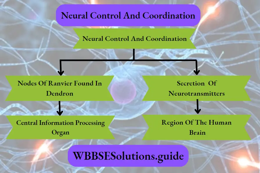
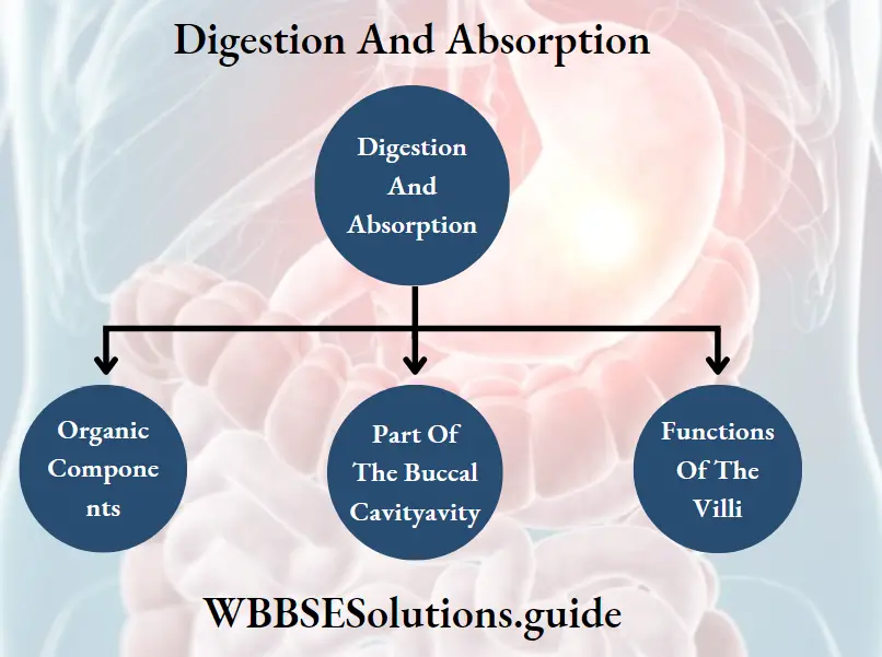
Neural Control And Coordination Introduction

As you are reading this book, Think about the organs That are Functioning with functioning within you right now! Your eyes are sensing the light reflected off this page.

Your brain is decoding the meanings of the words you are reading. Think about the processes that are running within your body. You are breathing, perhaps perspiring, feeling the chair on which you are sitting, and your heart is beating.

All of these are being regulated by an organ system inside your body. It is the neural system or nervous system.

The nervous system controls various metabolic and physiological functions of the body.

Different organs of the body are directly connected to the nervous system. The actions of these organs are coordinated by the nervous system. This is known as neural coordination.

Read and Learn More: WBCHSE Notes for Class 11 Biology

Neural System

All organ systems of animals are controlled and coordinated effectively by the neural system or the nervous system. The neural system allows the organism to interact with its environment, appropriately.

This system functions at the cellular level through nerve cells or neurons.

It receives sensory information, interprets it, and generates appropriate responses using organs like muscles. The neurons set up the neural system in a number of discrete anatomical locations in the brain.

These systems send and receive impulses from the sense organs and other receptors of the body.

Thus, neural control provides the basis for body functions such as perception of senses, attention, memory, emotions, rational thinking, etc.

Neural system of different animals

  1. Sponges (Porifera) do not have any neural system, Hydra (Cnideria) has a neural system that is composed of a network of neurons.
  2. Planaria (Platyhelminthes) have two nerve cords that converge to form a rudimentary brain.
  3. The neural system of earthworm (Annelida) is composed of a single nerve cord and paired nerve ganglia.
  4. The neural system of arthropods is better organized, It consists of the brain, ganglia, and neural tissue.
  5. The neural system of vertebrates is highly developed.

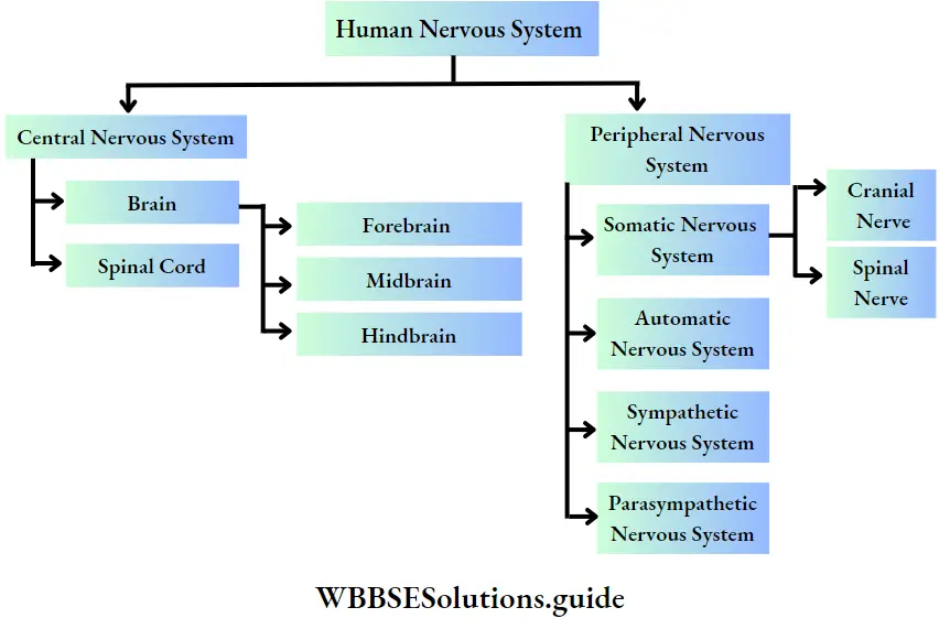
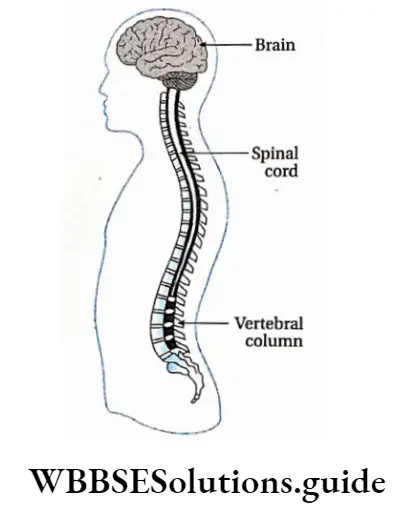
The nervous system of humans and other advanced vertebrates consists of three parts—

  1. Brain,
  2. Spinal cord and
  3. Nerve.

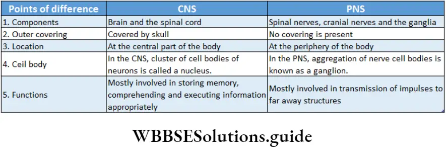
The brain and spinal cord form the central nervous system while nerves form the peripheral nervous system.

Neurons As Structural And Functional Units Of Neural System

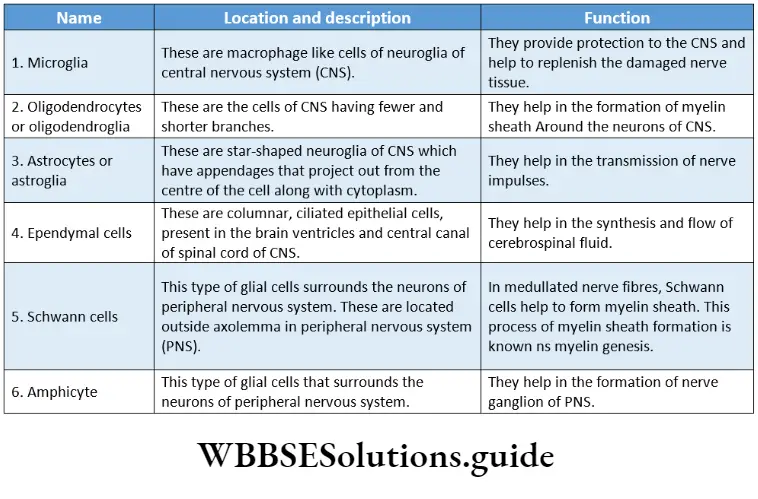
Though neurons are the main structural and functional components of the nervous system some other cells also take part in maintaining the proper functioning of this system.

The cells of the nervous system can be divided into two broad categories—

  1. Nerve cells (or neurons) and
  2. Supporting cells called neuroglia (or simply glia), are discussed below

Neuron

The human brain alone contains over 100 billion neurons. Each neuron can have up to 10,000 connections to other neurons in the brain.

Neuron Definition: The Structural And Functional units of the nervous system which are capable of Sending and receiving nerve impuluses is called a neuron.

Biology Class 11 Chapter 20 Neural Control And Coordination Typical Myelinated Neuron

Different Parts Of A Neuron

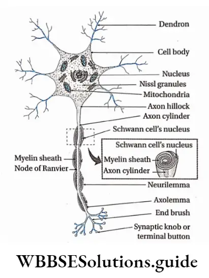
A typical neuron consists of a cell body (perikaryon or soma) and cellular processes called dendrons and axons. Different parts of a neuron are discussed below.

Cell Body or soma or perikaryon or neurocyton: The swollen part of a typical neuron, containing cytoplasm and nucleus, from which cellular processes emerge, is known as the cell body.

Different parts of the cell body are as follows—

  • The cell membrane forms the external boundary of the neuronal cell body and its processes. It is also known as a neurilemma.
  • The nucleus is located at the center of the cell body. The contents of the nucleus are enclosed within a double-layered nuclear membrane.
  • The cytoplasm is known as neuroplastic.

Neuroplasm contains—

  1. Microtubules—These are hollow rod-like filaments that are composed of a globular protein named tubulin.
  2. Neurofilaments— These are thinner than microtubules but thicker than microfilaments. They are a major Component Of the neuroral Cytoskeleton.
  3. Microfilaments— These Are Thinner Than The Neurofilaments. They consist of two intertwined strands, each of which is a polymer of actin protein subunits. They help in the maintenance of cell shape, changing of cell shape, and in cytoplasmic streaming.
  4. Neurofibrils— Neurofibrils are filamentous structures or microscopic fibrils that are found running in every direction through the nerve cell. They provide the neuron support and help to give it a shape,
  5. Nissl granules— These are the granular material present throughout the entire cell body and proximal portions of the dendrites. However, it is not present in the axon hillock (a portion of the soma from which the axon arises). The Nissl granules consist of ribosomes and endoplasmic reticulum.
  6. Centrosome— Nerve cells are incapable of division. So, it is assumed that neurons lack centrosomes. However, under the electron microscope, a non-functional centrosome has been observed,
  7. Other organelles—The cell organelles, commonly found in animal cells are also present within the neuroplasm. These include the Golgi complex, endoplasmic reticulum, mitochondria, lysosomes, etc.
  8. Storage material—The cytoplasm of a neuron contains glycogen, lipids, etc., as storage material They consist of two intertwined strands, each of which is a polymer of actin protein subunits. They help in the maintenance of cell shape, changing of cell shape, and in cytoplasmic streaming.
  9. Neurofibrils— Neurofibrils are filamentous structures or microscopic fibrils that are found running in every direction through the nerve cell. They provide the neuron support and help to give it a shape,
  10. Nissl granules— These are the granular material present throughout the entire cell body and proximal portions of the dendrites.  However, it is not present in the axon hillock (a portion of the soma from which the axon arises). The Nissl granules consist of ribosomes and endoplasmic reticulum.
  11. Centrosome— Nerve cells are incapable of division. So, it is assumed that neurons lack centrosomes. However, under an electron microscope, a non-functional centrosome has been observed,
  12. Other organelles—The cell organelles, commonly found in animal cells are also present within the neuroplasm. These include the Golgi complex, endoplasmic reticulum, mitochondria, lysosomes, etc.
  13. Storage material—The cytoplasm of a neuron contains glycogen, lipids, etc., storage material.

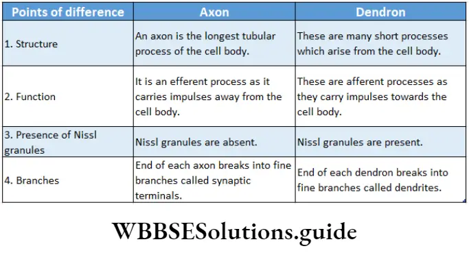
Cellular processes: The cytoplasmic, thread-like appendages that emerge from a cell body are known as cellular processes. These projections are of two types—dendrons and axons.

Dendron: The short cellular processes emerging from the cell body of a neuron that receives impulses from another neuron is called dendron (from the Greek dendron = tree branch).

Dendron Structure:

  1. The number of dendrons can be from one to many,
  2. Branches of dendrons are known as dendrites,
  3. Nissl granules, ribosomes, smooth endoplasmic reticulum, neurofilaments, micro-filaments, microtubules, and mitochondria are present in the dendrites,
  4. Dendron lacks myelin or medullary sheath.

Dendron Function:

  1. The dendrite is the impulse-receiving part of the neuron,
  2. Dendrites increase the surface area for receiving signals from other neurons.

Axon: A long cellular process emerging from a neuron that transmits nerve impulses from the cell body to another neuron is called an axon.

Dendron Structure:

  1. In most cases, a neuron consists of one axon. Apoiar neuron lacks any axon,
  2. The plasma membrane of the axon is called the axolemma and its cytoplasm is called axoplasm.
  3. The axoplasm does not contain the Nissl substance or Golgi apparatus. But the axoplasm does contain mitochondria, microtubules, and neurofilaments,
  4. Depending upon the presence or absence of myelin sheath around axons, they are either myelinated or non-myelinated, respectively.
  5. In myelinated axons, the part that lacks myelin forms a ridge due to the fusion of neurilemma and axolemma. This region is called the node of Ranvier. Generally, nodes of Ranvier are distributed at a distance of 1-3.
  6. At their distal ends, the axons branch extensively. Their terminal ends, which are mostly enlarged, are called synaptic terminals (synaptic buttons)

Biology Class 11 Chapter 20 Neural Control And Coordination Differences Between Axon And Dendron

Neurons are classified into different categories on the basis of certain features which are as follows-

Biology Class 11 Chapter 20 Neural Control And Coordination Types Of Neuron

Neuroglia Or Glial Cell

Neuroglia Or Glial Cell Definition: The cells of the nervous system other than neurons that are incapable of generating and transmitting nerve impulses but provide structural and functional support to the neurons are called neuroglia or glial cells.

Neuroglia Or Glial Cell Characterises:

  1. Neuroglia are incapable of transmitting nerve impulses.
  2. These cells can divide (unlike nerve cells).
  3. The brain contains more neuroglial cells than neurons.
  4. The appendages of some glial cells (astrocytes) are thinner at their terminal end and form special structures called end-feet that remain associated with blood capillaries.

Ganglion

The region outside the CNS where there is an aggregation of numerous nerve cell bodies, is known as ganglion.

Ganglion Example: 31 pairs of nerves have originated from the human spinal cord. Two main nerve roots—dorsal nerve root and ventral nerve root, combine to form spinal nerves.

But before their meeting point, a thinner region is seen where numerous neurons remain associated with each other. This region is known as the dorsal ganglion.

Biology Class 11 Chapter 20 Neural Control And Coordination Types Of Neuroglia

Nerve

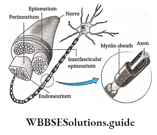
Nerve Definition: A cord-like structure, comprising a collection of neurons, blood vessels, lymph vessels, and adipocytes enclosed by a layer of connective tissue, that conveys impulses throughout the body is called a nerve.

Structure Of Nerves

Nerves emerge from the brain and spinal cord and branch out to almost all parts of the body. The nerves are composed of nerve fibers or neurons.

These nerve fibers are bundled together like strands of an electric cable enclosed within a connective tissue covering.

Connective tissue coverings: On the outer side of the nerve, there is a covering made up of connective tissue.

  • Blood vessels, lymph vessels, and adipocyte cells are found within it.
  • This covering consists of three layers—endoneurium, perineurium, and epineurium.

Endoneurium: It is the innermost layer that covers up the individual nerve fibers. This layer is relatively thin.

Perineurium: A collection of nerve fibers that are individually bound by endoneurium, is again surrounded by another connective tissue layer called the perineurium.

The bundle of nerve fibers bound together by perineurium is called fasciculus (plural: fasciculi).

Epineurium: Several fasciculi are again bound together by another layer of connective tissue. It is called epineurium. It is the outermost layer of connective tissue layer covering the nerve.

This layer is usually thick and rich in blood vessels, lymph vessels, adipocytes, and collagen fibers.

Biology Class 11 Chapter 20 Neural Control And Coordination Structure Of Nerve Fibre

Nerve fibers: A nerve may have a few to more than a million nerve fibers. These are actually axons of different neurons. Several of these fibers remain bound together beneath the connective tissue coverings.

Types Of Nerves

Nerves are of different types. Their classification on the basis of several criteria has been discussed below

Biology Class 11 Chapter 20 Neural Control And Coordination Type Of Nerves

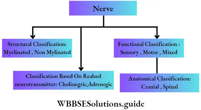
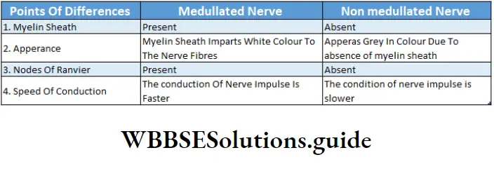
Structural classification of nerves: Based on the presence or absence of myelin sheath, nerves are classified as—

Myelinated nerve: Medullated or myelinated nerves are nerves with large diameters. The nerve fibers of medullated nerves remain wrapped with a protein-lipid sheath called myelin sheath, through which nerve impulse is conducted rapidly.

This type of nerve is made of nerve fibers consisting of three main components. These components are—

  1. Axis cylinder— It is the central core of the axon of a neuron. There is a thin covering called an axolemma that surrounds the axon. The axolemma is again enclosed by an outer covering called a myelin sheath.
  2. Myelin sheath—It is an external covering of the axon. It is made up of protein and lipids. It provides insulation to the nerve fiber and hence prevents the spreading of nerve impulses to adjacent tissues. In the nerves of the peripheral nervous system (PNS), the myelin sheath is produced by a type of glial cell called Schwann cell. Nodes of Ranvier are present in this type of nerve.
  3. Neurilemma—There is another covering external to the myelin sheath, which is called neurilemma.

Example: This type of nerve fiber is found in the white matter of the central nervous system.

Non-myelinated nerve: Non-medullated or non-myelinated nerves are nerves of small diameter. The nerve fibers of these nerves are devoid of the myelin sheath. Through these nerves, a nerve impulse is conducted very slowly. This type of nerve fiber is covered with an external neurilemma. They are devoid of nodes of Ranvier.

Example: This type of nerve fiber is found in the grey matter of the central nervous system.

Biology Class 11 Chapter 20 Neural Control And Coordination Differences Between Medullated And Non medullated Nerves

Functional classification of nerves: Based on the functions carried out by nerve fibers, nerves are classified into three kinds. These are—

Sensory nerves: These nerves contain sensory fibers. Sensory nerves carry impulses from sense organs to the brain or the spinal cord. These nerve fibers are also called afferent nerve fibers. The nerve impulse conducted by sensory nerve fibers is called sensory impulse.

Examples of sensory nerves are—the olfactory nerve, optic nerve, auditory nerve, etc fibers.

Sensory nerves carry impulses from sense organs to the brain or the spinal cord. These nerve fibers are also called afferent nerve fibers. The nerve impulse conducted by sensory nerve fibers is called sensory impulse.

Examples of sensory nerves are—the olfactory nerve, optic nerve, auditory nerve, etc.

Anatomical Classification: Based on the anatomical origin of nerves, these are classified as—

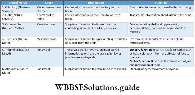
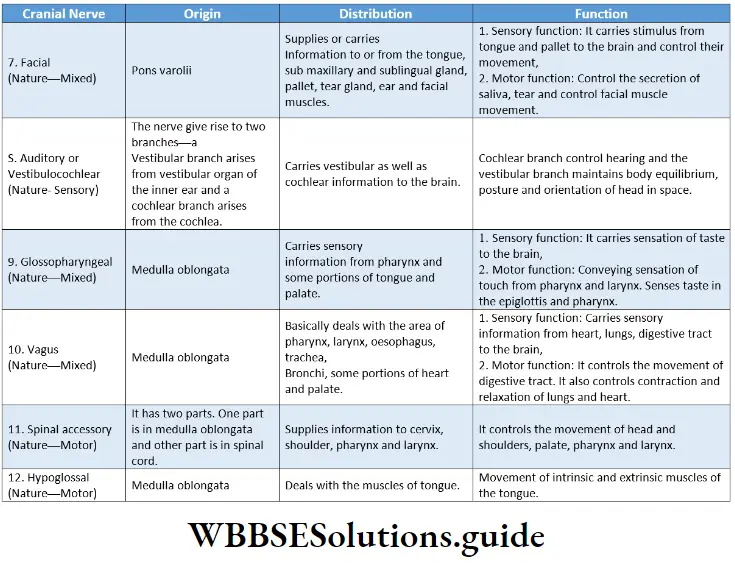
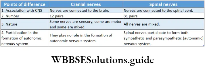
Cranial nerves: These nerves originate from the brain. They may be entirely motor-sensory or mixed.

The human nervous system consists of 12 pairs of cranial nerves.

Spinal nerves: It is always of a mixed type originating from the spinal cord. The human nervous system consists of 31 pairs of spinal nerves.

Classification based on neurotransmitter: Based on neurotransmitter released, nerves are classified as—

Adrenergic nerves: These nerves synthesize and release adrenaline and nor-adrenaline.

Cholinergic nerves: These nerves synthesize and release acetylcholine (ACh).

Nervous System In Humans

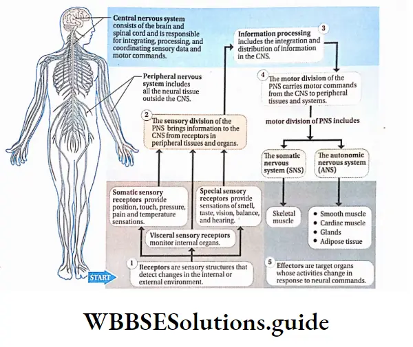
The nervous system in the human body is an organ system that coordinates the functioning of all the systems of the body and helps the body to respond to external stimuli.

Functions Of Nervous System

The main functions of the nervous system in humans and other animals are described below.

Response to external stimuli: The human nervous system is equipped to sense and respond to continuous changes within the body and in its external environment.

Control and coordination: The nervous system works along with the endocrine system to communicate, integrate, and coordinate the functions of various organs and organ systems in our body. It also helps the body to respond to the external stimuli.

Higher mental functions: It also acts as a seat of self-consciousness, intelligence, thoughts, and emotions.

Maintenance of balance with the environment: Humans And Other higher animals sense the changes in the environment and Act Accordingly, Both with the help of the nervous System.

Control of reflex action: Grey matter of the spinal cord, within the central nervous system, controls the reflex actions of the body.

Biology Class 11 Chapter 20 Neural Control And Coordination Neural Coordination In Human Nervous System

Mechanism Of Action Of The Nervous System

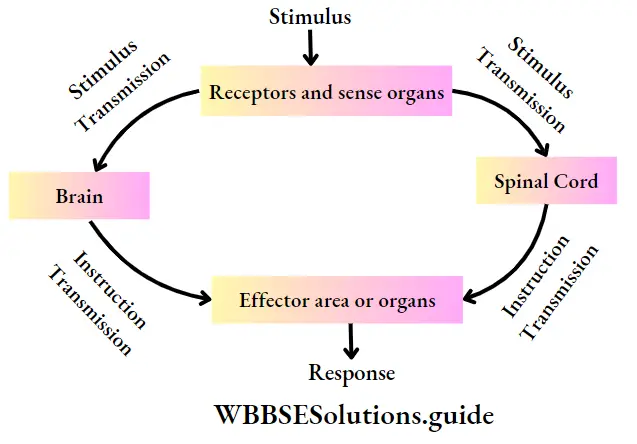
The action of the nervous system is accomplished in some stages. These are—

Receiving stimulus: Mechanical, chemical, or physical changes that excite the body or body parts, are known as stimuli.

Stimulus is received by the receptors present in the eyes, ears, nose, tongue, skin, and other parts of the body.

Transmission of impulse to the central nervous system: The received stimulus is transmitted to the central nervous system as a sensory nerve impulse, by sensory nerves.

Sending instructions from the central nervous; system to the target site: The sensory information is analysed in the brain and spinal cord of the central nervous system.

Instruction for necessary action is then sent as a motor impulse, through the motor nerves, to the effector organizer

The accomplishment of action: As per instruction from the central nervous system, actions are completed in the effector area or organizer:

Biology Class 11 Chapter 20 Neural Control And Coordination Mechasim Of Action Of Nervous System

Divisions Of Human Nervous System

The human nervous system comprises the following divisions—

Biology Class 11 Chapter 20 Neural Control And Coordination Human Nervous System

Central Nervous System

Central Nervous System Definition: The structural and functional center for the entire nervous system comprising the brain and spinal cord is called the central nervous system or CNS.

Biology Class 11 Chapter 20 Neural Control And Coordination Central Nervous System

Central Nervous System Characteristic features:

  1. The components of the CNS are the brain and the spinal cord.
  2. The brain is located within a bony cavity called the cranium or brain box. The dorsal part of the brain bears an opening called the foramen magnum.
  3. Through this opening, the spinal cord enters the vertebral column. The spinal cord is present within the neural canal of the vertebral column. It runs upto the end of the vertebral column.
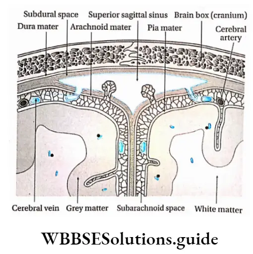
  4. Outside the CNS, three layers of protective tissues are present, which are referred to as meninges.
  5. These meninges surround the brain and the spinal cord providing additional layers of protection to. The three layers of meninges are—
    1. Dura mater,
    2. Arachnoid mater and
    3. Pia mater.
  6. The space between the dura mater and the arachnoid mater is called subdural space.
  7. The small space between the arachnoid mater and the pia mater is called subarachnoid space. Within this space cerebrospinal fluid (CSF) is present.
  8. At the end of the spinal cord, the pia mater extends upto the coccyx like a thin thread, termed filum terminale. This part is composed of fibrous connective tissue.
  9. Human CNS has grey matter and white matter beneath the layer of meninges. In the CNS of the brain, the white matter lies beneath the grey matter.
  10. But in the spinal cord, the grey matter lies beneath the white matter. CNS of vertebrates comprises four brain ventricles. The spinal cord consists of one central canal or neurocoel.
  11. Note that brain ventricles (chambers within the brain) and central canal or neuronal are completely filled by cerebrospinal fluid (CSF).
  12. Within CNS, grey matter is present like an island in white matter. This is known as the nucleus.

Biology Class 11 Chapter 20 Neural Control And Coordination Different Layers Of menings In Humans Brain And The Location Of Grey And White Matter

Central Nervous System Structure: The central nervous system is mainly composed of two parts—the brain and the spinal cord. These are described below in detail.

Brain Ventricles

  • The human brain contains four connected cavities called brain ventricles. The cerebrum of the forebrain has two cerebral hemispheres.
  • The first and second ventricles are located there laterally and are called lateral ventricles.
  • The diencephalon of the forebrain includes two parts—the hypothalamus and epithalamus.
  • The third ventricle is present between the epithalamus and the hypothalamus and is connected to the lateral ventricles by an interventricular pore, known as the foramen of Monroe.
  • The fourth ventricle is located within the cerebellum and pons of the metencephalon of the hindbrain.
  • The fourth ventricle is connected to the third ventricle by a tubule called the aqueduct of Sylvius.
  • The fourth ventricle remains connected to the central canal of the spinal cord. Two holes are present in the fourth ventricle—the foramen of Lusaka and the foramen of Magendie.
  • Through these pores, the cerebrospinal fluid moves upward to enter into the subarachnoid space.

Brain

Brain Definition: The swollen part of the central nervous system located in the brain box, having cavities filled with CSF and Procted By meninges, is called Brain.

Brain Location: The brain is located in the cranial cavity of the human skull.

Brain Structure: The brain of vertebrates is made up of 100 billion neurons and a large number of neuroglia.

The approximate weight of an adult human brain is 1.36 kg. The different parts of the human brain are shown below in the given chart.

Biology Class 11 Chapter 20 Neural Control And Coordination Human Brain

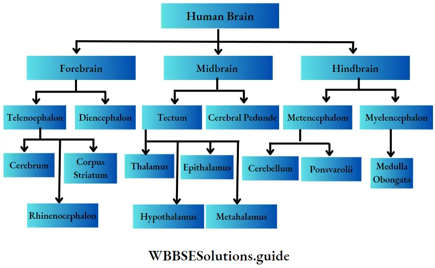
Forebrain or Prosencephalon

The forebrain has two subdivisions—the telencephalon and the diencephalon.

Telencephalon: Telencephalon is again subdivided into three parts namely, cerebrum, corpus striatum, and rhinencephalon.

Biology Class 11 Chapter 20 Neural Control And Coordination Different Regions Of Human Brain

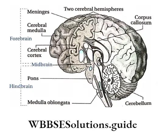
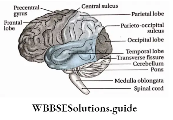
Brain Structure: Cerebrum is divided into two symmetrical semicircular parts called cerebral hemispheres.

The surface area of the cerebral hemisphere is greatly folded, accommodating several layers of neurons.

This area is called the cerebral cortex. Each fold is known as gyrus (plural: gyri) and the groove or depression between them is called sulcus (plural: sulci).

Deep and wide sulci are called fissures. Fissures divide the cerebral hemisphere into a number of lobes.

These are—

  1. Frontal lobe,
  2. Parietal lobe,
  3. Occipital lobe,
  4. Temporal lobe.

The fissure between the frontal and parietal lobes is called the fissure of Rolando or central sulcus.

The fissure between the parietal and temporal lobes is known as the Sylvian fissure. The fissure between the parietal and occipital lobe is called the parieto-occipital fissure.

Beneath the cerebral cortex, there lies the cerebral medulla. It is made up of white matter which is mainly composed of nerve fibres.

The two cerebral hemispheres are linked by a bundle of longitudinal nerve fibers called the corpus callosum. The cerebrum contains pyramidal (Betz), stellate, and spindle neurons.
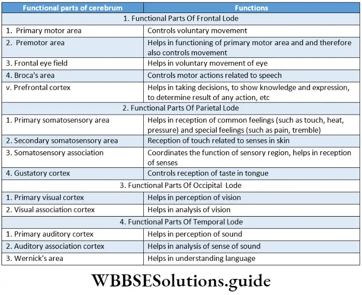
Brain Functions:

The cerebrum controls the action of different voluntary muscles, the movement of eyelids and eyeballs,

Different areas of the cerebral cortex control our ability to speak (Broca’s area) and write (Wernicke’s area),

The temporal lobe contains the auditory cortex and is involved in auditory perception,

Similarly, the occipital lobe contains the visual cortex and is responsible for visual processing.

Different areas of the cerebral cortex are also responsible for the perception of other sensations like taste, smell, touch, pressure, pain, heat, and cold.

The cerebrum is the center for memory, intelligence, judgment, conscious thought, and other activities associated with thinking and feeling.

Biology Class 11 Chapter 20 Neural Control And Coordination Organisation Of Cerebrum

Biology Class 11 Chapter 20 Neural Control And Coordination Different Functional Regions Of Cerebrum And Their Functions

Corpus striatum: It is made of grey matter suspended within white matter within the cerebrum.

This part of the brain constitutes a structure called basal ganglia. The Corpus striatum helps in the modification of functions of the motor region in the cerebrum.

Rhinencephalon: The ring-like structure around the brain stem is known as rhinencephalon. The rhinencephalon helps in sensing smell.

Limbic System

The system that is composed of parts of the telencephalon, diencephalon, and mesencephalon and is expanded at the periphery of the cerebral cortex and diencephalon, controlling human emotions, feelings, inspiration, etc., is called the limbic system. The term limbic’ means border or boundary.

The parts of the diencephalon that are associated with the limbic system are—the hypothalamus and the nucleus of the frontal thalamus.

The parts of the telencephalon such as the amygdala, hippocampus, septal nuclei, para-olfactory lobe, and limbic cortex form the structural components of the limbic system. Other than these, reticular formations of the midbrain are also associated with the limbic system.

In human beings, the primary function of the limbic system is to control emotions such as fear, hopelessness, anger, depression, happiness, etc.

Diencephalon: It is located between telencephalon and mesencephalon. The diencephalon is situated between the two cerebral hemispheres.

It has four parts, namely—hypothalamus, epithalamus, metathalamus, and thalamus.

Hypothalamus: It is a small cone-shaped structure of the brain lying below the thalamus. It makes up the floor of the third cerebral ventricle projects downwards from the brain and ends in the pituitary gland.

Location: The hypothalamus is located below the thalamus, just above the brain stem, and anterior to the third ventricle.

Structure: Hypothalamus consists of numerous neurosecretory cells. Near infundibulum (pituitary

Limbic System Structure: Hypothalamus consists of numerous neurosecretory cells. Near the Infundibulum (pituitary stalk) of the hypothalamus, two optic nerve crosses each other.

This region of crossing is known as optic chiasma. The hypothalamus also consists of numerous hypothalamic nuclei.

Limbic System Function:

  • The hypothalamus plays a significant role in the regulation of body temperature (thermoregulation),
  • The hypothalamus regulates our appetite and feeding behavior,
  • The hypothalamus also helps in the maintenance of water balance in our body.
  • The hypothalamus also helps to maintain the normal functioning of the cardiovascular system.
  • The hypothalamus regulates the action of the autonomic nervous system,
  • The neurosecretory cells situated in the hypothalamus secrete neurohormones.
  • These act as releasing factor or inhibitory factor, that controls the secretion of different hormones from adenohypophysis or anterior pituitary
  • The hypothalamus helps to regulate circadian rhythm. Different functions of our body like sleep, wakefulness, etc., take place in a cyclic way following the day-night cycle.
  • This rhythm inside our body is called circadian rhythm,
  • The hypothalamus is also involved in the expression of emotions like rage, fear, and extreme anxiety.

Epithalamus: It is a small region in the diencephalon.

It is located in the posterior (dorsal) segment of the diencephalon (superior and posterior to the thalamus). It is connected to the pineal gland and epiphysis by a stalk.

The functions of the epithalamus are—

  • It controls the function of the pineal gland.
  • It plays an important role in receiving smell impulses and analyzing them.

Thalamus: It is a mid-line symmetrical structure of two halves, within the brain, situated near the center of the brain, superior to the mid-brain.

The thalamus consists of a large mass of grey matter and is located on both the lateral walls of the third ventricle.

The thalamus acts as a relay station for sensory impulses such as pressure, temperature, touch, pain, etc. From there, the impulses are retransmitted to the different areas of the cerebral cortex.

Metathalamus: It is a small region located at the extreme dorsal end of the brain. It consists of the two, paired, lateral and medial geniculate bodies. The metathalamus lies between the epithalamus and the hypothalamus.

The functions of the metathalamus are—

The medial geniculate body of the metathalamus acts as a relay station of audition i.e., it helps us to hear.

The lateral geniculate body of the metathalamus acts as a relay station of vision i.e., it helps us to see.

Midbrain or Mesencephalon

The midbrain is located between the diencephalon and mid-region of the hindbrain. It is the smallest part of the human brain. It is almost not visible from the outside. It consists of two parts, namely— tectum and cerebral peduncle.

Biology Class 11 Chapter 20 Neural Control And Coordination Cross Section Of Midbrain

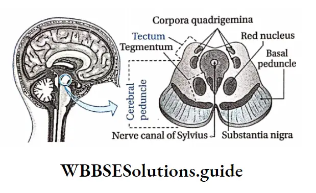
Tectum: The tectum of the midbrain is located in the dorsal region of the aqueduct of Sylvius. The primary structure of the tectum includes two swollen regions— superior colliculi and inferior colliculi.

The superior colliculi forms part of the visual system. The inferior colliculi is a part of the auditory system.

The structures appear as four small bumps on the brain stem. These bumps are together known as corpora quadrigemina.

Cerebral peduncle: The cerebral peduncles (or crus cerebri) are located anterior to the aqueduct of Sylvius in the midbrain.

From the interior, three layers are the visible tegmentum, substantia nigra, and basal peduncle.

Due to the presence of melanin pigment, the substantia nigra appears black or brown.

Function Of Midbrain:

  1. It coordinates the actions of different regions of the midbrain, forebrain, and hindbrain.
  2. The tectum region of the midbrain coordinates the reflex actions related to visual perception and olfaction.
  3. The midbrain plays an important role in controlling the body temperature.
  4. The red nucleus of the midbrain controls muscle tone.
  5. The midbrain controls the actions of different voluntary muscles.

Hindbrain Or Rhombencephalon

It is located at the posterior end of the midbrain. It consists of two parts metencephalon and myelencephalon.

Biology Class 11 Chapter 20 Neural Control And Coordination Different Regions Of Hindbrain

Metencephalon: The Metencephalon is located in the posterior region of the hindbrain.

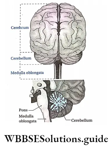
It has two parts— the cerebellum and the pons.

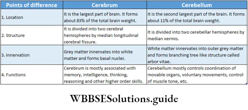
Cerebellum: Cerebellum is a major structure located at the top of the brain stem (where the spinal cord meets the brain) and at the back portion of the brain, lying under the occipital and temporal lobes of the cerebral cortex.

The cerebellum is the most important and largest part of the human hindbrain. It is also known as the ‘little brain’ or the ‘tree of life’.

Cerebellum Structure: Cerebellum is made up of two hemispheres called cerebellar hemispheres. Two cerebellar hemispheres are connected by a mid-region named vermis. The cerebellum contains Purkinje cells.

Cerebellum Functions:

  • The cerebellum plays an important role in controlling body balance,
  • The cerebellum controls the movement of the hands and legs. It receives impulses from the cerebral cortex. Depending on these impulses, it controls the movement of the hands and legs.
  • The cerebellum controls voluntary movement.
  • The cerebellum exerts control over the reflex arc involving muscles. In this way, it helps to maintain normal muscle tone.

Pons Varolii:

  • Pons Varolii is a region in the brain stem that lies between the midbrain (above) and medulla oblongata (below) and in front of the cerebellum.
  • The ventral part of the pons is convex and the dorsal part is plain. Pons is connected to 5, 6, 7, and 8 cranial nerves.

The main functions of pons Varolii are as follows—

  • Pneumotaxic and apneustic centers of pons act as respiratory centers for the regulation of respiration.
  • Pons serve as a relay station between several areas of the brain.
  • The nerve center of pons Varolii conducts contraction of the detrusor muscle of the urinary bladder, in order to carry out urination.

Myclencephalon or medulla oblongata:

  • The medulla oblongata is a cone-shaped structure located in the hindbrain, anterior to the cerebellum, below the pons, and above the foramen magnum. It is the lowest part of the hindbrain.
  • The medulla oblongata constricts at the posterior end and appears like a cone. Its length is approximately 2.4 cm. The spinal cord arises from its posterior end.
  • Cranial nerves 9, 10, 11, and 12 remain connected to it. Medulla oblongata consists of different nerve centers, such as—the vasomotor center, respiratory center, vomiting center, and salivary center.

Pons Varolii Functions:

  1. Medulla oblongata controls visceral reflexes such as—sneezing, coughing, vomiting, secretion of saliva, engulfing, etc.
  2. Medulla oblongata plays an important role in controlling respiration.
  3. Medulla oblongata plays an important role in controlling the action of the cardiovascular system including the heartbeat.

CSF Or Cerebrospinal Fluid

Cerebrospinal Fluid Location: CSF is present in the neuronal of the central nervous system and central canal of the spinal cord.

It is also present in the subarachnoid space of the meninges.

Cerebrospinal Fluid Origin: The first and second ventricles of the human brain have ciliated epithelium on their wall.

The wall has enough capillary-rich area known as choroid plexus. Plasma of the blood, in the capillaries of the choroid plexus, enters the brain ventricles by diffusion and forms CSF.

CSF content: The total CSF content of a human body is approximately 150 ml.

Components: CSF is clear, and slightly alkaline in nature (pH—7.33). The various components of CSF are—water, plasma proteins, glucose, mineral salts or ions, creatinine, etc.

Cerebrospinal Fluid Functions:

  1. It acts as a shock absorber and protects the brain from mechanical injury,
  2. It maintains the shape and structure of the brain by maintaining an outward pressure,
  3. It transports nutrients to the tissues of the brain and spinal cord,
  4. It moistens the brain and spinal cord and enables the exchange of substances within I nerve cells,
  5. It has white blood corpuscles that protect the brain from infections,
  6. CSF provides buoyancy to the brain.
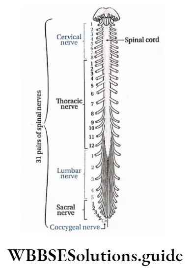
Biology Class 11 Chapter 20 Neural Control And Coordination Different Regions Of Hindbrain

Spinal Cord

Spinal Cord Definition: The long, thin, tubular bundle of neurons and supporting cells that originate from the posterior end of the medulla oblongata of the brain enters the vertebral column, and runs through it is known as the spinal cord.

Spinal Cord Location: The human spinal cord originates from the medulla oblongata of the brain and is present within the neural canal of the vertebral column.

Spinal Cord Length and breadth: It is elongated and about 45 cm long in men and around 43 cm in women. It is enlarged in the cervical and lumbar regions.

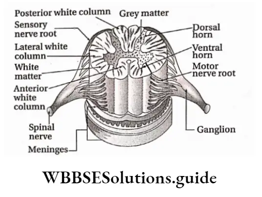
Spinal Cord Coverings:

Just like the brain, the spinal cord also has an outer covering of meninges.

From outside to inside, layers of meninges are—dura mater, arachnoid mater, and pia mater.

Biology Class 11 Chapter 20 Neural Control And Coordination Spinal Cord And Spinal Nerves

Spinal Cord External structure:

  1. The human spinal cord continues upto the conus medullaris (tapering posterior end of the spinal cord) near the first or second lumbar vertebra, terminating in a tapering fibrous extension, known as the filum terminale.
  2. It is hollow, elongated, and cone-shaped.
  3. The posterior and anterior sides of the spinal cord bear longitudinal grooves.
  4. The posterior groove is known as the posterior median sulcus and the anterior groove is known as the anterior median fissure. Along these two grooves, the spinal cord divides into two symmetrical parts.

Internal structure: The transverse section of the human spinal cord shows the following structures—

Central canal: The transverse section of the spinal cord shows two symmetrical hemispheres.

Its center constitutes the central canal. The canal and its ducts are together known as neurocoel.

The central canal is lined by ciliated epithelial tissue known as ependyma. Through this canal, the CSF flows.

Grey matter: Grey matter is present in the center of the spinal cord like ‘H’ or like the wings of a butterfly. In each segment of the spinal cord, grey matter is distributed in three grey columns—the posterior grey column or dorsal horn,

Anterior grey column or ventral horn and Lateral grey column or lateral grey horn. Two nerve roots originate from the human spinal cord. The dorsal nerve root originates from the dorsal horn and the ventral nerve root originates from a ventral horn.

White matter: White matter envelopes grey matter and is present at the periphery of the spinal cord. In each half of the spinal cord, white matter is distributed in three columns. These are—

  1. Posterior white column,
  2. Anterior white column and
  3. Lateral white column

Biology Class 11 Chapter 20 Neural Control And Coordination Spinal Cord

Functions:

  1. Spinal cord functions as the central control center for reflex actions.
  2. It partially controls the functions of the autonomic nervous system.
  3. Controls muscle tone.
  4. Through afferent nerve fibers, impulses from the peripheral nervous system are transmitted to the brain.

Again, motor impulses are transmitted from the brain to effector organs by efferent nerve fibers, many of which pass through the spinal cord.

This means the spinal cord maintains coordination between the peripheral system and the brain.

Peripheral Nervous System

Peripheral Nervous System Definition: The part of the nervous system that is constituted of nerves is called the peripheral nervous system.

Peripheral Nervous System Structural characteristics: The peripheral system is of two types—the somatic nervous system which is constituted of 31 pairs of spinal nerves and 12 pairs of cranial nerves and the autonomic or visceral nervous system which is constituted of sympathetic and parasympathetic nerves.

These nerves innervate various visceral organs of the body. These nerves carry sensory impulses from these organs to the CNS.

Also, they carry motor impulses from the CNS to the effector organizer As a result, a response is being given by that organ.
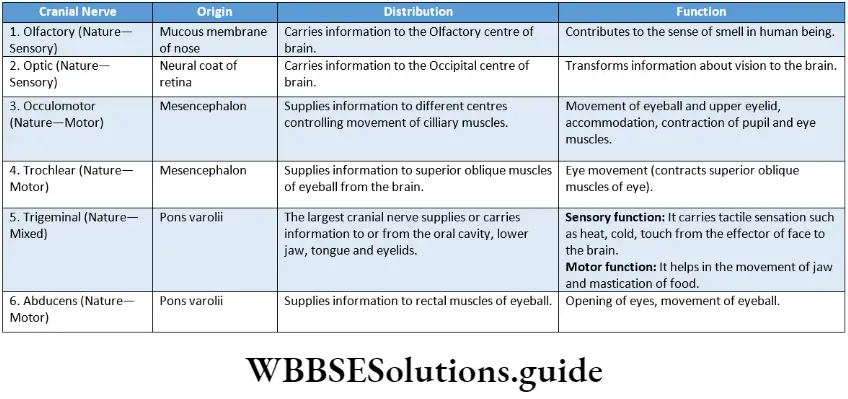
Cranial Nerves

Cranial Nerves Definition: The nerves that originate directly from the brain are known as cranial nerves. These nerves are of three types—sensory or afferent, motor or efferent, and mixed types.

Note that, sensory nerves or afferent nerves originate from receptor organs or sense organs and meet at the brain. But, efferent nerves or motor nerves originate from the brain and meet at the effector organizer
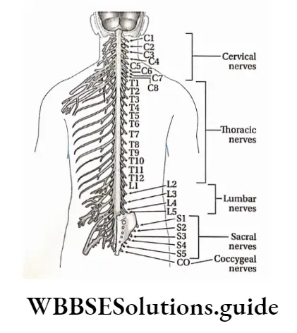
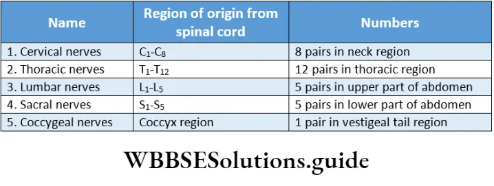
Biology Class 11 Chapter 20 Neural Control And Coordination Cranial Nerves At a Glance

Biology Class 11 Chapter 20 Neural Control And Coordination Cranial Nerves At a Glance 2

Spinal Nerves

Spinal Nerves Definition: The nerves that emerge from the spinal cord are known as spinal nerves.

The human body consists of 31 pairs of spinal nerves. They are named as per their origin

Biology Class 11 Chapter 20 Neural Control And Coordination Spinal Nerve And Plexuses

Features

  1. Dorsally, a branch of the spinal nerve called the dorsal root divides into six to eight nerve rootlets that enter the spinal cord.
  2. There is swelling, a little away from the rootlets. It is called the dorsal root ganglion.
  3. It contains cell bodies of afferent neurons.
  4. Ventrally, another six to eight rootlets leave the spinal cord and converge to form the ventral root.
  5. The ventral and dorsal roots merge, penetrate the dural sac, and enter the intervertebral foramen to form the spinal nerve proper.
  6. Spinal nerves are mixed nerves, with a two-way traffic of afferent (sensory) and efferent (motor) signals.

Function: Within the grey matter of the spinal cord, the sensory nerve enters carrying the sensory impulses.

These impulses are translated into motor impulses in the grey matter. Finally, the motor impulses leave the grey matter by traveling through the motor nerve.

Biology Class 11 Chapter 20 Neural Control And Coordination Spinal Nerve At A Glance

Biology Class 11 Chapter 20 Neural Control And Coordination Different difference between CNs And PNs

Biology Class 11 Chapter 20 Neural Control And Coordination Different difference between Cranial Nerve And Spinal Nerves

Divisions of the peripheral nervous system Based upon the consciousness of our body movements PNS can be classified into the somatic nervous system (SNS) and autonomic nervous system (ANS).

The somatic nervous system controls our voluntary movement. On the other hand, the autonomic nervous system works on its own, self-controlled and independent of our voluntary efforts.

Visceral Or Autonomic Nervous System

Visceral Or Autonomic Nervous System Definition: The part of the nervous system that controls bodily functions that are not controlled voluntarily such as breathing, heartbeat regulation, digestive processes, etc., is known as the autonomic nervous system.

It consists of a pair of chains of ganglions (a ganglion is a group of cell bodies of neurons outside the CNS) and nerves, found on either side of the backbone.

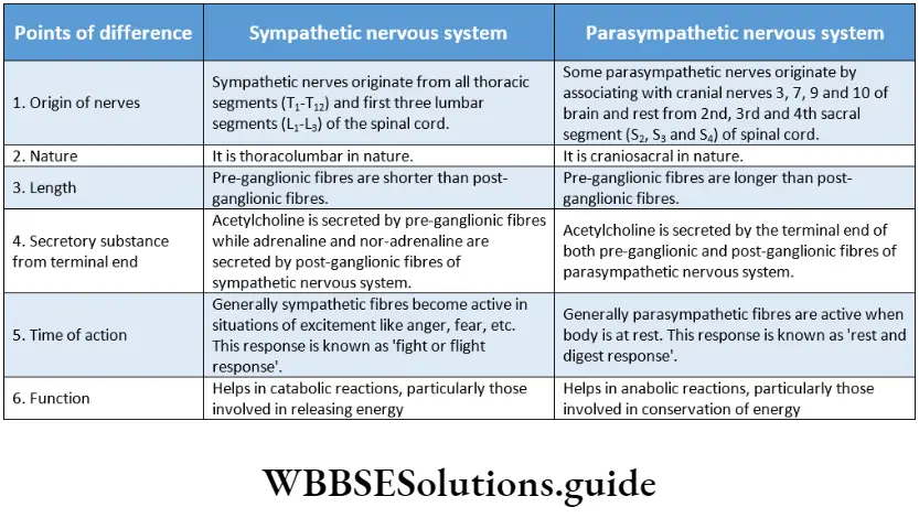
Two antagonistic and anatomically separate divisions comprise the autonomic nervous system.

These two are called the sympathetic and parasympathetic divisions, With a few exceptions, the two subdivisions influence and act upon the organs of the body with differential effects.

Functions of the autonomic nervous system:

The general functions of ANS are as follows—

General functions: The autonomic nervous system maintains homeostasis in the body. It controls the actions of cardiac muscles and muscles of different visceral organiser

Due to the antagonistic or opposite actions of the sympathetic and parasympathetic nervous system, a balance is maintained.

Fight Or Flight Response

The ‘fight or flight’ response was first described by an American physiologist, Walter Bradford Cannon to describe the physiological reaction that occurs within animals in response to a perceived attack or threat. In this response, the animal reacts to the threat by choosing either to fight or to flee from the area.

Rest and digest the response

The ‘rest and digest response occurs when the body is at rest, especially after feeding. In this response, energy is conserved as it slows heart rate, increases intestinal and glandular activity, and relaxes sphincter muscles in the gastrointestinal tract, thereby facilitating the process of digestion.

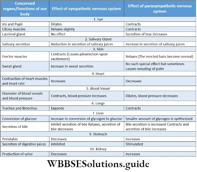
Functions of sympathetic division:

The sympathetic nervous system controls catabolic reactions associated with the release of energy.

It functions along with the adrenal medulla. Together they constitute the sympathetic-adrenal medullary system, It increases heart rate, blood pressure, etc., dilates the pupil, reduces the secretion of saliva, secretion of sweat, expands the bronchioles of the lungs, and also expands the gall bladder, urinary bladder, etc.

Functions of the parasympathetic nervous system:

The parasympathetic nervous system controls the anabolic reactions of the body. It enables digestion, absorption, storage of food, etc., and stores energy in the body.

The parasympathetic nervous system controls normal regular physiological activities of the body. It helps to secrete various digestive juices and thus helps in digestion. It also helps to absorb the digested food.

It increases the secretion of tears, reduces blood pressure, and contracts the urinary bladder.

Sympathetic Nervous System

The division that accelerates various activities of the body like heartbeat, breathing, etc., is called the sympathetic nervous system. The response given by this system is a ‘fight or flight’ response.

Sympathetic nerves originate from the thorax and lumbar region. During anger, fear, etc., the activity of the sympathetic nervous system is increased.

Biology Class 11 Chapter 20 Neural Control And Coordination Sympathetic And Parasympathetic Nervous Systems

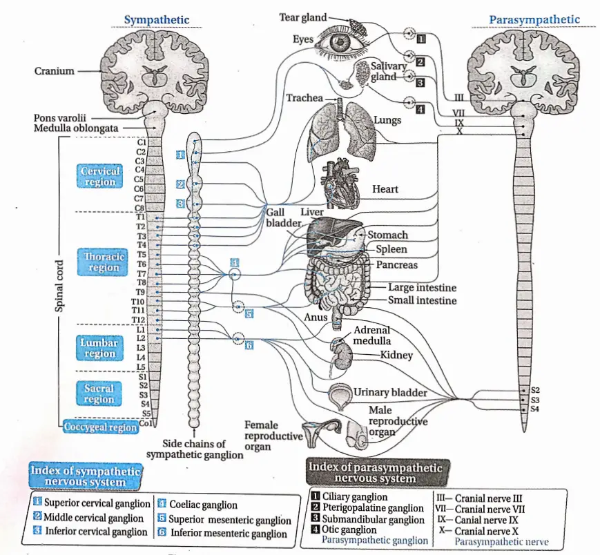
Origin: Sympathetic nerves originate from the lateral horn of grey matter of the first thoracic vertebra to the third lumbar vertebra.

Components: The four components of the sympathetic nervous system are—

Chain of lateral ganglia: This chain is composed of 21 ganglia which extend from the cervical to the sacrum region.

Pre-ganglionic fibers: These fibers are axons of neurons that are located in the spinal cord.

Collateral ganglia: Coeliac ganglion, superior mesenteric ganglion, and inferior mesenteric ganglion are the three collateral ganglia of the sympathetic nervous system which are located in the abdominal cavity.

Parasympathetic Nervous System

The part of the autonomic nervous system that decreases or slows down various activities of the body like heartbeat, respiratory process, etc., is called the parasympathetic nervous system.

The parasympathetic nervous system is anatomically of craniosacral type. The response given by this type of nervous system is Vest and digest’ response.

The nerves of the parasympathetic nervous system originate from the cranium and sacral vertebral segments of the spinal cord and are called the craniosacral nervous system.

Generally, the action of parasympathetic nerves increases when the body is at rest.

Parasympathetic Nervous System Origin: Some of the parasympathetic nerves originate from the brain by fusing with cranial nerves 3, 7, 9, and 10.

The rest of the parasympathetic nerves originate from the sacral vertebra 2nd, 3rd, and 4th (S2, S3, and S4) of the spinal cord.

Components: The three components of the parasympathetic nervous system are—

Pre-ganglionic fiber: These are the axons of neurons that are located in the midbrain, brain stem, and sacral segment of the spinal cord.

Parasympathetic ganglia: These ganglia are located very close to the peripheral tissue.

They do not form a chain of ganglia. The pre-ganglionic fibers synapse with the neurons of parasympathetic fibers.

Post-ganglionic fiber: These are the axons of neurons that are located in the sympathetic fiber.

Biology Class 11 Chapter 20 Neural Control And Coordination Effect Of Sympathetic And Parasympathetic Nervous System

Biology Class 11 Chapter 20 Neural Control And Coordination Differerences Between Sympathetic And Parasympathetic Nervous System

Nerve Impulse

A nerve impulse refers to an electrical or chemical signal that is transmitted along a nerve fiber.

The neurons of the nervous system are not connected directly i.e., they lack any cytoplasmic connections between them.

So nerve impulses are transmitted from one neuron to another by synapse.

So before going into the details about the process of transmission, we have to know about synapses which are as follows.

Synapse

The word ‘synapse’ came from ‘synaptein’, which Sir Charles Scott Sherrington et a!., had coined from the Greek word ‘syn-‘ (‘together’) and ‘haptein’ (‘to clasp’).

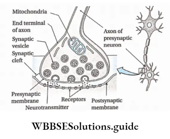
Synapse Definition: The microscopic gap present between the terminal end of the axon of one neuron and the dendron or axon or cell body of another neuron, through which nerve impulses are transmitted by neurotransmitters or electrical ions from the neuron to the post neuron is called a synapse.

Synapse Structure:

  1. In a neuron, an impulse travels along the length of the axon until it reaches the far end, called the axon terminal.
  2. The axon terminal branches profusely and gives a brush-like appearance hence called end brush.
  3. The terminal portion of each of these branches is swollen. It is called the synaptic knob or terminal knob.
  4. The axon terminal ends very close to the dendron or cell body or axon of another neuron, with which it forms the synapse.
  5. Here, the first neuron is called a presynaptic neuron while the second one is called a postsynaptic neuron.
  6. The nerve impulse is transmitted from the presynaptic neuron to the postsynaptic neuron.
  7. The membrane of the presynaptic neuron along the synapse is called the presynaptic membrane.
  8. The membrane of the postsynaptic neuron along the synapse is called the postsynaptic membrane. There is a gap between pre- and postsynaptic neurons. It is called the synaptic cleft.
  9. In the cytoplasm of the presynaptic axon terminal, there are small membrane-bound vesicles called synaptic vesicles which contain neurotransmitters.
  10. These substances act as chemical messengers and help to carry the neural signal from one neuron to another.

Biology Class 11 Chapter 20 Neural Control And Coordination Structure Of Synapse

Types of synapses: On the basis of the medium of transmission of impulses synapses can be classified into three types, such as—

  1. Chemical synapse: Impulses are transmitted by neurotransmitters.
  2. Electrical synapse: impulses are transmitted by ions.
  3. Conjoint synapse: Impulses are transmitted by both chemical as well as electrical signals.

On the basis of structure, synapses can be classified into four types, such as—

Axodendritic synapse: The synapse between the axon of the presynaptic neuron and the dendrite of the postsynaptic neuron.

Axosomatic synapse: The synapse between the axon of the presynaptic neuron and the cell body or soma of postsynaptic neurons.

Axoaxonic synapse: The synapse between the axons of both, pre and postsynaptic neurons.

Dendrodendritic synapse: The synapse between the dendrites of both, pre and postsynaptic neurons.

On the basis of participating nerve cells, synapses can be classified into 3 types—

One-on-one: In this case, one neuron forms a synapse with another neuron.

Many with one: In this case, the terminal end of a number of neurons forms a synapse with one neuron.

One with many: In this case, the terminal end of one neuron forms a synapse with many neurons.

Functions: Synapses have four main functions. They are as follows—

They transmit information between neurons or neurons and their target cells.

They filter out low-frequency impulses.

They act as valves to ensure that impulses pass across them in one direction only.

They also act as junctions allowing impulses to be divided up along many neurons or merge into one.

Generation Of Nerve Impulse

The generation of nerve impulses in nerve fibers depends on the distribution of Na+ and K+ in and out of the axolemma. Again, the distribution of Na+ and K+ is controlled by protein channels.

(such as Na+ and K+ channels) and carrier proteins like Na+/K+ -ATPase pump, in the axolemma. The different stages of the generation of nerve impulses are described below.

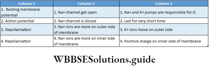
Resting Potential: It is the potential difference across the membrane of a resting neuron i.e., when nerve impulses are not transmitted.

At this stage, the axonal membrane is nearly impermeable to Na+ while comparatively more permeable to K+.

The ‘membrane is also impermeable to negatively charged proteins present in the axoplasm. As a result, inside the axoplasm, there is a high concentration of K+ and negatively charged proteins and a low concentration of Na+.

During resting conditions, this potential difference across the axonal membrane is called resting membrane potential. This state of the resting membrane is called the polarised state.

The average value of resting membrane potential is -70mV.

Action Potential: When a nerve receives a stimulus, a sequence of events occurs.

Due to the conduction of impulse in a region, Na+ channels of that region open.

This causes an influx of Na+ inside the axoplasm. This creates an increase in positive charge in the axoplasm.

Thus the polarisation across the membrane, as found under resting conditions is now reversed. The reversal of polarity is called depolarization. The potential difference across.

Biology Class 11 Chapter 20 Neural Control And Coordination Steps Of Generation Of Action Potential

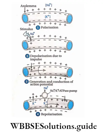
The membrane under this condition, is called action potential. Its value is +35mV.

After depolarisation, Na+ channels are closed. The wave of this reversed polarity of depolarisation (action potential) moving down an axon is called a nerve impulse or spike.

Repolarisation: When the action potential of the axon becomes +35 mV, the K+ channel of the axolemma opens. Due to this reason, K+ from the intracellular matrix moves out to extracellular fluid. As a result, electric potential is reduced.

Gradually, the positive charge in the extracellular fluid increases compared to td axoplasm.

After some time, the concentration of K+ increases in extracellular fluid so much that the K+ channel closes. At this time, the Na+/ K+-ATPase pump opens.

Through this pump, three Na+ move out from the axolemma, and two K+ move inside the axolemma.

In this way, after some time, the concentration of Na+ and K+ becomes the same as before (as in resting condition) on either side of the axolemma. Thus, nerve fibers again attain resting potential.

Conduction Of Nerve Impulse

The process of conduction of nerve impulses through medullated, and non-medullated nerve fibers and synapses follows various methods as described below.

Conduction of nerve Impuluses in Non-medullated nerve fibres: Nerve impulse or action potential is transmitted in small steps. In each step, repolarisation and depolarisation take place. This is known as continuous conduction.

Conduction of nerve impulses in medullated nerve fibers: Myelin sheath contains fat. Since fat is a poor conductor of heat and electricity, it cannot transmit impulses.

Only nodes of Ranvier allow transmission of impulses and ion exchange occurs at these points. So, depolarisation of nodes of Ranvier occurs.

The depolarised node of Ranvier triggers an increase in the permeability of ion channels leading to depolarisation in the adjacent node of Ranvier. Therefore, nerve impulses are transmitted by jumping from one node to the next.

Biology Class 11 Chapter 20 Neural Control And Coordination Saltatory Conduction Of Nerve Impulse in a Medullated Nerve Fibre

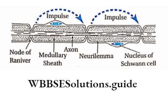
So, it is faster in the medullated nerve fibers than the non-medullated nerve fibers.

This type of conduction is called saltatory conduction because it has been derived from the Latin word saltare which means ‘leaping’.

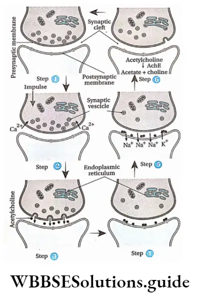
Synaptic transmission: Synaptic transmission is the process whereby electrical impulses pass through a synapse with the release of neurotransmitters.

Transmission of nerve impulses in the synapse region is a chemical process.

The sequence of events that lead to postsynaptic changes is as follows—

  1. As the action potential reaches the axon terminal of a presynaptic neuron, depolarisation occurs which causes voltage-gated Ca2+ channels to open. This leads to an influx of Ca2+ into the synaptic knob from the cisternal.
  2. Ca2+ influx in the axoplasm of the synaptic knob continues. This causes an increase in concentration of Ca2+ and eventually induces vesicles filled with neurotransmitters, to migrate towards the presynaptic membrane. There the vesicles fuse with the presynaptic membrane.
  3. The presynaptic membrane and vesicles now form a continuous membrane, so that the neurotransmitter i.e., acetylcholine, is released into the synaptic cleft also by exocytosis. There may be other types of neurotransmitters such as adrenalin, dopamine, etc., but the following discussion is given with respect to acetylcholine.
  4. Acetylcholine diffuses through the synaptic cleft and binds with receptor site on the postsynaptic membrane.
  5. As a result, acetylcholine-dependent sodium ion channels open, and movement of sodium ions takes place through the postsynaptic membrane. This creates a change in the membrane potential of the post-synaptic membrane.
  6. The postsynaptic membrane and synaptic cleft contain the enzyme acetylcholinesterase. This enzyme hydrolyses acetylcholine into acetate and choline. This causes repolarisation of the postsynaptic membrane.
  7. The entire cycle repeats again.

Biology Class 11 Chapter 20 Neural Control And Coordination Transimission Of Nerve Impulse At Synapse

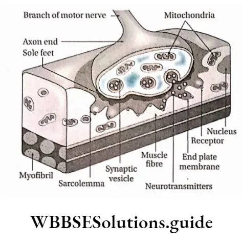
Neuromuscular transmission: The junction between a motor neuron and a muscle fiber by which the nerve can communicate with the muscle fiber is called the neuromuscular junction.

Through these junctions, neurotransmitters are transmitted from the neuron to the muscle. This type of transmission is known as neuromuscular transmission.

In this case, the terminal end of the axon extends and appears like a plate, which is located near the sarcolemma.

At the neuromuscular junction, the plate-like structure formed by the axon terminal is known as the motor end plate.

The extended part of the terminal end motor neuron is known as sole feet or end feet. The axoplasm of this part contains more vesicles.

These vesicles contain neurotransmitters like acetylcholine. This portion also contains endoplasmic reticulum, mitochondria, etc. Sarcolemma of muscle fibers are thick and contain grooves.

The following events occur at the neuromuscular junction—

The axon of the motor neuron carrying a vesicle containing acetylcholine reaches the motor end plate with the help of Ca2+ ions.

Acetylcholine from these vesicles is released between the membrane of the sole feet and muscle fibers. This causes depolarization of the end plate and creates action potential.

As a result, impulses from the end plate enter muscle fibers and cause contraction of muscle fibers.

Biology Class 11 Chapter 20 Neural Control And Coordination Neuromuscular Junction

Neurotransmitters

The organochemical substances that transmit nerve impulses from the presynaptic region to the postsynaptic region at the synapse are known as neurotransmitters.

They are of two types—excitatory and inhibitory.

Excitatory: These are capable of depolarising the postsynaptic membrane and transmitting nerve impulses. Examples—are acetylcholine, and noradrenaline.

Inhibitory: These prevent the transmission of nerve impulses in the synaptic region. Example—GABA (y-amino butyric acid), is Screted from the axon end of some neurons in CNS.

Reflex Action And Reflex Arc

Some neurons are organized to enable the human body to react rapidly at times of danger, even before consciously being aware of the threat. Under such conditions, impulses from sense organs are not transmitted to the brain but to the grey matter of the spinal cord.

There it is analyzed fast and action impulses are transmitted to the effector organiser These sudden, unlearned, involuntary responses to certain stimuli are called reflex action. The neural path along which reflex action takes place is known as the reflex arc.

Reflex Action

Reflex Action Definition: A spontaneous, automatic, involuntary response to a stimulus that occurs without any conscious effort or thought and requires the involvement of CNS, is known as a reflex action.

Examples:

  • Drawing back of the hand when pricked with a needle.
  • Sudden closing of eyes when flashed with a high-intensity light.
  • Secretion of saliva on seeing or smelling food.

Types Of Reflex Action

The types of reflex actions are described below.

Unconditioned or inborn reflexes: The reflexes which are automatic, instinctive, unlearned reaction and can be evoked even immediately after birth, is called inborn or unconditioned reflex action.

These reflex actions do not need previous exposure to the stimulus.

So these are inborn and inherited reflex actions. The reflex arc of this reflex action is stable (that is, it follows a simple reflex arc. These types of reflex actions cannot be changed or forgotten easily.

Types: On the basis of the type of organs involved with inborn reflex actions, these are divided into three types. These are as follows—

Superficial reflex action: The types of reflex action in which stimuli are exerted on superficial structures, mainly in skin or mucous membranes, are known as superficial reflex action.

Example:

  1. Sudden exposure to bright light causes us to close eyes due to pupillary reflex,
  2. Sudden exposure of the hand to fire causes it to draw it back.

Simple And Complex Reflex Arc

If a sensory neuron connects directly to a motor neuron in the special cord then it is known as a simple reflex arc.

However, in some reflex arcs, the sensory neuron connects to the motor neuron through association neurons, and such reflex arcs are known as complex reflex arcs.

Effect Of Cerebellum On Reflex Action

The role of the cerebellum is to finely tune the reflex arc. This role of the cerebellum is gradually archived and adjusted through the process of learning.

Based on past experiences, the cerebellum rectifies any motor error in the reflex arc and uses this prediction to adjust motor output, thereby helping in the process of reflex action.

Visceral reflex action: The type of reflex action in which stimuli are exerted on the visceral organs of the body is called visceral reflex action.

Biology Class 11 Chapter 20 Neural Control And Coordination Knee Jerk

Example:

Digestive reflexes such as secretion of digestive juices after engulfing food and movement of the bowel after digestion.

Cardiovascular reflexes such as regulation of heartbeat, blood pressure, etc.

Acquired or conditioned reflex action: The reflex actions that are acquired by training after birth and are subjected to changes are known as acquired or conditioned reflex actions.

These actions are acquired by humans and other vertebrates after birth. These actions depend on repeated conditioning. These actions are subjected to be forgotten or changed as per need.

Example:

  • Walking, writing by a child.
  • Riding bicycle.
  • Reciting poem.
  • Actions shown by animals in circuses, etc.

Pavlov’s experiment

The famous Russian scientist Ivan Petrovich Pavlov is known as the father of conditional reflex action.

He has performed an experiment to prove how an unconditioned reflex action can generate a conditioned reflex action. His experiment was as follows

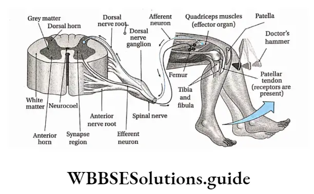
Deep reflex action: The type of reflex action in which stimuli are exerted on some underlying structures, mainly in muscles and tendons, is known as deep reflex action.

Example:

Knee jerk or patellar tendon reflex—When the knee is hit by a doctor’s hammer, the thigh and lower portion of the leg jerk,

Biceps jerk reflex—Suddenly hitting the biceps causes stretching in the tendon due to which elbow folds.

An unconditioned stimulus (food—meat) was presented to a hungry dog every day. It caused salivation (an unconditioned response).

At the same time, a conditioned stimulus (ringing a bell) was given. The dog learned to associate the conditioned stimulus (bell) with the unconditioned stimulus (food).

Eventually, after some days, it was observed that ringing of the bell alone caused salivation. This is because the dog adapted to the stimulus of a ringing bell that brought food.

Biology Class 11 Chapter 20 Neural Control And Coordination Pavlov Experiment

Biology Class 11 Chapter 20 Neural Control And Coordination Diffference Between Unconditioned Reflex action And Conditioned Reflec Action

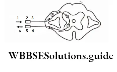
Reflex arc Definition: The path through which reflex action takes place and extends between the sensory organ to the effector organ through the grey matter of the spinal cord is called reflex arc.

Biology Class 11 Chapter 20 Neural Control And Coordination Components Of Reflax

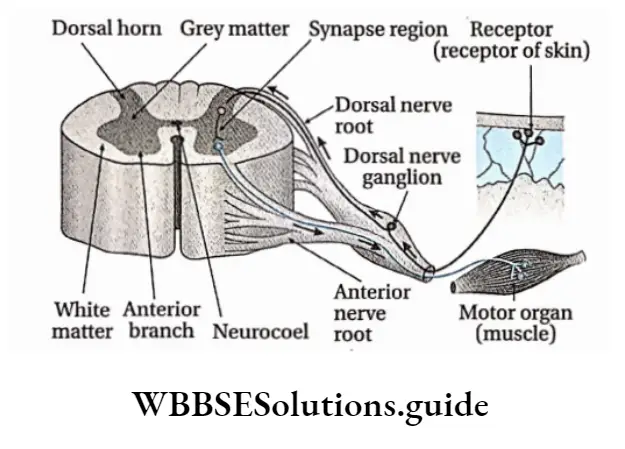
Different components of a reflex arc and their functional roles

The reflex arc is constituted of five parts that are described below.

Receptor: To accomplish a reflex action, the reflex arc must be directly linked with a receptor organ. Reflex action occurs when sense organs or receptors are excited by a stimulus.

Sensory or afferent neuron: These neurons of a reflex arc remain connected with the receptor. They carry impulses from the receptor to the grey matter of the spinal cord.

Grey matter of spinal cord: Grey matter of the spinal cord acts as the controller or center of reflex action. In the grey matter of the spinal cord, sensory impulses are analyzed and interpreted.

Effector organ: Effector organs are capable of responding to a stimulus to perform a particular action. Generally, muscles, glands, etc., participate as effector organs.

Sense Organs

Sense Organs Definition: The organs that are specialized to acquire and process sensory stimuli from different sources, both external and internal are called sense organizer

Sense Organs Types: Humans and other advanced vertebrates have different types of sense organizers The human body consists of five main types of sense organizer

These are—

  1. One Pair Of Eyes
  2. One Pair Of Ears
  3. Nose
  4. Tounge
  5. Skin

Sensory Receptors

Sensory Receptors Definition: The sensory nerve endings, a cell or group of cells, that respond to various kinds of stimulation are called receptors.

They are transducers that convert various forms of energy in the environment (internal or external) into action potentials in the afferent neurons. The awareness that results within the body is called sensation.

The sense organs—the eyes, ears, skin, mouth, and nose—have receptors that detect sensations. The sensory neurons present within them transmit the information to the central nervous system where it is interpreted as sight, sound, touch, taste, or smell.

Types of receptors: Receptors are classified into different types, as depicted in the chart given below.

On the basis of the location of the receptors, it can be classified into three types—

Exteroceptors: These receptors are located near the surface of the body. They can sense changes in the surrounding environment. Generally, they include receptors for touch, heat, cold, etc.

Interoceptors: These receptors are located within the body. They are stimulated by the internal stimuli which are generated inside the body.

There are several types, such as—chemoreceptors, stretch receptors, baroreceptors, etc.

Proprioceptors: The receptors which can detect the position and posture of the body.

They are located in skeletal muscle, bones, tendons, ligaments, etc. On the basis of the type of the received stimuli, receptors can be classified into five types, etc

Mechanoreceptors: These are receptors that are stimulated by mechanical forces, such as touch, pain, equilibrium, etc., received on muscles, tendons, ligaments, and joints. They comprise both exteroceptors and interoceptors.

Several types of mechanoreceptors are discussed in the following table.

Biology Class 11 Chapter 20 Neural Control And Coordination Types Of Mechanoreceptors

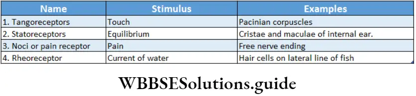
Telereceptors: Receptors sensing light and sound, which provide information about the distant environment, are called telereceptors.

Telereceptors, on the basis of their location, have been divided into three types which have been discussed below.

Biology Class 11 Chapter 20 Neural Control And Coordination Types of Telereceptors

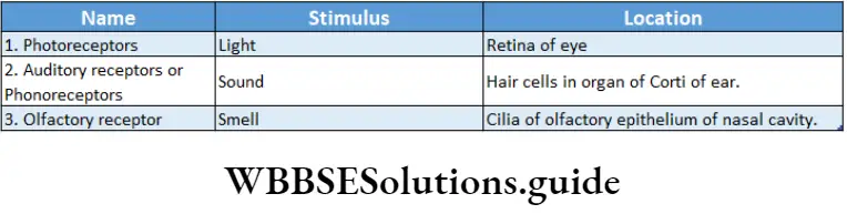
Chemoreceptors: These are receptors that are stimulated by chemical changes or by chemicals that are either inhaled or tasted (gustatory receptors).

They also modify hydrogen ion and carbon dioxide concentrations in blood. For example, chemoreceptors of the carotid artery can sense changes in blood partial pressure of 02 and C02 in the blood

Thermoreceptors: These are receptors that are sensitized by heat. For example, Ruffini’s end organs, are the end organ of Mazzoni.

Galvanoreceptors: These are receptors that are sensitized by gravity. For example, the organs of some fish have this type of receptor.

Human Eye

Our eyes are sense organs for vision. They also help in the perception of color.

Human beings have two simple eyes, present ventrally to the cranium and above both sides of the nose.

They are placed within the eye orbits. We have binocular vision (which involves two eyes to view an object). In the case of frogs, fish, etc., monocular vision (which involves one eye to view an object) is observed.
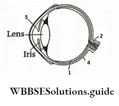
Biology Class 11 Chapter 20 Neural Control And Coordination Structure Of Human Eye

Eyeball

On the ventral portion of the brain box and above both sides of the nose, there are two cavities or orbits called the eye cavities within which eyeballs are located. These are supported by muscular tissue.

Each eyeball is almost spherical and has a diameter of 2.5 cm. The eyeball remains in position within the cavity with the help of six muscles. Eyeball actually is a fluid-filled sac-like structure.

It is composed of three parts—

  • Coats,
  • Eye chambers and
  • Refractory media
  • Coats

The human eyeball is covered with three layers or coats which are—

  • Fibrous coat,
  • Pigmented coat and
  • Neural coat.

Fibrous coat: The outermost layer of the wall of the eyeball is made up of fibrous connective tissue. This layer can be divided into two—

  • Sclera and
  • Cornea.

Sclera: This is the outer covering layer of the eyeball. The sclera is present in the posterior portion, comprising about five-sixth part of the external layer.

The sclera is a comparatively tough, translucent, and thick layer that is composed of white collagenous connective tissue. The posterior region of the eyeball from where the optic nerve emerges lacks sclera.

Cornea: It is the bulged, transparent anterior region of the eyeball. It covers the l/6th part of the fibrous coat of the eyeball. On the outer surface of the cornea, there is a thin and clear layer called the conjunctiva.

Functions of fibrous coat:

The sclera provides shape to the eyeball and forms a protective rigid layer for the eyeball.

Being transparent, the cornea acts as a refractive medium and permits light to pass through easily and fall on the lens. It acts as a convex lens to converge the diverging rays.

Pigmented coat or vascular coat: The middle layer of the human eyeball contains melanin pigment in its cells.

So, this layer appears brown. This layer is known as the pigmented layer. It is calculated and so, known as vascular coat. The different parts of this layer are discussed below.

Choroid: The melanin-containing brownish part beneath the sclera, in human eyeball is known as choroid.

Ciliary body and suspensory ligament: The thin layer that is present in the terminal region of the choroid, at the junction of the sclera and cornea of the human eyeball is known as the ciliary body. The thin fibers projecting from the ciliary body are known as suspensory ligaments.

Iris: The pigmented part present in front of the ciliary body, suspensory ligament, and lens of the human eyeball is called the iris. It controls constriction and dilation of the pupil.

At the center of the iris, a spherical opening is present, known as the pupil.

Iris contains two types of muscles—

Circular Muscles And Radial Muscles.

Functions of pigmented or vascular coat:

Due to its deep brown color, the choroid absorbs excess light and prevents its repeated reflection. Thus, it helps to form a clear image.

Suspensory ligaments hold the lens in its proper position.

While seeing objects nearer or farther, we cannot move the lens forward or backward, but with the help of ciliary muscles, the curvature of the lens can be changed. As a result, the focus of the lens can be changed as required. This phenomenon is known as accommodation.

The pupil dilates and constricts due to the action of circular muscles and radial muscles of the iris. In this way, the iris modifies the diameter of the pupil and controls the incident rays on the lens. The color of the iris is considered the color of the eye.

Neural coat or retina: The most internal layer of the human eye is the posterior part of the lens and the inner portion of the choroid where the optic nerve is found. This layer, made up of nervous tissue and connective tissue is known as the neural coat or retina

Structural features of the retina:

  • Retina, present posterior to the lens, is extended dorsally to ciliary bodies.
  • The different cells of the retina are arranged in 10 layers.
  • The most external layer among the 10 layers is pigmented and remains associated with the choroid.
  • There are two types of pigmented cells in this layered cells and cone cells. These cells are also described later.

The different neuronal cell layers of the retina bear four types of cells—bipolar cells, ganglion cells, horizontal cells, and amacrine cells.

All the nerve cells in the most internal retinal layer form the optic nerve.

The retina also contains one type of glial cell called Muller cell that helps in maintaining its structural integrity.

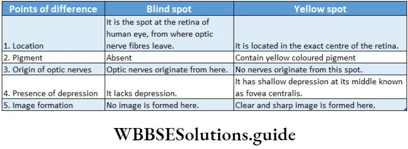
A depressed region is observed at the center of the retina. It is yellowish and so, known as yellow spot or macula lutea.

The clearest and brightest image is formed in this region. Its central region is depressed known as fovea centralis. It is the most photosensitive region of the retina.

The junction between the origin of the optic nerve and the posterior end of the retina lacks any photosensitive cells (rod and cone cells).

As a result, the image does not form in that region. This part of the retina is known as the blind spot or optic disc.

Different cellular layers of the retina: The retina consists of the following 10 layers—

Pigment epithelium: This outermost layer of the retina consists of pigmented cells. This is a non-neural layer that absorbs excess light which has passed through the other retinal layers and is immediately next to the choroid.

Photoreceptor layer: This layer includes the outer external segments of the photoreceptor cells, rods, and cones.

External limiting membrane: Although this appears to be a membrane, actually it is not. It is formed by intercellular junctions—the zonula adherent—binding the apical aspect of the Muller cells.

External nuclear layer: This consists of the cell bodies of the rods and cones.

External plexiform layer: This is a synaptic area that contains the terminals of the rods and cones.

Inner nuclear layer: This consists of the soma of the retinal interneurons (horizontal, bipolar, and amacrine cells). It integrates and modulates the activity of photoreceptors.

Inner plexiform layer: This is a synaptic area containing the terminals of the bipolar, amacrine, and ganglion cells.

Ganglion cell layer: This consists of the soma of the ganglion cells (multipolar neurons).

Optic nerve fiber layer: This layer is composed of the axons of the ganglion cells that gather to form a thick bundle, the optic nerve.

Internal (inner) limiting membrane: This membrane is composed of a basal lamina interposed between the vitreous body and the Muller cells.

Biology Class 11 Chapter 20 Neural Control And Coordination Structure Of Human Eye

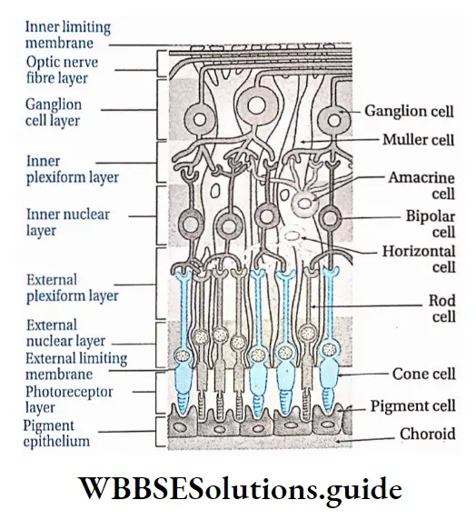
Photoreceptor cells of the retina: The human retina contains two types of photoreceptor cells—rod cells and cone cells. These are discussed below.

Rod cells: These cells are rod-shaped. They contain the pigment rhodopsin. Vitamin A is required for the synthesis of rhodopsin. The human eye contains 115-120 million rod cells.

Functions:

Rod cells help to see objects in dim light at night, which means they can absorb low-intensity light,

Rod cells generally form the black and white vision.

Cone cells: These cells are cone-shaped. They contain the pigment iodopsin. The human eye contains 6.0-6.5 million cone cells.

Functions:

Cone cells are capable of absorbing high-intensity light, so they help us to see objects in bright light,

They help in the perception of color and thus are responsible for color vision. The inactivity of cone cells leads to color blindness.

Biology Class 11 Chapter 20 Neural Control And Coordination Difference Between Rod And Cone Cells

Biology Class 11 Chapter 20 Neural Control And Coordination Differences Between Blind Apot And Yellow Spot

Functions of retina

The retina of the human eyeball acts as the photoreceptor organ. Rod cells of the retina help to see objects in dim light. On the other hand, cone cells help to see objects in bright light.

Light from different sources reflects on the lens and creates an image on the retina. This reflection is transmitted to the cerebrum by optic nerves. There it is translated and a sensation of vision is created.

The yellow spot of the retina, especially the fovea centralis, forms a clearer image of the object.

Rod cells of the retina cause black-and-white vision and cone cells causes colored vision.

Eye chambers

The human eyeball contains three chambers which are as follows—

  • Anterior chamber: The chamber between the iris and cornea of the human eyeball is known as the anterior chamber. This chamber is filled with fluid aqueous humor.
  • Posterior chamber: The thin chamber between the lens and iris of the eyeball is known as a posterior chamber. This chamber is also filled with aqueous humor.
  • Vitreous chamber: The large chamber at the posterior part of the lens, between the lens and the retina is known as a vitreous chamber. This chamber is filled with fluid vitreous humor.

Refractory Media

All those parts of the eyes that convert parallel or divergent light rays into convergent light rays are known as refractory media. The refractory media of human eyes are—lens, aqueous humor, vitreous humor, and cornea.

Lens: It remains suspended from ciliary bodies by the suspensory ligament in the human eyeball. It is the main refractory medium of human eyes.

The lens is elastic in nature and bi-convex in shape. It is composed of transitional columnar epithelial tissue. It is as clear as crystal. It contains the protein crystallin. It is surrounded by a capsule.

Functions:

It is the main refractory medium of human eyes.

It helps to focus on objects based on distance. While seeing a nearby object, ciliary muscles contract and the lens becomes thick. While seeing distant objects, ciliary muscles relax, thus reducing the thickness of the lens.

Aqueous humor and vitreous humor: The clear fluid substance present in the anterior and posterior chambers of the human eye is known as aqueous humor.

On the other hand, the fluid substance present in the vitreous chamber of the human eye is known as vitreous humor.

Both aqueous and vitreous humor is made of salt, sugar, and protein, vitreous humor is viscous i.e., jelly-like.

Functions:

  1. Both aqueous and vitreous humor maintain intraocular pressure and inflate the eyeball. This is how they maintain the shape of the eyeball.
  2. They act as refractory media of the eyes.
  3. Aqueous humor provides nutrients to the lens, cornea, iris, etc.

Cornea: It is the convex, transparent part of the human eyeball which also acts as a refractory medium to some extent.

Accessory Protective Part Of Human Eye

The structure and functions of accessory protective parts of the eyes are described below.

Accessory Protective Part Of Human Eye Eye orbit: In the anterior part of the human cranium, beside the nose, within two hollow cavities the eyeballs are placed.

These two hollow cavities are known as eye orbits. Note that eye orbit has a thick layer of muscular tissue, on which eyeballs are placed. Eyeballs are bound to the eye orbit by six muscles. The optic nerve too, helps to bind the eyeball to eye orbit.

Accessory Protective Part Of Human Eye Functions: Due to the presence of muscular tissue from the interior and bony covering from the outside, the eye orbit protects the eyeball from mechanical injury.

Eye muscles: The six muscles by which eyeball remains bound to the eye orbit are known as eye muscles.

These are—

  1. Superior rectus muscle,
  2. Inferior rectus muscle,
  3. Medial rectus muscle,
  4. Lateral rectus muscle,
  5. Superior oblique muscle,
  6. Inferior oblique muscle.

Functions:

  1. With the help of these muscles, we can move the eyeballs down and sideways.
  2. Eyeballs can also be rotated by these muscles.

Eyelids: The human eye is covered by a thin, extended, skin layer on the upper part and lower part.

These are known as upper eyelids and lower eyelids respectively. At the corner of the human eye (near the nose), a small, red, muscular nictitating membrane is present in a vestigial condition.

Accessory Protective Part Of Human Eye Functions:

Eyelids protect the frontal part of the eyeball from dust.

Under bright light eyelids close, thus protecting the eye, rather than the lens, from bright light.

Eyebrow and eyelash: Each eye of humans has two special parts on upper region of the eyelids and the lower region of the forehead.

This part is curved like a bow and is known as an eyebrow. The eyelids are again lined by fine hairs which are known as eyelashes.

Accessory Protective Part Of Human Eye Function: Due to the presence of an eyebrow, rainwater, and sweat cannot enter the eyeball from the forehead.

Conjunctiva: The thin and transparent layer present beneath the upper eyelid and which extends upto the lower lid, covering the anterior part of each human eye is known as conjunctiva.

Conjunctiva is actually made up of squamous epithelium. Inflammation of the conjunctiva is known as conjunctivitis.

Accessory Protective Part Of Human Eye Functions:

It protects the eyeball from dust particles.

Being transparent, it allows light rays to pass through it easily into the eyeball to reach the lens.

Lacrimal gland: Lacrimal gland or tear gland is present in the upper lateral region of each orbit or beneath the upper and outer border of each eyelid. It opens into the upper eyelid by some tubules.

The tear produced in the tear gland is a fluid containing NaHC03, NaCI, etc. Tear also contains a bacteriolytic enzyme called lysozyme.

Accessory Protective Part Of Human Eye Functions:

Tears keep the upper surface of the eyeball moist. If the lacrimal gland gets damaged, secretion of tears is reduced, and conjunctiva contracts thereby causing dryness and burning sensation in the eye.

Tear wash out the dust particles present in the open the surface of the eyeball.

Lysozyme, an antibacterial enzyme present in tears protects the eye from bacterial infection.

Physiological Function Of Eye

Vision is the main physiological function of the human eye. In case of the human eye helps us with two types of vision, such as—light vision and color vision.

Light vision: Eyes help us to see objects by providing a stereoscopic vision. The mechanism of vision by which eyes sense and visualize nearby and far away objects is given below.

Mechanism of vision:

  1. The human eye acts as a camera. Parallel or divergent light rays from objects pass through the conjunctiva, cornea, and aqueous humor and fall on the lens.
  2. After falling on the lens or incidence, light rays become convergent. These convergent rays pass through the vitreous humor, present behind the lens, and then fall on the yellow spot of the retina.
  3. Retina contains a photosynthetic pigment, called rhodopsin. Light splits rhodopsin and converts it into two chemical compound named retinene (retinal) and opsin.
  4. Light also brings changes in the structure of opsin which causes changes in membrane permeability.
  5. Changes in membrane permeability cause ion channels to open which results in potential difference and generation of action potential.
  6. This action potential i.e. impulse is carried by the optic nerve to the visual cortex of the cerebrum through the optic tract.
  7. Here the image is formed. Note, stimulus from the right eye goes to the visual cortex of the left cerebral hemisphere and stimulus from the left eye goes to the visual cortex of the right cerebral hemisphere. In the visual cortex, the stimulus is analyzed.
  8. By the special capability of the brain, the image becomes elongated. This results in the sensation of vision.

Color vision: Cone cells of the human eye help to determine the color of different objects.

Visual Accommodation

Human eyes cannot change the distance of the lens directly to observe objects nearer or farther. However, ciliary muscles and suspensory ligaments can adjust the curvature of the lens.

As a result, the focal length of the lens changes due to which the image of the object falls on the retina. This phenomenon is known as accommodation.

This is a type of reflex action. Human eyes have good visual accommodation. Light coming from any object located 25cm away can be easily sensitized by eyes.

This is known as the least distance of distinct vision. The ciliary bodies and suspensory ligaments of human eyes together are known as accommodation apparatus.

Some Important Eye Disorders

There are some important disorders related to the eyes which are as follows.

Myopia: It is the most common refractive error of the eye. Myopia is also referred to as short-sightedness as the person cannot see distant objects.

In this condition, parallel rays of light coming from distant objects, instead of focusing on the retina, focus in front of it.

As a result, objects located distantly cannot be seen clearly. It can be corrected by using glasses with a concave surface.

Hypermetropia or Hyperopia: It happens when the eyeball is shorter than normal and the parallel rays of light are brought to focus behind the retina.

It is also referred to as long-sightedness. In this case, nearby objects appear blurry while distant objects are seen clearly. It can be corrected by using glasses with convex lenses.

Astigmatism: Astigmatism is a common condition in which the curvature of the cornea or lens is not uniform.

This causes blurred vision of objects both near and far. This can be corrected by using glasses with cylindrical lenses.

Presbyopia: Presbyopia is an age-related eye condition that makes it more difficult to see near objects very clearly.

After the age of 40, the lens of the human eye becomes hard thereby losing the capacity of contraction-relaxation and thus people find difficult to see, particularly to read. It can be corrected by using glasses with bifocal or convex lenses.

Cataract: Due to aging, the lens becomes swollen, hard, and opaque. Vision becomes feeble and sometimes, is lost. This condition is known as cataract. It can be corrected by replacing the affected lens with an artificial one.

Glaucoma: The defects in the eyes that lead to damage of the optic nerve are known as glaucoma.

It happens due to an increase in intraocular pressure leading to the rupture of the optic nerve.

When blood flow is reduced in the optic nerve i.e., in the case of ischemia, the optic nerve is damaged.

Due to damaged optic nerve, vision is impaired and even blindness may occur.

Strabismus (squint): In this disorder, two eyes do not look exactly in the same direction at the same time.

It is caused due to dysfunction of the extraocular muscle. It is usually corrected by surgical procedures at an early stage.

Trachoma: It is a pathological condition caused by the bacterium Chlamydia trachomatis.

It is characterized by inflammation of the conjunctiva and cornea leading to pain and watering of the eyes.

In extreme cases, it may lead to complete blindness. Trachoma can be treated by antibiotics in the early stages and surgery in the late stages.
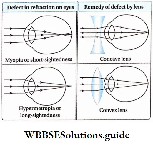
Biology Class 11 Chapter 20 Neural Control And Coordination Diseases Related To eye

Human Ear

The human ear is the sense organ of hearing and equilibrium. It consists of three main parts—

  1. External ear,
  2. Middle ear and
  3. Inner ear. The ear receives the sound waves and helps in its interpretation.

Parts Of Ear

Different parts of the human ear are discussed under Separte Heads.

Biology Class 11 Chapter 20 Neural Control And Coordination Parts Of Human Ear

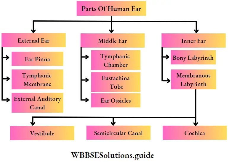
Sound waves can travel through solid, liquid, and gaseous medium. The human ear is the best example to Sound waves that can travel through solid, liquid and gaseous mediums. The human ear is the best example of solid bones in the middle ear and fluid medium in the inner ear.
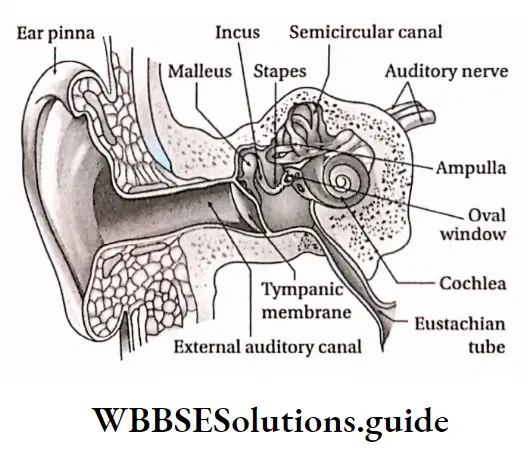
Biology Class 11 Chapter 20 Neural Control And Coordination Different Parts Of Human Ear

External Ear

The human external ear consists of three parts—pinna, external auditory canal, and tympanic membrane.

External Ear Pinna: The outer ridged cartilaginous part of the ear covered with skin, through which sound waves are received from the external environment is called the pinna.

External Ear Location: The pinna is an expanded cartilaginous portion, covered with skin that projects from both sides of the head.

External Ear Characteristics:

  • Pinna is made of elastic cartilage. It has an expanded leaf-like structure, it has a skinny covering on cartilage.
  • Along the upper periphery of the pinna, a fold is seen, known as a helix.
  • The lower part of the pinna is muscular and so, it is calculated. This flexible lobe is known as the lobule.
  • The muscles of the pinna are vestigial, hence, the human pinna cannot be moved.

Functions:

  • Pinna collects sound waves from different sources and funnels them to the auditory canal.
  • Therefore, pinna acts as the receiver of sound waves.
  • Due to the presence of folds in pinna, it inhibits dust, insects, etc., from entering the auditory canal.

External auditory canal: This is the S-shaped passage through which sound waves travel from outside to the tympanum.

Location: External auditory canal or external auditory meatus, a part of the external ear, extends from an opening in the pinna to the tympanic membrane.

Characteristics:

  • The external auditory canal is a slightly bent, tubular structure. It runs upward and then downward.
  • It is supported by cartilage and temporal bone in the outer and inner portions respectively. The junction of the outer and inner portions is referred to as the osteocartilaginous junction.
  • The inner wall of the auditory canal is covered by skin. The sweat gland of this layer has transformed into a ceruminous gland or wax gland which secrete cerumen or earwax.

Functions:

  • It conducts the sound waves towards the tympanic membrane. 0 Since it is a bit bent, it protects the tympanic membrane from external mechanical injuries.
  • The ceruminous gland in its wall secretes a waxy substance, cerumen that contains lysozyme and immunoglobulin. So, dust particles, insects, and germs cannot reach upto the tympanic membrane.
  • Tympanic membrane or tympanum or ear drum: It is the membrane that acts as a partition between the external and middle ear and vibrates in response to sound waves.

Location: It is located in the inner region of the auditory canal i.e., as a separating membrane between the auditory canal and the middle ear.

Characteristics:

It is an elliptical, smooth, and translucent separating membrane. It separates the middle ear from the external ear.

It stretches from the frontal to the posterior end, across the end of the external auditory canal.

Its outer surface is concave and inner surface is
convex. It remains in stretched condition.

The tympanic membrane consists of three layers—the outer layer of stratified epithelium, a middle fibrous layer, and an inner layer of cuboidal epithelium.

Its inner layer is connected with the arm of the ear ossicle, the malleus.

Functions: The tympanic membrane is considered as the eardrum. Vibrating sound waves, transmitted by air strike the tympanic membrane.

This creates vibrations in the membrane. These vibrations are transmitted to the inner ear through ear ossicles in the form of waves.

Middle ear

Inner to the tympanic membrane, within the temporal bone of the human skull, the middle ear is located.

The three parts of the middle ear are—

  1. Tympanic cavity,
  2. Eustachian tube,
  3. Three ear ossicles.

Tympanic cavity: The small space in the middle ear surrounding the ear ossicles is called the tympanic cavity.

Location: It is an irregularly shaped, air-filled chamber, embedded in the inner portion of the temporal bone.

Characteristics:

  • The tympanic cavity is an air-filled chamber.
  • The inner wall of the chamber is lined by cuboidal epithelial tissue.

Functions:

  • The lower part of the tympanic cavity is connected to the eustachian tube.
  • It houses three little bones—malleus, incus, and stapes.

Eustachian tube: The tubular canal through which the tympanic cavity communicates with the nasopharynx is called the eustachian tube.

Location: It is located in the middle ear, from the lower portion of the tympanic membrane to the nasopharynx. It is also known as a pharyngotympanic tube or auditory tube.

Characteristics:

  • The eustachian tube opens into the nasopharynx through an opening. So, the eustachian tube is also known as pharyngo-tympanic tube.
  • Its inner wall is lined by ciliated epithelial tissue.

Functions:

Through this tube air from the mouth cavity and pharynx enters the tympanic cavity. As a result, air pressure on both sides of the tympanic membrane remains the same. So, the tympanic membrane does not burst even in loud sounds.

Due to the presence of ciliated epithelium in the inner wall, dust particles and mucous are present in the air. from the mouth cavity and pharynx, do not reach the tympanic cavity.

Auditory ossicles: These are the three bones in the middle ear, which are articulated from a chain for the transmission of sound from the external ear to the internal ear.

Location: The tympanic cavity of the middle ear contains three ear bones, connected with each other by bony joints. Ligaments bind the joints of these three bones.

Types: Three ear ossicles of the middle ear, from the end of the tympanic membrane, are—malleus, incus, and stapes.

Malleus: Malleus looks like a hammer. It bears a spherical head and an elongated arm. This arm is connected to the inner portion of the tympanic membrane and the spherical head joins with the next ossicle, incus.

Incus: Incus is the second ossicle. It appears like an anvil. Its frontal part has a concave cup-like ridge.

Within it, the head of a malleus is placed and it forms a joint. Its long appendage helps it to join with the third ossicle, stapes.

Stapes: It is the third and smallest ossicle of the human ear. It is also the smallest bone of the human body. It appears like a stirrup. It has a head, neck, two arms, and an oval base.

Its head remains bound to the appendage of the incus. On the inner surface of the tympanic cavity, an oval opening is seen, known as an oval window or fenestra ovalis. The base of the stapes is attached to this opening.

Biology Class 11 Chapter 20 Neural Control And Coordination Ear Ossicle

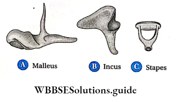
Functions: When sound waves strike the tympanic membrane, the vibrations are transmitted through the ossicles—malleus, incus, stapes, via the oval window to the inner ear.

Inner Ear

The inner ear is located next to the middle ear and is a double-walled structure. It has two main parts— a bony labyrinth and a membranous labyrinth.

Bony labyrinth: Bony labyrinth is located in a cavity, within the temporal bone. The space between the bony and membranous labyrinth is called perilymphatic space. This space is filled with perilymph.

Membranous labyrinth: It is a complex structure, containing canaliculi and membranous sacs.

The fluid present in it is known as endolymph. This part is connected to the auditory nerves.

It is composed of three parts—

  1. Vestibule,
  2. Three semicircular canals and
  3. Cochle

Biology Class 11 Chapter 20 Neural Control And Coordination Membranous Labyrinth

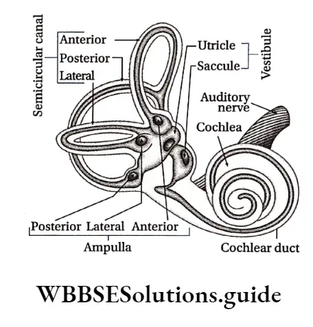
Vestibule: The extended portion of the membranous labyrinth of the inner ear, connected to the three semi-circular canals is known as the vestibule.

It is central sac-like part, which is again formed of two unequal parts— utricle and saccule and is known as the otolith organ.

Structural features:

The vestibule is an endolymph-filled, thin membrane-bound chamber,

This part contains two endolymph-filled sacs. The larger one is called utricle and the smaller one is called saccule.

The endolymph of vestibule contains granules of CaC03 and silica. These are known as otoliths,

Its inner wall is lined by ciliated epithelium and the base of the cells of this part are connected to nerve fibers of vestibular branches of the auditory nerve.

There are 2 sensory spots in the vestibule—the macula of the utricle and the macula of the saccule. The macula consists of sensory hair cells and supporting cells.

Functions: Movement of head causes displacement of the otolith. Thus, sensory cells of the utricle and saccule are stimulated.

This stimulation reaches the cerebellum through auditory nerves. In this way, vestibule of the inner ear maintains body balance with the help of the cerebellum.

Vestibular Apparatus

The three semicircular canals, endolymph containing sacs—utricle and saccule altogether are known as vestibular apparatus. It plays an important role in maintaining body balance.

Utricle And Saccule

Utricle: It is a large sac whose frontal part consists of three semicircular canals, containing ampullae. A duct opens at the dorsal part of the utricle which is known as utricosaccular duct.

Saccule: It is a comparatively smaller sac. The duct opening from the posterior end of the saccule is known as ductus endolymphaticus.

This duct joins with the utriculosaccular duct extends more at the dorsal surface and fuses into a closed sac called saccus endolymaphaticus. Another small duct from the saccule, known as ductus reuniens joins with the cochlear duct.

Three semicircular canals: The frontal part of the membraneous labyrinth contains three bow-like canals. These are known as semicircular canals.

Structural features:

  1. The three semicircular canals of the inner ear are located in three planes. These planes are located equilaterally.
  2. The base of each canal is swollen and is known as ampulla (plural: ampullae)
  3. Due to the presence of the common arm of two semicircular canals, each ear contains five ampullae. Each ampulla contains a sensory spot, called crista ampullaris. Each crista consists of longer hair cells and a gelatinous mass, called a cupula (lacks otolith)
  4. Endolymph is present between the semicircular canals, ampullae, and vestibule
  5. The inner wall of an ampulla contains sensory ciliated epithelial tissue.

Functions: The semicircular canal is under the control of the auditory nerve. Displacement of otolith maintains body balance.

Cochlea: The coiled part that appears like a snail and is located posterior to the membraneous labyrinth is known as the cochlea.

Structural features:

The cochlea’s interior is divided into three parallel, fluid-filled compartments by two membranes, the vestibular (Reissner’s) membrane and the basilar membrane,

Each compartment is referred to as a scala (staircase) since each one spiral about an axis or the central pillar of the cochlea like a winding staircase.

They are the (bony) scala vestibuli, the (membranous) scala media or cochlear duct, and the (bony) scala tympani.

The interiors of the scala vestibule and scala tympani are lined by a simple squamous epithelium. Inside scala vestibuli and scala tympani, perilymph is present,

The scala vestibule and scala tympani communicate with each other by a narrow passage, helicotrema. This structure permits perilymph to flow from the scala vestibule into the scala tympani.

At the proximal end of the cochlea, the scala vestibule, and scala tympani communicate with the oval and round window respectively,

The cochlear duct also referred to as the scala media, contains endolymph. In the cross-section of the cochlea, the triangular-shaped cochlear duct appears to be wedged between the scala vestibule and the scala tympani.

The Reissner’s membrane, the roof of the cochlear duct, serves as a division between the cochlear duct and the scala vestibuli. This membrane is composed of squamous epithelium.

Its function is unclear; however it is believed to play a role in the transmission of vibrations from the perilymph of the scala vestibule to the endolymph of the cochlear duct,

The floor of the cochlear duct, which separates it from the scala tympani, is the basilar membrane. The basilar membrane is an elastic structure.

It shows a gradual decrease in stiffness from the oval window (at the cochlear base) to the helicotrema (at the cochlear apex),

The gradual decrease in stiffness permits this membrane to be sensitive to high-frequency vibrations near the cochlear base, and low-frequency vibrations near its apex,

Thus, the Reissner’s membrane is interposed between the scala vestibule and the cochlear duct, and the basilar membrane is interposed between the cochlear duct and the scala tympani.

The receptors associated with the sense of hearing form a structure, the organ of Corti, which rests along the floor of the cochlear duct upon the basilar membrane,

The organ of Corti consists of supporting epithelial cells (pillar cells, cells of Hensen, cells of Claudius, etc.) and neurosensory receptor cells, called ‘hair cells’.

Biology Class 11 Chapter 20 Neural Control And Coordination Cross Section Of Cochlea

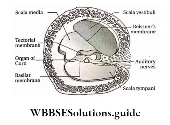
Each hair cell displays numerous stereocilia and a single kinocilium projecting from its apical cell surface,

The free ends of the stereocilia project into the tectorial membrane, an acellular, gelatinous sheet,

The hair cells are the receptors of the auditory system that act as transducers. They serve to convert mechanical energy into electrical energy that can be relayed to the brain-stem,

Hair cells synapse with the peripheral ends (dendritic processes) of the bipolar neurons whose cell bodies are housed in the spiral ganglion.

The central processes of these bipolar neurons form the cochlear nerve, which joins the vestibular nerve to form the vestibulocochlear nerve (VIII cranial nerve).

This nerve courses through the internal acoustic meatus to enter the cranial vault, and then enters the brainstem

Functions: Cochlea is stimulated by sound vibrations produced in the organ of the Corti. This sensation is transmitted to the cerebrum by auditory nerves. In this way, the cochlea helps in hearing.

Biology Class 11 Chapter 20 Neural Control And Coordination Organ Of Corti

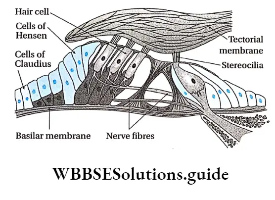
The Physiological Function Of The Ear

Physiological functions of the human ear are as follows— Hearing: The ear enables us to hear external sound.

Mechanism of hearing:

Sound waves from vibrating bodies are transmitted through air to the pinna.

From there, sound waves, through the auditory canal, reach the tympanic membrane. This vibration is transmitted through the ear ossicles of the tympanic cavity to the inner ear via an oval window.

Vibrations are transmitted in the form of waves to the perilymph of scala vestibuli through the fenestra ovalis and then transferred to the scala media through Reissner’s membrane.

The waves in the endolymph cause the basilar membrane to vibrate. This causes the hair cells to bend, pressing them against the tectorial membrane.

As a result of stimulation of the receptor cells, nerve impulses are generated in the associated afferent neurons.

These are conducted by auditory nerves to the auditory cortex of the brain. There it is analysed and we are able to hear.

Biology Class 11 Chapter 20 Neural Control And Coordination Physiological Function Of Ear

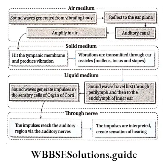
Understanding pitch of sound: The pitch of sound depends on the vibrations of sound waves.

Sounds of different vibrations cause stimulation along different regions of the basilar membrane depending on the pitch of the sound.

So, we can understand sounds of varied pitch. In the case of high-pitched sounds or sounds of high frequency, the receptor cells at the base of the cochlear duct are stimulated.

In the case of low-pitched sound, the receptor cells towards the tip are stimulated.

Understanding loudness of sound: Loudness of sound depends on the distribution of sound. Loudness is expressed by decibels (dB).

The greater the amplitude of sound, the more receptor cells of the organ of Corti in the basilar membrane are stimulated.

Hence, sound can be heard louder. Too loud sound damages the cells of the basilar membrane, causing deafness.

Maintenance of body balance:

  • The vestibule and three semicircular canals of the human inner ear maintain the body balance.
  • The vestibule and semicircular canals have sensory epithelial tissue. This part contains CaC03 and silica suspended in endolymph.
  • These substances are displaced upon a change in body posture, This causes stimulation of cristae and maculae present in the ampulla and vestibule respectively.
  • This stimulation is transmitted through auditory nerves of the vestibular branch that join with a cochlear branch to form the vestibulocochlear nerve or auditory nerve.
  • The vestibular branch transmits the stimulus first to the vestibular nucleus and next to the cerebellum.
  • The cerebellum receives stimuli from proprioceptors of the eyes, skeletal muscles, and joints. These stimuli are then transferred finally to the cerebrum and thus body balance is maintained.

Notes

  1. Ampulla: Swollen part of semicircular canal.
  2. Ganglion: A structure containing a number of nerve cell bodies, linked by synapses and often forming a swelling on a nerve fiber.
  3. Kinocilium: A special type of cilium on the apex of hair cells located in the inner ear of the vertebrates.
  4. Motor end plate: The plate-like formation at the nerve terminal where an axon of a motor neuron establishes synaptic contact with the sarcolemma of the striated muscle fiber.
  5. Neurilemma: The plasma membrane of a neuronal cell body.
  6. Neurotransmitter: Chemicals that help in the transmission of nerve impulses throughout the nervous system.
  7. Nuclei: Aggregation of nerve cell bodies within the CNS (not to be confused with the nucleus of a cell).
  8. Ossicles: A very small bone usually occurring in the middle ear.
  9. Photoreceptor discs: The outer segment of rods and cone cells lack, ER, Golgi, and mitochondria and are filled with a hundred to a few thousand flattened membrane organelles, called photoreceptor discs.
  10. Plexus: A structure in the form of a network of nerves.
  11. Stereocilia: Mechanosensing organelles of hair cells, having a similar structure to microvilli.
  12. Threshold stimulus: The minimum stimulus required for a reflex action to occur.

Points To Remember

  1. Neurons are the basic structural and functional units of the nervous system.
  2. Each nerve cell (neuron) consists of a cell body or soma from which numerous processes arise.
  3. The human nervous system is divided into two types—central nervous system (CNS) and peripheral nervous system (PNS).
  4. The cell bodies within the central nervous system (CNS) are frequently clustered into groups called nuclei (not to be confused with the nucleus of a cell).
  5. The plasma membrane of the axon is called the axolemma, and the cytoplasm contained in it is called the axoplasm.
  6. Association neurons or interneurons are located entirely within the CNS and serve the associative, or integrative functions of the nervous system.
  7. The glial cells support neurons structurally and maintain their long-term neuronal integrity.
  8. The junction between two neurons is called a synapse. It is also called physiological value.
  9. A nerve is a cord-like structure composed of nerve fibers (axons) and connective tissue.
  10. A sensory nerve is composed of afferent fibers only a motor nerve of efferent fibers only, and a mixed nerve is composed of both. Most nerves are mixed.
  11. There are 12 pairs of cranial nerves and 31 pairs of spinal nerves.
  12. Meninges surround the brain and spinal cord and act as a try layered protective sheath or covering.
  13. The spinal cord is divided into cervical, thoracic, lumbar, and sacral regions, each served by spinal nerves.
  14. The autonomic nervous system deals with the visceral organs, like the heart, stomach, glands, and intestines.
  15. Any changes in the resting membrane potential (which is -70 mV), due to a stimulus of sufficient magnitude, that cause the membrane potential to move towards zero potential (less negative) mean depolarisation.
  16. A reflex is a quick, involuntary, stereotyped reaction of a gland or a muscle to a stimulus.
  17. Somatic (spinal) reflexes are responses of skeletal muscles.
  18. The retina contains the receptors for sight—rod and cone cells.
  19. The center of the retina contains a special region called the fovea centralis. It is an oval, yellowish area with a depression containing only cone cells.
  20. The ear is divisible into three parts—the external ear, the middle ear, and the inner ear, each performing a necessary function for the perception of sound.
  21. The external ear is comprised of the cartilaginous structure—pinna, external auditory canal, and the tympanic membrane.
  22. The middle ear is embedded in the temporal bone of the skull.


Neural Control And Coordination Questions And Answers

Biology Class 11 WBCHSE Neural Control And Coordination Questions And Answers

Question 1. What are the nodes of Ranvier? Are they found in Dendron also?
Answer:

The Myelin sheath of medullated nerve fibers is found on axons at intervals. Such periodic gaps of the myelin sheath are called nodes of Ranvier. Nodes of Ranvier help in the saltatory conduction of impulses.

The dendron of neurons is always hon-medullated, so the dendron lacks a node of Ranvier.

Neural Control And Coordination

Question 2. Mention the location and process of secretion of neurotransmitters in the synaptic cleft.
Answer:

Neurotransmitters (e.g., ACh) are present in the synaptic vesicles within the axolemma of the synaptic knob of axon in the presynaptic neuron.

Synaptic vesicles fuse with the presynaptic membrane and by exocytosis they are released in the synaptic cleft.

Question 3. Which organ is considered a ‘central information processing organ’? Where is it located?
Answer:

The human brain is considered the ‘central information processing organ’.

It is located within a special cavity called the cranium or brain box in the human skull.

Read and Learn More WBCHSE Solutions For Class 11 Biology

Question 4. Which is the largest and most important region of the human brain? What are the names of its two symmetrical parts?
Answer:

The largest and most important region of the human brain is the cerebrum.

The two symmetrical parts of the cerebrum right and left cerebral hemispheres.

Biology Class 11 WBCHSE

Question 5. What is the corpus callosum? Is it found in the brains of other species?
Answer:

The transverse, bundle of nerve fibers, that holds the two cerebral hemispheres together is known as the corpus callosum.

Yes, not only humans, but all vertebrates (except platypus) have corpus callosum in their brains.

Question 6. What are grey matter and white matter? Why they are named such?
Answer:

The central nervous system consists of two types of tissues—grey matter and white matter.

The grey matter is mainly composed of neuronal cell bodies, unmyelinated axons, glial cells, synapses, and capillaries. Due to the absence of a myelin sheath, this tissue appears grey. Hence, this name was given.

The white matter is composed of myelinated axons, hence, they appear white and are named such

Class 11 Biology WBCHSE

Question 7. What is known as the cerebral medulla? What is its composition?
Answer:

The region interior to the cerebral cortex, in each cerebral hemisphere of the human cerebrum, is known as the cerebral medulla.

The cerebral medulla is composed of white matter. This region contains numerous medullated nerve fibers. Note that due to the presence of myelin sheath in the medullated nerve fibers, this region appears white.

Question 8. What is a cerebral aqueduct? orWhat is corpora quadrigemina?
Answer:

The third and fourth ventricles of the human brain are connected by a duct of the midbrain. This duct is known as the cerebral aqueduct or aqueduct of Sylvius.

On the tectum of the human midbrain, four spherical swellings or lobes are observed. Together, these are known as corpora quadrigemina.

Biology Class 11 WBCHSE

Question 9. Which part of the human brain is known as the brain stem? or Which part of the human brain is involved in breathing and secretion of saliva?
Answer:

The part of the brain that remains connected to the spinal cord is known as the brain stem. The human brain stem is composed of the midbrain (mesencephalon), pons varolii (included in metencephalon), and medulla oblongata (myelencephalon).

The medulla oblongata of the hindbrain controls breathing and secretion of saliva.

Question 10. What is a yellow spot or macula lutea? What is fovea? Mention one use of fovea.
Answer:

The convex region at the middle of the retina is known as the yellow spot or macula lutea.

The central part of the yellow spot is more depressed. This part is known as fovea centralis or fovea in short.

The fovea is the most photoreceptive area of the retina. Image is formed on this region when an object is minutely tracked and observed.

Question 11. Where is Organ of Corti located? or Where is stereocilia present? or What is known as the tectorial membrane?
Answer:

The organ of Corti is located in the basilar membrane of the membranous labyrinth in the human inner ear.

The organ of Corti contains hair cells. Their terminal ends are connected to nerve fibers and their free ends contain numerous cilia. These cilia are called stereocilia.

The elastic membrane present above the hair cells of the organ of Corti is known as the tectorial membrane.

Biology Class 11 WBCHSE

Question 12. What is known as crista ampullaris? or What is its function?
Answer:

The sensory spots present in the ampullae of the semicircular canals are called crista ampullaris.

Crista ampullar is the sense organ of rotation.

Neural Control And Coordination Multiple Choice Question and Answers

Neural Control And Coordination Multiple Choice Question and Answers

Question 1. Receptor sites for neurotransmitters are present on—

  1. Pre-synaptic membrane
  2. Tips of axons
  3. Post-synaptic membrane
  4. Membranes of synaptic vesicles

Answer: 3. Membranes of synaptic vesicles

Question 2. Myelin sheath is produced by—

  1. Astrocytes and Schwann Cells
  2. Oligodendrocytes and Osteoclasts
  3. Osteoclasts and Astrocytes
  4. Schwann Cells and Oligodendrocytes

Answer: 4. Schwann Cells and Oligodendrocytes

Question 3. Good vision depends on adequate intake of carotene-rich food.

  1. Vitamin A derivatives are formed from carotene.
  2. The photopigments are embedded in the membrane discs of the inner segment.
  3. Retinal is a derivative of Vitamin A.
  4. The retina is a light-absorbing part of all visual photopigments.

Options:

  1. 1,3,4
  2. 1,3
  3. 2,3, and 4
  4. 1,2

Answer: 1. 1,3,4

Question 4. Photosensitive compound in the human eye is made up Of—

  1. Opsin and Retinal
  2. Opsin and Retinol
  3. Trunsducin and retinene
  4. Guanosine and Retinol

Answer: 1. Opsin and Retinal

Read and Learn More WBCHSE Multiple Choice Question and Answers for Class 11 Biology

Question 5. Choose the correct statement.

  1. Nociceptors respond to changes in pressure.
  2. Meissner’s corpuscles are thermoreceptors.
  3. Photoreceptors in the human eye are depolarised during darkness and become hyperpolarised in response to the light stimulus.
  4. Receptors do not produce graded potentials.

Answer: 3. Photoreceptors in the human eye are depolarised during darkness and become hyperpolarised in response to the light stimulus.

Question 6. In the mammalian eye, the ‘fovea’ is the center of the visual field, where—

  1. More rods than cones are found
  2. The high density of cones occurs but has no rods
  3. The optic nerve leaves the eye
  4. Only rods are present

Answer: 2. High density of cones occur, but has no rods

Question 7. Destruction of the anterior horn cells of the spinal cord would result in loss of—

  1. Integrating impulses
  2. Sensory impulses
  3. Voluntary motor impulses
  4. Commissural impulses

Answer: 3. Voluntary motor impulses

Question 8. Stimulation of a muscle fiber by a motor neuron occurs at—

  1. The neuromuscular junction
  2. The transverse tubules
  3. The myofibril
  4. The sarcoplasmic reticulum

Answer: 1. The neuromuscular junction

Question 9. Which one of the following statements is not correct?

  • Retinol is the light-absorbing portion of visual photopigments
  • In the retina, the rods have the photopigment rhodopsin, while, cones have three different photopigments
  • Retinol is a derivative of vitamin C
  • Rhodopsin is the purplish red protein present in rods only

Answer: 3. Retinol is a derivative of vitamin C

Question 10. Which excitatory neurotransmitter is involved in the transmission of impulses at the neuromuscular junction?

  1. Epinephrine
  2. Serotonin
  3. Acetylcholine
  4. Glycine

Answer: 3. Acetylcholine

Question 11. Which area of the cerebral cortex is responsible for the

  1. Broca’s area
  2. Wernicke’s area
  3. Premotor area
  4. Association area of sensory cortex

Answer: 2. Wernicke’s area

Question 12. Which one of the following statements is true for ‘Motor cortex’?

  • It is located in the frontal lobe of the cerebral cortex.
  • It contains pyramidal cells.
  • It is responsible for all visual functions.
  • It is essential for our thought process.
  • It stimulates wakefulness.

It regulates voluntary muscular movements. Select the correct answer using the codes given below.

  1. 1,2,3 And 4
  2. 2,3,4 and 5
  3. 2,4,5 and 6
  4. 1,2,4 and 6

Answer: 4. 1,2,4 and 6

Question 13. The statoacoustic receptor responds to changes in the—

  1. Light and pressure
  2. Pressure and touch
  3. Pain and pressure
  4. Sound and equilibrium

Answer: 4. Sound and equilibrium

Question 14. Select the correct statement regarding the Schwann cells.

  1. Surround axon of myelinated nerve fiber
  2. Support muscle fibers.
  3. Found in the Haversian system of bones.
  4. From the basement membrane of the epithelium.

Answer: 1. Surround axon of myelinated nerve fiber

Question 15. Which of the following statements is wrong regarding the conduction of nerve impulses?

  1. In a resting neuron, the axonal membrane is more permeable to K+ ions and nearly impermeable to Na+ ions.
  2. The fluid outside the axon has a high concentration of Na+ and a low concentration of K+ in a resting neuron.
  3. Ionic gradients are maintained by Na+-K+ pumps across the resting membrane which transport 3 Na+ ions outwards for 2K+ ions into the cell.
  4. Resting potential is the electrical potential difference across the resting membrane.

Answer: 5. Resting potential is the electrical potential difference across the resting membrane.

Question 16. A neuron is polarised only when the outer surface of the axonal membrane possesses a negative charge and its inner surface is positively charged. Color blindness is due to defects in—

  1. Cones
  2. Rods
  3. Rods and cones
  4. Rhodopsin

Answer: 1. Cones

Question 17. Which one of the following does not act as a neurotransmitter?

  1. Acetylcholine
  2. Glutamic acid
  3. Epinephrine
  4. Tyrosine

Answer: 4. Tyrosine

Question 18. Which cranial nerve has the maximum branches?

  1. Auditory
  2. Trochlear
  3. Trigeminal
  4. Vagus

Answer: 4. Vagus

Question 19. The myelin sheath around the axon is produced by which type of neuroglial cells?

  1. Satellite glial cells
  2. Radial glial cells
  3. Dendrocytes
  4. Schwann cells

Answer: 4. Schwann cells

Question 20. Choose the functions of the sympathetic nervous system.

  1. Constricts bronchi and pupil of the eye
  2. Increases heart rate, relaxes bronchi
  3. Decreases heart rate, increases peristalsis
  4. Dilates blood vessels stimulate salivary glands Which option is correct for the correctly matched groups for column 1, column 2, and column 3?

Answer: 2. Increases heart rate, relaxes bronchi

Question 21. Which option is correct for the correctly matched groups for column 1, column 2, and column 3?

Biology Class 11 Chapter 20 Neural Control And Coordination Multiple Choice Question 21

  1. 1-3-1, 2-4-2, 3-1-4, 4-2-3
  2. 1-2-4, 2-1-2, 3-3-1, 4-4-2
  3. 1-4-2, 2-2-2, 3-1-1, 4-1-4
  4. 1-4-1, 2-3-2, 3-2-3, 4-1-3

Answer: 1. 1-3-1, 2-4-2, 3-1-4, 4-2-3

Question 22. Which of the following options is correct for statements ‘X’ and ‘Y’? Statement X: Immediately after repolarisation, the ionic imbalance is created on both sides of the nerve fiber.

  • Statement Y: During repolarisation K+ ion channel opens up and the K+ ion moves on the inner side of the plasma membrane.
  • Statements X and Y are correct and Y is not correct for X
  • Statements X and Y are correct and Y is correct for X
  • Statement X is correct and statement Y is incorrect
  • Statement X is incorrect and statement Y is correct

Answer: 3. Statement X is correct and statement Y is incorrect

Question 23. The part of the brain where the center for hunger and thirst is located in—

  1. Cerebrum
  2. Hypothalamus
  3. Cerebellum
  4. Medulla oblongata

Answer: 2. Hypothalamus

Question 24. The reflex arc, which is made of two neurons is known as—

  1. Monosynaptic reflex arc
  2. Disynaptic reflex arc
  3. Polysynaptic reflex arc
  4. Asynaptic reflex arc

Answer: 1. Monosynaptic reflex arc

Question 25. The following is the scheme showing the path of the reflex arc. Identify the different labelings 1,2,3,4,5 and Fin the reflex arc.

Biology Class 11 Chapter 20 Neural Control And Coordination Multiple Choice Question 25

  1. 1—Stimulus, 2-Effector,3—Motor nerve, 4-Sensory nerve,5—Receptor 6-Response
  2. 1—Stimulus, 2-Receptor,3—Motor nerve, 4-Sensory nerve,5—Effector 6-Response
  3. 1—Stimulus, 2-Effector,3—Sensory nerve, 4-Motor nerve,5—Receptor 6-Response
  4. 1—Stimulus, 2-Receptor, 3—Motor nerve, 4-Sensory nerve,5—Effector 6-Response

Answer: 4. 1—Stimulus, 2-Receptor, 3—Motor nerve, 4-Sensory nerve,5—Effector 6-Response

Question 26. The cornea is a very important component of the human eye. The main function of the cornea is to—

  1. Bend the light before it reaches the lens
  2. Provide structural support to the eye
  3. Contain a concentrated amount of cone cells in the correct orientation
  4. Change the shape of the lens to enable the image to be focused on the retina

Answer: 4. Change the shape of the lens to enable the image to be focused on the retina

Question 27. A diagram showing the axon terminal and synapse is given. Justify correctly at least two of A-D.


  1. 1—Receptor, 3—Synaptic vesicles
  2. 2—Synaptic connection, 4—K+
  3. 1—Neurotransmitter, 2—Synaptic cleft
  4. 3—Neurotransmitter, 4—Ca 2+

Answer: 1. 1—Receptor, 3—Synaptic vesicles

Question 28. Parts A, B, C, and D of the human eyes are shown in
the diagram. Select the option, that gives correct Identification along with its function characteristics

Biology Class 11 Chapter 20 Neural Control And Coordination Multiple Choice Questions 28

  • 1—retina; contains photoreceptors— rods and cones
  • 2— blind spot; has only a few rods and cones
  • 3—aqueous chamber; reflects the light, which does not pass through the lens
  • 4—The choroid, its anterior part forms the ciliary body.

Answer: 1. 1—retina; contains photoreceptors— rods and cones

Question 29. The thermoregulatory center of the human body is Associated with

  1. Cerebrum
  2. Cerebellum
  3. Hypothalamus
  4. Medulla Oblongata

Answer: 3. Hypothalamus

Question 30. Which of the following is the smallest cranial nerve?

  1. Abducent
  2. Optic
  3. Trochlear
  4. Facial

Answer: 3. Facial

Question 31. Hearing is controlled by—

  1. Cerebellum
  2. Diencephalon
  3. The frontal lobe of the cerebrum
  4. The temporal lobe of the cerebrum

Answer: 4. Temporal lobe of cerebrum

Question 32. Adrenaline is Equivalent To Which Neurotransmitter?

  1. Epinephrine
  2. Acetylcholine
  3. Dopamine
  4. GABA

Answer: 1. Epinephrine

Question 33. Sensory neurons in the retina of the eye care—

  1. Rods
  2. Cones
  3. Ciliary Body
  4. Both 1 And 2

Answer: 4. Both 1 And 2


Digestion And Absorption Notes

Digestion And Absorption Introduction

Your body Requires Energy To Carry Out the Various metabolic processes.  This energy is provided by food. It contains nutrients such as carbohydrates, fats, proteins, vitamins and minerals, that are required for overall nutrition and growth of the body.

What happens to the food after eating? But how does the body get these nutrients from food? Here, the digestive system comes into play.

The overall function of the digestive system is to convert the complex food molecules that are ingested, into smaller and simpler molecules.

These simpler molecules can be absorbed by the intestinal cells and are distributed throughout the body via the blood.

These nutrients are utilised for cellular metabolism and also as the building blocks needed for cell growth and repair. In this chapter, you shall study more about digestion and absorption of food.

Digestion

Digestion Definition: Digestion is a physiological process by which enzymes break down complex and insoluble organic components of food, into simpler, soluble substances that can be absorbed and assimilated easily by the cells of the body.

Digestion Can Be Of Different Types Based On—

  1. Site of Occurrence
  2. Process Of Digestion.

Biology Class 11 Chapter 16 Digestion And Absorption Digestion

Types Of Digestion Based On Site Of Occurrence: Based on the site of occurrence, digestion can be of two types—intracellular digestion and extracellular digestion.

Intracellular Digestion: The process by which the digestion of food takes place within the cell, is called intracellular digestion. In this process, the cell engulfs the food materials and encloses those inside food vacuoles or endosomes. The cytoplasm, surrounding the food vacuoles, secrete digestive enzymes. This type of digestion is mainly found in protozoa, cnidaria, etc.

Intracellular Digestion Can Be Further Divided Into Two Types—

Heterophagic digestion involves breaking down external substances engulfed by the cell via pinocytosis or phagocytosis.

Autophagic digestion involves consuming components present within the cell.

Extracellular Digestion: The process in which the digestion of food takes place outside the cell is called extracellular digestion.

In this process, the food materials are digested within the alimentary canal, with the help of enzymes containing digestive juices. This type of digestion is found in multicellular organisms such as human beings.

In humans, the mode of nutrition is holozoic.

This Type Of Nutrition Has Five Parts—

  1. Food intake or ingestion,
  2. Digestion of food,
  3. Absorption of nutrients,
  4. Assimilation of nutrients and
  5. Egestion or defecation of unabsorbed, undigested food residues.

Types Of Digestion Based On The Process: Based on the process, digestion is of two types— mechanical or physical digestion and chemical digestion.

Mechanical Or Physical Digestion: The process of breaking down larger food particles into smaller particles is called mechanical or physical digestion. This increases the surface area of the food so as to quicken further processes of digestion. It takes place within the mouth by the mastication process.

Chemical Digestion: The process of breaking large particles of food into simple and absorbable form, by the action of the digestive enzymes is called chemical digestion. It takes place in the mouth, stomach and small intestine.

Absorption

Absorption Definition: Absorption is the process by which digested products and nutrients present in the food move across the epithelium of the alimentary canal into the blood and lymph.

The absorbed nutrients then enter the blood or lymph for circulation to the tissues.

Absorption Site Of Occurrence: Absorption of digested food occurs mainly at three sites of the digestive system— The stomach (a little bit), the Small intestine, and the large intestine.

Absorption Types: The small intestine absorbs the end products of digestion, by two mechanisms of absorption—

  1. Active absorption,
  2. Passive absorption.

Absorption Active Absorption: It is an energy-requiring process by which nutrients are absorbed against a concentration gradient from the intestinal lumen by the intestinal epithelial cells.

Passive Absorption: In this process, the nutrients are absorbed along the concentration gradient from the intestinal lumen by the intestinal epithelial cell.

Digestive System

Digestive System Definition: The physiological system in the animal body that is responsible for ingestion and digestion of food, absorption and assimilation of nutrients and digested products and ingestion of undigested food is called the digestive system or alimentary system.

The human digestive system is divided into—

  1. Alimentary canal or food canal and
  2. Digestive glands. Different parts of the human alimentary system are given in the following chart.


Biology Class 11 Chapter 16 Digestion And Absorption Alimentary System

Alimentary Canal

Alimentary Canal Definition: The long, muscular tube-like structure that is part of the alimentary system through which the food passes during digestion is called the alimentary canal.

The nutrients are absorbed from the digested part of food into the blood whereas the undigested or non-essential portion is eliminated as waste. The alimentary canal is also called the digestive tract.

Biology Class 11 Chapter 16 Digestion And Absorption Human Alimentary System

Histological structure human alimentary canal

The histological structure of the human alimentary canal consists of the following layers—

Mucous layer or tunica mucosa: It is the innermost layer of the alimentary canal. The mucus, secreted from the cells of this layer, keeps the inner wall of the alimentary canal slimy. Thus, the wall is protected from mechanical shocks and enzymatic activity of the digestive enzymes.

The glandular cells of the mucous layer secrete several hormones and digestive enzymes. This layer is thrown into circular folds or plicae circulares (or valves of Kerkring or valvulae conniventes) in the intestine.

The intestinal wall also bears numerous finger-like projections called villi. Longitudinal folds observed in an empty stomach are called rugae. The mucous layer itself is made up of three sub-layers.

They are as follows—

Epithelial layer: This is the innermost layer of mucosa. It remains in direct contact with the food passing through the alimentary canal. It consists of different types of cells lined on a basement membrane.

For example, the oral cavity, tongue, pharynx and oesophagus contain squamous epithelial cells. On the other hand, in the stomach, small intestine and large intestine this layer is made up of columnar epithelial cells.

Lamina propria: This is a thin layer of areolar connective tissue that lies beneath the epithelial layer.

This layer contains blood and lymph vessels. The cell population is variable in this layer and can include fibroblasts, macrophages, lymphocytes, eosinophils, etc. These cells provide immunity to the alimentary canal.

Muscularis mucosa: This is the outermost layer of mucosa, made up of smooth muscle fibres. Its contraction produces movement of the mucosa independent of the peristaltic movement of the entire wall of the digestive tract. This layer is absent in the oral cavity and at the pharynx.

Submucous layer or tunica submucosa: This layer remains surrounding the mucous layer and is made up of fibrous and areolar connective tissue. Numerous blood vessels, lymph vessels and nerve fibres are present in this layer. Parasympathetic and sympathetic nerves are present in clusters at different places, forming the submucosal plexus or Meissner’s plexus.

Biology Class 11 Chapter 16 Digestion And Absorption Histological Structure Of Human Alimentary Canal

Muscular layer or muscle layer or tunica muscularis: This layer lies next to the submucous layer and is made up of smooth muscle fibres.

This layer is further subdivided into two layers—

  1. Inner circular
  2. Muscle layer and
  3. Outer longitudinal muscle layer.

A thin connective tissue layer containing blood vessels, lacteals and nerve fibres separates these two layers. In the stomach, another layer called the oblique layer is present between these two sub-layers.

Sympathetic and parasympathetic nerves form clusters at different parts within this layer, forming Auerbach’s plexus or myenteric plexus. The muscular layer is responsible for peristaltic contractions. The circular muscle layer forms sphincters at specific locations of the digestive tract.

Serous layer or tunica serosa: It is the outermost layer of the alimentary canal, made up of a layer of simple squamous epithelium (or mesothelium), supported by an underlying areolar connective tissue layer.

This layer is also called tunica adventitia. The parts of the alimentary canal present in the abdominal and pelvic cavity, are covered with a serous layer known as peritoneum. It contains a large number of blood vessels, lymph vessels and lymph nodes.

The peritoneum is made up of two membranes—

  1. Visceral peritoneum, which protects the internal organs present in the peritoneal cavity.
  2. Parietal peritonium, that forms the inner lining of the abdominal cavity.

Different parts of the alimentary canal have been discussed separately

Different Parts Of The Human Alimentary Canal

The different parts of the human alimentary canal have been discussed below.

Mouth

The mouth is the opening present at the beginning of the alimentary canal through which the intake of food takes place.

Mouth Location: It is present at the beginning of the alimentary system, just below our nose.

Mouth Structural features:

  1. The mouth is a transverse slit, bounded by two movable lips.
  2. The lips are covered with skin on the outer side and mucous membranes on the inner side.
  3. The inner wall of the mouth is covered with stratified squamous epithelium.
  4. The small vertical groove present in the middle region of the upper lip is called the philtrum. The philtrum extends and forms another structure called a tubercle.

Mouth Functions:

  1. The mouth helps in food intake. The lips help in opening and closing of the mouth. They assist in the intake of food as it passes into the mouth.
  2. As lips have a large number of nerve endings, they are considered tactile organs, and sensitive to touch and temperature.
  3. The mouth also plays an important role in communication, making sound and speech.

Buccal or oral cavity

The cavity that lies just behind the mouth where the ducts from the salivary glands open is called the buccal or oral cavity.

  1. Location: It is present just behind the mouth.
  2. Structural features: The structural features of the different parts of the buccal cavity are as follows.

Mouth Hard and soft palate:

  1. The concave roof of the mouth is called the palate.
  2. The anterior bony part of it is called the hard palate. The posterior muscular region that is continuous with the hard palate is called the soft palate.
  3. The hard palate is covered by a thick mucous membrane. The mucous membrane covering the floor of the nasal cavity is continuous with a soft palate.
  4. A small projection called the uvula or velum palati, hangs free from the posterior region of the soft palate. During the act of swallowing or deglutition, it prevents pieces of food from entering the nasal cavity.

Mouth Jaw:

  1. The oral cavity is lined by an upper and a lower jaw. They are hard, bony and contain teeth. The upper jaw is attached to the skull, hence immovable.
  2. However,- the lower jaw can be moved both sideways and down.
  3. The jaw bones are covered by gums or gingivae (singular: gingiva) inside the mouth. Gums surround the teeth and provide a seal around them.

Teeth:

  1. Both the upper and lower jaws have the teeth embedded in them.
  2. Human adults have 32 teeth. 16 teeth in each jaw. In humans, there are four
    types of teeth—8 incisors (cuts food), 4 canines (C) (tears food), 8 premolars (PM) (grinds food) and 12 molars (M) (crushes and grinds food).
  3. The third molar present on both sides of each jaw are together called wisdom teeth. These teeth generally grow at the age of 20-21 years.
  4. This set of 32 teeth is called permanent teeth. However, there is a set of 20 teeth in children. They are called milk teeth. They develop at about the age of 6 months and remain till they are 5-6 years old.

The nature of dentition and structure of teeth in humans is given below.

  1. Nature of dentition in human: In human 4 different types of teeth are present, hence, human has heterodont type of dentition,
  2. Two sets of teeth arise within a lifetime of a human, so it is called diphyodont type of dentition,
  3. Teeth are embedded into the jaws, so it is called the codon type of dentition.

Biology Class 11 Chapter 16 Digestion And Absorption Human Dentition And different types of teeth

Structure of a tooth: Although the shapes of different teeth vary, they have a similar basic structure.

Its structural features are as follows—

  1. A tooth is formed of two parts—the exposed part or crown and the part that is embedded in the jaw bone or root.
  2. The crown is covered with a whitish, hard substance called enamel. It is composed of calcium salts and hydroxyapatite crystals,
  3. The root is formed by a mineralised matrix called dentin.
  4. Dentin is covered by a calcified layer called cementum. It anchors the tooth to the bone,
  5. The central cavity of dentine is filled with soft, gelatinous and vascular connective tissue called pulp. The cavity is called a pulp cavity,
  6. The portion of the pulp cavity that extends into the root is called a root canal.

Biology Class 11 Chapter 16 Digestion And Absorption Structure Of Tooth

Tongue: The tongue is present on the floor of the mouth.

Its structural features are as follows—

  1. It is a soft muscular, flattened sensory organ.
  2. Its anterior two-third part lies in the oral cavity which is free in movement.
  3. Its one-third posterior part is attached to the floor of the buccal cavity in the pharynx by a fold of the mucous membrane called the lingual frenulum.
  4. The oral and pharyngeal parts of the tongue are separated by a ‘V-shaped furrow, the sulcus terminalis.

Biology Class 11 Chapter 16 Digestion And Absorption Different Types Of Papillae In The Human Tounge

The dorsal surface of the tongue is covered with minute, round-shaped raised structures called lingual papillae.

Each papilla is surrounded by a number of taste buds. The average person has about 10,000 taste buds. Sensory receptors present within these taste buds are responsible for various tastes.

Lingual papillae are of four types—

  1. Circumvallate papillae are present in a V-shaped row at the posterior region of the tongue. They are 8-12 in number,
  2. Filiform papillae are the cone-shaped papillae that are the smallest in size. They are the most abundantly distributed at the anterior part of the tongue. They contain receptors for touch instead of taste,
  3. Fungiform papillae, the mushroom-shaped papillae are distributed on the tip and side of the tongue. Each papilla contains five taste buds,
  4. Foliate papillae are located on the posterior part of the tongue.
  5. The tongue has voluntary muscles, hence its movement can be controlled.

Functions: The functions of the buccal cavity—

Digestion: After ingestion, food mixes with the saliva secreted by salivary glands inside the buccal cavity. The food transforms into a soft rounded mass or bolus. The carbohydrates present in the food get metabolised by the action of the salivary enzyme, ptyalin.

Taste: The tongue which is present in the buccal cavity, helps to analyse of texture and taste (bitter, sweet, sour and salt) of the food.

Mastication and swallowing (deglutition) of food: Mechanical processing (mastication) of food occurs inside the buccal cavity with the help of teeth.

Pharynx

Pharynx is the cone-shaped musculo membranous cavity structure, leading from the mouth to the oesophagus.

Pharynx Location: It is present in the posterior region of the buccal cavity.

Pharynx Structural features:

  • It is a cone-shaped, small passage where the alimentary canal and air passage cross each other. It is 12-14 cm long. It is divided into three parts.
  • The upper part of the pharynx has openings of a pair of internal nares and a pair of eustachian tubes. This part of the pharynx is called the nasopharynx.
  • The oropharynx lies behind the buccal cavity and acts as the passage for food bolus.
  • The last part of the pharynx or laryngopharynx is connected to the larynx and the trachea (windpipe).
  • An aperture called glottis in its anterior portion connects it to the trachea. The glottis is closed by a triangular flap of elastic cartilage called epiglottis. It prevents the entry of food and water into the larynx.

Biology Class 11 Chapter 16 Digestion And Absorption Buccal Cavity And Pharynx

Pharynx Functions:

  1. The pharynx helps to transfer the food bolus from the mouth to the oesophagus (helps in Deglulition or the act of swallowing).
  2. The pharynx joins the nasal passage to the larynx and helps in breathing.

Oesophagus

The Oesophagus is the part of the alimentary canal that extends beyond the pharynx, upto the stomach.

Oesophagus Location: From the pharynx, it extends downwards leading to the stomach.

Biology Class 11 Chapter 16 Digestion And Absorption Wall Of The Oesophagus

Oesophagus Structural features:

  1. The oesophagus is about 25 cm in length.
  2. It is a long and thin tube that enters the thoracic cavity and runs parallel to the windpipe.
  3. The inner lining of the oesophagus is generally smooth.
  4. No glands are attached to the oesophagus The oesophagus consists of four layers—the mucosa Submucosa, muscular mucosa and tunica serosa or adventitia (from inside to outwards).

Oesophagus Functions: The oesophagus helps the food bolus to travel down to the stomach, through peristalsis. No digestion of food takes place here.

Stomach

The stomach is the largest part of the alimentary system. It has a sac-like appearance. It is the part of the alimentary canal next to the oesophagus, where digestive juices and hydrochloric acid are released for further digestion of the food.

Stomach Location: It is located below the diaphragm, next to the oesophagus, with the liver on its right.

Stomach Structural features:

  1. The stomach is a wide, J-shaped organ.
  2. It is about 25 cm long, while breadth and height both is about 10 cm.
  3. The stomach has two curvatures. The lesser curvature makes up the right border of the stomach. It is slightly depressed. The greater curvature makes up the left border of the stomach. It is slightly elevated.

Biology Class 11 Chapter 16 Digestion And Absorption Stomach

The stomach is divided into four parts—

Cardiac stomach: The part of the stomach that joins the oesophagus is called the cardiac stomach, it is called so because of its location near the heart.

Fundus: The semicircular region of the stomach, that extends a little beyond the level of the cardiac stomach, is called the fundus

Body: The part of the stomach that extends in between the fundus and the pyloric region of the stomach is called the body,

Pyloric stomach or Pylorus: The distal part of the stomach, that extends beyond the body region is called the pyloric stomach or pylorus. It is divided into two parts.

The part of the pylorus that joins the body of the stomach is called the pyloric antrum while the part which joins the duodenum is called the pyloric canal.

The stomach has two openings the cardiac orifice, through which the oesophagus opens into the stomach and the pyloric orifice, through which the stomach opens into the duodenum.

The sphincter muscle present at the junction of the oesophagus and the stomach i.e. at the cardiac orifice, is called the cardiac sphincter.

The pyloric orifice is surrounded by a well-defined muscular sphincter, called the pyloric sphincter. It controls the movement of food out of the stomach but prevents reflux of duodenal contents into the stomach.

The stomach wall consists of mucosa, submucosa, muscular and serosa (serous) layers. The inner surface of the stomach, mainly the mucosal region, is thrown into longitudinal folds called rugae. These folds appear only when the stomach is empty.

The inner lining of the stomach has numerous microscopic glands called gastric glands which are present in groups of 2-7, which together link with the gastric lumen through the gastric pits.

Cardiac stomach Functions: The stomach performs the following functions in the human body.

  • Digestion: Major enzymatic digestion is initiated in the stomach, particularly that of proteins. The gastric glands secrete digestive juices. Different enzymes are present in these digestive juices that help to digest fats and proteins. HCI creates an acidic environment within the stomach. This allows the enzymes to work properly. The stomach is responsible for the formation of chyme, which is a semi-fluid mass of partly digested food.
  • Storage: The stomach acts as a storage for food until it moves into the duodenum.
  • Mechanical breakdown: The vigorous contractions of smooth muscles present in the stomach help to grind food materials into smaller bits and also to mix with gastric secretions.
  • Absorptive function: Small amounts of salts, water Alcohol, medicine etc get absorbed in the stomach.
  • Other secretions: The stomach secretes a substance called Castle’s intrinsic factor. It is required for the absorption of vitamin B12 in the small intestine. Moreover, gastrin hormone is secreted by G cells and stimulates the glands to secrete the digestive juices.
  • Protection: HCI released by the oxyntic cells can kill the microbes that enter our body along with food and provide protection against them.

Production of HCI in the stomach

CO2 present in the interstitial fluid diffuses into the parietal cells of the stomach and reacts with water to form carbonic acid (H2CO3).

This reaction is facilitated by carbonic anhydrase. This H2CO3 dissociates into H+ and HCO-3. H+ ions enter the lumen of digestive glands.

Chloride ions already present in the interstitial fluid, enter the digestive glands via the parietal cells. H+ and Cl react to form HCI.

Small Intestine 

The long, narrow and coiled part of the alimentary system that starts just below the stomach, where both digestion and absorption take place, is called the small intestine.

Cardiac stomach Location: It extends from the pyloric valve of the stomach to the large intestine.

Cardiac stomach Structural features:

  1. The small intestine is a narrow and extended tube-like structure. It has a length of slightly more than 5m and a diameter ranging between 3.5-4.5 cm.
  2. The walls of the small intestine are made up of four layers, from inside to outwards—mucosa, submucosa, muscularis externa and serosa.
  3. The entire mucosa layer is lined with numerous, densely packed finger-like extensions called villi.
  4. The submucosa layer contains Brunner’s glands at the duodenum. It secretes a slimy alkaline substance which neutralises the acidity of the chyme.
  5. Numerous circular or oblique folds (or folds of kicking) called plicae circulares line the inner wall of the small intestine.
  6. At the bases of villi, glandular areas called crypts of Lieberkuhn, are present. These contain mucus and hormone-secreting cells.
  7. It has the following regions—
    1. Duodenum: It is part of the small intestine. It forms a C-shaped arch with the stomach. Its length is around 25 cm. Its lateral walls have a slightly swollen region called the ampulla of Vater.
    2. The hepatopancreatic duct (the common duct formed by the pancreatic duct and bile duct) enters the duodenum through this ampulla- The internal wam contains circus folds and villi,
    3. Jejunum: It is the coiled, middle region of the small intestine. It is about 2 m long. The inner wall is thicker and contains more folds and villi than the duodenum. Glands are absent in this region.
    4. Ileum: It is the last part of the small intestine. It is about 3 m long. It remains in a coiled condition within the abdominal cavity. The inner wall ileum contains a lesser number of plicae and villi than the jejunum.

Small masses of lymphatic tissues called Peyer’s patches are found on the ileum wall. These tissues produce lymphocytes which prevent infection. It joins with the colon region of the large intestine.

At its junction with the colon, a valve called the ileocecal valve is present. It allows the undigested parts of the food to enter the colon but prevents the reverse.

Villi

  1. Villi Location: The mucosa of the small intestine.
  2. Villi Structure:
    1. A mucous layer of the small intestine folds into small finger-like projections called villi.
    2. Each villi is about 0.5-1 mm long and has an outermost layer of columnar epithelial cells called enterocytes. The free surface of these cells has numerous 1 pm long microvilli and form a brush-like appearance. This is called border structure,
    3. At the base of the villi crypts of Lieberkuhn are present which contains endocrine cells. These cells secrete intestinal juice.
    4. Each villus has a central lacteal,
    5. Several blood capillaries form a network around lacteal. The veins present at the ends of these capillaries join to form the hepatic portal vein.
  3. Villi Function: The villi as well as the microvilli increase the surface area for absorption in the small intestine.

Biology Class 11 Chapter 16 Digestion And Absorption Structure Of Villus

Villi Functions: Functions of the small intestine are—

Digestion

  1. The small intestine receives bile from the liver. Bile does not contain enzymes. It contains salts like sodium glycolate and sodium taurocholate, which emulsifies fat causing fat droplets to disperse in water.
  2. It also receives pancreatic juice from the pancreas and intestinal juice from intestinal glands. Both of these juices contain enzymes necessary for the digestion of proteins, carbohydrates and fats.

Absorption: The small intestine is the site for the absorption of nutrients. The circular folds, villi, and microvilli increase the surface area for absorption.

Segmentation and peristalsis: The wall of the small intestine shows two types of movements—segmentation and peristalsis. Segmentation causes small and regular, localised contractions that bring chyme in contact with digestive juices. Peristalsis of the small intestine allows undigested food to move to the large intestine.

Protection: The germs that have survived the effect of HCI in the stomach get killed by the lymphocytes produced at the lymph nodes in the small intestine.

Hormonal secretion: Hormones like cholecystokinin (CCK) and secretin are secreted within the small intestine.

Biology Class 11 Chapter 16 Digestion And Absorption Difference Between Jejunum And ileum

Large intestine

The large intestine is the part of the alimentary tube, present between the small intestine and the anus, where the undigested food remains stored for some time.

Location: The large intestine lies next to the ileum, in the abdominal cavity.

Structural features: The diameter of the large intestine is 6.5 cm and its length is about 1.5 m. The inner wall contains some semilunar folds. These are called plicae semilunaris.

The large intestine is divided into the following parts—

Caecum: It is a small sac-like structure present below the junction of the small intestine with the large intestine.

The caecum has a small (about 8-9 cm length) finger-like blind tube called the vermiform appendix at its lower end. Infection of the vermiform appendix is known as appendicitis. In human beings, it is a vestigial organ.

Colon: It is a wide tube-like structure present between the caecum and rectum. The wall of the colon has longitudinal muscles that form three separate bands called taenia coli.

Contraction of these muscles produces a series of bulges or pouches called the haustra, in the colon. The colon is divided into four parts—ascending, transverse, descending and sigmoid colon.

Rectum: It is the enlarged structure at which the sigmoid colon opens. It is about 13 cm in length.

Anal canal and anus: The rectum opens into a short canal-like structure called the anal canal. It is generally 3.8 cm long. This canal opens outside by an orifice called the anus. Anus is guarded by two sphincter muscles called anal sphincter.

Functions: The large intestine performs the following function in human beings.

Production of faecal matter: The faeces are formed by the action of the different bacteria present in the colon region of the large intestine.

Absorption: In the large intestine, Na+, Cl and H2O are absorbed from the undigested food matter. Some vitamins and medicines also get absorbed in this region.

Biology Class 11 Chapter 16 Digestion And Absorption Different Parts Of Large Intestine

Mucus secretion: The goblet cells present in the large intestine secrete mucus. The mucus softens the faecal mass and makes it slippery.

Storage of faecal matter: The faecal mass is stored in the rectum for some time before egestion.

Egestion of faecal matter: The faecal matter is removed from the body through the anus.

Biology Class 11 Chapter 16 Digestion And Absorption Difference Between Small And Large Intestine

Digestive Glands

Digestive glands Definition: The glands related to the digestive system which secrete digestive juices containing enzymes that convert complex food substances into simpler absorbable forms are called digestive glands.

Digestive glands Types: Digestive glands in humans include salivary glands, gastric glands, intestinal glands, liver and pancreas. The different types of digestive glands, along with their secretions are discussed below.

Salivary glands

The salivary glands are present in the buccal cavity. They secrete saliva. Enzymes present in it help to break down the carbohydrate-rich food.

Biology Class 11 Chapter 16 Digestion And Absorption Location Of Salivary Glands

Salivary glands Types: Salivary glands are of three types—parotid, submandibular and sublingual glands.

Parotid gland: The paired parotid glands are the largest of the salivary glands and weigh about 15-30 g.

They are present just below the ears. They open inside the mouth, at the root of the second molar on both sides of the upper jaw, through Stensen’s ducts.

Biology Class 11 Chapter 16 Digestion And Absorption Histological Structure Of Parotid Glands

Mumps

  1. Mumps is a viral disease that affects salivary glands, mainly parotid glands. Children at the age of 5-9 years are more vulnerable to this disease.
  2. If mumps occurs after adolescence, then it may lead to infection in the testes. This may further lead to sterility. Sometimes pancreas may also get affected by the mumps virus. This is the risk of diabetes mellitus in the patient.

Submandibular gland: The submandibular or submaxillary gland is the second largest salivary gland. It weighs about 7-16 g. The gland is located at the angle of the lower jaw, at the junction of the lower and upper jaws. The duct, by which each submaxillary gland opens into the mouth, is called Wharton’s duct.

Sublingual gland: The smallest of the salivary glands is the sublingual gland, weighing about 2-4 g. It is located below the tongue. It opens into the mouth through several fine ducts called ducts of Rivinus. Structure: The anatomical and histological structures of the salivary glands are discussed below.

Anatomical structure:

  • The salivary glands are divided into several lobes. The lobes are separated by interlobular septum.
  • Each lobe is further divided into numerous acini (singular: acinus) or alveoli (singular: alveolus).
  • Each acinus or alveolus has an outer membrane. The inner surface of the membranes has glandular cells.
  • The central portion of the acinus is empty, where the secretions are stored. The ducts that arise from these cavities join to form the salivary duct.

Histological structure:

  1. Histologically, the salivary glands are made up of two types of cells—
    1. Serous cells and
    2. Mucous cells.
  2. The parotid glands are composed primarily of serous cells. The sublingual glands contain mainly mucous cells. On the other hand, the submandibular glands contain both serous and mucous cells, hence they are called mixed types of glands.
  3. Mucous cells release a thick secretion called mucus.

A few star-shaped contractile cells are situated in between the basement membrane and the secretory cells called myoepithelial cells which help in the expulsion of saliva from the gland.

Mumps Function: The function of the salivary glands is to secrete saliva.

Saliva: The colourless, watery fluid secreted by the salivary glands, that contains mainly a starch-digesting enzyme called ptyalin (salivary amylase) in the buccal cavity is called saliva.

Characteristics and components: Saliva is a colourless and viscous liquid. The saliva maintains a slightly acidic pH ranging from 6.0 to 7.0. The Components of Saliva Are listed.

Biology Class 11 Chapter 16 Digestion And Absorption Components Of Saliva

Other Components Of Saliva

  1. Cellular components: Saliva contains yeast cells, bacteria, parasites, protozoa, polymorphonuclear leucocytes (granulocytes), etc., as their cellular components.
  2. Gaseous components: Every 100 ml of human saliva contains 1 ml oxygen, 2.5 ml nitrogen and 50 ml CO2 in dissolved condition.
  3. Function: Saliva plays an important role in the digestion of food. it also has some other functions.

Role in digestion:

Saliva contains the enzyme ptyalin which acts in a slightly acidic medium, in the presence of chloride ions. It breaks down boiled starch, glycogen, etc., into maltose, maltotriose, limited dextrin and isomaltose,

The water present in the saliva helps to break down the protein molecules present in the food.

Non-specific immunity: Human saliva contains an enzyme called lysozyme. This enzyme helps to break down the polysaccharide units of the cell walls of bacteria like Streptococcus spv Staphylococcus sp., etc., that enter the mouth via food. This kills these bacteria and provides protection.

Role in excretion: Saliva acts as a medium to release urea, heavy metals,(Hg, Pb, Bi, As, etc.), isocyanate, excess antibiotics, etc., from the body.

Helps to taste: Saliva aids in moistening dry food. This enables us to feel its taste.

Mechanical and other functions:

  1. Saliva contains a glycoprotein, called mucin. It mixes with the food and makes it slippery so that swallowing becomes easier,
  2. It lubricates the mouth and helps in speaking,
  3. It softens the food and makes chewing easier,
  4. Saliva has a high concentration of bicarbonate ions. It acts as a buffer and protects the mucous layer inside the oral cavity and teeth from the acids produced by the microbes.

Gastric Glands

Gastric glands are the small tubular glands, present on the mucous layer of the stomach. They secrete digestive enzymes.

Types: Gastric glands are of three types—

  1. Cardiac gastric glands are located in the proximal part of the stomach,
  2. Fundic or intermediate gastric glands in the central stomach areas and
  3. Pyloric gastric glands in the terminal stomach portion.
  4. Secrete mucus that protects the stomach lining from the digestive action of gastric juice.
  5. Gastrin cells, also called G cells secrete a hormone, gastrin.

Gastric glands Function:

  1. Gastric glands secrete gastric juices that help to break down the food and digest it.
  2. The mucus secreted by these glands helps to protect the tissues of the stomach from the digestive juices.

Gastric juice: The highly acidic fluid, secreted by the gastric glands, that contains digestive enzymes is called digestive juice or gastric juice.

Gastric glands Characteristics and components: The pH of gastric juice is low, ranging from 0.7 to 3.8. About 1200-1500 ml of gastric juice is produced every day. Around 500- 1000 ml is secreted after every meal. The composition of gastric juice is given in the following chart.

Biology Class 11 Chapter 16 Digestion And Absorption Components Of Saliva

Gastric glands Function: The different components of gastric juice have the following functions—

Role of HCI:

  1. HCI found in gastric juice converts inactive pepsinogen, and prorennin into their active forms.
  2. It provides an environment with the optimal pH required for the action of pepsin,
  3. It converts some of the ingested proteins into acid metaproteins,
  4. It kills the bacteria that have entered the food.
  5. It also helps in hydrolysis of sucrose.

Role of mucin:

  1. Mucin produced by the gastric glands forms a protective barrier on the inner lining of the stomach. It prevents the HCI from damaging the cells of the stomach,
  2. It acts as a buffer and lowers the acidity of the stomach.

Role of pepsin: Pepsin is a protease, that is the principal enzyme in gastric juice. The precursor, pepsinogen, is converted to the enzyme pepsin when exposed to HCI. This pepsin now hydrolyses proteins, acid metaproteins, etc., into proteoses and peptones.

Role of chymosin: This enzyme also known as rennin is absent in the gastric juice of adults. In children, this enzyme converts milk protein, casein into paracasein, under an acidic medium.

Role of Castle’s intrinsic factor: Castle’s intrinsic factor, present in the gastric juice, helps in the absorption of vitamin B12, in the intestinal lumen. Its absence decreases vitamin B12 absorption.

Role of gastric lipase: Gastric lipase is a fat hydrolysing enzyme. It acts on emulsified fats and hydrolyses them into fatty acids and glycerol.

Role of other enzymes:

Other enzymes present in the gastric juice include—

  1. Lysozyme kills bacteria by breaking the polysaccharide layer of their cell membrane,
  2. Urease hydrolyses urea into ammonia,
  3. Carbonic anhydrase stimulates the oxyntic cells of the stomach to synthesise HCI.
  4. Gelatinase breaks down gelatin into peptones.

Biology Class 11 Chapter 16 Digestion And Absorption Difference Between saliva and gastric juice

Liver

The largest gland of the body, normally weighing about 1.5 kg, which takes part in digestion and excretion is called the liver. it is present in the upper part of the abdominal cavity, just below the diaphragm, on the right side of the body.

Structure: The anatomical and histological structures of the liver are discussed below.

Anatomical structure:

  1. The liver is the largest gland (exocrine) in the body.
  2. It is reddish-brown in colour.
  3. It has a larger right lobe and a smaller left lobe, separated by a ligament.
  4. The ligament is called the falciform ligament. It is continuous with the peritoneum.
  5. The lower part of the liver is further divided into two lobes—quadrate lobe and caudate lobe by transverse fissures.
  6. The liver is covered by a connective tissue layer, called Glisson’s capsule.
  7. The lower part of the right lobe has a slightly depressed region, called cystic fossa. It contains a thin-walled, pyriform (pear-shaped) sac-like structure called a gall bladder.
  8. The duct arising from the gall bladder is called the cystic duct. Two ducts arising from the two lobes of the liver join to form the common hepatic duct.
  9. When the common hepatic duct joins the cystic duct, it forms the common bile duct.
  10. The common bile duct joins the pancreatic duct and opens into the duodenum at the ampulla of Vater.

Biology Class 11 Chapter 16 Digestion And Absorption Liver

Liver Histological Structure:

  1. A capsule made of connective tissue surrounds the liver. This capsule invaginate the liver to divide it into numerous hepatic lobules.
  2. Lobules of the liver are polygonal in shape. They are separated from each other by connective tissue. Each lobule is made up of hepatocytes (hepatic cells).
  3. Hepatocytes are arranged as plates (hepatic plates) of one cell thickness with vascular channels (sinusoids) on either side. These plates remain surrounding a central vein like the spokes of a bicycle wheel.
  4. The hepatic artery, hepatic portal vein and a branch of the bile duct are present in the connective tissue, within the liver. Liver Together, they are called the hepatic or portal triad.
  5. Hepatocytes secrete a fluid called bile into a network of narrow channels between the opposing membranes Of adjacent liver cells. These passageways are called bile canaliculi.
  6. Large phagocytic cells known as Kupffer cells or Stellate cells are attached to the inner lining of hepatic sinusoids. They act as macrophages. They remove pathogens and debris that may have entered the sinusoids.
  7. Small spaces called perisinusoidal spaces (also known as the space of Disse) are present in between a hepatocyte and a sinusoid. Transport of proteins and other plasma components from sinusoids to hepatocytes occurs through these spaces.

Biology Class 11 Chapter 16 Digestion And Absorption Microscopic Veiow Of Any Lobe Of Liver

Liver Function: Functions of the liver in humans are given below.

  • Secretion: It produces bile, which remains stored within the gall bladder before entering the bile duct.
  • Storage: It stores iron (Fe2+) and fat-soluble vitamins such as vitamins A, D, E, and K. Some quantities of fat also remain stored within the liver.
  • Metabolism: The liver takes part in the metabolism of carbohydrates, proteins and lipids.

Carbohydrate metabolism: It helps in carbohydrate metabolism and maintains the concentration of glucose in the blood.

It converts glucose to glycogen by glycogenesis (under the influence of the hormone insulin). It converts glycogen into glucose by glycogenolysis (under the influence of the hormone glucagon). The liver also converts fats and proteins into carbohydrates by gluconeogenesis, when required.

Fat metabolism: The liver helps in the oxidation of fats, production of ketones, and synthesis of fats from carbohydrates and proteins. It also helps in the synthesis of phospholipids. It can also regulate the blood cholesterol level by converting some of them to bile acids. These bile acids form bile salts, which help in emulsification of fats.

Protein metabolism: It produces plasma proteins, It also synthesises urea (by ornithine cycle) and uric acid by deamination of amino acids.

Transamination: Synthesis of new amino acids by transfer of amino group from an amino acid to another compound takes place in the liver.

  1. Role in excretion: The liver acts as an accessory excretory organ. Ammonia produced due deamination of proteins is converted into urea in the liver. It also detoxifies blood by metabolising and eliminating poisonous substances such as toxins, metals, bacteria, etc., from the body. It also excretes bilirubin, produced by the breakdown of haemoglobin.
  2. Role in synthesis: The liver synthesises plasma proteins, such as prothrombin, fibrinogen, etc., from amino acids. Synthesis of anti-coagulation factor, heparin, takes place within the hepatic cells
  3. Immunity: It plays an important role in the immunity of the body. Kupffer cells, present in the sinusoids, engulf the bacteria that enter the liver. The liver also produces antibodies that help in the development of the immune system.
  4. Regulation of body temperature: The metabolic reactions taking place in the liver produce heat, that helps in the regulation of body temperature.
  5. Bile: It is the greenish-yellow (in the case of humans)coloured, non-enzymatic digestive juice secreted by the 1 hepatocytes of the liver. It enables the breakdown and absorption of fats.
  6. Characteristics and components: Bile is generally greenish-yellow in colour. It is alkaline in nature, with a pH of about 7.7. Sometimes, the alkalinity is slightly less (pH ranging between 7.0-7.6). An average of 500-1000 ml of bile is produced every day. The components of bile are described in the following chart
  7. Function: Bile take part in different functions in the human body

1. Role in digestion:

  1. Bile salts help in the emulsification of fats, i.e., break down fat into small droplets. It increases the surface area for the enzyme lipase to act on. Lipase breaks down the fat droplets into fatty acids and glycerol,
  2. Bile also contains glycocholic acid and taurocholic acid that stimulate the action of the enzyme lipase,
  3. Bile also acts as a medium for the reaction between fats and lipase enzymes.

Biology Class 11 Chapter 16 Digestion And Absorption Role In Digestion

2. Role in absorption:

  • Bile also enhances the absorption of fats (digested or undigested), fatty acids, metal ions such as Na+, K+, Ca2+ and the fat-soluble vitamins A, D, E, and K.

3. Role in excretion:

  • Bile also serves as the route of excretion for heavy metals such as Znz+ Cu, etc., bile helps in the regulation of body temperature. pigments, toxins, bacteria, cholesterol and lecithin, etc..

4. Choleretic Action:

  • Bile contains bile salts that are coloured, non-enzymatic digestive juice secreted by the 1 hepatocytes of the liver. It enables the breakdown and absorption of fats.

5. Laxative action:

  • Bile salts act as natural laxatives by stimulating peristaltic movements of the intestine.

6. Role as a lubricant:

  • Mucin present in bile, acts as both a lubricant and a buffer.

7. Neutralisation of stomach acid:

  • The sodium carbonate present in bile neutralises any excess stomach acid before the chyme enters the ileum. Besides these, bile salts also act as bactericides. They can destroy many microbes present in the food.

Biology Class 11 Chapter 16 Digestion And Absorption Difference between bile secreted by liver and bile secreated by gall bladder

Pancreas

The pancreas is the organ that is present below the stomach, in j the abdominal cavity. It has both exocrine (ductal) and j endocrine (ductless) functions.

Pancreas Structure: The pancreas is a slightly pink-coloured light organ. Its length is around 12-15 cm and it weighs around 60 g. It is a slightly flattened and stretched structure.

Its ends are slightly edged. Its narrow end is free, while its broad end has a duct arising from it, called the pancreatic duct. This duct joins the ampulla of Vater, present in the duodenum.

Biology Class 11 Chapter 16 Digestion And Absorption Location Of Pancreas

Pancreas is a mixed gland. Its exocrine and endocrine functions are discussed below

Exocrine region of the pancreas:

  1. The exocrine region I of the pancreas is composed of numerous small, sac-like I structures called acini. They are covered by membranes.
  2. Each acinus is composed of a single layer of pyramidal glandular cells. These cells are arranged in groups around a central lumen, from which several ducts (ducts of acini) arise.
  3. These acinar ducts together form a duct of Wirsung or the pancreatic duct. The pancreatic duct, in turn, joins the common bile duct and enters the duodenum at the ampulla of Vater.
  4. An accessory pancreatic duct called the duct of Santorini is found in some individuals. It rises from the main pancreatic duct and opens in the duodenum just above the common bile duct.
  5. The blood flow through the ampulla of Vater is regulated by a sphincter muscle called hepato-pancreatic sphincter (sphincter of Oddi).
  6. The enzyme-containing granules present in the exocrine region are called zymogen granules.

Biology Class 11 Chapter 16 Digestion And Absorption Histological Structure Of Pancreas

Endocrine region of the pancreas:

  1. The endocrine cells are arranged in groups, which are interspersed among the acini. These groups are called islets of Langerhans (named after the discoverer scientist P. Langerhans).
  2. There are five types of endocrine cells found in islets—alpha-beta, delta, epsilon and P-P cells.
  3. Different hormones (from a, (3, 5, e cells) and pancreatic polypeptide (P-P cells) are secreted by this region. The islets are surrounded by networks of blood capillaries.
  4. The hormones, that are secreted, enter the blood capillaries and get transported to the target organs.

The pancreas is one of the main digestive glands in humans. It performs the following functions.

  1. Digestive juice secretion: The enzymes, secreted by the exocrine region of the pancreas, break down. carbohydrates, fats, proteins, and acids in the duodenum.
  2. Secretion of bicarbonate ions: The exocrine tissue also secretes bicarbonate ions that neutralise the stomach acid in the duodenum.
  3. Hormone secretion: Insulin, glucagon, and gastrin hormones are secreted by the endocrine region of the pancreas. P-P cells secrete pancreatic polypeptides that regulate the secretory activity of pancreatic cells carbohydrates, fats, proteins, and acids in the duodenum.
  4. Secretion of bicarbonate ions: The exocrine tissue also secretes bicarbonate ions that neutralise the stomach acid in the duodenum.
  5. Hormone secretion: Insulin, glucagon, and gastrin hormones are secreted by the endocrine region of the pancreas. P-P cells secrete pancreatic polypeptides that regulate the secretory activity of pancreatic cells.

Biology Class 11 Chapter 16 Digestion And Absorption Components Of Pancreatic Juice

Function: The digestive functions of the pancreatic juice are given below.

  1. Activation of trypsin and its role: Inactive trypsinogen present in the pancreatic juice gets activated into trypsin, under the influence of enzymes like enteropeptidase or enterokinase.
    1. Trypsin hydrolyses proteins, acid metaproteins and peptones into polypeptides and amino acids.
    2. Trypsin activates chymotrypsinogen.
  2. Activation of chymotrypsin and its role: Inactive chymotrypsinogen present in pancreatic juice gets activated into chymotrypsin, by trypsin. Chymotrypsin helps to convert milk protein, casein, into paracasein which on reacting with Ca2+, precipitates calcium para caseinate as curd, It also helps in hydrolysing other proteins into polypeptides.
  3. Activation of carboxypeptidase and its role: Inactive procarboxypeptidase present in the pancreatic juice, gets converted into active carboxypeptidase, by the action of trypsin, in the duodenum. Carboxypeptidase hydrolyses long polypeptide chains into shorter ones and releases terminal amino acids from the peptide, within the cavity of the small intestine.
  4. Activation of elastase and its role: Inactive proenzyme, proelastase is present in the pancreatic juice. It gets activated by trypsin, to form elastase enzyme. Elastin protein present in yellow fibres (found in connective tissue matrix) gets hydrolysed by this enzyme to form amino acids.
  5. Role of collagenase: Collagenase is a proteolytic enzyme present in the pancreatic juice. It hydrolyses the fibrous protein collagen into amino acids under a slightly alkaline medium.
  6. Role of steapsin: Lipase or steapsin present in the pancreatic juice, hydrolyses emulsified fats into fatty acids and glycerol. 1 molecule of triglyceride gets hydrolysed into 3 molecules of fatty acids and 1 molecule of glycerol.
  7. Role of cholesterol esterase: Cholesterol esterase present in the pancreatic juice, hydrolyses cholesterol ester into cholesterol and fatty acids, under the influence of bile. It may catalyse the esterification of cholesterol esters with free fatty acids.
  8. Role of phospholipase: Phospholipase present in the pancreatic juice, hydrolyses phospholipids into lipophospholipids and fatty acids.
  9. Role of nuclease: Dnase and RNase present in I pancreatic juice, hydrolyse DNA and RNA j respectively, into specific nucleotides.
  10. Role of amylase: Pancreatic juice contains an amylolytic enzyme called pancreatic amylase or amylopsin which converts different polysaccharides (glycogen, starch, etc.) into maltose.

Biology Class 11 Chapter 16 Digestion And Absorption Difference Between trysin And pepsin

Biology Class 11 Chapter 16 Digestion And Absorption Difference Between Amylopsin And Ptyalin

Intestinal Glands

Intestinal glands are the small glands present on the inner epithelium of the small intestine. It secretes enzyme-containing intestinal juice.

Intestinal glands Structure: I small intestine. At the base of the mucosal villi, numerous microscopic intestinal glands are present.

These are called Lieberkiihn’s glands(crypts of Lieberkuhn). These glands are tubular and unbranched and secrete enzymes and mucus.

The submucosa layer of the duodenum has another type of gland called Brunner’s glands. These glands are branched and secrete alkaline watery fluid, enzymes and mucus.

They open into the crypts of Lieberkuhn. The mixture of secretion of these two glands is known as intestinal juice or succus enteric.

Intestinal glands Function: The main function of the intestinal glands is the secretion of intestinal juice and mucus.

Intestinal juice or succus entericus: The secretion of intestinal glands, that contains several digestive enzymes is called intestinal juice or succus entericus.

Characteristics and components: Intestinal juice is alkaline in nature. Its average pH is 8.3. An average of 1-2L of intestinal juice is produced every day. The composition of the intestinal juice.

Biology Class 11 Chapter 16 Digestion And Absorption Components Of Intestinal Juice

Intestinal glands Function: The functions of the components of intestinal juice are as follows.

  1. Role of enteropeptidase or enterokinase: Enterokinase, secreted by the intestinal glands into the intestinal juice, activates trypsinogen into trypsin, This enzyme, trypsin, further activates other enzymes.
  2. Role of erepsin: Erepsin present in the intestinal juice is a mixed enzyme. It is a mixture of aminopeptidase, tripeptidase and dipeptidase. Their functions are as follows—
  3. Aminopeptidase: It hydrolyses long chains of polypeptides into smaller polypeptides (such as tripeptides and dipeptides),
  4. Tripeptidase: It hydrolyses specific tripeptides into dipeptides and amino acids,
  5. Dipeptidase: It hydrolyses specific dipeptides into amino acids.
  6. Role of amylolytic enzymes: Amylolytic enzymes act on starch and related oligosaccharides and polysaccharides. The most known amylolytic enzymes are or-amylase, amylase and glucoamylase.

The activity of the amylolytic enzymes I are as follows—

Maltase: It hydrolyses maltose into 2 molecules of glucose,

Lactase: It hydrolyses the milk sugar, and lactose into 1 molecule of glucose and 1 molecule of galactose,

Invertase or sucrase: It hydrolyses sucrose/into 1 molecule of | glucose and 1 molecule of fructose,

Isomaltase or oligo-l,6-glucosidase: It hydrolyses isomaltose and branched dextrin into maltose and glucose. It breaks the a-1,6 linkage in dextrin.

Role of lipase: It hydrolyses emulsified fats within the lumen of the small intestine, into fatty acids and glycerol. 1 molecule of triglyceride on complete hydrolysis produces 3 molecules of fatty acids and 1 molecule of glycerol.

Role of lecithinase: Lecithinase removes the fatty acid residue from lecithin. It hydrolyses a phospholipid, lecithin, present within the lumen of the small intestine into fatty acids, glycerol, choline and inorganic phosphate.

Role of nucleotidase and nucleosidase: Nucleotidase (DNase and RNase), hydrolyses nucleotide units of DNA and RNA respectively, into nucleosides. Nucleosidase hydrolyses nucleoside units of DNA and RNA, which leads to the release of basic or nitrogenous components of the chain.

Role of mucin: Mucin proteins are known for providing protection and lubrication to epithelial surfaces of the intestine.

Role of cholesterol esterase: It releases cholesterol from fat-containing food.

Biology Class 11 Chapter 16 Digestion And Absorption Comparison Between Different Digestive Juices Found In Human BeingsBiology Class 11 Chapter 16 Digestion And Absorption Comparison Between Different Digestive Juices Found In Human Beings 2

Role Of Digestive Enzymes And Gastrointestinal Hormones In Digestion

Several digestive enzymes and gastrointestinal hormones are secreted by the different glands. Let us study them and their role in digestion in detail

Digestive Enzymes

Digestive Enzymes Definition: The enzymes that act on the complex food substances in the digestive tract and convert them into simple absorbable forms are called digestive enzymes.

Different digestive enzymes and their roles

Different types of digestive enzymes are—

Biology Class 11 Chapter 16 Digestion And Absorption Types Of Digestive Enzymes

Proteolytic enzymes: The enzymes that hydrolyse the protein molecules present in the food are called proteolytic enzymes or proteases.

Role in digestion: Proteolytic enzymes gradually hydrolyse protein molecules into amino acids, which are absorbed by the cells.

⇒ \(\text { Proteins } \stackrel{\text { Proteolytic enzymes }}{\longrightarrow} \text { Amino acids }\)

Examples: Pepsin, trypsin and erepsin.

Biology Class 11 Chapter 16 Digestion And Absorption Digestive Enzymes

Amylolytic enzymes: The enzymes that degrade or hydrolyse complex carbohydrates present in the food, into simpler products are called amylolytic enzymes.

Role in digestion: Amylolytic enzymes facilitate the breakdown of complex carbohydrates such as fibres starches and sugars into simple sugars, mainly mono and disaccharides and monosaccharides like glucose, fructose, etc. are absorbed by the cell.

Biology Class 11 Chapter 16 Digestion And Absorption Role In Digestion 2

Examples: Salivary amylase (ptyalin), amylopsin, intestinal sucrase, maltase, lactase, etc.

Lipolytic enzymes: The enzymes that hydrolyse  fats or lipids present in the food, into fatty acids and glycerol are called lipolytic enzymes or lipases

Role in digestion: Lipases are responsible for the hydrolysis of lipids into fatty acids and glycerol that can be absorbed by the blood as well as the cells.

Biology Class 11 Chapter 16 Digestion And Absorption Role In Digestion 2

Examples: Gastric lipase, pancreatic lipase, intestinal lipase, Etc.

Biology Class 11 Chapter 16 Digestion And Absorption Role Of Different Type Of Enzymes In Digestion

Gastrointestinal Hormones

Gastrointestinal Hormones Definition: The hormones secreted mainly by mucous cells of the digestive tract that control various processes of digestion are called gastrointestinal hormones.

The main gastrointestinal hormones are—

  1. Gastrin,
  2. Enterogastrone,
  3. Secretin,
  4. Cholecystokinin pan¬creozymin (CCK-PZ),
  5. Villikinine,
  6. Duocrinin,
  7. Enterocrinin,
  8. Gastric inhibitory peptide,
  9. Vaso-active intestinal peptide,
  10. Somatostatin.

These hormones, along with their roles are discussed below.

Biology Class 11 Chapter 16 Digestion And Absorption Different GI Hormones And Their Role In Digestion

Biology Class 11 Chapter 16 Digestion And Absorption Different GI Hormones And Their Role In Digestion. 2

Peristalsis

Peristalsis Definition: The basic propulsive movement of the digestive tract, including its periodic contractions and relaxations, that enable the food to pass through it, is called peristalsis.

Peristalsis Types: The three types of peristalsis are—

Primary peristalsis: It is the peristaltic wave triggered by the swallowing centre in the brain. It is generally slower and mild in nature. The peristaltic contraction wave travels at a speed of 2 cm/s.

Secondary peristalsis: It is generally faster in nature. It travels around 2-25 cm/s. It extends across the entire small intestine. Scientist Alvarez had named it as true peristalsis. Due to its speed, it is also known as rush peristalsis.

Reverse peristalsis: It is slightly different than the other two types of peristalsis. It occurs in the lower part of the duodenum. Due to this peristaltic movement, the food bolus enters the stomach from the duodenum.

Hence, it is called antiperistalsis or reverse peristalsis. Sometimes, this process occurs in the ileum.

Depending on the condition of the food bolus, the receptors on the inner walls of the duodenum get stimulated. This leads to the antiperistaltic movement. Vomiting is caused due to this antiperistaltic movement.

Biology Class 11 Chapter 16 Digestion And Absorption Mechanism Of Peristalsis Occuring In The Intestine

Mechanism of peristalsis: Peristalsis is a propulsive reflex activity that occurs in the digestive tract. Both circular and longitudinal muscle layers are involved in this motion.

Peristalsis is initiated when the outer wall is distended by a bolus or chyme. Bolus of food stimulates mechanoreceptors and chemoreceptors at a specific region in the gastrointestinal tract, ultimately resulting in peristalsis.

The semi-digested food (chyme), after passing through the stomach, enters the small intestine and stimulates the muscles of its inner lining. Due to this, circular muscles of muscularis externa layer, present within the inner lining of the small intestine, contract.

Stimulation at any point in the gastrointestinal tract can cause a contractile ring to appear in the circular muscle. This contractile ring then spreads along the gastrointestinal tract.

This allows the chyme to move downwards. In the next step, these muscles relax and return to their normal state. Due to the alternate contraction and relaxation, chyme gradually moves through the intestine.

Receptive relaxation of the gastrointestinal tract takes place on the distal side of the contractile ring. It allows chyme to be propelled more easily towards the anus. This completes the peristalsis.

Significance of peristalsis: The significances of peristalsis are—

  1. Transportation of food: In the oesophagus, peristalsis helps to propel the swallowed food or fluid into the stomach. It helps to transport the semi-digested food (chyme) from the stomach to the colon.
  2. Digestion: Due to peristalsis, different juices like gastric juice, pancreatic juice, bile, etc., can easily mix with the bolus and chyme and stimulate the process of digestion.
  3. Absorption: It stimulates the absorption of nutrients through the walls of the small intestine, into the bloodstream.
  4. Blood circulation: Peristalsis or waves of muscular contraction in the gastrointestinal tract also drives the blood flow
  5. Ejection of faecal matter: Peristalsis occurs about 1-3 times in the large intestine, making the faeces enter the rectum and ultimately helping in its ejection.

Digestion Absorption And Assimilation Of Carbohydrate Protein And Fat

Different substances (such as carbohydrates, protein, fat, etc.) present in food get digested, absorbed and assimilated by the digestive system. These processes are explained below.

Digestion Of Carbohydrates

The process by which complex carbohydrates, present in the food, are broken down into simpler sugars, using amylolytic enzymes is called digestion of carbohydrates.

Carbohydrates comprise 60-80% of our food. An adult human being takes about 300-500 g of carbohydrates every day, which releases about 1200- 2000 kcal of energy.

Carbohydrates present in the food are of three types mainly—

  1. Monosaccharides (glucose, fructose),
  2. Disaccharides (lactose, maltose, sucrose),
  3. Polysaccharides (starch, glycogen, dextrin, cellulose)

Importance of cellulose

Cellulose-containing foods are difficult to digest. But they are an important part of the diet. They provide roughage to the body, which is important for the formation and egestion of faeces.

Roughage is also required for proper peristaltic movements of the intestines and thereby, prevents constipation.

Several bacteria are present within the stomach, small intestine, large intestine, etc., that ferment the cellulose present in the food.

Site Of Digestion

Digestion of carbohydrates is initiated in the mouth by the action of saliva and is completed in the small intestine by I alkaline medium. action of the intestinal juice.

Mechanism of digestion

Carbohydrate digestion by amylolytic enzymes results in the formation of simple carbohydrates, through different steps. These are described in detail below.

Digestion in mouth: The two types of amylolytic enzymes present in saliva are—

  1. Ptyalin
  2. Maltase.

Role of ptyalin: This enzyme hydrolyses starch or glycogen into maltose, maltotriose, and isomaltose and limits dextrins in a slightly alkaline medium. But ptyalin can hydrolyse only ar-1, 4-linkages but not ar-1, 6-linkages.

Biology Class 11 Chapter 16 Digestion And Absorption Digestion In Month

Role of maltase: This enzyme acts on maltose to produce two units of glucose.

Biology Class 11 Chapter 16 Digestion And Absorption Role of maltase

Digestion in the stomach: There are no carbohydrate hydrolysing enzymes in gastric juice. But HCI present in the gastric juice hydrolyses sucrose molecules into glucose and fructose.

Digestion in the small intestine: Two types of juices, that is, pancreatic juice and intestinal juice, along with bile, are mixed with the chyme in the small intestine.

Digestion by Pancreatic juice: The pancreatic juice contains pancreatic amylase or amylopsin.

It hydrolyses remaining starch, glycogen, dextrin, etc., present in ‘ the food, into disaccharides such as maltose isomaltose, small dextrins, etc., and even glucose. This enzyme functions in the presence of Cl- ions, in an alkaline medium.

Biology Class 11 Chapter 16 Digestion And Absorption Digestion by Pancreatic juice

It is to be noted that Cl ions act as activators for both ptyalin and pancreatic amylase.

Digestion by Intestinal juice: The intestinal juice contains the following amylolytic enzymes. These are

  1. Intestinal amylase
  2. Isomaltase
  3. Maltase
  4. Sucrase
  5. Lactase.

All these enzymes act in an alkaline medium at pH 8. Their roles in carbohydrate digestion are discussed below.

  1. Role of intestinal amylase: a-amylase is present in very small quantities in the intestinal juice. The starch and dextrin that somehow escape the action of pancreatic lipase, become the substrate for intestinal lipase. It converts them into disaccharides like maltose, isomaltose, maltotriose, etc.
  2. Role of isomaltase or oligo-l,6-glucosidase or r-dextrinase: Isomaltase or oligo-l,6-glucosidase or ar-dextrinase breaks ar-1, 6-glycosidic bond of ar-dextrin into maltose and maltotriose. It also hydrolyses isomaltose into glucose.

Biology Class 11 Chapter 16 Digestion And Absorption Role Of isomaltase Or Aligo

Role of maltase: Maltase present in the intestinal juice hydrolyses disaccharide, maltose, into 2 molecules of monosaccharide, glucose.

1 molecule of maltose (C12H22O11) →Maltase +H2O Glucose (C6H12O6) + Glucose (C6H12O6)

The enzyme hydrolyses trisaccharide, maltotriose into 3 molecules of glucose. The maltase breaks the 1, 4-glycosidic bond between maltose and maltotriose.

1 molecule of maltotriose (C18H32O16) →Maltase 3Molecules of glucose (C6H12O6)

Role of sucrase or invertase: Sucrase or invertase, present in intestinal juice, hydrolyses sucrose into 1 molecule of glucose and 1 molecule of fructose.

Biology Class 11 Chapter 16 Digestion And Absorption Role Of Sucrase Or Invertase

Role of lactase: Lactase, present in intestinal juice, hydrolyses milk sugar lactose, into 1 molecule of glucose and 1 molecule of galactose.

Biology Class 11 Chapter 16 Digestion And Absorption Role Of Lactase

Role of limit dextrinase: Limit dextrinase, present in intestinal juice, hydrolyses dextrins into glucose.

Digestion Of Proteins

The process by which complex protein molecules, are broken down into simpler, absorbable forms (amino acids), using proteolytic enzymes is called protein digestion.

Proteins are required for the proper growth and nutrition of the body. Protein requirement for the body is about 0.5-0.8 g per kg body weight of an adult human being.

For children, this requirement is around 4 g per kg body weight.

The different types of proteins present in the food include—

  1. Different types of albumin and globulin,
  2. Milk-protein called casein (phosphoprotein), lactalbumin and lactoglobulin,
  3. Collagen present in white fibres and elastin in yellow fibres,
  4. Mucin and
  5. Nucleoproteins.

Site Of Digestion

Digestion of proteins does not take place in the oral cavity due to the absence of any proteolytic enzymes in saliva. It begins in the stomach and ends in the small intestine.

Mechanism Of Digestion

Protein digestion by proteolytic enzymes results in the formation of amino acids, through different steps. Along with amino acids, some quantities of dipeptides and tripeptides are also generated. These are absorbed by the cells, in their peptide form.

Digestion in the stomach: Protein digestion in the stomach is carried out by the gastric juice.

Gastric juice contains the following proteolytic enzymes—

  1. Pepsinogen,
  2. Chymosin,
  3. Gelatinase.

Besides these, it also contains HCI. Their roles in protein digestion are discussed below.

Role of pepsinogen: It is present in the gastric juice as an inactive proenzyme form. It is initially activated by HCI to form the active enzyme, pepsin.

⇒ \(\text { Pepsinogen } \stackrel{\mathrm{HCl}}{\longrightarrow} \text { Pepsin }\)

Its optimum pH ranges from 1.5-2.2 and hence, can act within the acidic environment inside the stomach.

It is an endopeptidase that cleaves the peptide bond in proteins. It converts metaproteins into proteoses and peptones. It is also capable of digesting collagen, a fibrous protein which is the main constituent of connective tissue in animals.

Biology Class 11 Chapter 16 Digestion And Absorption Role Of Pepsinogen

Role of chymosin: An enzyme, called rennin, is present in the digestive juice of cattle, that helps in the digestion of milk.

It is present in a proenzyme form, prorennin, that gets activated into rennin, in the presence of HCI.

Rennin is present as chymosin in the digestive juice of infants but not in adult human beings. It acts on casein, present in the milk, and converts it to soluble paracasein.

This paracasein binds to calcium ions forming insoluble calcium paracaseinate that is finally digested by pepsin.

Biology Class 11 Chapter 16 Digestion And Absorption Role Of Chymosin

Role of gelatinase: In the presence of HCI in the stomach, gelatinase hydrolyses gelatin into peptones

⇒ \(\text { Gelatin } \stackrel{\text { Gelatinase }}{\longrightarrow} \text { Peptone }\)

Role of hydrochloric acid: It is secreted by the oxyntic cells in the stomach.

Its functions in protein digestion are—

  • It causes denaturation and disintegration of proteins into small peptides and amino acids.
  • It converts proteins to acid metaproteins, which are easily digested.

⇒ \(\text { Protein } \stackrel{\mathrm{HCl} \text { present in gastric juice }}{\longrightarrow} \text { Acid metaprotein }\)

It activates proenzyme forms of the enzymes into their active forms, such as pepsinogen to pepsin and prorennin to rennin.

It lowers the pH in the stomach, making the medium acidic that is suitable for the action of pepsin.

Digestion in the small intestine: Protein digestion continues in the small intestine under the action of pancreatic juice and intestinal juice. Their roles in protein digestion are discussed below.

Role of pancreatic juice: Trypsin, chymotrypsin, carboxypeptidase, elastase and collagenase are pancreatic proteolytic enzymes. Their roles are as follows

Role of trypsin: It is secreted as trypsinogen. It is an inactive form of trypsin. It is initially activated by enterokinase or enteropeptidase enzyme, present in the intestinal juice.

\(\text { Trypsinogen } \stackrel{\text { Enterokinase }}{\longrightarrow} \text { Trypsin }\)

Its optimum pH is 8.0. It hydrolyses proteins, proteoses, acid metaproteins, peptones, etc., into simpler and smaller peptides such as aminopeptides, dipeptides, tripeptides, etc.

Biology Class 11 Chapter 16 Digestion And Absorption Role Of Trypsin

Role of chymotrypsin: It is secreted as chymotrypsinogen. It is an inactive form of chymotrypsin.

It is activated by trypsin. Its optimum pH is 8.0. Chymotrypsin converts casein, present in milk, into paracasein in the slightly alkaline medium of the small intestine. Thus, its function is to coagulate milk.

Role of carboxypeptidase: It is secreted in an inactive form called procarboxypeptidase.

It is activated by trypsin. Carboxypeptidase has an optimum pH of 7.4. It hydrolyses long polypeptide chains into smaller ones.

It specifically hydrolyses the amino acid from the carboxyl end of peptide chains, to yield individual amino acids.

⇒ \(\begin{array}{ll}
\text { Long chain } \stackrel{\text { Carboxypeptidase }}{\longrightarrow} & \text { Small peptides + } \\
\text { polypeptides } & \text { Amino acids }
\end{array}\)

Role of elastase: It acts on a protein, named elastin. It hydrolyses the carboxyl group of the terminal amino acid, producing smaller peptide chains.

⇒ \(\text { Elastin } \stackrel{\text { Elastase }}{\longrightarrow} \text { Small peptides }\)

Role of collagenase: It acts on a protein, named collagen. It hydrolyses the collagen fibres, producing smaller peptide chains.

⇒ \(\text { Collagen } \stackrel{\text { Collagenase }}{\longrightarrow} \text { Small peptides }\)

Role of intestinal juice: The main proteolytic enzyme present in intestinal juice is erepsin.

Erepsin is actually a mixture of aminopeptidases, dipeptidases, tripeptidases and enterokinase. These protein hydrolysing enzymes serve a major role in alkaline medium.

Role of aminopeptidase: It is an exopeptidase (peptidase that catalyses the cleavage of terminal peptide bonds).

It acts on the amino terminal of the polypeptides and finally releases an amino, acid. It results in the formation of dipeptides and tripeptides.

⇒ \(\begin{array}{ll}
\text { Long chains of } \stackrel{\text { Aminopeptidase }}{\longrightarrow} & \text { Tripeptide, }+ \text { Amino } \\
\text { polypeptide } & \text { dipeptide } \text { etc. acids }
\end{array}\)

Role of tripeptidase: Tripeptidases act on tripeptides and hydrolyse them into dipeptides and amino acids.

⇒ \(\text { Tripeptide } \stackrel{\text { Tripeptidase }}{\longrightarrow} \text { Dipeptide + Amino acid }\)

Role of dipeptidase: Dipeptidases act on dipeptides and hydrolyse them into two amino acids.

⇒ \(\text { Dipeptide } \stackrel{\text { Dipeptidase }}{\longrightarrow} \text { Amino acid }+ \text { Amino acid }\)

Digestion Of Fats

The process by which fats and lipids present in the food are broken down into simple and absorbable forms like fatly acid and glycerol using lipolytic enzymes, is called digestion of fat.

Fats are the main source of energy. They provide us with the energy for various metabolic processes.

The different fats that are present in the food are—

  1. Neutral fats or triglycerides,
  2. Phospholipids,
  3. Cholesteroids,
  4. Free cholesterol,
  5. Fatty acids and
  6. Glycerol.

About 25% of the fats get converted into fatty acids and glycerol. Some of the fats undergo partial hydrolysis to form monoglycerides and diglycerides.

Site of digestion

Digestion of fats does not take place in the oral cavity due to the absence of any lipolytic enzyme in the saliva. Its digestion begins in the stomach, by gastric juice and is fully digested in the small intestine, by intestinal juice.

Mechanism of digestion.

The key issue in the digestion of fats is that of solubility—lipids are poorly soluble in water. Thus digestion is greatly aided by emulsification (the breaking up of fat globules into much smaller emulsion droplets). It enhances the surface area of the lipid so that water-soluble.

lipase can work on the globule surface of triacylglycerol (TAG). The mechanism of digestion of fats is as follows.

Digestion in the stomach: The fat-digesting enzyme present in gastric juice is gastric lipase.

Role of gastric lipase: The action of gastric lipase is inhibited by the absence of any emulsifying substance and the presence of hydrochloric acid in the stomach.

So, it has mild action over the fats and converts them into fatty acids and monoglycerides.

⇒ \(\text { Fat } \stackrel{\text { Gastric lipase }}{\longrightarrow} \text { Fatty acids }+ \text { Monoglyceride }\)

Digestion in the small intestine: The main digestion of fats takes place in the small intestine. When the chyme enters the small intestine, three alkaline secretions— bile, pancreatic juice and intestinal juice, are mixed with it.

Role of bile: It plays an important role in fat digestion. The chyme mixes with the bile salts due to peristalsis.

The bile salts such as sodium glycocholate and sodium taurocholate have the ability to break down large fat droplets into smaller ones. This process is called emulsification of fats.

It increases the surface area of the fat droplets and decreases the surface tension of the fat droplets to prevent the adherence of the fat droplets together again. This makes the action of the enzyme lipase, easier.

Biology Class 11 Chapter 16 Digestion And Absorption Emulsification Of Fats

Role of pancreatic juice: The enzymes for fat digestion present in pancreatic juice, are—steapsin, phospholipase, cholesterol esterase. Their roles are discussed as follows.

Role of steapsin: Steapsin present in pancreatic juice, hydrolyses emulsified fats under a slightly alkaline medium. 1 molecule of triglyceride gets hydrolysed into 3 molecules of fatty acids and a molecule of glycerol.

Biology Class 11 Chapter 16 Digestion And Absorption Role Of Steapsin

Role of phospholipase: The enzyme, phospholipase, acts upon two phospholipids, lecithin and cephalin

Both lecithin and cephalin release 1 molecule of fatty acid from each of them, producing lysolecithin and lysocephalin, respectively.

Further action of phospholipase B on these products releases another fatty acid molecule and glycerol-phosphoryl choline.

When phosphodiesterase acts upon them, glycerol is released. On the other hand, when phosphatase acts on them, choline and sphingosine are released.

Biology Class 11 Chapter 16 Digestion And Absorption Role Of Phospolipase

Role of cholesterol esterase: Cholesterol esterase is also termed as cholesterol ester hydrolase.

Cholesterol esterase present in the pancreatic juice, acts on cholesterol ester or cholesteroid to form cholesterol and fatty acids, on hydrolysis under slightly alkaline medium.

Biology Class 11 Chapter 16 Digestion And Absorption Role of Cholesterol Esterase

Intestinal juice: The intestinal juice contains the following lipolytic enzymes—intestinal lipase, and lecithinase. Their roles in fat digestion are as follows.

Role of intestinal lipase: It breaks down emulsified fats into glycerol and fatty acids. It produces 3 molecules of fatty acids and 1 molecule of glycerol from each molecule of triglyceride.

Role of lecithinase: It hydrolyses a phospholipid named lecithin into fatty acids, glycerol, choline and inorganic phosphate.

⇒ \(Lecithin \stackrel{\text { Lecithinase }}{\longrightarrow} Fatty acid + Glycerol + Choline + Inorganic phosphate\)

Absorption Of Carbohydrates

The process by which end products of carbohydrate digestion pass through the interstitial mucosa into the lymph or blood, is known as absorption of carbohydrate.

Simple carbohydrates (such as glucose, fructose, and galactose) do not require further digestion.

Complex carbohydrates (such as starch, glycogen, etc.,) are absorbed mostly as monosaccharides (such as glucose, galactose, fructose, levulose).

Some quantities of carbohydrates are also absorbed as disaccharides (such as sucrose, maltose, lactose, etc.)

Site of absorption

Even though some amount of monosaccharides may be absorbed through the mucosa of the stomach, most of the soluble products of digestion are absorbed in the small intestine.

The maximum amount of carbohydrates is absorbed in the duodenum and jejunum, leaving a very small amount for the ileum to absorb. Other than this, some amounts of glucose and galactose are absorbed in the large intestine.

Absorption pathway

The products obtained by the digestion of carbohydrates get absorbed by the microvilli or brush-bordered enterocytes, into the lumen of the small intestine.

These substances enter the network of blood capillaries and then into the hepatic portal system. Finally, they enter into the liver. A small amount of these products pass through the lacteals and flow through the lymph.

Process of absorption

Different types of carbohydrates are absorbed by different processes. These have been discussed below.

Absorption of monosaccharides:

Most of the glucose gets absorbed by this mechanism. This means that their absorption requires the expenditure of energy.

It takes place against the concentration gradient, i.e., occurs even when their concentration in the intestinal

lumen is less than that in the blood. For the active absorption of glucose, carrier proteins and Na+ ions are required.

This mechanism is also known as the active sodium-monosaccharide symport process.

In this case,  the carrier protein is sodium-glucose transporter (SGLT-1). Na+ ions are present in the intestinal lumen while carrier proteins are present on the membrane covering the microvilli, located on epithelial cells of the villi.

The carrier protein has 2 separate sites—one for Na+, and the other for glucose. A complex is formed by SGLT-1, glucose molecule, Na+ and carrier protein on the microvilli membrane, towards the intestinal lumen.

This complex passes through the microvilli membrane to the cytoplasmic end of the cells. Here the complex breaks down and glucose molecules and Na+ enter the cells.

It transports 2 Na+ ions (along the concentration gradient) and 1 glucose molecule (against the concentration gradient) to the cytoplasm of the cell. The carrier protein separates from the complex and the same mechanism is repeated.

Biology Class 11 Chapter 16 Digestion And Absorption Active Absorpation Of Glucose

Some amounts of glucose and galactose enter the enterocytes by sodium-independent, facilitated diffusion.

During high glucose concentration in the enterocytes, absorption of glucose occurs through a facilitated diffusion process. In this case, a carrier protein named glucose transporter-2 or GLUT-2 is involved.

Some amounts of glucose and galactose also enter the small intestine by simple diffusion.

Absorption of fructose takes place by diffusion. It is passive in nature, i.e., no energy is required for the process and takes place along the concentration gradient.

Among the monosaccharides, pentose and tetrose-like carbohydrates (such as arabinose) get absorbed by simple diffusion.

Absorption of disaccharides: Some amount of disaccharides like sucrose, maltose and lactose get absorbed in the jejunum and ileum. The absorption of these carbohydrates takes place by simple diffusion, which occurs passively, along the concentration gradient.

Absorption Of Proteins

The process by which end products of protein digestion pass through the interstitial mucosa into the lymph or blood, is known as absorption of protein.

Digestion products of proteins are absorbed mainly as amino acids. A small quantity of protein is absorbed as dipeptides or tripeptides.

Site of absorption

Absorption of amino acids occurs in the small intestine. It is rapid in the duodenum and jejunum, but slow in the ileum.

Absorption pathway

The amino acids get absorbed by the epithelium of the villi in the small intestine. They enter the blood flowing through the network of blood capillaries around the villi.

They are then transported to the liver by the hepatic portal vein. From the liver, they are transported to the different organs of the body. A very small amount of amino acid after being absorbed by the villi, gets absorbed into the lacteal and flows through the lymph.

Process of absorption

Amino acids dipeptides and tripeptides are absorbed by different processes. These have been discussed below

Absorption of amino acids:

Active absorption of amino acid in the gastrointestinal tract involves a mechanism similar to that of glucose absorption.

At least four sodium-dependent amino acid transporters are present in the luminal plasma membrane of enterocyte. This process—

  1. Takes place against the concentration gradient,
  2. Requires cellular energy and
  3. Requires carrier proteins (na+-amino acid co-transporter) and na+ ions the carrier protein is present on the microvilli membrane. These transporters bind na+ and amino acid molecules to form a complex.
  4. This complex passes through the microvilli membrane, towards the cytoplasmic end of the cell.
  5. On reaching the cytoplasmic end, the complex undergoes conformational change and breaks down to release Na+ and amino acid into the cytoplasm.
  6. The earner problem separates and repeats the same process with another Na and ammo acid molecule.

Biology Class 11 Chapter 16 Digestion And Absorption Active Absorption Of Amino Acids

Some of the amino acids get absorbed through the brush-bordered enterocytes by sodium-independent amino acid-transporter.

Once amino acids are in the enterocyte, however, they accumulate and eventually move by facilitated diffusion through the base of the cell and into the blood.

Some amino acids get absorbed within the small intestine by simple diffusion.

Absorption of dipeptides and tripeptides:

Dipeptides and tripeptides are mainly absorbed by the epithelium of the villi and then to blood capillaries.

Absorption Of Fats

The process by which the end product of fat digestion passes through the interstitial mucosa into the lymph or blood is known as the absorption of fat.

About 75% of hydrolysed fat is absorbed mainly as higher fatty acids, glycerol, monoglycerides and cholesterol. Only 25% of the hydrolysed fat is absorbed as fatty acids and glycerol.

Site of absorption

The products of digestion of fats enter the circulatory system from the small intestine, mainly in the jejunum and ileum.

Absorption pathway

  1. Digested as well as semi-digested fats get absorbed by the epithelium of the small intestine.
  2. From The epithelium, they get Absorbed into lymph vessels called lacteals. These fats continue to flow within the lymph Present In the Lacteals. The fats next enter the bloodstream from this lymph.
  3. Only a small amount of fat gets absorbed by the blood in the blood capillaries, surrounding the villi. They are then transported to the liver by the hepatic portal vein.

Biology Class 11 Chapter 16 Digestion And Absorption Modern Theory Of Absorption Of Fats

Process of absorption

Fats are absorbed by different processes. These have been discussed below.

Absorption of lower fatty acids and glycerol by portal veins:

  1. Water-soluble, lower fatty acids and glycerol get absorbed into the epithelial cells of villi from the intestinal lumen.
  2. From the villi, they enter the nearby blood capillaries. They are then carried to the liver by the hepatic portal vein.
  3. Some amount of fatty acids, phospholipids and proteins, get absorbed through the villi into the intestinal lumen.
  4. They combine with each other to form VLDL (very low-density lipoproteins), that enter the lacteal and flow into the lymph. Finally, they enter into the blood flow.

Absorption of higher fatty acids by lacteal: Higher fatty acids being insoluble in water, cannot be directly absorbed into the blood. They are absorbed by the intestine.

In the lumen of the small intestine, higher fatty acids, monoglycerides and fat-soluble vitamins associate to form water-soluble droplets called micelles.

Bile salts and potassium bicarbonate ions help the micelle to penetrate through the microvilli of enterocytes, into the cells.

After entry inside the enterocytes, the bile salts are released and fats (triglycerides) are re-synthesised.

Those triglycerides are then coated with a layer of phospholipids and proteins to form small globules called chylomicrons.

These are about 1 micrometre in diameter. They enter lymphatics (lacteals) instead of blood capillaries due to their larger size. From the lymph, they enter into the bloodstream.

Different hypotheses regarding the absorption of fats

Lipolytic hypothesis:

  • This hypothesis was postulated by Verzar and McDougall. According to this hypothesis, the fatty acids and glycerol generated during digestion of fats, are absorbed separately within the body.
  • The fatty acids combine with the bile salts to form a complex, which is absorbed by the epithelial cells of the small intestine.
  • Especially fatty acids combine with glycerol to synthesise triglycerides that enter the lymph vessels.

Partition hypothesis:

  • This hypothesis has been postulated by Frazer. According to this hypothesis, 30% of the fat is hydrolysed in the small intestine to form mono and diglycerides. The rest 70% forms an emulsion of unhydrolysed fats.
  • The small hydrolysed fatty acids get absorbed by the portal system. Some part of the hydrolysed as well as unhydrolysed fat gets absorbed by the central lacteal in the villi of the small intestine.
  • Finally, they enter the lymph vessels. The unhydrolysed fat is now transferred to the blood vessels through the thoracic duct.

Biology Class 11 Chapter 16 Digestion And Absorption Differences Between Micelle And Chylomicron

Assimilation Of Carbohydrates

Assimilation is the biological process through which absorbed food materials become the integral component of the protoplasm and extracellular materials.

After digestion of carbohydrates, mainly monosaccharides are obtained. Glucose is the most important monosaccharide obtained, that undergoes various metabolic processes in the body. The metabolic processes of the body that utilise glucose, are as follows.

Glycogenesis

The enzyme-controlled, biochemical process by which glycogen is synthesised within the cell, from glucose and other monosaccharides is called glycogenesis.

Site of occurrence: Liver and muscle fibres

Ribose-5-phosphate and other pentose phosphates are synthesised from glucose and glycogen.

This occurs by a hexose monophosphate (HMP) shunt. These pentose phosphates are used to synthesise DIMA and RNA.

Site of occurrence: Liver, adipose tissue, adrenal cortex, mammary glands, etc.

Glycogenolysis

The enzyme-controlled biochemical process by which glycogen, mainly stored in the liver, gets converted into glucose by several steps within the cell is called glycogenolysis.

Site of occurrence: Liver and cytoplasm of muscle fibres

Normal glucose concentration in the blood

Some amount of glucose is always present in the blood. It is known as blood sugar. An adult human being has 70-100 mg of glucose per 100 ml of blood. This is known as normal blood sugar.

Hyperglycemia, glycosuria and hypoglycemia

Insufficient secretion of the hormone insulin in the body causes diabetes mellitus. Under such conditions, blood sugar level rises (hyperglycemia) and thereby excess glucose gets excreted through the urine (glycosuria).

It is generally seen when there is more than 180 mg of glucose/100 ml of blood. During fasting, the blood glucose level falls, resulting in the condition called hypoglycemia.

Biology Class 11 Chapter 16 Digestion And Absorption Differences Between Glycogenesis And Glycogenolysis

Assimilation Of Proteins

The final products of protein digestion are amino acids. They are absorbed in the small intestine and then pass through the liver before reaching the main bloodstream.

This amino acid are released from the liver to the cells for the synthesis of protein, enzymes, and non-proteinaceous substances (like fat, carbohydrates etc.) and for the production of energy. Excess amino acids get deaminated by the separation of amino groups which combines with CO2 and form urea.

Amino acid pool

The amino acids, resulting from the digestion of proteins, get absorbed in the small intestine and get added to the amino acid pool. The amino acids required for the synthesis of different proteins or non-protein nitrogen (NPN) are taken up from this amino acid pool.

Different uses of amino acids produced from proteins are as follows-

Biology Class 11 Chapter 16 Digestion And Absorption Different Uses Of Amino Acids

Assimilation Of Fats

Complete digestion of fats or lipid molecules within the alimentary canal produces fatty acids and glycerol.

Partial hydrolysis of lipids produces monoglycerides, diglycerides, etc Fatty acids, glycerol, monoglycerides, diglycerides, etc., get absorbed in the small intestine.

After passing through lacteals, they enter the blood. These components take part in several catabolic and anabolic reactions in the body.

Some important examples are—

  1. Most of the fats or lipids get stored within the body in several regions like under the skin, mesentery, etc. They prevent the loss of heat from the body.
  2. A layer of fat is present around the organs, to protect them from mechanical shock.
  3. Fats or lipids, combine with phosphate and proteins to form phospholipids and lipoproteins. Lipids are essential for the synthesis of steroid hormones.
  4. Triglycerides or neutral fats are generated from fatty acids and glycerol, mainly in the hepatic cells. This process is known as lipogenesis.
  5. These are synthesised by intestinal cells and passed into the chyle (milky body fluid consisting of lymph and emulsified fats). Then they go to blood as neutral fat being suspended in plasma.
  6. Ketone bodies are produced from the fats by ketogenesis. It is used as fuel by the body.
  7. Energy is released from fatty acids by β-oxidation.

β -Oxidation Of Fatty Acids

The oxidation of fatty adds at β-carbon (third carbon) considering —the COOH group as the first carbon is known as β-oxidation of fatty adds.

Site of occurrence: It takes place in the mitochondria of hepatic cells. It may also take place in cells of the brain, cardiac muscles, kidneys, etc. Additionally, peroxisome also contains the enzyme for this process.

Calorific Value Of Food

Definition: The amount of energy produced by oxidation of lg of protein, fat or carbohydrate, within the cell is called its calorific value.

Unit of calorific value: The solar energy remains stored as chemical energy within the molecules of proteins, fats and carbohydrates. On oxidation, the chemical energy gets converted into heat energy.

This heat energy is utilised by the cells to carry out the various metabolic and physiological processes in the body.

The unit of energy is calorie (cal). It is the heat required to raise the temperature of lg of pure water by 1°C. In biological processes, the unit is generally used in kcal.

1 Kcal=1000 cal

Calorific values of proteins, carbohydrates and fats: Different types of food provide different fat yields of 9.3 kcal (1 kcal = 4.1868 x 10³ joules). One gram of carbohydrate yields 4.1 kcal while, one gram of protein yields 4.3 kcal.

Bomb Calorimeter

A bomb calorimeter is used to measure the calorific values of proteins, carbohydrates and fats. The specific amount of the food sample is placed within a funnel-shaped container inside the instrument.

This sample is oxidised by passing electricity in the presence of oxygen. The energy that is released is measured according to the principle of calorimetry.

Later, scientists Benedict and Atwater determined the amount of energy released by the food inside the cell

Biology Class 11 Chapter 16 Digestion And Absorption Bomb calorimeter

Egestion

Egestion Definition: The process by which remaining undigested, unabsorbed parts of the food are removed from the body, through the anus is called egestion.

Colon during the last phase of digestion. After the nutrients are absorbed in the small intestine, the undigested part of the food passes through the ileocecal valve into the large intestine.

By the peristaltic movement, these substances pass through the ascending, transverse, descending and sigmoid colon, Finally, they reach the rectum where they are stored for some time before egestion.

Composition of faeces: About 1500 ml of chyme enters the large intestine of an adult human being, out of which only 200 ml is excreted as faeces. Faeces are composed of water (75%) and undigested food, fibres, inorganic substances, epithelial cells, etc. It also contains microorganisms, mainly bacteria (5%), secretions of the gastrointestinal tract and mucus secreted by goblet cells,

Egestion of faeces:

  1. Egestion of faeces occurs as a reflex action. It is excreted from the body through the anus.
  2. The anus is normally closed by a pair of sphincter muscles. The internal anal sphincter is composed of smooth muscle, and the external anal sphincter is composed of skeletal muscles.
  3. The external anal sphincter is voluntary in nature, therefore, the opening and closing of the anus are under control.
  4. Faecal matter enters the rectum due to strong contractions of the sigmoid colon. This causes sudden distension of the walls of the rectum. The receptors in the rectum get stimulated due to this distension.
  5. This stimulation is sent to the brain, via the nerves. It is interpreted by the brain and the need to defecate is felt. Increased contractions of the colon and rectum help to push faeces through the anal canal, and out of the anus.

Significance of egestion:

  1. Undigested food, edible fibres (cellulose, hemicellulose, etc.), bacteria, etc., are released from the body by faeces.
  2. If the egestion of faeces is not regular, then the ability of the colon to undergo peristaltic contractions decreases.
  3. This leads to the accumulation of faeces in the colon. This condition is known as colonostasis or atonia constipation.

Disorders associated with improper egestion

  1. Irritable bowel syndrome (IBS): It includes stomach ache, constipation, flatulence in the intestines due to accumulation of gas, etc.
  2. Inflammatory bowel disease: It includes stomach ache, diarrhoea, wound in the colon, fever, loss of body weight, etc.
  3. Colorectal cancer: It includes the formation of a malignant tumour (cancerous in nature) in the colon or rectum
  4. Haemorrhoids or piles: It includes thickening of the inner lining of the rectum or anal canal, which may lead to bleeding and painful egestion of faeces.

Nutritional And Digestive Disorders

Nutritional And Digestive Disorders Definition: The disorders that pertain to the gastrointestinal tract and are caused due to nutritional deficiency or improper functioning of the digestive system are called nutritional and digestive disorders.

Some of these nutritional and digestive disorders are discussed below in detail

Protein Calorie Energy Malnutrition (PCM ) Or Protein Energy

The group of clinical conditions, observed mainly in children, which occur due to deficiency of protein or energy-giving nutrients or both in the diet, are called Protein Calorie Malnutrition (PCM) or Protein Energy Malnutrition (PEM).

Protein Calorie Energy Malnutrition Causes:

  1. A deficiency of protein-rich food during the foetal stage as well as during infancy and childhood is a major cause of PEM. Besides proteins, deficiency of vitamins, minerals, etc., can also lead to such disorders.
  2. The inability of the body to digest, absorb and assimilate the nutrients that are generated due to improper functioning of the digestive system is another important cause of such disorders.
  3. Lack of health consciousness and inadequate breastfeeding by the mother also cause PEM.

Protein Calorie Energy Malnutrition Types: Two types of disorders are caused by PEM—

  1. Kwashiorkor
  2. Marasmus.

Protein Calorie Energy Malnutrition Causes:

  1. Kwashiorkor is caused by protein deficiency. This condition usually occurs at about the age of 12 months to 4 years.
  2. Marasmus is caused by total calorie/energy depletion. It usually develops in children between the ages of six months and one year.

Protein Calorie Energy Malnutrition Symptoms:

  1. Symptoms of kwashiorkor include improper growth and weight loss, liver enlargement, oedema, dry or peeling skin, hair discolouration, abdominal distension due to accumulation of fluid, anaemia, diarrhoea, ulcers in the tongue and at the corner of the lips, weak immune system, irritation, moon-like face, etc.
  2. Symptoms of marasmus include stunted growth and loss in weight. It is also characterised by weak limbs, profound weakness, frequent infections, loss of appetite, diarrhoea, dry skin, loosening of the skin due to degeneration of fats, and thin and discoloured hair.

Biology Class 11 Chapter 16 Digestion And Absorption kwashiorkor Affected And Marasums Affected

Preventive measures: PEM and PCM can be prevented by the following preventive measures—

  1. Adequate nutrition, specifically, intake of adequate amounts of protein in the diet must be taken.
  2. Proper care of the mother should be taken right from the beginning of her pregnancy. Adequate amount of proteins must be there in her diet.
  3. Breastfeeding for the first six months of the child should be ensured, along with proper nutrition.

Indigestion

The disorder that arises due to improper digestion of food, leading to a loss of appetite and feeling of stomach satiety is called indigestion or dyspepsia.

Indigestion Causes:

  1. Eating too spicy or fat-containing heavy meals, eating too heavy meals may be an important cause of indigestion.
  2. Decreased secretion of the enzymes required for digestion also leads to indigestion.
  3. Indigestion may also be caused due to mental anxiety.
  4. Food poisoning is another cause of indigestion.

Indigestion Symptoms: Symptoms of indigestion include loss of appetite, feeling of stomach satiety, sour taste, nausea, stomach ache and feeling of discomfort. Sometimes constipation is also a symptom of indigestion.

Preventive measures: Indigestion can be prevented by adopting the following measures—

  1. Eating very rich, spicy foods should be avoided.
  2. Too heavy meals must be avoided. Small, regular meals must be taken.
  3. Enough consumption of water must be ensured. This helps in proper digestion.
  4. Regular exercise ensures the proper functioning of the digestive system. Hence, exercise should be a part of the daily routine. Mental anxiety must be avoided.

Constipation

The disorder that is marked by difficult or infrequent passage of faeces is called constipation.

Constipation Causes:

  1. Decreased contractions of the intestines lead to slower movement of bowels through the colon. Hence, constipation occurs.
  2. Lack of an adequate amount of fibre in the diet causes constipation.
  3. A low amount of water intake causes constipation.
  4. Mental anxiety or depression, and lack of physical activity also lead to constipation.
  5. It may also be caused by tumours or ulcers in the intestines, which block the pathway of the faeces.
  6. Taking laxatives or medications against constipation can make it chronic.

Constipation Symptoms:

  • Symptoms of constipation include very less bowel movements, with the stool being very hard and dry.
  • Blood may also be discharged along with the faeces. Loss of appetite and stomach discomfort are its other symptoms.

Vomiting Or Emesis

Forceful oral expulsion of gastric or intestinal content, due to reverse peristalsis caused by accumulation of fluid in the stomach or intestine, is called vomiting.

Vomiting Causes:

  1. Due to improper absorption of water from the food during digestion, food in semi-liquid or liquid form accumulates in the stomach or small intestine. This stimulates the mucous layer leading to vomiting.
  2. Vomiting may also be caused by stimulation of the inner walls of the pharynx due to any chemical or foreign body that has entered the body along with food.
  3. Over-consumption of alcohol or infection in the gall bladder and intestines also causes vomiting.
  4. In the case of some people, travelling to high altitudes (high altitude sickness) or by sea (sea sickness due to rolling to sea) may initiate vomiting.

Vomiting Symptoms: Symptoms of vomiting include a feeling of discomfort in the stomach, nausea and loss of appetite.

Preventive measures: Vomiting can be prevented by the following measures—

  1. Particular chemicals or odours that may initiate vomiting for a person should be avoided.
  2. Consumption of small and light meals must be ensured if vomiting has occurred due to indigestion.

Jaundice

The disorder caused due to the accumulation of bile pigments (bilirubin, biliverdin) in blood is called jaundice,

Jaundice Causes and types: Based on the causes, jaundice can be categorised into three types—

  1. Haemolytic jaundice is caused by to excessive destruction of RBCs within the spleen. This leads to the rupturing of the plasma membrane of the RBCs, releasing haemoglobin (haemolysis) formation of dark yellow-coloured stool.
  2. Hepatocellular jaundice is caused due to improper functioning of the liver, when the person is suffering from viral hepatitis or drug-induced hepatitis cirrhosis of the liver, etc. This leads to an accumulation of bile pigments in blood and other cells of the body.
  3. Obstructive jaundice is caused due to blockage in the bile duct. This leads to less bile secretion and more bile absorption by the hepatic portal vein and lymphatic ducts. As a result, the concentration of bile pigments increases in the blood, colour of the stool becomes pale mud Causing Obstructive jaundice.

Jaundice Symptoms: Symptoms of jaundice include yellowing of skin, mucous membranes and conjunctiva of eyes. Dark yellow or yellowish-green coloured urine is also released in jaundice.

Preventive measures: Jaundice can be prevented by the following measures—

  1. Since this disease is mainly caused due to improper functioning of the liver, hence, its proper functioning must be checked regularly.
  2. Bilirubin excretion test, liver function test, etc., are some of the tests that can check liver functioning.
  3. According to the results of the above tests, proper medical assistance should be taken.
  4. Water that is consumed must be clean and filtered. Contaminated and impure water consumption can cause jaundice.

Diarrhoea

Gastrointestinal infections caused by a wide range of pathogens including bacteria, viruses and protozoa are called diarrhoea.

Diarrhoea Causes and Types: Diarrhoea can be categorised into the following types—

  • Infectious diarrhoea—Diarrhoea that is caused mostly due to contamination by bacteria (such as Campylobacter sp., Salmonella sp., Shigella sp., and Escherichia coli.) and viruses (such as Rotavirus, Calicivirus, and Adenovirus).
  • Psychogenic diarrhoea— Tension, fear, etc., are some of the emotions that can cause diarrhoea.
  • Ulcerative colitis—In this case, ulcer and inflammation occur in most of the large intestine.

Diarrhoea Symptoms: Symptoms of diarrhoea can range from slightly watery stools to more frequent extremely watery stools.

The frequency of bowels may reach upto 15-20 times a day. Due to the loss of water and minerals like sodium, and potassium salts through the stools, the body gets dehydrated and becomes weak.

Diarrhoea Preventive measures: Diarrhoea can be prevented by the following measures—

  1. Proper hygiene and cleanliness must be maintained to prevent infection by germs.
  2. Food should always be kept covered.
  3. In the case of diarrhoea-affected patients, ORS solutions must be given frequently to replenish the water content in the body.
  4. In case of severe diarrhoea, intravenous saline must be given after proper medical assistance.

Digestion And Absorption Notes

  1. Bile salts: A mixture of sodium salts of bile acids, cholic acid and chenodeoxycholic acid, synthesized in the liver as a derivative of cholesterol. It helps in emulsification of fats.
  2. Bolus: A small rounded mass of chewed food.
  3. Castle’s Intrinsic Factor: A glycoprotein produced by the parietal cells of the stomach and is necessary for the absorption of vitamin B12. It has been named after American physiologist W.B. Castle.
  4. Emulsification: A process by which two immiscible substances are mixed or one is suspended in another
  5. Enterocytes: The tall columnar cells that are found in the luminal side of the small intestine and colon and are capable of absorbing water and nutrients from the digestive tract
  6. Lymph Node: Each of the small swellings present in the lymphatic system where lymph is filtered and lymphocytes are formed.
  7. Mucin: A class of glycoprotein present in mucus having water-retaining properties.

Points To Remember

  1. The major function of the GL tract is the digestion and absorption of nutrients.
  2. The alimentary canal is the continuous muscular digestive tube comprising of mouth, pharynx, oesophagus, stomach, small intestine, and large intestine.
  3. Saliva, secreted by salivary glands, helps in swallowing food, carbohydrate digestion and transport of immunoglobulins.
  4. The pharynx provides a common path for food, fluids, and air.
  5. The stomach is the part of the alimentary canal that can be distended the most. It is continuous with the oesophagus.
  6. The inner surface of the stomach consists of long folds called rugae.
  7. The chyme is prepared by digestion of food in the beginning small intestine.
  8. Parietal cells produce hydrochloric acid and intrinsic factors, and chief cells secrete pepsinogen.
  9. Gastrin helps to stimulate the secretion of gastric acid.
  10. The small intestine is the part of the gastrointestinal tract extending between the stomach and the large J intestine. It has three regions—duodenum, jejunum j and ileum.
  11. The small intestine is the site where digestion and absorption of nutrients are completed. Digestion of all the contents of food—fats, proteins, and carbohydrates occurs in the small intestine to convert them into soluble molecules that can be absorbed,
  12. The liver is the largest gland in the body and it produces bile, which is a fat emulsifier.
  13. Bile is a yellowish-green, alkaline solution. It is composed of bile salts, bile pigments, cholesterol, neutral fats, phospholipids and a variety of electrolytes.
  14. Bile is received by the small intestine from the gall bladder through the common bile duct and pancreatic i juice is received from the pancreas via the pancreatic duct.
  15. Bile does not usually enter the small intestine until the gallbladder contracts by stimulation of cholecystokinin (CCK).
  16. Pancreatic juice mainly contains water and enzymes that break down all categories of foodstuffs and electrolytes.
  17. Pancreatic secretion helps in the neutralisation of the acids present in chyme. It also has enzymes involved in the digestion of carbohydrates, fats and proteins.
  18. The pancreas is stimulated by secretin to secrete a bicarbonate-rich fluid for neutralising acidic chyme.
  19. Glycerol and fatty acids enter the epithelial cells of the villi and are packed as lipoprotein droplets. These droplets then enter a lacteal.
  20. The large intestine comprises of caecum, colon, rectum and anal canal. The diameter of the large intestine is larger than the small intestine.
  21. The large intestine absorbs water from undigested food residues and eliminates the latter as faeces.
  22. When the vagus nerve gets damaged, peristalsis within the oesophagus decreases drastically. This condition is known as achalasia.
  23. The large intestine has thick mucosa, that has crypts with a large number of mucus-producing goblet cells.
  24. Bacteria enter the colon via the small intestine and anus. They colonise in the colon and ferment some of the indigestible carbohydrates.
  25. On digestion, carbohydrates form maltose, maltotriose, and a-limit dextrins. These are cleaved by enzymes to monosaccharides and are taken up by enterocytes.
  26. Enterocytes absorb the lipids. These lipids are packed and secreted as chylomicrons into the lymph.
  27. On digestion, proteins break down into amino acids, dipeptides, and tripeptides. These are taken up by enterocytes and transported into the blood.

Digestion And Absorption Question and Answers

Class 11 Biology WBCHSE Digestion And Absorption Questions and Answers

Question 1. What are the organic components of food from which energy can be derived? or What are the nutrients required in less amount for normal growth and development?
Answer: Proteins, carbohydrates and fats these are –

  1. The three organic components of food, from which energy can be derived for various physiological processes. So they are known as energy-giving or calorie-containing food.
  2. Vitamins (organic compounds), water and mineral salts (inorganic components)—these three types of nutrients do not provide energy, but are required for normal growth and development of the body.

Digestion And Absorption

Question 2. What attaches the tongue to the lower part of the buccal cavity?
Answer: The frenulum attaches the tongue to the lower part of the buccal cavity.

Read and Learn More WBCHSE Solutions For Class 11 Biology

Question 3. Name a vestigial organ connected with the large intestine.
Answer: Vermiform appendix is a vestigial organ that is connected to the cecum.

Question 4. Mention the location and functions of the villi.
Answer: Location: They are small finger-like projections, present within the inner lining of the small intestine.

Functions: Digested nutrients, vitamins, water, mineral salts or ions, etc., are absorbed from the lumen of the small intestine by the villi. These substances are then transported either to blood or lymph.

Question 5. Villi also help to increase the surface area of the small intestine. Or where are the crypts of Lieberkuhn located? What are their functions?
Answer:

  1. Location: They are located in the inner lining of the small intestine.
  2. Function: Intestinal juice or succus entericus is secreted by the crypts of Lieberkuhn.

Question 6. Why is the liver considered as a digestive gland even though no digestive enzymes are secreted by it?
Answer: The liver secretes bile, which does not contain any digestive enzyme. But bile salts (sodium taurocholate and sodium glycocholate), present in the bile, break down fats into smaller particles. Hence, the emulsification of fats is brought about. Lipase can act on these emulsified fats and hydrolyse them into fatty acids and glycerol. So, even though the liver does not produce any digestive enzyme, it is considered a digestive gland.

Question 7. What are Glisson’s capsules? Which type of cells secrete bile?
Answer:

  1. The connective tissue sheath that covers the liver and ensheaths the hepatic artery, portal vein and bile ducts within the liver is called Glisson’s capsule.
  2. The numerous hepatic cells that are present in the middle region of each lobe of the liver, secrete bile.

Question 8. What is the function of the gallbladder? or What is the cystic duct?
Answer:

  1. The function of the gall bladder is to store and concentrate the bile produced by the liver. This is why the gall bladder is also known as the storehouse of bile.
  2. The duct that arises from the gall bladder, and joins the hepatic duct to form the common bile duct, is known as the cystic duct.

Question 9. What is lactose intolerance?
Answer: The milk-digesting enzyme, lactase, is present in large amounts in childhood but decreases with age. Due to this, some people are not able to digest milk or milk products. This phenomenon is known as lactose intolerance.

Question 10. Mention the role of amylase enzyme in saliva.
Answer: About 30% of the starch gets hydrolysed into disaccharides (mainly maltose), by the action of the amylase, present in the saliva.

Biology Class 11 Chapter 16 Digestion And Absorption Salivary amylase

Question 11. Which type of cell, present in the inner lining of the stomach, secretes mucin? Or why doesn’t the inner lining of the stomach get digested by the cone? Is HCI present in gastric juice?
Answer:

  1. Mucin is secreted by the mucous neck cells present in the gastric glands, in the inner lining of the stomach.
  2. Gastric juice contains mucin and bicarbonate ions. Mucin provides a slimy layer on the inner lining of the stomach. This prevents the epithelial cells from coming in contact with the HCI present in the gastric juice. Mucin and bicarbonate ions neutralise HCI, thereby protecting the inner lining of the stomach from digestion.

Question 12. Mention the role of saliva in the destruction of harmful microbes.
Answer: Saliva contains a bacteriolytic enzyme, lysozyme. This enzyme breaks down the cell wall of the bacteria that have entered the mouth, along with the food. This, in turn, kills the bacteria, thereby providing protection.

Biology Class 11 WBCHSE

Question 13. What is known as peristalsis? Or Where are Brunner’s glands located?
Answer:

  1. The basic propulsive movement of the digestive tract, including its periodic contractions and relaxations, that enable the food to pass through it, is called peristalsis.
  2. Brunner’s glands are present in the submucosa layer of the inner lining of the alimentary canal, specifically the duodenum.

Question 14. What are goblet cells? Or  Name two systems of the body where goblet cells are present.
Answer:

  1. Goblet cells are mucus-producing cells. They are also known as unicellular glands.
  2. The digestive and respiratory systems both contain goblet cells.

Question 15. What are the substances that can be absorbed in the stomach?
Answer: Substances like simple carbohydrates, water, alcohol, etc., get absorbed in the small intestine.

Question 16. Which organ’s improper functioning leads to jaundice? Or Give two symptoms of jaundice.
Answer: Jaundice is caused by improper functioning of the liver.

Two symptoms of jaundice include—

  1. Accumulation of bile pigments (bilirubin and biliverdin), which causes yellowish colouration of the skin, eyes, etc., of the patient.
  2. The release of bile pigments through the urine gives it a deep yellow colour.

Question 17. What type of reaction is vomiting? Which part of the brain regulates it?
Answer: Vomiting is a type of reflex action. It is regulated by the vomiting centre located in the medulla oblongata of the brain.

Question 18. What is a peptic or gastric ulcer?
Answer: The mucous lining of the anterior portion of the stomach and small intestine, i.e., duodenum secretes excess HCI and proteolytic enzymes called pepsin. This leads to the formation of wounds or ulcers, called peptic ulcers.

Biology Class 11 WBCHSE Digestion And Absorption Very Short Answer Type Questions

Question 1. Define digestion.
Answer: The process by which” complex food substances are converted into a simple, absorbable form that can be absorbed by the body is called digestion.

Question 2. Which organ has an acidic environment within it?
Answer: The stomach has an acidic environment within it

Question 3. What are the two organs where mechanical digestion occurs?
Answer: Mouth, oesophagus.

Question 4. Define the term chyme.
Answer: The thick semi-fluid mass of partially digested food, mixed with digestive juices and enzymes formed during digestion is called chyme.

Question 5. What are the components of a portal triad?
Answer: Branches of the hepatic artery, branches of the portal vein and bile duct.

Question 6. Name the site of action of a pancreatic enzyme.
Answer: Duodenum.

Question 7. What do we call the type of teeth attachment in which each tooth is embedded in a socket of the jaw of bones?
Answer: Thecodont

Question 8. Give the name of the enzymes involved in the breakdown of nucleotides into sugars and bases.
Answer: Nucleosidases

Biology Class 11 WBCHSE

Question 9. Trypsinogen is an active enzyme of pancreatic juice. An enzyme, enterokinase, activates it. Which tissues/cells secrete this enzyme? How is it activated?
Answer: The cells of the duodenum secrete enterokinase. It is activated by the food that enters the duodenum.

Question 10. Where does the venous blood go after leaving the small intestine?
Answer: Liver

Question 11. Name the four layers that compose the wall of the alimentary canal from innermost to outermost.
Answer: Mucous, submucous, muscular and serous.

Question 12. What is a proteolytic enzyme?
Answer: Proteolytic enzyme is the enzyme, that breaks the long polypeptide chains into shorter fragments (peptides).

Question 13. Gastric juice contains—

  1. Pepsin, lipase and rennin
  2. Trypsin, lipase and rennin
  3. Trypsin, pepsin and lipase
  4. Trypsin, pepsin and renin

Answer: 1. Pepsin, lipase and rennin

Question 14. Succus entericus is the name given to—

  1. A junction between the ileum and large intestine
  2. Intestinal juice
  3. Swelling in the gut
  4. Appendix

Answer: 2. Intestinal juice

Digestion And Absorption Multiple Choice Question and Answers

Digestion And Absorption Multiple Choice Questions

Question 1. A baby boy aged two years is admitted to play school and passes through a dental check-up. The dentist observed that the boy had twenty teeth. Which teeth were absent?

  1. Canines
  2. Pre-molars
  3. Molars
  4. Incisors

Answer: 2. Pre-molars

Question 2. Which cells of Crypts or Lieberkuhn secrete antibacterial lysozyme?

  1. Paneth cells
  2. Zymogen cells
  3. Kupffer cells
  4. Argentaffin cells

Answer: 1. Paneth cells

Question 3. Which of the following options best represents the enzyme composition of pancreatic juice?

  1. Amylase, pepsin, trypsinogen, maltase
  2. Peptidase, amylase, pepsin, rennin
  3. Lipase, amylase, trypsinogen, procarboxypeptidase
  4. Amylase, peptidase, trypsinogen, rennin

Answer: 3. Lipase, amylase, trypsinogen, procarboxypeptidase

Read and Learn More WBCHSE Multiple Choice Question and Answers for Class 11 Biology

Question 4. Which of the following guards the opening of the hepatopancreatic duct into the duodenum?

  1. ileocecal valve
  2. Pyloric sphincter
  3. Sphincter of Oddi
  4. Semilunar valve

Answer: 3. Sphincter of Oddi

Question 5. In the stomach, gastric acid is secreted by the—

  1. Parietal cells
  2. Peptic cells
  3. Acidic cells
  4. Gastrin secreting cells

Answer: 1. Parietal cells

Question 6. Primary dentition in humans differs from permanent dentition in not having one of the following types of teeth—

  1. Incisors
  2. Canine
  3. Premolars
  4. Molars

Answer: 3. Premolars

Question 7. The enzymes that are not present in succus entericus are—

  1. Lipase
  2. Maltase
  3. Nucleases
  4. Nucleosidase

Answer: 3. Nucleases

Question 8. The initial step in the digestion of milk in humans is carried out by—

  1. Lipase
  2. Trypsin
  3. Rennin
  4. Pepsin

Answer: 3. Rennin

Question 9. Fructose is absorbed into the blood through mucosa cells of the intestine by a process called—

  1. Active transport
  2. Facilitated transport
  3. Simple diffusion
  4. Co-transport mechanism

Answer: 2. Facilitated transport

Question 10. Which of the following does not produce any digestive enzymes?

  1. Salivary gland
  2. Pancreas
  3. Liver
  4. Stomach

Answer: 3. Pancreas

Question 11. The disease that occurs in mature adult human beings due to deficiency of calciferol is—

  1. Keratomalacia
  2. Osteomalacia
  3. Glossitis
  4. Pernicious anaemia

Answer: 2. Osteomalacia

Question 12. Absorption of vitamin B12 in humans requires glycoprotein ‘P’ secreted from ‘Q’. The correct choices of P and Q are—

  1. P-extrinsic factor and Q-stomach
  2. P-intrinsic factor and Q-stomach
  3. P-intrinsic factor and Q-small intestine
  4. P-exopolysaccharide and Q-small intestine

Answer: 2. P-intrinsic factor and Q-stomach

Question 13. The salivary amylase shows maximum digestive action at pH—

  1. 3.6
  2. 6.8
  3. 7.5
  4. 8.5

Answer: 2. 6.8

Question 14. In the following process of digestion, the enzymes at location ‘X’ and ‘Y’ Define digestion respectively,

Proteins \(\stackrel{\mathrm{x}}{\longrightarrow}\) protease And Peptones \(\stackrel{\mathrm{Y}}{\longrightarrow}\) Dipeptides

  1. Chymotrypsin and pepsin
  2. Pepsin and trypsin
  3. Ptyalin and pepsin
  4. Trypsin and dipeptidase

Answer: 2. Pepsin and trypsin

Question 15. Choose the wrong statements among the following—

  1. Trypsinogen is activated by enterokinase
  2. The optimum pH for salivary amylase activity is 8.9
  3. Rennin helps in the digestion of milk proteins
  4. Goblet cells secrete mucus
  5. Submucosal glands of the intestine are also known as Brunner’s glands

Answer: 2. The optimum pH for salivary amylase activity is 8.9

Question 16. Chylomicrons are—

  1. Small fat globules coated with protein
  2. Protein molecules coated with fat
  3. Small granules found in gastric juice
  4. Neural signals that stimulate intestinal secretions
  5. Aerobic microbes

Answer: 1. Small granules found in gastric juice

Question 17. Match Column 1 With 2 And Column 3

Biology Class 11 Chapter 16 Digestion And Absorption Match The Following Coloumn Question 17

  1. 1-1-1, 2-3-2, 3-2-3, 4-4-4
  2. 1-4-1, 2-1-2, 3-4-3, 4-3-4
  3. 1-3-1, 2-1-3, 3-4-2, 4-2-4
  4. 1-3-1, 2-1-2, 3-4-3, 4-2-4
  5. 1-2-1,2-4-2, 3-3-3, 4-1,4

Answer: 3. 1-3-1, 2-1-3, 3-4-2, 4-2-4

Question 18. The middle part of the small intestine is—

  1. Duodenum
  2. Jejunum
  3. Ileum
  4. Pyloric

Answer: 2. Jejunum

Question 19. Release Of Pancreatic Juice From Pancreas is Stimulated by

  1. Secretin
  2. Trypsinogen
  3. Cholecystokinin
  4. Enterokinase

Answer: 2. Trypsinogen

Question 20. Pernicious Anaemia Results Due to Deficiency Of

  1. Vitamin B1
  2. Vitamin B12
  3. Vitamin A
  4. Iron

Answer: 2. Vitamin B12

Question 21. Which is incorrectly matched?

  1. Rennin—liver
  2. Ptyalin—mouth
  3. Pepsin—stomach
  4. Trypsin—intestine

Answer: 1. Rennin—liver

Question 22. Emulsified fats are digested by—

  1. Gastric juice and pancreatic juice
  2. Bile juice and intestinal juice
  3. Pancreatic juice and bile juice
  4. Pancreatic juice and intestinal juice

Answer: 3. Pancreatic juice and bile juice

Question 23. Which ‘enzyme’ initiates the digestion of proteins?

  1. Trypsin
  2. Aminopeptidase
  3. Pepsin
  4. Carboxypeptidase

Answer: 2. Aminopeptidase

Question 24. Select the correct match of the digested products in humans given in column 1 with their absorption site and mechanism in column 2

Biology Class 11 Chapter 16 Digestion And Absorption Match The Following Coloumn Question 24

  1. 1-3,2-2,3-4,4-1,5-5
  2. 1-3,2-4,3-1,4-5,5-2
  3. 1-2,2-5,3-1,4-4,5-3
  4. 1-4,2-3,3-1,4-2,5-5

Answer: 1. 1-3,2-2,3-4,4-1,5-5

Question 25. Which of the following is a gastrointestinal hormone?

  1. Prolactin
  2. Enterogastrone
  3. GH
  4. FSH

Answer: 2. Enterogastrone

Question 26. Digestion is brought about by—

  1. Enzymes
  2. Hormones
  3. Water
  4. Mucus

Answer: 1. Enzymes

Question 27. If the pH of the stomach is 1.6, then which enzyme will digest the protein?

  1. Trypsin
  2. Pepsin
  3. Amylase
  4. Erepsin

Answer: 2. Pepsin

Question 28. In rabbits, the digestion of cellulose takes place in—

  1. Colon
  2. Ileum
  3. Caecum
  4. Rectum

Answer: 3. Caecum

Question 29. What type of teeth are absent in rabbits?

  1. Molars
  2. Premolars
  3. Canines
  4. Incisors

Answer: 3. Canines

Question 30. Which part of our body secretes the hormone secretin?

  1. Ileum
  2. Stomach
  3. Duodenum
  4. Oesophagus

Answer: 3. Duodenum

Question 31. Salivary amylase, a digestive Enzyme Begins the digestion Of

  1. Carbohydrates
  2. Fats
  3. Proteins
  4. All Of these

Answer: 1. Carbohydrates

Question 32. Most digestion and absorption of food take place in

  1. Stomach
  2. Small intestine
  3. Caecum
  4. Large intestine

Answer: 2. Small intestine

Question 33. A balanced diet does not include—

  1. Carbohydrates and fats
  2. Nucleic acids and enzymes
  3. Proteins and vitamins
  4. Minerals and salts

Answer: 2. Nucleic acids and enzymes

Question 34. Column I contains the names of the sphincter muscles of the alimentary canal and column II contains their locations. Match them properly and choose the correct answer.

Biology Class 11 Chapter 16 Digestion And Absorption Match the Following Colounm Quesation 34

  1. 1-3,2-2,3-4,4-1,5-5
  2. 1-3,2-4,3-1,4-5,5-2
  3. 1-2,2-5,3-1,4-4,5-3
  4. 1-4,2-3,3-1,4-2,5-5

Answer: 4. 1-4,2-3,3-1,4-2,5-5

Excretory Products And Their Elimination Notes

Introduction Excretion Excretory Organs And Excretory Products

You throw your household waste every day into the dustbin. Even your body produces different types of waste every day. Have you ever thought about what happens to those wastes? How are they expelled from the body? This work is carried out by the excretory system.

Different types of wastes are produced in the body -through various metabolic processes. For example, proteins present in our diet get metabolized to produce ammonia, ureSÿand uric acid.

Carbon dioxide, produced due to respiration, is also a waste product. These waste products need to be removed from the body.

The organ system, that is responsible for the removal of wastes from our body, is called the excretory system.

Different types of excretory products or wastes are produced by the metabolic activities of cells. They are removed from the body by the process of excretion, carried out by the excretory organizer

Exteriors: The biological process by which the metabolic wastes (nitrogenous and other harmful substances) are removed from the body is called excretion.

Excretory organs: The organs responsible for producing, collecting, and removing the metabolic wastes (both nitrogenous and non-nitrogenous) from the body are called the excretory organizer

Biology Class 11 Chapter 19 Excretory Products And Their Elimination Excretory Organs In Human Body

The organ of the excretory system, that produces most of the wastes is called the primary excretory organ.

The organs that take part in excretion though they have other major roles in the body, are called accessory excretory organisers Kidney is the primary excretory organ in our body, whereas the lungs, salivary glands, skin, liver, and large intestine are accessory excretory organisers

Excretory products: The unwanted and harmful substances, generated by metabolic activities in the body, are called excretory products. For example, urea, ammonia, uric acid, etc.

Types of excretory products: Waste products produced are generally of the following types—

Nitrogenous waste products: These are nitrogen-containing waste products generated by the catabolism of proteins and nucleic acids. For example, ammonia, urea, hippuric acid, etc., are some of the nitrogenous waste products of animals.

Biology Class 11 Chapter 19 Excretory Products And Their Elimination Types Of Excretory Products

Removal of waste products: Like us, unicellular organisms also produce metabolic wastes and toxic compounds. These wastes are excreted directly from the cells, into the external environment.

Multicellular organisms require different excretory organs to remove the wastes. The wastes produced in the body enter the excretory system through different body fluids, such as blood and lymph. Finally, they are removed from the body through the excretory organs as excretory products.

Importance of excretion:

  1. The harmful metabolic wastes, if not removed may cause several disorders and malfunctioning of different organs of the body. Excretion serves to remove these and helps the body to function properly.
  2. Excretion also maintains proper metabolic conditions in the cell. It helps to
    maintain the concentrations of ions and water within the cells, thus maintaining homeostasis.

Biology Class 11 Chapter 19 Excretory Products And Their Elimination Different Excretory Products In living Organisms

Biology Class 11 Chapter 19 Excretory Products And Their Elimination Different Excretory Products In living Organisms 2

Modes Of Excretion In Animals

Depending on the excretory product, there are Different Modes of excretion in animals.

Principal Modes Of Excretion

Ammonia, urea, and uric acid are the principal excretory products in animals. Based on the type of nitrogenous wastes released, excretion can be of the following types—

  1. Ammonotelism,
  2. Ureotelism and cd uricotelism.

Ammonotelism: The excretion of nitrogenous wastes from the body, mainly in the form of ammonia, is called ammonotelism.

The animals, that release ammonia as the main nitrogenous waste product, are called ammonotelic animals.

For example, aquatic animals like sponges, coelenterates, crustaceans, cnidarians, Platyhelminthes, echinoderms, bony fish, tadpoles, salamanders, etc.

Ureotelism: The excretion of nitrogenous wastes from the body, mainly in the form of urea, is called ureotelism.

The animals that excrete urea as the main nitrogenous waste are called ureotelic animals. For example, man, whales, desert mammals like kangaroo rats, camels, terrestrial amphibians, aquatic and semi-aquatic reptiles like alligators, etc.

Uricotelism: The excretion of nitrogenous wastes mainly in the form of uric acid, is called uricotelism.

The animals that excrete uric acid as the main nitrogenous waste product are known as uricotelic; animals. For example, birds, land reptiles, insects, land snails, and some land crustaceans, etc.

Biology Class 11 Chapter 19 Excretory Products And Their Elimination Comparsion Among The Processes Of Proncipal Modes Of Excreation

Dual Excretion

Some of the ammonotelic organisms can become partially ureotelic under adverse conditions, especially at times of shortage of water in the environment and in the organism’s body.

For example, Earthworms, when present in moist soil, release ammonia as the excretory product, but release urea, when present in dry soil.

Other Modes Of Excretion

Besides the principal modes of excretion, there are some other modes of excretion as well. They are as follows—

Aminotelism: The excretion of excess amino acids, without being converted to any other substance, is called aminotelism.

Animals showing this mode of excretion, are called aminotelic animals. Some members of Phylum Mollusca and Echinodermata are amniotic.

Guanotelism: The excretion of guanine as the main nitrogenous waste product is called guanotelism.

Animals showing this mode of excretion are called guanotelic animals. Spiders, some bony fish, and some members of Phylum Mollusca are guanotelic.

Excretory Organs Of Invertebrates

The excretory system in different organisms varies according to their complexity.

It shows the process of evolution from simple to complex organisms. Freshwater and terrestrial invertebrates have a wide variety of adaptations for maintaining salt and water balance (osmoregulation), and also excreting nitrogenous wastes.

Three well-known invertebrate excretory organs are—protonephridia, metanephridia, and Malpighian tubules.

Protonephridia and flame cells: The tubular excretory structures of Platyhelminthes (flatworms) are called protonephridia. It contains specialized excretory cells called flame cells.

Also found in rotifers, some annelids, and the cephalochordata—Amphioxus/ Branchiostoma.

Nephridia: The simple tubular excretory structure, usually found in annelids, is called nephridia. It removes nitrogenous wastes and maintains a fluid and ionic balance in the body.

Malpighian tubules: The tubular excretory structure of insects is called Malpighian tubules.

Green glands: Green glands or Antennal glands remove excretory products in crustaceAnswer:

Human Excretory System

Human Excretory System Definition: The organ system that is responsible for collecting and removing the metabolic wastes from the human body is called the human excretory system.

The human excretory system consists of a pair of kidneys, a pair of ureters, the urinary bladder, and the urethra.

Biology Class 11 Chapter 19 Excretory Products And Their Elimination Human Excretory System

Kidneys

Kidneys Definition: The pair of bean-shaped organs, located in the abdominal cavity, that are responsible for the production of urine are called kidneys.

Kidney Location: The kidneys are located near the rear wall of the abdominal cavity.

They are considered retroperitoneal, which means they lie behind the peritoneum.

Each kidney extends between the 12th thoracic vertebra and the 3rd lumbar vertebra, on both sides of the backbone. The right kidney is situated a little lower than the left kidney.

Kidneys Shape And Size: Each kidney measures about 10-12 cm in length, 5-6 cm in breadth, and 3-4 cm in thickness. The right kidney is slightly smaller than the left one.

Kidneys Weight: The kidney usually weighs 120-170 g. In healthy adult males, each kidney weighs an average of 170 g while in healthy adult females it weighs around 135 g.

Kidneys Structure: Each kidney is covered with a tough capsule made up of fibrous connective tissue. It is known as the renal capsule. The renal capsule is surrounded by a layer of fat, the adipose capsule, and another outer fibrous membrane, the renal fascia.

Adhering to the surface of each kidney, there is a layer of fat (pararenal fat) that protects the kidney from mechanical shock. One side of the kidney is concave in nature. This region has a depression, where renal artery enters while the renal vein and ureter exit the kidney.

This depression is called the hilum. The longitudinal section of a kidney shows three distinctive layers. The outer, granulated layer is called the renal cortex.

It stretches down through a radially striated middle layer. It is called the renal medulla. A hollow cavity, called the renal pelvis, connects the kidney to the ureter, through which urine passes to the urinary bladder.

The renal pelvis branches into 2-3 compartments, inside the kidney. These are called major calyces (singular: calyx).

The major calyces are further divided into smaller compartments, called minor calyces. The renal medulla is divided into 8-18 triangle-shaped compartments, called renal pyramids.

Several renal pyramids join to form the renal papilla. The renal cortex extends into the renal medulla between the renal pyramids, forming long column-like structures, called renal columns of Bertin.

The kidneys are made up of several tubular structures called nephrons. According to their location, nephrons may be—superficial or cortical and juxtamedullary.

Each nephron is made up of the following parts—Malpighian corpuscle and Renal tubule.

Several renal tubules from adjacent nephrons join to form a larger tubule, called collecting tubule.

These are generally found in the renal medulla. This makes the medulla appear striated. Several collecting tubules join further to form the ducts of Bellini.

Biology Class 11 Chapter 19 Excretory Products And Their Elimination Longitudinal Cross Section Of Kidney

Kidneys Functions: Kidneys in the human body carry out Serval important Functions. They Are-

Kidneys Formation of urine: The primary function of the kidneys is to produce urine. Several nitrogenous and non-nitrogenous wastes get excreted from the body Through urine. urine is actually produced within the Nephrones Present in the kidneys.

Kidneys Maintenance of concentration of H+ and other inorganic ions in blood: Kidneys help to maintain the pH of the blood by releasing hydrogen (H+) ions and reabsorbing bicarbonate (HCO3) ions.

They also play an essential role in regulating the amount of several important inorganic ions in the body. These include Na+, K+, C, HCO3,H+,Ca2+ And PO43-.

Kidneys Maintenance of plasma volume: Kidneys help in the absorption of Na+ ions as well as water. This helps to maintain the volume of plasma.

Kidneys Reabsorption: The nephrons selectively reabsorb certain important ions as well as water. This helps to maintain the proper concentration of ions in the blood.

Kidneys Removal of wastes and harmful substances: Wastes such as ammonia, urea, uric acid, hippuric acid, etc., that are generated within the body, get excreted through urine. Besides these, several other harmful substances such as toxins, drugs, etc., also get removed from the body by urine.

Kidneys Maintenance of acid-base balance in the body: Another important role of the kidneys is the regulation of acid-base balance. Many of the metabolic functions in the body are sensitive to pH.

Thus, the pH of the body fluids must be maintained within a narrow range. This function is carried out by the coordinated action of the lungs, liver, and kidneys.

Controlling blood pressure: The kidneys produce an enzyme, called renin. When the blood decreases, this enzyme is secreted. This enzyme activates the renin-angiotensin system.

This constricts blood vessels and helps to increase blood pressure. It also helps in the absorption of Na+ ions as well as water. This influences the plasma volume.

It again influences the total blood volume, which in turn regulates blood pressure.

Production of RBCs: Kidneys secrete a hormone called erythropoietin in response to low oxygen levels in tissues Erythropoietin stimulates red blood cell formation in the bone marrow. Decreased erythropoietin secretion leads to low erythrocyte production which may cause anaemia.

Homeostasis: Kidneys are able to maintain homeostasis by varying the concentration of urine. They reabsorb water and ions from urine when blood plasma volume is low.

They do not reabsorb ions and water from urine (hence, producing dilute urine) when blood plasma volume is high. This regulates the water content in the blood, which in turn maintains homeostasis.

Secretion of different substances: Kidneys also serve as important glands that secrete renin, calcitriol, erythropoietin, etc.

Renin activates the renin-angiotensin-aldosterone system, which helps to regulate blood pressure and sodium-potassium balance.

Calcitriol, on the other hand, is a metabolite of vitamin D3. It is necessary for normal reabsorption of Ca2+ by the gastrointestinal tract.

It is also important for the deposition of calcium in bones and the synthesis of collagen tissue. Erythropoietin regulates RBC production in bone marrow.

Ureters

Ureters Definition: The muscular tubes that carry urine from the kidneys to the urinary bladder are called the ureters.

Ureter Location: Each ureter descends downwards from the hilum of each kidney, to enter the urinary bladder posteriorly at its inferior surface.

Ureters Structure: The ureters are muscular tubes about 25 cm long and 30 mm in diameter. The lumen of the ureter is narrow and branched.

Ureters Functions:

Both the ureters transport urine produced in the kidney, from the renal pelvis to the bladder.

Peristaltic contractions by the involuntary muscles, cause urine to enter the bladder.

Since the ureters pass behind the urinary bladder, and enter the bladder at its base, the bladder compresses the openings of the ureters. This prevents the back-flow of urine into the ureters when pressure in the bladder is high during urination.

Urinary Bladder

Urinary Bladder Definition: The pear-shaped, muscular, sac-like organ that collects the urine and stores it till urination is called the urinary bladder.

Urinary Bladder Location: The urinary bladder is located in the pelvic cavity, just posterior to the pubic symphysis.

In males, it is directly anterior to the rectum. In females, it is anterior to the vag*na and inferior to the uterus.

Urinary Bladder Anatomical structure: The urinary bladder is triangular in shape when empty, but appears oval when filled with urine.

It is composed of transitional epithelium. The bladder has three openings—two for the ureters and one for the urethra, which drains out the urine.

The trigone is a smooth triangular area at the base of the bladder bordered by these three openings. Two sphincters are found at the junction of the urethra and the bladder. They are internal and external sphincters.

The internal sphincter is present around the opening of the urethra. Inferior to the internal sphincter, the external sphincter is present. It is composed of skeletal muscle that can be voluntarily controlled. The bladder has several features that allow it to retain urine.

After urine enters the bladder through each ureter, small folds of bladder mucosa act like valves to prevent its backflow.

These folds are made up of transitional epithelial tissues that prevent the reabsorption of urine from the urinary bladder. About 600 ml of urine can be stored in the urinary bladder.

Biology Class 11 Chapter 19 Excretory Products And Their Elimination Part Of A Ureter And A Uninary Bladder

Urinary Bladder Functions: The function of the urinary bladder is to store urine prior to its elimination from the body.

When the urinary bladder gets filled with 300 – 400 ml of urine, an adequate stimulus is generated for its elimination. The bladder then expels urine into the urethra by a process, called micturition.

Urethra

Urethra Definition: The small tube-like structure, extending from the urinary bladder, through which the urine is excreted out of the body is called the urethra.

Urethra Location: It begins at the neck of the urinary bladder and ends at the external urethral orifice.

Urethra Anatomical structure: The urethra is a fibromuscular tube that conducts urine from the bladder (and s*men in males) to the exterior. It is different in structure in males and females.

In males, the urethra is about 20 cm in length. It can be divided into 4 parts. The pre-prostatic urethra is about the first 0.5-1.5 cm in length from the internal urethral orifice.

At this orifice a sphincter muscle is present. The next part extends through the prostate gland. This part is called the prostatic urethra.

At this region, the ejaculatory duct (receives sperms) and several prostatic ducts (receives prostate fluid) meet the urethra.

The urethra now passes the external urethral sphincter, through the perineum at an angle of 90°, before entering the penis.

This portion is called the membranous urethra. Then the urethra enters the penis. This portion is called the penile urethra. Urethra opens to the exterior by a small orifice at the apex of the penis which is called urinogenital aperture or urinary meatus.

In males, the urethra carries urine during urination and sperm during ejaculati*n. The female urethra is about 4-5 cm in length. It extends from the internal to the external urethral orifice.

The urethra is fused with the anterior wall of the vag*na. It ends at the urethral orifice or urinary meatus located between the clitor*s and the vag*na.

There is an external urethral sphincter present near this opening. In females, the reproductive and urinary systems are not connected.

Urethra Functions: The urethra serves as the passageway through which urine is discharged from the body. In males, the urethra also serves as the duct through which s*men is ejaculat*d.

Biology Class 11 Chapter 19 Excretory Products And Their Elimination Different Between Male And Female Urethra

Nephron

Nephron Definition: The structural and functional unit of the kidney is called the nephron.

The name ‘nephron’ comes from the Greek word ‘nephros’, meaning ‘kidney’. Each nephron has two parts a globular renal corpuscle or malpighian body and a tubular structure called renal tubule.

Nephron Location: Most (85%) of the nephrons are present in the renal cortex. The rest (15%) of them are present in the juxtamedullary region of the renal medulla.

Number: Each human kidney has approximately 1.3 million nephrons.

Nephron Types: There are two types of nephrons—

  1. Superficial and
  2. Juxtamedullary.

Superficial nephron: The nephron, in which the renal corpuscle remains in the outer region of the cortex and the loop of Henle (a part of renal tubule) is at the outer zone of the medulla, is called a superficial nephron.

This is also called the cortical nephron. About 85% of the nephrons are superficial in nature.

They are mostly located in the periphery of the renal cortex. A few species, including humans, possess very short superficial nephrons whose loop of Henle is short and never enters deep into the medulla.

The efferent arterioles from adjacent nephrons form a network of peritubular capillaries. These are the nephrons that are responsible for the production of urine under normal conditions.

Juxtamedullary nephron: The nephron, in which the renal corpuscle is located in the inner region of the cortex next to the medulla, and the loop of Henle enters the medulla, is called juxtamedullary nephron.

They are generally located in the inner cortex and medulla. They can produce urine during emergency periods.

They differ from superficial nephrons in two important ways—

  1. The loop of Henle is longer and extends deeper into the medulla.
  2. The efferent arterioles of adjacent nephrons form not only a network of peritubular capillaries but also a series of vascular loops called the vasa recta around the loop of Henle.

Parts Of Nephron

Nephrons are differentiated into the following regions—

  1. Malpighian body or renal corpuscle
  2. Renal tubule.

Biology Class 11 Chapter 19 Excretory Products And Their Elimination Different Parts Of The Nephron

Biology Class 11 Chapter 19 Excretory Products And Their Elimination Countercurrent Mechanism

Malpighian body or Renal corpuscle

Malpighian body or Renal corpuscle Definition: The flattened spherical part of the nephron is called the malpighian body or renal corpuscle.

The Malpighian body is present in the renal cortex only. It has a diameter of about 200 pm.

It consists of a tuft of interconnected capillaries, called glomerulus (plural: glomeruli), that is encased in a hollow sphere called Bowman’s capsule

Glomerulus: A network of capillaries within the Bowman’s capsule that performs ultrafiltration of blood is known as glomerulus.

The afferent arteriole (50 pm in diameter) that leads to the glomerulus has a larger diameter than the efferent arteriole (25 pm in diameter) that leaves it. So the blood pressure in the glomerulus remains very high.

Biology Class 11 Chapter 19 Excretory Products And Their Elimination Malpighian Corpuscle Or Body

Bowman’s capsule: The hollow, double-layered, cup-shaped part of the renal corpuscle that holds the glomerulus is called the Bowman’s capsule (also called the glomerular capsule)

It is composed of visceral (inner) and parietal (outer) layers

Visceral layer: It is a single layer of squamous epithelium that lies next to the glomerular capillaries. The visceral layer is made of specialized cells, called podocytes.

These podocytes send small projections or foot processes over the length of the glomerulus. The foot processes are called end-feet or pedicels. They interdigitate with one another forming filtration slits.

The size of the filtration slits (generally 25 pm) restricts the passage of large molecules (e.g., albumin) and cells (e.g., RBCs and platelets).

Parietal layer: It lies next to the visceral layer. It is made up of a single layer of simple squamous epithelial cells.

Biology Class 11 Chapter 19 Excretory Products And Their Elimination Podocyte Present In The Viscreal Layer Of Bowmans Capsule

A common basement membrane is present in between the inner layer of the Bowman’s capsule and the endothelium of the glomerulus.

Renal tubule Definition: The tubular part of the nephron, extending from the parietal layer of the Malpighian body, is called the renal tubule.

The renal tubule is about 3 cm in length. It has three parts—The proximal convoluted tubule (PCT), a loop of Henle, and a distal convoluted tubule (DCT).

Proximal convoluted tubule (PCT): The longest and the most convoluted part of the nephron is called the proximal convoluted tubule

Proximal convoluted tubule Characteristics:

  1. It is about 15 mm long and has an external diameter of about 60 mm.
  2. The walls of PCT are made up of a single layer of closely-placed epithelial cells called podocytes.
  3. The cells have projections called pedicels or end feet. These cells interdigitate with one another and are united by apical tight junctions (intercellular junctions between adjacent epithelial cells at the apex of the cells).
  4. The PCT has a layer of cuboidal cells surrounding the lumen.
  5. The cells, lining the lumen, have numerous finger-like projections from their free ends. These are called microvilli. They are generally 1-1.3 urn in length, The presence of the microvilli gives them a brush-border appearance.
  6. Several pinocytic cavities are present within the cell membrane of these microvilli. The microvilli help the lysosome, in protein endocytosis. They serve to increase surface area for reabsorption.
  7. The upper and lower portions of the cell membrane extend into the cytoplasm as structures, called canaliculi.
  8. The presence of elongated mitochondria is an important feature of this region. It provides energy for the process of selective reabsorption.

Loop of Henle: The U-shaped portion of the nephron, next to the PCT is called the loop of Henle.

Proximal Convoluted Tubule Characteristics:

  1. It is about 20 mm in length.
  2. The loop of Henle (sometimes known as the nephron loop) begins in the cortex, extends into the medulla, and then returns to the cortex.
  3. Its descending as well as ascending limbs, both have thin and thick regions.
  4. Like PCT, this region also has elongated mitochondria attached to the basement membrane.
  5. It has a lesser microvilli

Biology Class 11 Chapter 19 Excretory Products And Their Elimination Henle's loop

Distal convoluted tubule (DCT): The next convoluted portion of the nephron that arises from the loop of Henle, is called the distal convoluted tubule.

Distal convoluted tubule Characteristics:

  1. The DCT is about 5 mm long and has an external diameter of 50 mm.
  2. It is lined by simple cuboidal epithelium, with shorter and fewer microvilli. Hence, there is no distinct brush border.
  3. A region of this tubule lies very close to the afferent arteriole. This region is called macula densa.
  4. Macula densa region has tightly packed columnar epithelial cells.
  5. Cells lining the tubule have mitochondria, attached to the basement membrane. This enables the active transport of ions to take place by the energy supplied by ATP. The plasma membrane has a large number of Canaliculi.

Distal convoluted tubule Collecting duct Definition: The duct that the distal convoluted tubule ends into is called the collecting duct.

Collecting duct Characteristics:

  1. The collecting duct is about 20 mm long and pass through both the renal cortex and medulla.
  2. It is actually not the part of a nephron, rather, the DCTs of several nephrons join together to form one collecting duct.
  3. Several collecting ducts, carrying urine, join to form the duct of Bellini. This duct enters the renal pelvis.
  4. The epithelium of the collecting ducts is made up of principal cells (P cells) and intercalated cells (I cells).
  5. P cells, which predominate, are relatively longer and have few organelles. They are involved in Na+ reabsorption and vasopressin-stimulated water reabsorption.
  6. I cells, which are lesser in number, have more microvilli, cytoplasmic vesicles, and mitochondria.
  7. They are concerned with acid secretion and HC03- -transport. Though the collecting duct was previously considered part of a nephron, modern scientists consider it a separate part of the kidney.

Peritubular Capillaries And Vasa Recta

Blood supply in the tubules of nephrons is carried out by peritubular capillaries and vasa recta.

Peritubular capillaries: The network of capillaries, formed by the branching of the afferent arteriole, that runs alongside the nephron and extends over the renal tubule are called peritubular capillaries.

Vasa recta: In the case of juxtamedullary nephrons, peritubular capillaries surround the proximal tubules and distal tubules, as well as Henle’s loop, in a reticulate manner. This is called vasa recta.

Although less than 0.7% of the renal blood enters the vasa recta, these vessels perform many important functions. These include—

Transportation of oxygen and important nutrients to different parts of the nephron.

Delivery of substances to the nephron for secretion.

It serves as a pathway for the reabsorbed water and solutes to enter the circulatory system.

Modification of the concentration of the urine.

Functions Of A Nephron

Nephrons play a very important role in the process of urine formation. Different parts of the nephron perform different functions such as filtration, reabsorption, renal tubular secretion, formation of new substances, etc. These have been discussed in detail in the next section.

Biology Class 11 Chapter 19 Excretory Products And Their Elimination Different Between Proximal And Distal Convoluted Tubles

Biology Class 11 Chapter 19 Excretory Products And Their Elimination Different Between Cortical And Juxtamedullary Nephrons

Urine Formation Role Of Different Parts Of Nephron

Both nitrogenous and non-nitrogenous wastes are excreted through the urine. Let us study some more about urine and the role of the nephron in urine formation.

Urine Definition: The transparent, yellowish, acidic fluid, with a characteristic pungent odor, that contains nitrogenous and non-nitrogenous excretory products, is called urine.

Urine Characteristics: The following are the characteristics of urine—

Urine Color: Due to the presence of a pigment, called urochrome or urobilin, the urine attains a yellowish color. This color changes in case of different diseases.

Urine Quantity: About 1-1.5 L of urine is produced per day by an adult human being. This quantity may vary with water intake, external environment, physical and mental conditions, etc.

Urine Chemical properties: It is a transparent and acidic fluid. Its pH ranges from 4.5-8.2. Excess intake of animal proteins may increase the acidity, while a diet rich in vegetables, citrus fruits, etc., may decrease the acidity

Odor: Its characteristic odor is that of aromatic compounds. When urine is left for some time, the urea present in the urine gets converted to ammonia, thereby releasing the pungent smell of ammonia.

Constituents of urine

The urine has normal and abnormal constituents. These constituents have been discussed below-

Normal constituents of urine: Urine contains both water and solid constituents. Most of the urine is water (94-96%), with 4-6% of solid constituents.

The solid constituents can be further divided into organic and inorganic constituents. Inorganic constituents are (in 1.5 I L urine volume in 24 hours)— Cl- (6-9 g), NaCI (10-15 g), P043- (0.8-1.3 g), S042- (0.8- 1.4 g), K+ (2.5-3 g), Na+ (4-5 g), Ca2+ (0.1-0.3 g), Mg2+ (0.1-0.2 g), I (50-250 μg), As (50 μg), Pb (50 μg). Organic constituents are further divided into nitrogenous and non-nitrogenous constituents.

Nitrogenous constituents— urea (25-30 g), ammonia (0.7 g), uric acid (0.7 g), hippuric acid (0.1-1.0 g), creatine (60-150 g), creatinine (1.2-1.7 g), amino acid (150-200 mg).

Non-nitrogenous constituents—ketone bodies (0.002-0.012 g), glucose (0.01-0.04 g), oxalic acid (0.014-0.023 g), ascorbic acid (0.014-0.05 g), phenolic compounds (0.2-0.3 g).

Abnormal constituents of urine: These constituents are not found in urine under normal conditions, but are seen in case there is a disease.

These abnormal constituents are discussed as follows—

Glucose: When the glucose concentration in the blood Exceeds 180 mg/100 mL, it results in the release of glucose in the urine. This condition, marked by the presence of glucose in urine, is called glucosuria.

Other carbohydrates: Due to various reasons, fructose, lactose, and galactose may also be released with I urine. These conditions are called fructosuria, lactosuria, and galactosuria, respectively.

The reason behind the more acidic nature of the urine compared to the blood plasma

The glomerular filtrate generated from the blood plasma has an alkaline pH of 7.4, similar to that of blood plasma.

As the filtrate passes through the renal tubule, reabsorption of the sodium ions from the filtrate takes place. Hence, the alkaline nature of the filtrate decreases.

Simultaneously, cells lining the inner walls of the renal tubule secrete hippuric acid, ammonium ion, hydrogen ion, etc., that get mixed with the filtrate. This makes the urine more acidic in nature (pH of 6.0).

Protein: Exhaustive physical work, intake of a high-protein diet, improper metabolism of proteins, inflammation of kidneys, etc., causes the release of excess protein in urine. This condition is known as proteinuria.

Ketone body: The concentration of ketone bodies in the urine increases in case of diabetes mellitus, pregnancy, or improper metabolism of carbohydrates. This condition is known as ketonuria.

Fat: Due to kidney disorder, the poisonous effect of alcohol, etc., excess fat gets released as abnormal constituents of urine. This condition is known as lipuria.

Pigment: Due to a disorder in liver function, bilirubin and biliverdin are excreted through the urine. This condition is mainly found in cases of jaundice or hepatitis.

Blood: Sometimes blood is found in urine, due to internal wounds, inflammation of kidneys, bacterial infection, etc. This condition is known as haematuria.

Alkaptones: The release of excess alkaptones through the urine, occurs in a congenital or hereditary disorder, called alkaptonuria.

Pus: The presence of pus as an abnormal constituent in the urine is seen in the case of some diseases such as urinary tract infections. This condition is called pyuria.

Role Of Different Parts Of The Nephron In Urine Formation

Urine formation in the nephron is the result of successive processes—ultrafiltration, selective reabsorption, and tubular secretion. The different parts of the nephron carry out these processes. Let us study each in detail.

Role of Malpighian corpuscle

The Malpighian corpuscle performs the following functions—

Ultrafiltration: Ultrafiltration is a variety of membrane filtration in which a difference in pressure or concentration across the semipermeable membrane leads to the retention of colloidal particles while smaller particles and the solvent are forced to move across the membrane.

Water, ions, and small nitrogenous molecules from the blood flowing through the glomerulus are filtered into the Bowman’s capsule by this process.

Ultrafiltration Description: The process of ultrafiltration is as follows—

  1. The visceral layer of the Bowman’s capsule behaves as an ultrafilter. The cells of the Bowman’s capsule and surrounding capillaries form a selectively permeable membrane.
  2. This membrane allows only water, ions, small nutrient molecules (such as glucose and amino acids), and nitrogenous waste molecules (primarily urea) to pass through it.
  3. Blood enters the afferent arteriole and flows into the glomerulus. The high pressure of blood in the glomerulus is called glomerular capillary pressure (GCP). It forces the fluid, containing these molecules and ions, into the capsule.
  4. Blood cells, platelets, and plasma proteins are too large to pass through. Therefore, they are retained in the capillaries. The charge on molecules as well as their diameters affect their passage into Bowman’s capsule.
  5. Foot processes have a negatively charged coat (glycocalyx) that limits the filtration of negatively charged molecules, such as albumin. This action is called electrostatic repulsion. All these features of Bowman’s capsule help in the process of filtration of blood.
  6. The fluid that enters the Bowman’s capsule is an ultrafiltrate of the blood. It is also known as glomerular filtrate or primary urine. The filtrate leaving the Bowman’s capsule is similar to blood plasma in composition, as it passes into the proximal convoluted tubule.

Some factors responsible for ultrafiltration

  1. The active surface area of the filtration membrane: The active surface area of the filtration membrane depends on the number of active glomeruli and functional blood capillaries present in each glomerulus.
  2. Permeability of the filtration membrane: Generally, the pores present in the filtration membrane have a diameter of 20-25 nm. Most of the plasma proteins, blood corpuscles, and blood platelets cannot pass through these pores.

However, the colloids present in the blood pressure are called glomerular colloidal osmotic pressure (GOP).

  • This pressure along with capsule hydrostatic pressure (CHP) creates a hindrance to ultrafiltration. Both GOP and CHP are known as the opposing pressure. If their total value exceeds Glomerular capsule pressure (GCP) the filtration will occur. The pressure at which the filtration pressure (EFP). The average value of GCP is 75 mm of Hg, while the average values of GOP and CHP are 30 and 20 mm of Hg respectively. The average value of EFP is 25 mm Hg.
  • About 1400 L of blood passes through the kidneys every day. The Bowman’s capsule filters about 180 L of fluid from this blood.
  • The glomerular filtrate contains wastes such as urea, uric acid, creatinine, excess ions, etc. It also contains glucose, amino acids, etc., some of which get reabsorbed later.
  • The amount of glomerular filtrate produced per minute is called the glomerular filtration rate (GFR). The rate of filtration is 125 ml/min. (Jo) Ultrafiltration is driven by Starling forces across the glomerular capillaries. Changes in these forces alter the glomerular filtration rate (GFR). GFR and renal blood flow (RBF) are normally controlled within narrow ranges by a phenomenon called autoregulation.

Storage of ultrafiltrate: The ultrafiltrate is stored in the cavity of Bowman’s capsule for a temporary period.

Biology Class 11 Chapter 19 Excretory Products And Their Elimination Ultrafiltration

Role Of Renal Tubule

The glomerular filtrate passes through the renal tubule into the collecting ducts.

The renal tubule carries out three functions—

  1. Selective reabsorption,
  2. Tubular secretion,
  3. Synthesis of some substances.

Selective reabsorption: The reabsorption of filtered molecules, such as H+, Na+, and K+ ions from the glomerular filtrate in the tubules to the blood is called selective reabsorption.

About 85% of the 180 L of glomerular filtrate, formed per day, is reabsorbed by this process.

This reabsorption, as well as the regulated reabsorption of the remaining volume of filtrate, occurs by osmosis. It results in the transport of Na+ and Cl ions across the tubule wall.

A concentration gradient is created between tubular filtrate and the plasma, in the surrounding capillaries. This promotes the osmosis of water back into the vascular system.

Reabsorption in proximal convoluted tubule: The proximal convoluted tubule reabsorbs approximately 67% of the filtered water, Na+, K+, CP, and other solutes.

In addition, the PCT also reabsorbs most of the glucose and amino acids filtered by the glomerulus.

The key element, in proximal tubule reabsorption, is the Na+K+ATPase, which is found in the basolateral membrane. The reabsorption of every substance, including water, is linked in some way to the operation of this enzyme.

Specialized ion pumps transport K+, Na+, and C from the filtrate into the fluid surrounding the tubule.

Active transport proteins, embedded in the walls of the tubule, reabsorb the amino acids, glucose, and other nutrients out of the filtrate.

Water, chloride ions, and urea are absorbed passively, whereas substances like hormones, amino acids, sulfates, phosphates, etc., are absorbed through active transport.

The coiled structure and microvilli greatly increase the total surface area that is available for the reabsorption of solutes.

The reabsorption of solutes makes the filtrate hypoosmotic to the interstitial fluid. This causes water to flow out of the tubule, into the interstitial fluid by osmosis.

The reabsorption of water in PCT is obligatory in nature. The movement of water is further facilitated by membrane proteins, called aquaporins.

The aquaporins ensure that the maximum water is removed from the tubule during the reabsorption process. The presence of a large number of mitochondria provides the energy to carry out the process of reabsorption.

Reabsorption in a loop of Henle: The fluid, which has a high concentration of urea and other unabsorbed wastes, moves through the proximal convoluted tubule into the descending portion of the loop of Henle.

The descending limb of the loop of Henle is permeable to water, due to the presence of aquaporin-1, in both the apical and basolateral membrane. About 15% of the filtered water is removed from the descending limb.

Therefore, the fluid in the descending limb of the loop of Henle becomes hypertonic, to plasma.

However, the ascending limb is impermeable to water. Na+, K+, and Cl are co-transported out of the thick segment of the ascending limb.

Bivalent ions like Mg2+, Ca2+, etc., are also reabsorbed actively in this region. This dilutes the solution. In the ascending limb, it becomes more dilute because of the movement of Na+ and Cl out of the tubular lumen.

Thus, the fluid becomes hypotonic to plasma, when it reaches the top of the ascending limb Hence, this portion of the nephron is called the diluting segment.

Reabsorption in distal convoluted tubule: The distal tubule is relatively impermeable to water. So, water reabsorption is facultative here.

The reabsorption of water is regulated by ADH or antidiuretic hormone (vasopressin). Due to sweating, continued removal of the solute further dilutes the tubular fluid.

Under such conditions, ADH is secreted. It stimulates the cells of the DCT and the collecting duct, which are otherwise impermeable to water, to reabsorb water.

After passing through the distal convoluted tubule, only 3% of water remains in the filtrate.

The salt content remaining in the filtrate is negligible. About 97.9% of the water in the glomerular filtrate gets absorbed in the PCT, loop of Henle and DCT.

Obligatory Water Loss

Although about 180 L of glomerular ultrafiltrate is produced every day, the kidneys normally excrete only 1 to 2 L of urine in a 24-hour period.

The urine volume, however, varies according to the needs of the body.

When a well-hydrated person drinks more water, urine production increases to 16 ml per minute. If this situation continues, 23 L of urine may be excreted per day.

On the other hand, in case of severe dehydration, when the body needs to conserve water, only 0.3 ml of urine per minute, or 400 ml per day, is produced.

A volume of 400 ml of urine per day is the minimum needed to excrete the metabolic wastes produced by the body; this is called obligatory water loss.

Tubular secretion: The secretion of certain substances, such as H+, urea, etc., into the glomerular fluid, when it passes through the DCT is called tubular secretion.

Tubular secretion leads to the removal of waste products from the blood and interstitial fluid.

These waste products are hydrogen ions, creatinine, para amino hippuric acid, certain pigments, and drugs.

H+ ions are actively secreted, while the products of detoxified toxins, produced by the liver, are passively secreted. Many water-soluble drugs, such as penicillin and other medications, along with their metabolites are also secreted into the nephron.

Some amount of ammonia is also secreted into the tubule. H+ secretion is coupled with HCO reabsorption from the filtrate to the plasma in the peritubular capillaries. The release of certain hormones triggers changes in the salt concentration in the body.

This varies the secretion of K+ and H+ ions into the filtrate in the DCT. Additionally, active secretion of H+ occurs in the collecting ducts. The balance of ions established in the blood, urine, and interstitial fluid, regulates the pH level of the blood and body fluids.

In this way, the kidney acts as a “safety valve.” If the acidity of the body fluid rises, the excess H+ ions are secreted into the collecting ducts. These ions are then excreted through the urine.

Biology Class 11 Chapter 19 Excretory Products And Their Elimination Selective Reabsorption And Tubular Secretion In Different Parts Of The Nephron

New product formation: Different substances like ammonia, hippuric acid, etc., are synthesized by the cells, lining the renal tubule. These substances are released into the lumen of the tubule and pass out of the body a,ong with the urine.

Biology Class 11 Chapter 19 Excretory Products And Their Elimination Conditions Affecting Urine Formation

Biology Class 11 Chapter 19 Excretory Products And Their Elimination Comparsion Differences Between Reabsorption And Secretion In Renal Tuble

Mechanism For Concentration Of Glomerular Flarte The Countercurrent Mechanism

Mechanism For Concentration Of Glomerular Flarte Definition: The mechanism, present within the nephron that regulates the balance of water, ions, and pH within the blood and interstitial fluid, is called the countercurrent mechanism.

The loop of Henle and vasa recta play important roles in the countercurrent mechanism. In this mechanism, the interplay occurs between the isotonic tubular fluid and the blood flowing in the opposite direction through the vasa recta. The name ‘countercurrent’ indicates the opposite direction of flow of the two fluids.

Mechanism For Concentration Of Glomerular Flarte Types: This system involves two separate types of subsystems.

Countercurrent multiplier: The gradient of increasing osmolarity produced by the activity of the loop of Henle, in the renal medulla, acts as a countercurrent multiplier. This makes the interstitial fluid in the renal medulla, hypertonic.

Countercurrent exchanger: In this system, the hypertonicity of the interstitial fluid is maintained in the renal medulla, by exchanging ions between the blood and the tubular fluid. The Vasa Recta Along The loop of henle acts as a countercurrent exchanger.

Significance of countercurrent mechanism:

Due to the Countercurrent mechanism, the nephrons are able to produce hyper or hypotonic urine. This maintains the osmolarity of the body fluids and blood.

Biology Class 11 Chapter 19 Excretory Products And Their Elimination Countercurrent Mechanism

Countercurrent multiplier mechanism in the loop of Henle:

  1. It is an energy-driven process that generates an osmotic gradient along certain sections of the renal tubule (Henle’s Loop and DCT, collecting duct).
  2. The loop of Henle is bathed in hypertonic interstitial fluid in the medulla. Its descending limb is permeable to water.
  3. Thus, water moves out of the tubular fluid making it concentrated. This water readily enters the vasa recta and is carried away. This rapid removal of water from interstitial fluid maintains its hypertonicity.
  4. As the fluid gradually moves through the descending limb, more water gets reabsorbed, but, urea and other ions such as Na+, and K+ do not get reabsorbed. This increases the osmotic concentration of tubular fluid up to 1200 mOsmol L-1.
  5. This hypertonic filtrate moves up to the ascending limb of Henle’s loop. Here water does not get reabsorbed as its wall is impermeable to it. But, a large amount of Na+ and Cl” is reabsorbed via active transport. Hence, the filtrate becomes isotonic (with respect to blood).
  6. Consequently, tubular fluid rises through the ascending limb, along with active reabsorption of NaCI, while, urea passively diffuses into the tubular fluid. The osmotic concentration of the filtrate lowers to about 100 mOsmol L-1.
  7. Some reabsorption takes place in the DCT due to the action of ADH hormone, this increases the osmotic concentration of the filtrate to about 300 mOsmol L-1.It depends on
    • The high permeability of the thin descending limb to water (via aquaporin-1),
    • Active transport of na+ and clout of the thick ascending limb, and
    • Inflow of tubular fluid from the PCT, and its outflow through the DCT.

Biology Class 11 Chapter 19 Excretory Products And Their Elimination Countercurrent Multiplier System

Countercurrent Exchanger Mechanism in the vasa recta:

  1. Blood enters and leaves the medulla through the vasa recta present at the boundary of the cortex and the medulla. These capillaries are highly permeable to solute and water.
  2. As the blood descends into the medulla towards the papillae via the descending limb of the vasa recta, it becomes more concentrated by gaining solutes and losing water to the hypertonic interstitial fluid.
  3. By the time blood reaches the tip of the ascending limb of the vasa recta, it has an osmotic concentration of 1200 mOsmol L-1.
  4. As this hypertonic blood moves towards the ascending limb of the vasa recta (towards the cortex) solutes diffuse back into the interstitial fluid and the water diffuses back into the blood, making it isotonic.
  5. Under normal conditions, vasa recta carries away only as much solute and water as is absorbed from the tubules. In this way, the high concentration of solutes is established in the interstitial fluid and the countercurrent mechanism is maintained.
  6. The ability of the vasa recta to maintain the osmotic gradient of interstitial fluid in the medullary region is flow-dependent.
  7. A substantial increase in blood flow through the vasa recta will ultimately disrupt the medullary osmotic gradient.
  8. Alternatively, if blood flow is reduced, the nephron segments within the medulla will receive inadequate oxygen.
  9. This will disrupt the active transport of ions across the ascending limb of Henle’s loop. As a result, the medullary osmotic gradient will be disrupted.

Biology Class 11 Chapter 19 Excretory Products And Their Elimination vasa recta- countercurrent exchanger

Osmotic concentration

The osmotic concentration of a solution is the concentration of particles that remain dissolved in that solution. It is generally expressed as milli osmoles 1 per liter (mOsmol L-1).

Osmorecepter

There are certain receptors in the body that can sense the change in the osmolarity of the body fluids. These receptors are called osmoreceptors. These are located in the anterior hypothalamus in the brain.

They respond only to certain solutes that are called effective osmoles. Certain solutes such as urea, do not affect the function of the osmoreceptors; These are called ineffective osmoles.

Role of countercurrent mechanism in producing hypertonic urine:

  • As the tubular fluid passes through the descending limb of the loop of Henle, more water gets reabsorbed into the blood. But Na+ and other ions do not get reabsorbed, hence the osmolarity of the tubular fluid increases.
  • As the tubular fluid passes through the ascending limb, some of the Na+ ions get reabsorbed, lowering the osmolarity of the tubular fluid.
  • Finally, when the tubular fluid passes through the DCT, hormones like ADH and aldosterone, cause some more of the Na+ ions to be reabsorbed in the renal medulla. This increases the osmolarity of the tubular fluid again.
  • Some amount of water also gets reabsorbed when the tubular fluid passes deeper into the renal medulla. Along with this, some amount of urea diffuses into the tubular fluid from interstitial fluid by passive transport.
  • This leads to the release of hypertonic or hyperosmotic urine (1200 mOsmol L-1). Hypertonicity of urine is measured against blood and plasma.

Micturition

Micturition Definition: The biological process by which the urine stored in the urinary bladder gets excreted by the urethral orifice to the exterior, is called micturition.

Process: Micturition is initiated through the coordinated contraction of certain muscles. These are smooth muscles present within the walls of the urinary bladder, thoracic and pelvic region, diaphragm, and internal and external sphincter muscles.

The process may be described as follows—

  • The urine produced within the nephron is carried by the ureters to the urinary bladder, where it is stored temporarily.
  • The urine storage capacity of the urinary bladder in humans is about 800 ml.
  • However, stretch receptors (nerve endings under the control of the autonomic nervous system), present within the walls of the urinary bladder, get stimulated when
    about 300-400 ml of urine gets stored within it.
  • When the amount of urine in the urinary bladder increases further, this stimulus is carried to the brain and this generates the urge to urinate.
  • The detrusor muscles, present within the walls of the urinary bladder, contract under the influence of efferent autonomic nerves.
  • At the same time, sphincter muscles associated with the urethra, relax. As a result, the urine passes through the urethra and gets excreted out of the body through the urethral orifice.
  • It is to be noted that contraction of the diaphragm and the muscles of the thoracic region, help in micturition. In the case of infants, some elderly adults, and those with neurological injuries, micturition is an involuntary reflex action.

Osmoregulation

Osmoregulation Definition: The regulatory mechanism, by which the living organisms maintain the balance of water and different ions within the cells as well as body fluids (blood, lymph, etc.), is called osmoregulation.

Osmoregulation Discussion: Water and ions are lost by various metabolic processes.

  1. Even the external environmental factors lead to the imbalance of water and ions. Water loss through sweat, feces, and during respiration from the lungs cannot be regulated.
  2. In contrast, renal excretion of water is regulated to maintain water balance in the body.
  3. Our body has well-developed mechanisms to maintain the osmotic concentration- of the body fluids.
  4. The water intake must precisely match the water loss from the body. When water intake is low or water loss is high, the kidneys conserve water by producing less urine.
  5. The urine becomes hyperosmotic with respect to plasma. When water intake is high, a large volume of hypoosmotic urine is produced.
  6. In a normal individual, the osmolarity of urine may vary from approximately 50 to 1200 mOsmL-1 of water. The corresponding urine volume may vary from as much as 18 L/day to as little as 0.5 L/day.

Significance: The significance of osmoregulation is—

Maintenance of osmotic concentration of body fluids: The osmotic concentration of the body fluids such as blood, lymph, etc., can be maintained by osmoregulation. If intake of water exceeds loss of water, then the water balance is positive. Conversely, when the intake of water is less than the loss of water, then the water balance is negative.

Maintenance of Homeostasis: It is maintained in extracellular fluids, by osmoregulation.

Rate of metabolic reactions: The rate of metabolic reactions is maintained properly within the cells by osmoregulation.

Regulation Of Kidney Function

Neural signals, hormonal signals, and intrarenal chemical messengers combine to regulate the renal processes according to the needs of the body.

Neural signals: They originate in the celiac plexus of the sympathetic nervous system. These signals exert major control over renal blood flow and glomerular filtration.

They also influence the release of vasoactive (affecting blood vessels) substances, like angiotensin, that affect both the kidneys and the peripheral blood vessels.

Hormonal signals: They originate in the adrenal gland, pituitary gland, heart, and kidney itself. Their effects are as follows—

The juxtaglomerular apparatus of the kidney secretes renin and affects the GFR and blood pressure.

The posterior pituitary gland secretes the hormone vasopressin (also called antidiuretic hormone or ADH).

ADH is a major regulator of excretion. Through its influence on the renal blood vessels and principal cells of the collecting duct, it probably affects sodium excretion as well.

The heart muscles secrete hormones such as atrial natriuretic peptides (ANP), a vasodilator that increases sodium excretion by the kidneys. Hormones like aldosterone, secreted by the adrenal cortex, also influence the sodium and potassium ion balance.

Intrarenal chemical messengers: They also show some effect on the process of urine formation.

It is clear that an array of substances (e.g., nitric oxide, purinergic agonists, superoxide, etc.) influence basic renal processes, but, for the most part, the specific roles of these substances are not well understood.

Juxtaglomerular Apparatus

Juxtaglomerular Apparatus Definition: The organization of certain types of cells, present near the region, where the DCT lies close to the afferent arteriole, and responsible for renin secretion is called juxtaglomerular apparatus (JGA).

Structure: Each JGA is made up of the following three cell types—

Granular cells: Granular cells juxtaglomerular cells), are the smooth muscle cells, present in the walls of the afferent arterioles.

The granular cells are so named because they contain secretory vesicles that appear granular under a light microscope. These granules contain the hormone renin.

Extraglomerular mesangial cells: Extraglomerular mesangial cells are morphologically similar and continuous with the glomerular mesangial cells, but lie outside Bowman’s capsule.

They lie in the region extending between the afferent and efferent arteriole. These cells are also known as lacis cells.

Macula densa cells: These are specialized thick epithelial cells. They are present in the region where the DCT lies close to the afferent arteriole.

These cells can detect the composition of the fluid within the nephron at the extreme end of the thick ascending limb of the loop of Henle. They help to control renin secretion.

Biology Class 11 Chapter 19 Excretory Products And Their Elimination Components Of juxtaglomerular Apparatus (JGA)

Juxtaglomerular Apparatus Functions:

When the blood pressure falls due to reduced blood flow to the kidneys, the sympathetic nervous system generates a stimulus. This stimulus causes the JGA to secrete renin.

It generates a decapeptide angiotensin-l by splitting angiotensinogen, a plasma protein. This under the influence of angiotensin-converting enzyme (ACE), forms angiotensin 2. This, in turn, stimulates the secretion of aldosterone from the adrenal cortex.

Aldosterone increases the reabsorption of water and Na+ ions in the DCT, thereby increasing the blood flow which raises the blood pressure. It finally increases the glomerular filtration rate (GFR).

During the shortage of oxygen in tissues, JG cells secrete a hormone called erythropoietin or renal erythropoietic factor (REF). This stimulates the production of RBCs in the bone marrow.

Biology Class 11 Chapter 19 Excretory Products And Their Elimination Renin Angiotensin mechanism Controlling kIdney Function

Atrial natriuretic peptide (ANP)

Atrial natriuretic peptide (ANP) Definition: The proteinaceous substance, secreted by the atrium of the heart, that acts as a vasodilator and maintains the balance between water and sodium ions in blood, is called an atrial natriuretic peptide (ANP)

Atrial natriuretic peptide Description: Earlier, it was considered that there are some factors that regulate the excretion of Na+ through urine. The process was termed natriuresis and the factors were called natriuretic factors.

Later, it was discovered, that cardiac atria secrete a factor with such a function. The first natriuretic hormone isolated from the heart was atrial natriuretic peptide (ANP).

The hormone ANP is also known as atrial natriuretic factor (ANF). ANP was subsequently isolated from other tissues, including the brain, where it exists in two forms that are smaller than circulating ANP.

Its secretion is stimulated by a rise in blood pressure and an increase in the effective i circulating volume.

Biology Class 11 Chapter 19 Excretory Products And Their Elimination Regulation Of Blood Volume By ANP

Functions: It has effects both on the kidney and the blood pressure. The effects are as follows—

On kidneys:

  • It inhibits NaCI reabsorption by the medullary portion of the collecting duct.
  • It inhibits ADH- ADH-stimulated water reabsorption across the collecting
    duct.

On blood pressure:

It reduces blood pressure by dilating the walls of the heart and enhancing urinary NaCI and water excretion.

It inhibits the secretion of renin by the JGA which in turn inhibits the secretion of aldosterone by the adrenal cortex. This promotes the excretion of sodium along with water. This causes blood pressure and blood volume to decrease.

It reduces the secretion of ADH from posterior pituitary.

ANP secretion by the heart rises with hypertension and expansion of extracellular fluid volume, causing vasodilation of the afferent arteriole and vasoconstriction of the efferent arteriole.

The net effect of ANP is therefore to produce a mild increase in GFR with little change in RBF (renal blood flow).

Disorders due to sodium imbalance in the blood

Hypernatremia: If the sodium level in blood plasma is above normal (130-145 mmol/L), the condition is called hypernatremia.

Hyponatremia: If the sodium level in blood plasma is below normal, the condition is called hyponatremia.

Antidiuretic Hormone (ADH) And Diabetes Insipidus

The hormone, secreted by the pituitary gland, that acts on the kidneys to regulate the volume and osmolarity of the urine, is called antidiuretic hormone (ADH) or vasopressin.

Source of ADH: It is synthesized by neuroendocrine cells within the hypothalamus region of the brain. The synthesized hormone is packed in granules. These granules are stored at the nerve terminals in the posterior pituitary.

Functions of ADH:

  • ADH increases the reabsorption of water in the DCT and the collecting duct.
  • When plasma ADH levels are elevated, less volume of concentrated urine is excreted. This condition is called antidiuresis.
  • When plasma ADH levels are low, more volume of urine is excreted. This condition is called diuresis. The urine, that is released, is dilute.
  • It lowers the reabsorption of Cl” ions and thus large quantities of CP ions are excreted through the urine.

Biology Class 11 Chapter 19 Excretory Products And Their Elimination Regulation Of Osmotic Pressure By ADH

Diabetes insipidus Definition: The syndrome, that is characterized by the release of a large volume of hypotonic urine due to deficiency of ADH hormone in the blood or due to malfunctioning of kidneys, is called diabetes insipidus.

Causes: The causes of diabetes insipidus are—

Damage or infection of pituitary gland or associated neurons: If certain regions of the nervous system (the supraoptic and paraventricular nuclei, the hypothalamohypophysial tract), or the posterior pituitary gland gets damaged or infected, diabetes insipidus results.

The disease may develop after surgical removal of the posterior lobe of the pituitary. However, this condition may be temporary. The fibers may recover, make new vascular connections, and begin to secrete vasopressin again.

Failure of ADH to bind to receptors: If the ADH cannot bind to V-2 receptors (a type of vasopressin receptor) due to kidney malfunctioning, this disease may result. It mainly occurs as a genetic defect.

Mutation of water channel proteins aquaporin-2: It inhibits the reabsorption of water in the DCT and collecting duct. This situation may also develop this disease.

Types: Diabetes insipidus is generally of the following two types—

Vasopressin-sensitive or neurogenic or central diabetes insipidus: The type of diabetes insipidus caused by the damage of the hypothalamus or posterior pituitary gland, leading to less ADH secretion is called vasopressinsensitive or neurogenic or central diabetes insipidus.

Low vasopressin secretion decreases water and salt reabsorption in the DCT and collecting duct. This leads to the frequent discharge of dilute urine.

Nephrogenic diabetes insipidus: The type of diabetes insipidus that occurs due to malfunctioning of the kidneys, is called nephrogenic diabetes insipidus. Two forms of nephrogenic diabetes insipidus are generally seen.

In one form, the gene for the type 2 vasopressin receptor (V2) is mutated, making it unresponsive.

In the other case, mutations occur in the autosomal gene for aquaporin-2. This in turn, gives rise to mutated aquaporin-2 proteins.

These mutated aquaporins get trapped in intracellular locations, instead of reaching the collecting duct. This impairs the reabsorption of water and leads to frequent urination, releasing more volumes of urine.

Nephrogenic diabetes insipidus Symptoms:

  1. This disorder is marked by frequent urination. This condition is called diuresis.
  2. The symptoms of diabetes insipidus include the passing of large amounts of dilute urine. This condition is called polyuria.
  3. ADH stimulates thirst. Increased ADH secretion generates excessive thirst which leads to the intake of large quantities of water, which may lead to a medical condition called polydipsia. But, this is what keeps these patients healthy.
  4. If their sense of thirst is depressed for any reason and their intake of dilute fluid decreases, they develop dehydration that may be fatal.
  5. Excessive thirst causes dryness of the mouth.

Nephrogenic diabetes insipidus Treatment: Desamino D-Arginine Vasopressin (DDAVP) is a medication prescribed for this disorder. It has similar functions as ADH.

Biology Class 11 Chapter 19 Excretory Products And Their Elimination Different between Diabets Mellitus And Diabets Insipidus

Role Of Other Organs In Ecreation

Besides the kidneys, there are certain other organs that remove waste products from the body. These include liver, skin, lungs, etc.

Role Of Skin

Skin is the largest organ in the human body. It is an accessory excretory organ. The skin contains sweat glands and sebaceous glands. The sweat glands secrete sweat while the sebaceous glands secrete sebum.

The different excretory functions of the skin have been discussed below.

Sweating:

There are numerous sweat glands present in the skin. They are microscopic, tubular structures that remain coiled within the dermis.

They are surrounded by a network of blood capillaries. They open up on the skin through minute pores.

These glands absorb several metabolic wastes such as urea, salts, and amino acids along with excess water from the blood and excrete them through the sweat.

The sweat glands remain very close to the blood capillaries. Thus, the metabolic wastes can diffuse out of the blood and enter the sweat glands. From here, these are excreted through the sweat.

In addition to excreting metabolic wastes, sweat also provides a cooling effect to the body.

About 1-1.5 L of sweat is excreted per day.

Types of sweating: Sweating is of two types.

Sensible sweating or perspiration: Sensible sweating or perspiration is a form of sweating or perspiration that can be sensed by the body. This perspiration absorbs the latent heat of the body, providing a cooling effect.

Insensible sweating or perspiration: Insensible sweating or perspiration is a kind of sweating or perspiration that occurs before the body can sense it. This can happen through the skin, mucous membranes of the lungs, and nasal cavity, in the form of vapor only. It is an obligatory process and has no connection with the sweat glands.

Other roles: A small amount of carbon dioxide gets excreted through the skin. It is about 1/100th of that excreted through the lungs. The skin of the external auditory meatus, in the external ear, contains ceruminous glands. They secrete a wax-like substance called cerumen. It contains several waste products.

Sebum, secreted by sebaceous glands, is an oily fluid. Different types of, sterols, fatty acids, etc., are excreted through the sebum.

Role Of Lungs

The lungs act as an accessory excretory organ besides being the principal respiratory organ.

The functions of; the lungs as excretory organs are as follows—

  1. About 18 L of C02 is produced every day by respiration. This is eliminated from our body through the air expired by the lungs.
  2. About 400 ml of water vapor is released from the body to the environment per day, through the expired air.
  3. Small amounts of acetone, ammonia, alcohol, etc., get excreted from the body by the lungs.

Role Of Liver

The liver is responsible for detoxifying and breaking down any toxin that enters our bodies.

Ammonia gas is generated within the body by deamination reactions. It is harmful to the body. The liver converts ammonia into urea, by the ornithine cycle. The liver filters the blood and recycles older and damaged RBCs.

It removes the following substances through the bile—heavy metals like copper, zinc, etc., certain drugs, bacteria, toxins from the blood (like alcohol), bile pigments and cholesterol, lecithin, etc.

Role Of Salivary Glands

The salivary gland produces saliva, which contains certain waste products, for example, inorganic salts, organic substances like urea), some drugs, and heavy metals such as lead, mercury, etc People who consume alcohol, excrete it through saliva. Large amounts of thiocyanates are present in the saliva of smokers. Even the microbes causing several diseases get released through the saliva.

Role Of Large Intestine

The feces, which are formed in the large intestine and eliminated from the body through the anus, contain heavy metals (e.g., bismuth, mercury, etc.), bile pigments, drugs, and many more waste products. Therefore, the large intestine also serves as an accessory excretory organ.

Disorders Related To Excretory System

Some of the different disorders related to the excretory System-

Uremia Definition: The disorder caused due to profound renal malfunction, that leads to the accumulation of urea and other nitrogenous wastes in the blood instead of being excreted with the urine is called uremia.

Excretory System Causes:

  1. It develops if urea concentrates in the blood instead of getting released through the urine due to renal malfunction.
  2. It develops when the byproducts of protein metabolism other than urea, such as creatinine, amino acids, etc., accumulate in the blood.

Excretory System Symptoms:

  • Anorexia, Symptoms of vomiting, and uremia include mental deterioration, confusion, muscle twitching, convulsions, and coma.
  • The blood urea nitrogen (BUN) and creatinine levels are high. The blood levels of these substances are used as an index of the severity of uremia.
  • It includes the accumulation of urea and creatinine along with that of other toxic substances (organic acids or phenols)

Excretory System Treatment:

  • Patients or the ones vulnerable to uremia are often haemodialysed by using a dialysis machine. This procedure has been described later in detail.
  • Another method to treat uremia is haemofiltration. It involves filtering the blood under high pressure.

Renal failure

Renal failure Definition: The malfunctioning of the kidneys either acute or chronic, caused due to low glomerular filtration rate (GFR) resulting in the accumulation of nitrogenous substances in the blood, is called renal failure.

Renal failure Causes: Renal Failure may occur due to

  1. Bacterial infection
  2. Injuries
  3. Hemorrhage in the kidneys
  4. A disease called glomerulonephritis (inflammation of glomeruli),
  5. less electrolyte concentration in the body fluids.

Renal failure Types: It is generally of the two types—

Acute renal failure (ARF) —in this case, the kidneys lose their ability to excrete wastes and to regulate the homeostasis of blood volume, pH, and electrolytes. It deteriorates over a relatively short period of time (hours to days).

Chronic renal failure—it is the slow deterioration of the renal functions, that extend for a much longer period and finally lead to symptoms of uremia.

Symptoms: It results in failure to excrete different waste products from the body. Its symptoms include uremia, accumulation of wastes in the blood, disruption of water-salt balance in the body, and failure to secrete erythropoietin.

Renal failure Treatment:

  1. Avoiding medications that may cause renal failure as a side-effect.
  2. Haemodialysis may also be prescribed.

Nephritis

Nephritis Definition: Inflammation of the kidneys is called nephritis.

Nephritis Causes: It can be caused by infection, but is most commonly caused by autoimmune disorders that affect the major organiser

Nephritis Types: It may involve inflammation of the glomeruli, renal tubules, or interstitial tissue surrounding the glomeruli and tubules.

It is generally of two types—

  1. Glomerulonephritis and
  2. Pyelonephritis.

Nephritis Symptoms:

Symptoms of glomerulonephritis include proteinuria and haematuria.

Nephritis causes additional problems like water retention, as the kidneys cannot function properly to excrete water. Water retention or edema, can further cause swelling of the feet, ankles, legs, and hands.

In the case of pyelonephritis, the concentration of the urine decreases, leading to painful urination and severe body ache.

Nephritis Treatment: Primarily, nephritis tends to be treated with antibiotics. Occasionally it is treated with steroids, particularly in those cases when it is caused by lupus (an immune system disease). However personal hygiene is important as microbial contamination is one of the major causes of this disease.

Renal Calculi

Renal Calculi Definition: The solid aggregations of the minerals in the urine, that do not get excreted, are called kidney stones or renal calculi.

Renal stone calculus or lithiasis is one of the most common diseases of the urinary tract. It occurs more frequently in men than in women. It is rare in children.

It shows a familial predisposition. Urinary calculus is a stone-like body, composed of urinary salts bound together by a colloid matrix of organic materials.

Renal Calculi Causes: The causes of renal calculi are—

Renal Calculi Dehydration: Due to dehydration, water reabsorption increases but reabsorption of the ions does not take place. This leads to their accumulation as calculi.

Changes in pH of the urine: If the buffering system in the kidneys gets affected, the urine that is released becomes alkaline. Substances like phosphates, etc., that form calculi, are released with such urine.

Microbial contamination: When crystalline substances form layers over pus, tissue fragments, etc., produced by microbial contamination, kidney stones are formed.

Metabolic conditions: Hyperparathyroidism, gout, etc., are also other causes of the formation of kidney stones.

Accumulation of calcium, uric acid, and oxalates leads to the formation of kidney stones.

Renal Calculi Types: Basically the renal stones can be divided into two major groups—

Primary stones: They appear in apparently healthy urinary tract without any preceding inflammation.

Secondary stones: They are usually formed as a result of inflammation.

Renal Calculi Symptoms: The symptoms include pain in the pelvic area and lower abdomen. This condition is called renal colic. It is associated with haematuria and painful urination. If the kidney stones descend and block the urethra, it may prevent the passing of urine through it. Retention of urine inside the bladder may result in urinary tract infection.

Renal Calculi Treatment: The kidney stones may be removed by surgery. Nowadays, laparoscopy is used to remove kidney stones.

In this process, the stones are collected from the kidney or the ureter through a small incision made in the respective organ. It is also called ‘keyhole surgery’.

The use of medicines like allopurinol (decrease uric acid production), diuretics, etc., may prevent the formation of stones in the kidney.

Another measure for treatment of the kidney stones is lithotripsy. It involves mechanical destruction of the kidney stones. Fragments of the stones are excreted through the urine.

Some other renal disorders body towards the dialyzer

Glomerulonephritis: This involves inflammation of the glomerulus.

Pyelonephritis: This involves inflammation of the renal pelvis due to infection in the ureters.

Some Important Procedures For Treatment Of Renal Disorders

There are some treatment procedures that are important for treating the above disorders. Some of them are discussed below.

Dialysis And Artificial Kidney

Dialysis and artificial kidney Definition: The clinical procedure by which the blood is purified artificially is called dialysis.

Dialysis uses the principle of diffusion of molecules, from high to low concentration.

In this process the diffusion occurs through a natural or synthetic semipermeable membrane, that allows only small molecules to pass through. This membrane is called a dialysis membrane. Dialysis is generally of two types.

Haemodialysis

The term dialysis refers to the separation of molecules on the basis of their ability to diffuse through an artificially selectively permeable membrane.

The principle of ultrafiltration is used in the “artificial kidney machine”, which is generally used for hemodialysis. This machine is also called the dialysis machine.

Haemodialysis Process:

In a dialysis machine, the patient’s blood with heparin at 0° C flows slowly through many small channels made of semipermeable membranes, through which only small molecules can diffuse.

A dialyzing solution or dialysate flows on the other side of these membranes.

The composition of this dialysis solution is crucial. It contains water, glucose, and electrolytes like sodium, potassium, calcium, magnesium, and chloride. This composition varies with the patient’s needs.

The total osmotic potential of the dialysis fluid must be equal to that of the plasma.

Molecules such as urea, uric acid, creatinine, etc., and ions that need to be removed from the blood are absent in the dialysis fluid.

These molecules and ions diffuse from the area of higher concentration (patient’s blood) to the area of lower concentration (dialyzing fluid). Thus, the blood in the patient’s body is purified by the removal of harmful products.

About 500 ml of the patient’s blood is in the dialysis machine at any one time, and the unit processes several hundred milliliters of blood per minute.

A patient with severe renal failure must be on the dialysis machine for 4 to 6 hours, three times a week.

Biology Class 11 Chapter 19 Excretory Products And Their Elimination Haemodialysis Or Artifical Kidney Arrangement

Importance of hemodialysis: Loss of kidney function or renal failure, results in uremia.

A person who suffers complete renal failure may die within 2 weeks, if not treated.

A drastic but highly successful treatment for such patients is a kidney transplant, but it needs a donor with proper compatibility with the patient. Therefore, artificial kidneys may be used as an immediate option.

Peritoneal dialysis

When the peritoneum inside the person’s body is used as the dialysing membrane, it is known as peritoneal dialysis.

Kidney Transplantation

Hemodialysis is not the permanent solution to complete renal failure. The damaged kidneys need to be transplanted by healthy kidneys from donors. This procedure is called kidney transplantation.

Excretory Products And Their Elimination Notes

  • Catabolism: A series of metabolic reactions which break down complex molecules into simpler units.
  • Chemical messengers: Metabolites that transport messages from one part of the body to the other, e.g., hormones, neurotransmitters, etc.
  • Endocytosis: This is a form of active transport in which a cell engulfs foreign particles.
  • Hemorrhage: Escape of blood from a ruptured blood vessel.
  • Homeostasis: The tendency towards a relatively stable equilibrium between interdependent elements, especially as maintained by physiological processes.
  • Peritoneum: The membrane that forms the lining of the abdominal cavity or coelom.

Points To Remember

  1. The organ system, that is responsible for the release of waste products from the body, is called the excretory system.
  2. Wastes generated from the body may be nitrogenous (urea, ammonia, uric acid, etc.) and non-nitrogenous (C02, oxalic acid, etc.).
  3. Organisms can be ammonotelic, ureotelic or uricotelic. Human beings are ureotelic.
  4. Protonephridia, metanephridia, and malpighian tubules are excretory organs found in different invertebrate organisms.
  5. The human excretory system consists of a pair of kidneys, a pair of ureters, a urinary bladder, and a urethra.
  6. According to the structure, the kidney can be pronephric (primitive structure), mesonephric (slightly modified), or metanephric (modified and differentiated).
  7. The structural and functional unit of the kidney is the nephron. It is where the urine is synthesised. 8- The different steps of urine formation, i.e., ultrafiltration, selective reabsorption, and tubular secretion, take place within the different parts of the nephron.
  8. The urine is a slightly yellowish, acidic fluid, that has a characteristic pungent odor and is involved in removing the nitrogenous and non-nitrogenous wastes from the body.
  9. The concentration of different constituents of urine varies according to different conditions of the body.
  10. The countercurrent mechanism regulates the proper concentration of water, several ions in blood and urine.
  11. The process of release of urine from the body is called micturition.
  12. Urine formation is also responsible for maintaining osmotic concentration of the blood and other body fluids. This process is called osmoregulation.
  13. A disease, named Bartter syndrome, is developed due to the absence of a channel protein called Barttin, present within the thick ascending loop of Henle. As a result, reabsorption of ions does not take place, which severely impairs water reabsorption in collecting ducts. Thus, liquid content of the body decreases.
  14. The functioning of the kidney is regulated by the juxtaglomerular apparatus (JGA), atrial natriuretic peptide (ANP), and antidiuretic hormone (ADH).
  15. About 2/3rd to 7/8th fraction of glomerular filtrate gets reabsorbed in the PCT, by passive absorption. This process is called obligatory water reabsorption Some amount of water reabsorption takes place in the DCT, due to the effect of ADH. This process takes place by active absorption. This process is called facultative water reabsorption.
  16. Hyposecretion of ADH causes diabetes insipidus.
  17. The accessory excretory organs include skin, lungs, liver, salivary glands, et
  18. Some of the disorders related to excretion include Uremia, nephritis, renal failure, etc.
  19. Renal calculi or kidney stones is another disorder related to the excretory system.
  20. Dialysis and kidney transplantation are important procedures involved in the treatment of these diseases.
  21. In the case of diseases like multiple myeloma, leukemia, lymphosarcoma, etc., a different globulin-type protein is found in the urine. These proteins are known as Bence-Jones proteins.
  22. The amount of any substance that is released through the urine per minute, is also present in the plasma. The amount of plasma in which this amount of substance is present is known as plasma clearance.


Excretory Products And Their Elimination Question And Answers

Class 11 Biology WBCHSE Excretory Products And Their Elimination Question And Answers

Question 1. With respect to location, what is the difference between both kidneys? Give the reason.
Answer: Both kidneys are present between the 12th thoracic vertebra and the 3rd lumbar vertebra, enclosed by the peritoneum. However, the right kidney is placed slightly lower than the left kidney. This is due to the presence of the liver towards the right side, just below the diaphragm.

Excretory Products And Their Elimination

Question 2. Aquatic animals are generally ammonotelic, whereas terrestrial animals are not—explain.
Answer: Most of the aquatic animals are ammonotelic. These organisms produce ammonia as the main nitrogenous waste product. Ammonia requires large amount of water to be excreted from the body.

To remove lgm of ammonia about 300-500 ml of water is required. This much water is available only to the aquatic animals. Moreover, ammonia being highly soluble in water, diffuses out of the body surface, gills, etc., of the aquatic organisms.

It mixes with water, rendering itself harmless. On the other hand, terrestrial animals do not have such a large supply of water, that is required to remove ammonia. Hence, most of them are ureotelic or uricotelic.

Read and Learn More WBCHSE Solutions For Class 11 Biology

Question 3. How does JGA control GFR?
Answer: When the rate of ultrafiltration decreases within the nephron, GFR also falls. At this point in time, JG cells, present within JGA, secrete renin. This renin increases the blood flow within the glomerulus, increasing the GFR to the normal limits.

Question 4. How does urine formation take place within DCT and collecting tubule, by the process of conditional reabsorption?
Answer: Reabsorption of water and Na+ within the DCT and collecting tubule, depends upon several conditions or factors.

Class 11 Biology WBCHSE

These conditions or factors are discussed below—

Neurosecretory cells present within neurohypophysis secrete ADH hormone, which influences water reabsorption in DCT and collecting tubules. When the secretion of ADH decreases, the quantity of water in urine also increases, causing diuresis.

Reabsorption of water and Na+ within the DCT and collecting tubule is mainly regulated by the hormone, aldosterone, secreted by the adrenal cortex.

Therefore, hormone-regulated water reabsorption results in concentrated urine.

Question 5. Describe the relation between renin and angiotensin.
Answer: When GFR decreases, renin is secreted from the JG cells, which converts angiotensinogen to angiotensin-1 and further into angiotensin-2. This angiotensin-2 causes contraction of the blood capillaries of the glomerulus, thereby increasing the GFR.

Question 6. Describe the relation between angiotensin 2 and aldosterone.
Answer: Angiotensin 2 stimulates the adrenal cortex. This results in an increase in the synthesis and secretion of a mineralocorticoid hormone, called aldosterone. Aldosterone increases the Na+ reabsorption within the DCT. This increases both GBP and GFR.

Question 7. What are mesangial cells? What are their functions?
Answer: Mesangial cells: The cells found in the middle region of the endothelium of glomerular capillaries and epithelium of Bowman’s capsule, are known as mesangial cells.

Functions:

  • These cells are contractile and help in glomerular filtration.
  • They are involved in renal secretion and absorption of immune complexes.

Question 8. Why does the colour of the urine become more yellow in summer?
Answer: Normal coloration of urine is due to the presence of urobilin and urobilinogen. More water is lost from the body as sweat, in summer.

This results in the deficiency of water within the body. This makes the blood hypertonic in nature, thereby increasing the osmotic concentration in the blood. Due to this, secretion of ADH from neurohypophysis increases.

As a result, water reabsorption in DCT increases causing hypertonic urine to be released. The presence of less water causes a darker coloration of urine.

Question 9. Which nitrogenous waste product is excreted through the saliva?
Answer: Urea is a nitrogenous waste product excreted
through the saliva.

Question 10. What are the conditions known when glucose concentrations in the blood as well as in the urine exceed the normal level?
Answer: When glucose concentration increases in the blood, the condition is known as hyperglycemia. When glucose concentration increases in the urine, the condition is known as glucosuria.

Question 11. What is the condition known as, when the glomerulus undergoes inflammation?
Answer: The condition when the glomerulus undergoes inflammation is called glomerulonephritis

Class 11 Biology WBCHSE Excretory Products And Their Elimination Very Short Answer Type Questions

Question 1. Define excretion.
Answer: The process by which harmful metabolic wastes and other byproducts are eliminated from the body, is called excretion.

Question 2. Define ammonotelism.
Answer: Ammonotelism is the process of excretion of nitrogenous wastes, mainly in the form of ammonia.

Question 3. What are uriotelic animals?
Answer: Animals who excrete uric acid as the main nitrogenous waste material are called uricotelic animals.

Question 4. What is meant by the term osmoregulation?
Answer: Osmoregulation is the process of maintaining water and salt balance in the body.

Question 5. What is hilum in the kidneys?
Answer: Hilum is a fissure in the concave region of the kidney where renal arteries enter into and ureter and veins exit out of the kidney.

Question 6. What are medullary pyramids?
Answer: The conical-shaped compartments that make up the renal medulla are known as medullary pyramids.

Class 11 Biology WBCHSE

Question 7. Name the fine branch of the renal artery that brings blood to Bowman’s capsule for filtration.
Answer: Afferent arteriole

Question 8. What are cortical nephrons?
Answer: Cortical nephrons are nephrons found in the renal cortex which have a shorter loop of Henle.

Question 9. What are peritubular capillaries?
Answer: A network of capillaries, formed by branching of efferent arteriole, that surrounds the segments of its own nephron and adjacent nephrons, are known as peritubular capillaries.

Question 10. What is vasa recta?
Answer: The series of vascular loops, formed by branching of the efferent arteriole, that surrounds the loop of Henle, are called the vasa recta.

Question 11. Why is glomerular filtration also called ultrafiltration?
Answer: Ultrafiltration is the process by which colloidal substances are separated from the solvent using a semipermeable membrane by application of pressure. Glomerular filtration is a similar process where impurities are separated from blood under high pressure of glomerular capillaries. Here, membrane of glomerular capillaries and Bowman’s capsule serve as the semipermeable membranes.

Question 12. Define glomerular filtration rate (GFR).
Answer: Glomerular filtration rate (GFR) is the volume of filtrate obtained through glomerular filtration per unit time.

Question 13. What is tubular secretion?
Answer: Tubular secretion is the secretion of materials, such as H+, K+, etc., from peritubular capillaries to renal tubular lumen, when the filtrate passes through the DCT. It occurs through the active transport process.

Class 11 Biology WBCHSE

Question 14. Which part of the kidney tubule has brush-bordered epithelium?
Answer: Proximal convoluted tubule.

Question 15. What are osmoreceptors?
Answer: The osmoreceptors are the sensory receptors present in the hypothalamus, which respond to changes in the osmotic pressure of the blood.

Question 16. What is the main excretory product of reptiles?
Answer: Uric acid

Question 17. How does sweat regulate body temperature?
Answer: Sweat provides a cooling effect on the body. It is released out on the surface of the skin and is evaporated using latent heat from the body. This lowers the body temperature.