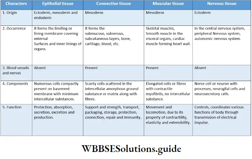
Morphology Of Flowering Plants Introduction
As we look around, we see different kinds of plants. They are of different forms and structures. Each of them has certain structural features that distinguish them from others. The branch of biology dealing with the study of these different forms and structural features of plants is called plant morphology [Greek: morphe = form and logos = discourse].
Importance Of Studying Plant Morphology:
- The study of morphology is important for understanding phytogeography, phylogeny, and evolution of plants.
- Plants are usually identified and classified on the basis of their morphological characters.
- Modification of different parts of a plant can be identified by comparing the morphological characters.
- It also helps in the study of plant breeding, genetics, crop production, etc. This chapter deals mainly with the morphology of angiosperms or flowering plants.
Read and Learn More: WBCHSE Notes for Class 11 Biology
Different Parts Of An Angiosperm
Angiosperm or flowering plants are the plants in which the seeds are embedded within the fruits. These types of plants appeared on Earth many years ago. These plants are most diverse and are found all over the world.
plant morphology
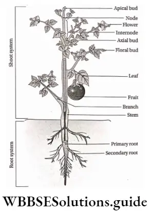
The body of an angiosperm is divided into two systems—the root system (the underground portion) comprising roots and their branches and the shoot system (the aerial or sub-aerial portion) comprising stem, branches, leaves, flowers, and fruits.
Root system: The root system usually consists of a main axis—the tap root, and its lateral branches—the branch roots. This system helps in water and mineral absorption from the soil. It also anchors the stem to the soil.
Morphology of flowering plants notes for NEET
Shoot system: The shoot system consists of the main axis of the plant body—the stem. It bears the branches and the leaves. This system helps in reproduction as it bears flowers and fruits. Shoot system also helps in the transportation of water and minerals, food production, gaseous exchange, transpiration, etc.
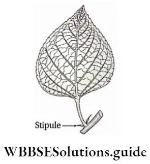
Stipules
Stipules Definition: The lateral outgrowths from both sides of the leaf base are known as stipules.
Stipules Location: Present mainly in pairs at the leaf base.
According to their presence or absence, leaves are of the following types—
Stipulate leaves: These leaves bear stipules. example china rose.
Exstipulate leaves: These leaves do not bear any stipule. example mango, Psidium guajava, etc.
morphology of angiosperms

Stipules Function:
- Stipules protect the axillary buds from mechanical injury at the young stage.
- They protect the leaves as bud scales.
- Photosynthesis occurs in foliaceous stipule.
- Spiny stipule provides protection to the plant.
- Tendrillar stipule helps the plants to climb.
Stipules Types: On the basis of life span, shape, location, and special functions, stipules are of different types. Those are discussed under separate heads.
According to life span
Caducous: The stipules that fall off before leaf maturation, are called caducous stipules. example Ficus benghalensis, Michelia champaca, etc.
Deciduous: The stipules that fall off soon after the maturation of the leaf, are called deciduous stipules. example Dillenia indica.
Persistent: The stipules that persist throughout the life span of a leaf, are called persistent stipules. example, Rose.
According to position
Free-lateral: When two distinct small stipules grow on two sides of the leaf base, it is called the free-lateral type. example china-rose.
Adnate: When the stipules grow and remain attached on both sides of the petiole up to a certain distance, then they are called adnate type. example Rosa centifolia (rose).
Morphology of flowering plants class 11 notes PDF
Intrapetiolar: When two stipules, occurring on both sides of opposite leaves, unite together by their inner margins and remain at the axils of leaves, then it is known as intrapetiolar stipule. example Paederia foetida and Gardenia jasminoides, etc.
Interpretiolar: Weakhen the stipules, occurring on both sides of opposite leaves, fuse along their outer margins, and remain on both sides of the stem between the petioles of opposite leaves, then it is known as interpetiolar stipule. Examples are Anthocephalus indicus, Ixora coccinia, Strychnos nux-vomica, etc.
Ochreate: In this type, two stipules emerge from a single leaf base and fuse along both of their margins to form a tube-like structure around the lower portion of the internode. Examples are Polygonum orientate, Rumex vesicarius, etc.
Modified Stipule
Sometimes the stipules are modified for various special functions.
Modifications in stipules are as follows—
Foliaceous: These stipules are modified into leaf-like structures that perform the functions of foliage leaves. example wild peas (Lathyrus aphaca) and peas (Pisum sativum).
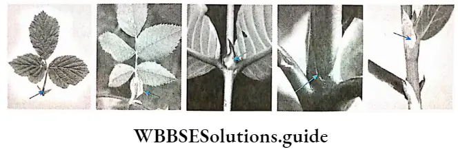
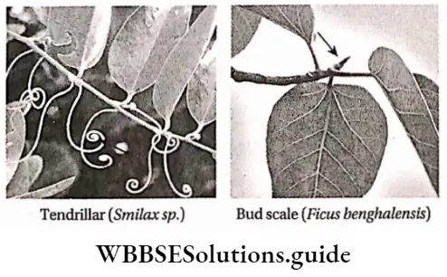
Tendrillar: These stipules are modified into tendrils and help in climbing. example Smilax macrophylla.
Spiny: These stipules are modified into spines and protect the plants from herbivorous animals. example Ziziphus mauritiana, Acacia arabica, etc.
Convolute or bud scales: These stipules are membranous and remain a protective covering of the buds. They fall off as the bud matures. example Ficus religiosa (peepal) and F. benghalensis (banyan), etc.
Winged: These stipules are modified into an expanded wing-like structure. example Crotalaria aiata.
Venation
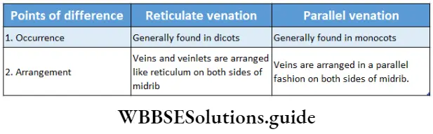
Venation Definition: The arrangement of veins in leaves is known as venation.
The arrangement of veins varies with plant species.
Usually, two types of venations are observed in leaves—
- Reticulate and
- parallel (striate).
Reticulate venation
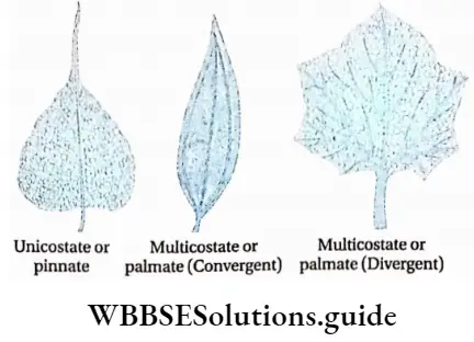
In this type of venation, the main vein or midrib of the leaf divides into numerous branches which again branch repeatedly to develop a network throughout the lamina.
Depending on the number of main veins, reticulate venation may be of two types
- Unicostate and
- Multicostate.
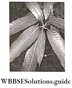
Unicostate or pinnate: In this type of venation, leaves bear one midrib. This runs centrally to the lamina and develops branches laterally resembling the pattern of a feather. These branches again form veinlets which form a network. example Artocarpus heterophyllus, Mangifera indica, etc.
morphology of plants
Multicostate or palmate: in this type of venation, leaves bear many main veins. These develop from the tip of the petiole and run either towards the apex or towards the margin of the lamina.
Venation is of two types—
- Convergent and
- Divergent.
Convergent type: In this type of venation, the main veins initially diverge out in different directions from the base of the lamina, and gradually converge at the apex. example Ziziphus mauritiana, cinnamon, etc.
Divergent type: In this type of venation, the main veins diverge in different directions from the base of the lamina towards the margin. example Cucurbita maxima, Gossypium herbaceum, etc.
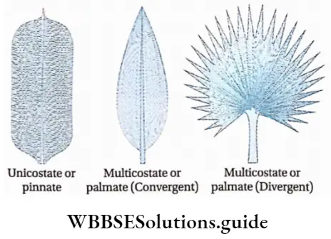
Parallel or striate venation
In some leaves, all the veins grow parallel to each other, either vertically or horizontally without forming any network. This type of arrangement of veins is known as parallel venation. Based on the number of mid veins,
It may be of two types—
- Unicostate and
- Multicostate,
Unicostate or pinnate: Leaves of some plants only bear a single midrib at the center of the lamina. Several parallel lateral branches arise from the midrib, on both sides. The lateral branches are perpendicular to the midrib. example Zingiber officinale, Canna indica, etc.
Multicostate or palmate: Leaves of some plants bear many major parallel veins. These develop from the base of the lamina and run either towards the margin or towards the apex.
It is further divided into two types—
- Convergent and
- Divergent.
Convergent type: In this type of venation, all the major veins run parallel to each other from the base of the lamina and converge at the apex of the lamina. example rice, bamboo, etc.
structure of a flowering plant
Divergent type: In this type of venation, all the major veins arise from the base of the lamina and diverge towards the margin in a parallel fashion. example Borassus flabellifer.
Types Of Leaves
Based on the origin, nature and incision of lamina leaves are of different types.
On the basis of nature and origin
Foliage leaves: These are the green, expanded leaves that perform physiological functions such as photosynthesis, respiration transpiration, etc. These are the lateral appendages of the aerial shoot growing at the nodes.
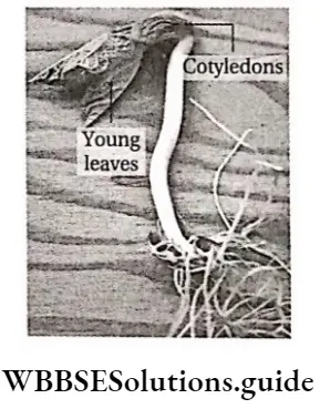
Seed leaves or cotyledons: The embryonic leaf, present within the seed, is known as seed leaf or cotyledon. Dicotyledonous plants contain two seed leaves and monocotyledonous plants contain only one seed leaf.
Morphology of flowering plants handwritten notes
Usually, cotyledons are swollen and fleshy due to the reserved food stored in them. During germination, they provide food to the growing embryo.
Scale leaves or cataphylls: These leaves are usually achlorophyllous, scaly or membranous and usually brown in color. In some cases, these leaves become fleshy due to food reserved in them. Sometimes they are chlorophyllous, such as, in aerial shoots of young bamboo, Casuarina, Asparagus, etc. The scale leaves are reduced forms of foliage leaves.
Prophylls: The first few leaves of a branch which modify into different structures other than the foliage leaves, are known as prophylls. Generally, prophylls are modified into one (orange) or two (wood apple) spines or tendrils (Cucurbita sp.).
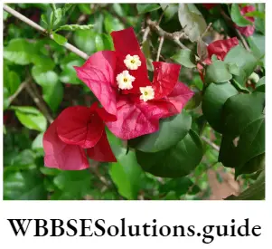
Bract leaves or hypsophylls: These are the reduced form of foliage leaves and bear floral buds at their axils. They can be colored or colorless. They may be leafy (Acalypha indica), petaloid (Bougainvillea spectabilis), spathe (Colocasia antiquorum), glume {Triticum aestivum), epicalyx i.e., present below calyx (Hibiscus rosa-sinensis), involucre or cupule (Quercus spicata, Betula bhojpatra).
Floral leaves: These are the specialised leaves of a typical flower which constitute sepals, petals, stamens, and carpels. They are discussed in detail while discussing the flower.
Homophyllous and heterophyllous plants
On the basis of the shape of leaves, plants are of two types—
Homophyllous plant: When a plant bears leaves of similar shape, then it is known as homophyllous plant. example Hibiscus rosa-sinensis.
Heterophyllous plant: When a plant bears leaves of more than one shape, it is known as a heterophyllous plant. example, Brassica campestris.
Sporophyll: Some leaves of gymnosperms and ferns, which bear spore-producing structures called sporangia, are known as sporophylls. They mainly produce and protect the sporangia.
On the basis incision of the lamina
On the basis of the incision of the lamina,
The leaves may be divided into two groups—
- Simple leaf and
- Compound leaf.
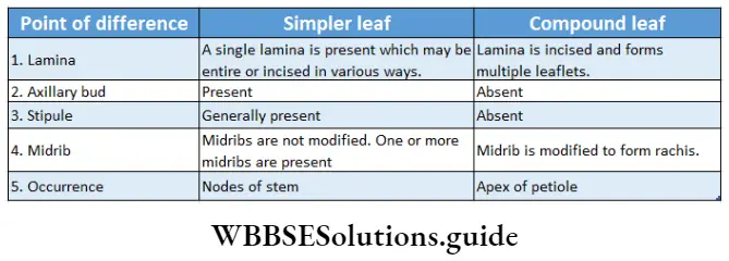
Simple Leaf
Simple leaf Definition: When a leaf is formed of a single lamina with usually entire or incised margin, but the incision never touches the mid-rib, the leaf is referred to as a simple leaf.
Simple leaf Characteristics:
- These leaves have entire or incised margins.
- The incision does not touch the midrib.
- They may have axillary bud in their axil and stipules at their base.
Simple leaf Types: On the basis of incision, simple leaves are of two types-
- Simple pinnate leaf and
- Simple palmate leaf
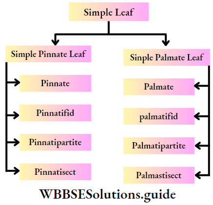
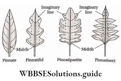
Simple pinnate leaf: In this leaf, the direction of the incision is towards the midrib. According to the extent of incision they are of different types.
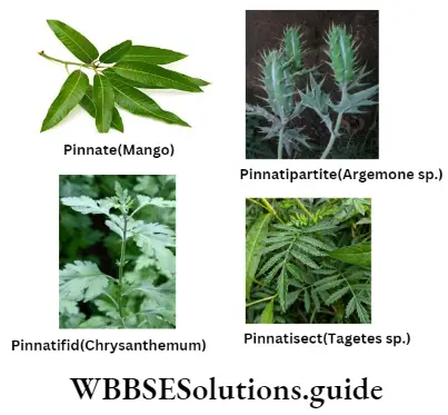
- Pinnate: In this type, the leaf blade is entire, i.e., the leaf does not have an incision. example mango leaves.
- Pinnatifid: In this type, the incision extends halfway towards the midrib. exampleChrysanthemum coronarium.
- Pinnatipartite: In this type, the incision extends more than halfway towards the midrib. example, Argemone mexicana.
- Pinnatisect: In this type, the incisions almost touch the midrib but the lamina is not separated into leaflets. Example Tagetes patula.
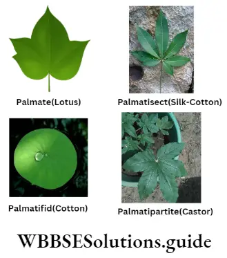
Simple palmate leaf: In this leaf, the incision is directed towards the petiole from the margins. According to the extent of incision they are of different types.
Palmatifid: In this type, the incision extends halfway towards the petiole from the margins. example Gossypium herbaceum.
Palmatipartite: In this type, the incision extends more than halfway towards the petiole from the margins. Example Ricinus communis.
Palmatisect: In this type, the incision almost touches the tip of the petiole. example Ipomoea paniculata.
Compound Leaf
Compound Leaf Definition: When the lamina of a leaf becomes completely incised and the incision reaches up to the midrib or petiole forming separate leaflets, then the leaf is known as a compound leaf.
Compound Leaf Characteristics:
- these leaves do not have any apex or apical buds.
- they have axillary buds in their axil and stipules at their base.
- leaflets do not have any axillary bud or stipule.
Compound Leaf Types: According to the arrangement of leaflets,
Compound leaves are of two types—
- Pinnate compound leaf and
- Palmate compound leaf.
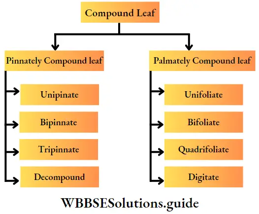
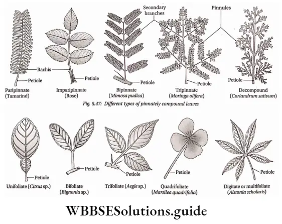
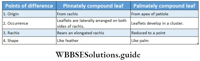
1. Pinnately compound leaf: In this type, the incision of the lamina extends towards the rachis (main midrib), and the leaflets are arranged on both sides of it or on its branches.
On the basis of the arrangement of leaflets on the rachis, leaves are of four types—
- Unipinnate: In this type, the leaflets are directly attached on both sides of the rachis, as in feathers.
It is of two types—
- Paripinnate: In this type, the leaflets are arranged in pairs on opposite sides of the rachis. The terminal end of the rachis contains an even number of leaflets. example, Tamarindus indicus.
- Imparipinnate: In this type, the leaflets are arranged in pairs on the rachis, and the terminal end of the rachis contains an odd or unpaired leaflet. example Azadirachta indica.
- Bipinnate: In this type, the rachis gives rise to secondary branches laterally. The leaflets are arranged on both sides of these secondary branches. These leaflets are known as pinnules. example , Mimosa pudica, Caesalpinia pulcherrima.
- Tripinnate: In this type, the secondary branches of rachis further divide to produce tertiary branches, The leaflets are laterally arranged on these tertiary branches. example Moringa oleifera, Oroxylon sp.
- Decompound: In this type, the tertiary branches are again branched in an indefinite manner. The J branches become flat and the pinnules become highly suppressed. example, Daucus carota, and Coriandrum sativum.
2. Palmately compound leaf: In this type of leaf, the incision of the leaf lamina extends towards the petiole. As a result, all leaflets seem to be attached to the apex of the petiole.
It does not consist of any rachis and may be of the following five types—
- Unifoliate: In this type, only one leaflet is attached to the apex of the petiole. example lemon, orange.
- Bifoliate or bipinnate: In this type, two leaflets are attached to the apex of the petiole. example of Bignonia grandiflora.
- Trifoliate or ternate: In this type, three leaflets are attached to the apex of the petiole. example Vitex negundo.
- Quadrifoliate or quadrinate: In this type, four leaflets are attached to the apex of the petiole. example, Marsilea quadrifolia.
- Multifoliate or digitate: In this type, more than four leaflets are attached to the tip of the petiole. example Bombax ceiba.
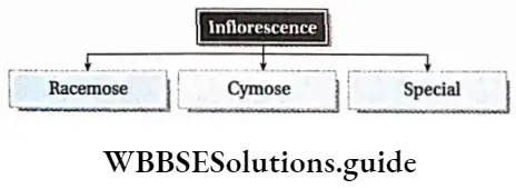
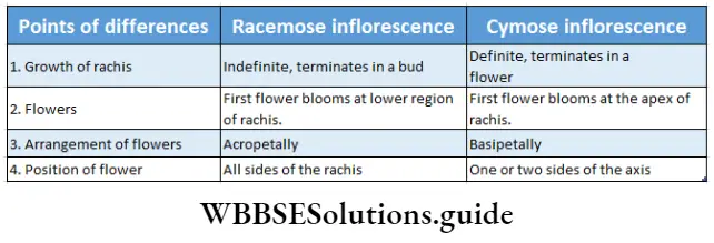
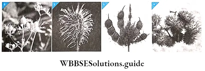
Racemose (Indefinite Or Ndeterminate) Inflorescence
Racemose Definition: The inflorescence, where the rachis grows indefinitely by producing lateral flowers acropetally or centripetally is known as racemose inflorescence.
Racemose Characteristics:
- The floral axis increases indefinitely in length.
- It is terminated by a bud.
- Sessile or stalked flowers are borne acropetally or centripetally on the floral axis. It means the mature flowers remain at the lower region of the axis and immature flowers at the upper region.
- Sometimes the rachis is condensed and develops into a round structure known as a receptacle. The flowers open from periphery J to center, centripetally.
Racemose Types:
The racemose inflorescence is divided into the following groups—
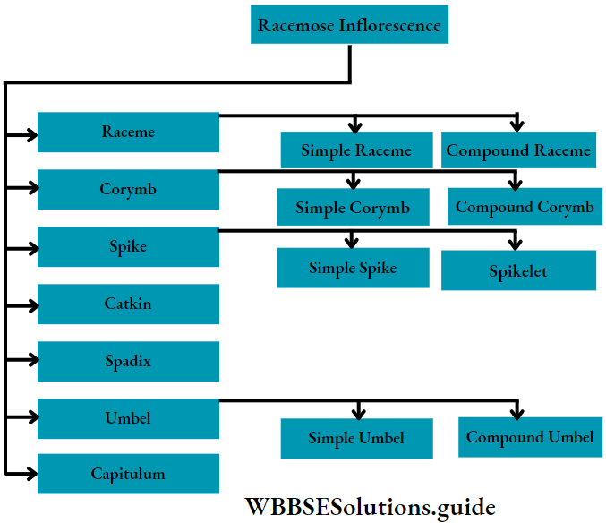
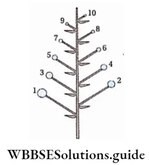
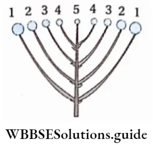
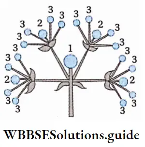
Raceme: In this type, the main axis or the rachis grows indefinitely, and bears pedicellate flowers. The flowers grow acropetally on the axis. example Raphanus sativus and Brassica nigra.
Raceme is of two types—
- Simple raceme and
- In compound raceme or panicle, compound raceme, the floral axis is branched and each branch appears like a simple raceme.
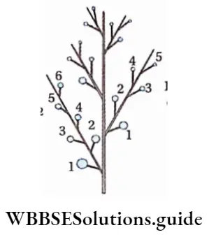
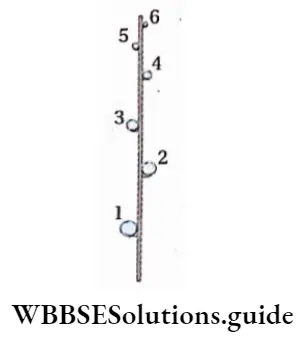
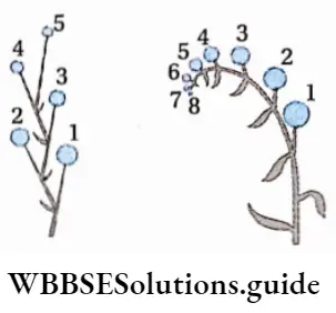
(Numbers in the figures given indicate the gradual growth of the flowers. For example, the flower which is numbered as 1 grows first followed by 2, and so on.)
Corymb: In this type, the floral pedicels or stalk are unequal in length. (The main floral axis is shorter than the axis of the basal flowers. All flowers grow almost at the same plane.) The flowers grow centripetally. example Prunus cerasus (cherry), Cassia sophera, Iberis amara.
Corymb is of two types—
- Simple or unbranched and
- Compound or branched. In compound corymb, the floral axis is branched and each branch appears like a simple corymb.
Spike: In this type, the floral axis grows indefinitely and bears sessile flowers. The flowers grow acropetally on the rachis and are bracteate. example Piper longum, Achyranthes aspera, Adhatoda vasica, etc.
Spike is of two types—
- Simple or unbranched and
- Compound or branched or spikelet. In compound spike, the floral axis is branched and each branch appears like a simple spike.

Umbel: In this type, the main floral axis is much reduced and the pedicels of all flowers are of equal length. The flowers grow centripetally.
Flowers are usually bracteate and the bracts unite to form an involucre (covering) at the base of the pedicellate flowers. example Centella Asiatica and Coriandrum sativum of Apiaceae, Prunus cerasus during the young stage.
Umbel is of two types—
- Simple and
- Compound In a compound umbel, the floral axis is branched and each branch appears like a simple umbel.
Spikelet or Locusta
It is a small spike with one or more flowers on the rachilla (secondary rachis). Usually, many flowers are borne on each inflorescence as in Triticum aestivum but in Oryza sativa, it has a single flower. In Zea mays, the male inflorescence bears a spikelet of two flowers.
In grasses like Panicum sp., two scaly bracts are present at the base of the entire inflorescence. These are known as glumes or empty glumes. Above them, there are one or more fertile glumes. These are known as the flowering glumes or lemmas.
Each lemma contains a single sessile flower in its axil, opposite to which a small glume is present, known as palea.
Catkin: In this type, the sessile, unisexual flowers grow acropetally on a pendulous peduncle. It is a modified compact spike. Example Acalypha hispida.

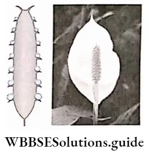
Spadix: It isCatkina special type of spikeFig. with fleshy rachis having both male and female flowers. The whole inflorescence is surrounded by a large bract called spathe. The spathe is absent in some cases such as in the Acorus calamus.
The female flowers are always borne towards the base of the rachis whereas the male flowers are towards the apex. This inflorescence also bears sterile flowers which are present in between the male and female flowers.
Best notes on morphology of flowering plants
The terminal region does not bear any flowers and is infertile. This region is termed an appendix. example Coiocasia antiquorum.
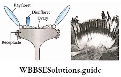
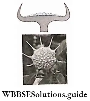
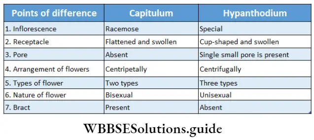
Capltulum: In this type, the small sessile flowers grow centripetally on the modified thick, fleshy, and flattened rachis, known as the receptacle.
Mostly two types of flowers are found in the capitulum—
- Ray florets on the margin of the receptacle and
- Disc floret in the center. Each floret is covered with green-colored scaly bracteoles. The whole inflorescence is surrounded by a cover of bracts. The disc florets are bisexual whereas the ray florets are sterile. example Helianthus annuus.
Cymose Inflorescence
Cymose Inflorescence Definition: The inflorescence, in which the rachis or peduncle grows up to a definite point and is terminated with a flower, is known as cymose inflorescence.
Cymose Inflorescence Characteristics:
- The growth of the floral axis is limited.
- The first flower grows at the tip of the axis, thus pausing the growth of the peduncle.
- The flowers are borne basipetally or centrifugally on the axis.
Cymose Inflorescence Types: Cymose inflorescence is of four types.
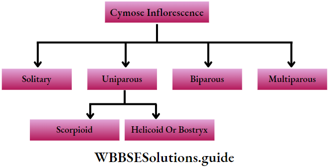
Solitary: In this type, the terminal or apical bud grows into a single flower. E.g. Hibiscus rosa-sinensis.

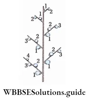
Uniparous or dichasial cyme: In this type, the main floral axis is terminated by a single flower. The main axis gives rise to a single lateral branch which is also terminated by a single flower.
The other lateral branches grow in the same manner. This type is again divided into the following groups—Scorpioid cyme and helicoid cyme or bostryx.
- Scorpioid cyme: In this type, the lateral branches bearing flowers grow alternately on both sides of the main axis forming a zigzag structure. example Hamelia patens, and Commelina benghalensis.
- Helicoid cyme or bostryx: In this type, the lateral branches bearing flowers grow successively on the same side forming a curved structure. example, Heliotropium indicum, and Ranunculus bulbosus.
Biparous or monochasial cyme: In this type, the main floral axis is terminated by the flower. Two lateral branches develop from the same point of the main axis in opposite directions. They are also terminated by flowers. This process continues. example Jasminum sp., Clerodendrum infortunatum.
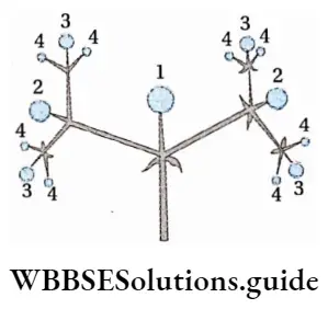
Multiparous or polycrystal cyme: In this type, the main axis is terminated by a flower and two or more lateral branches develop from the main axis. They are also terminated by flowers.
The lateral branches also behave like the main axis and the branching process continues. Examples are Caiotropis procera, and Carissa carandas.
Special Inflorescence
Special Inflorescence Definition: The highly modified and condensed cymose inflorescence.
Special Inflorescence Types: they are of the following types
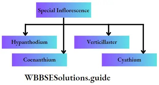
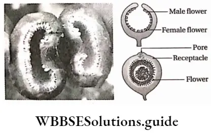
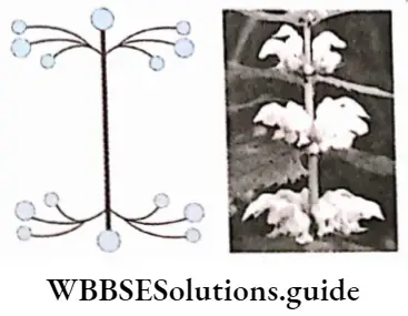
Hypanthodium
Hypanthodium Characteristics:
- In this type, the main axis condensed to form a hollow, fleshy, cup-like receptacle,
- The receptacle has an apical pore (ostiole) which is guarded by scales.
- The cup-like cavity of this inflorescence bears three types of flowers—male flowers in the apical region, female flowers in the basal region, and neutral sterile flowers, in between male and female flowers. The flowers grow on the margins of the receptacles. example Ficus benghalensis, Ficus hispida.
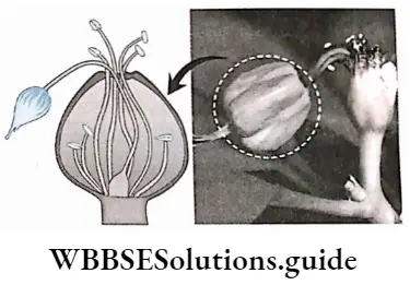
Cyathium
Cyathium Characteristics:
- The floral axis condenses and forms a conical receptacle.
- There is only one female flower in the central part of the receptacle and it hangs down due to the long pedicel.
- This flower is represented by a single pistil.
- Surrounding the female flower, several male flowers grow centrifugally on the receptacle.
- Male flowers bear a single stamen.
- A bright-colored involucre surrounds the whole floral arrangement on the receptacle. Thus, it appears as a single flower. example Poinsettia pulcherrima, and Pedilanthus tithymaloides.
Verticillaster
Verticillaster Characteristics:
- It is a condensed, biparous cymose inflorescence.
- This inflorescence occurs at the axil of two opposite decussate leaves surrounding the stem.
- At first, the inflorescence is dichasial cyme and then each branch of dichasium is reduced to scorpioid cyme.
- Here, the sessile and bilabiate flowers develop in clusters around the stems. example Leonurus sibiricus.
Coenanthium
Coenanthium Characteristics:
- This is like the hypanthodium but here the floral axis becomes a saucer-shaped receptacle, with slightly curved margins.
- The small flowers are arranged with cymose inflorescence on the receptacles. For example, found in Dorstenia sp.
The Flower
The flower is the main organ for sexual reproduction in angiosperms. The axillary or apical bud, from which the flower develops is known as the floral bud. Flower is a modified shoot.
Fruits and seeds both develop from flowers. So it is also known as the reproductive shoot. It is mainly composed of four parts—corolla, calyx, androecium and gynoecium.
Morphology of flowering plants short notes
The Flower Definition: The highly condensed and modified shoot responsible for reproduction is known as a flower.
Morphological characteristics of a flower:
- The flower has definite growth.
- This modified shoot is a temporary part of the plant.
- Flowers grow at the shoot apex or axils of the leaves or bract.
- A typical flower grows on a stalk called a pedicel.
- The swollen upper part of the pedicel is known as the thalamus. The thalamus is also differentiated into nodes and internodes. The internodes are highly condensed, thus the nodes are packed closely together.
- On the thalamus, the floral parts remain arranged in four concentric whorls.
- The outer two whorls, i.e., calyx and corolla, are the accessory whorls. The inner two whorls, i.e., androecium and gynoecium are the essential or reproductive whorls.
Different Parts Of A Typical Flower And Their Functions
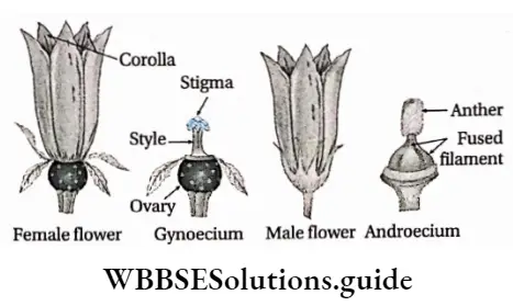
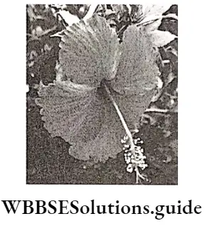
The flower that bears all the floral parts, i.e., pedicel, thalamus, corolla, calyx, androecium, and gynoecium with their proper functions is known as a typical flower. example, Hibiscus rosa-sinensis.
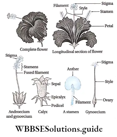
Pedicel: The green-colored stalk below the flowers, which bears the thalamus is known as the pedicel. The flowers with pedicel are known as pedicellate flowers (Example Hibiscus -sp.) and the flowers without pedicel are known as sessile flowers (Example Tube rose).
Pedicel Function: It connects the flower with the stem.
Thalamus: The disc-like, flattened structure above the pedicel, which bears all the floral whorls in concentric rings is known as the thalamus. It is also called the torus axis or receptacle. Usually, it is convex or concave and often it is very short or condensed. Sometimes it becomes elongated (axis) and shows distinct internodes.
Thalamus Function: It bears different floral whorls.
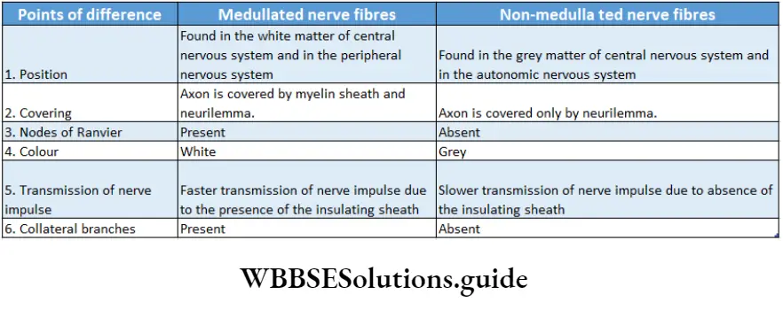
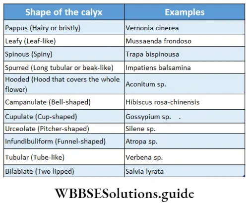
Calyx
Calyx Definition: A generally green-colored lowermost or outermost whorl of the flower, formed of sepals is known as calyx.
Calyx Characteristics:
- It is the first and outermost floral whorl on the thalamus.
- The individual member of the calyx is known as a sepal.
- The sepals may be free i.e., polysepalous, or may be partially or completely united i.e., gamosepalous.
- Generally, sepals are sessile and green, but sometimes they are of different colors. The number of sepals in the whorl is variable.
Calyx Function:
- It protects the other whorls in their bud stage.
- Green calyx takes part in photosynthesis.
- Colored calyx, helps in reproduction by attracting insects for pollination.
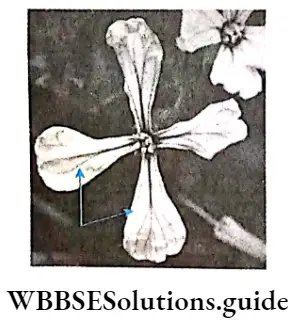
Corolla
Corolla Definition: The white or bright-colored, second whorl of the flower, formed of petals is known as a corolla.
Corolla Characteristic:
- It is the second whorl from the outside, situated above the calyx.
- The individual member of this whorl is known as the petal.
- The petals are mostly bright in color or white and have a sweet smell.
Corolla Function:
- Either with their bright color or smell, petals attract insects for pollination.
- They protect the essential reproductive whorls i.e., the androecium and the gynoecium.
- Usually, the base of the petals contains, nectaries(the nectar-secreting glands).
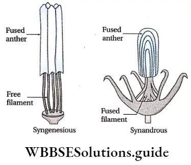
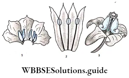
Androecium
Androecium Definition: Androecium is the essential, male reproductive whorls formed of one or more stamens.
Androecium Characteristic:
- It is the first essential and third whorl of the flower.
- The structural unit of the androecium is the stamen. They are placed on the thalamus either in a cyclic or in a spiral fashion.
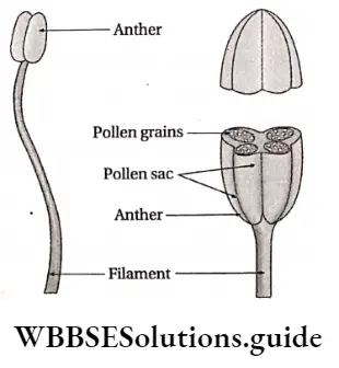
- A stamen appears quite different from sepals and petals.
- Stamen is generally composed of an elongated narrow stalk called a filament. A sac-like structure, called anther, is present at the tip of the filament.
- Each anther usually consists of two anther lobes connected by connective formed by the extension of the filament.
- Each anther lobe has two chambers, called pollen sacs or microsporangia.
- Each stamen, therefore, consists of four microsporangia and each microsporangia contains a large number of pollen grains or microspores.
Androecium Function:
- Stamens produce pollen grains (male gametophytes) inside the anther lobes.
- They help in fertilization.
Gynoecium or pistil
Gynoecium or pistil Definition: The essential, female reproductive whorl of flower that lies innermost and terminates the thalamus.
Gynoecium or pistil Characteristic:
- It is the second essential and fourth whorl of the flower.
- The gynoecium, also called the pistil, is the most essential reproductive whorl of the flower. The sterile pistil or gynoecium is known as pistillode.
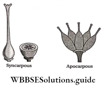
- The gynoecium consists of one or more carpels, which is the structural unit of the gynoecium.
- Each carpel consists of three parts—ovary, style, and stigma.
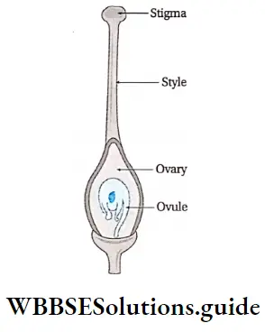
1. Ovary: It is the lowermost swollen, pitcher-like part of the gynoecium. It contains ovules (structures that later form the seeds) inside. It may be made up of single (monocapellary) or many (polycapellary) carpels. It may be of the following types.
2. Style: It is the slender and elongated stalk-like structure formed by gradual tapering of the tip of the ovary. Style development may be apical or terminal (For example Adhatoda vasica), lateral (For example, Mangifera indica), or basal (gynobasic—for example, Ocimum sanctum).
3. Stigma: It is the extreme tip of the style, which receives the pollen grains during pollination. The shape of stigma varies among species.
Stigma Function:
- Carpels produce female gametophytes.
- After fertilization embryo is produced the ovary matures to form fruits and seeds.
Perianth
In some flowers, the accessory floral whorls can not be differentiated into calyx and corolla. Such an accessory floral whorl is known as perianth. example Polyanthes tuberosa etc. The individual member of the perianth is known as tepal.
Again in some flowers, only one set of these accessory whorls are present. These members of the perianth can be like sepal (sepaloid), for example, Borassus flabellifer and Cocos nucifera, etc., or like petal (petaloid), for example, Michelia champaca, etc.
The flower that contains only one accessory whorl, i.e., either calyx or corolla or perianth, is called monochlamydeous or haplochlamydeous. example Polyanthes tuberosa. Ordinary flowers with both calyx and corolla are called dichlamydeous. example Pisum sativum etc.
Flower is a modified shoot
The vegetative shoot is composed of an elongated stem differentiated into nodes and internodes with leaves arranged at the nodes. The flower is morphologically similar to the shoot.
It has been modified for similar to the shoot. It has been modified for parts in different flowers proving that, flowers are the modified shoots.
The modifications of various parts are as follows
- Axis nature of thalamus,
- The leaf-like nature of the floral members and
- Homology of floral buds.
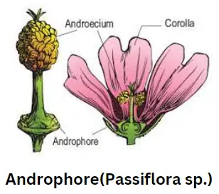
Axis nature of thalamus: Each flower bears a condensed axis—thalamus, on which the floral leaves remain arranged in concentric whorls. In some exceptional cases, it gets modified into an elongated axis and shows the stem characters.
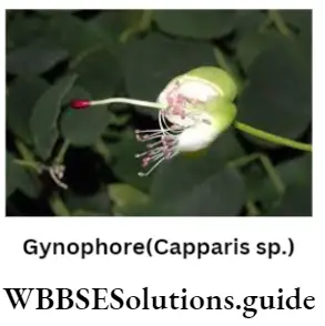
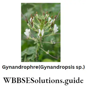
1. The floral leaves develop from the nodes. The elongated internodal region between petal and androecium is referred to as androphore, as seen in Passiflora suberosa.
The elongated axis between the androecium and gynoecium is referred to as gynophore as in Capparis septaria. Gynandropsis gynandra contains gynandrophore, i.e., both androphore and gynophore.
Anthophore is produced due to the elongation of the internode between the calyx and corolla and is found in Sielene sp.

2. The growth of the thalamus usually stops and is terminated by the gynoecium. But sometimes the thalamus develops beyond the gynoecium bearing either a leafy vegetative shoot or a flower above the first one. This growth is known as proliferation or monstrous development. example Found in pears, roses, etc.
3. In some plants, after fertilization the thalamus elongates like an ordinary stem and gives rise to an aggregate fruit. example Michelia champaca and Polyalthia longifolia.
Leaf nature of the floral members: The sepals, petals, stamens, and carpels are the modified leaves. They exhibit the same type of ptyxis and aestivation.
These may be proved from various instances, which are as follows
Gradual transition of floral members: The floral leaves are spirally arranged on the thalamus. The outermost sepals are green with distinct venations and they gradually transform into petals.
The petals gradually become narrow bearing anther at the tip and then are transformed into a typical stamen. It is commonly found in water lily, Nymphaea sp.
Modification of sepal into leaf: In Mussaenda frondosa, out of five sepals one remains as the leaf with prominent venation, but instead of being green, it is colored like petals.
In Mussaenda philippica, all five sepals maintain leaf-like structures and have prominent venations but are colored.
Transformation of leaf to petal: In Paeonia officinalis, a gradual transition of leaves to sepals and sepals to petals can be observed, supporting the leafy nature of perianths.


Leafy petals: In some flowers, sepals, and petals appear as foliage leaves, as in green roses.
Petaloid stamen: In Canna indica, the stamens become petaloid staminodes. However, in some cases, a part of the anther lobe becomes petaloid and the other part remains fertile.
Petaloid and sepaloid carpel: In Zinnia sp., the carpels become petaloid or sepaloid.
Leafy nature of carpel: In Pisum sativum, the gynoecium is formed by the folding of a single leaf along its midrib. The leaf develops a single-chambered ovary containing seeds. The upper elongated part of the leaf develops into the style and its apex forms the stigma.
Homology of floral buds: Floral buds are homologous to some organs. In some cases, the floral buds get transformed into vegetative buds or bulbils. example Agave sp., Allium sativum, Globba bulbifera, etc. Floral buds occupy the terminal or axillary positions, like the vegetative buds.
Types Of Flower
Flowers are of different types based on different characteristics of their whorls.
Classification of flowers based on the presence or absence of the floral whorls:
Flowers based on the presence or absence of floral wholes are of the following types.
Complete flower: The flower that bears all the four floral whorls i.e., calyx, corolla, androecium, and gynoecium, is known as a complete flower. example Datura metel.
Incomplete flower: A flower that lacks one or more floral whorls is known as an incomplete flower. example Polianthes tuberosa
Naked or achlamydeous flower: The flowers which bear either androecium or gynoecium, or both but do not have calyx and corolla, are known as naked or achlamydeous flowers. example Beta vulgaris.
Classification of flowers on the basis of the absence or presence of essential whorls: Flowers on the basis of presence or absence of essential wholes are of the following types.
Bisexual (perfect or hermaphrodite or monoclinous) flower: The flower that bears both the essential whorls i.e. androecium and gynoecium, is known as bisexual flower. Common examples of bisexual flowers are china-rose, mustard, etc.
Morphology of flowering plants revision notes
Unisexual (imperfect or diclinous) flower: The flower which bears only one essential whorl i.e. either androecium or gynoecium, is known as an unisexual flower.
Thus they are called—pistillate or female flower and staminate or male flower. In Cucurbita maxima, the flower is incomplete and unisexual due to the absence of either androecium or gynoecium.
Sterile flower: The flower in which both the androecium and gynoecium are either absent or non-functional, is known as a sterile or neutered flower. example Amorphophallus companulatus.
Classification of flowers on the basis of symmetry: Flowers on the basis of symmetry are of the following types.
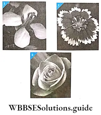
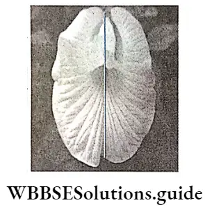
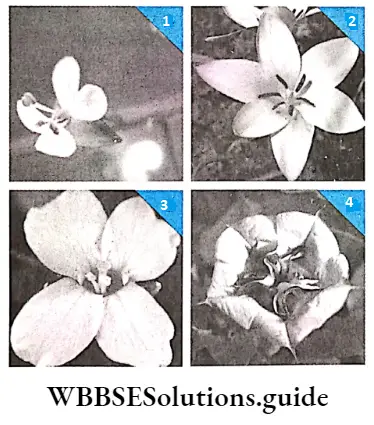
Actinomorphic or regular flower: The flower that can be divided into two equal and symmetrical halves if cut through any vertical plane passing through the axis, is known as actinomorphic or regular flower. example Vinca rosea, china rose.
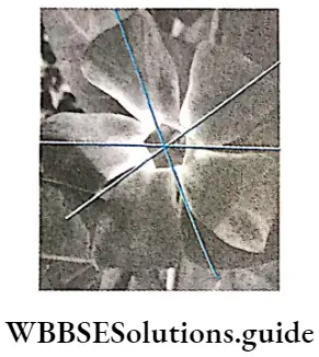
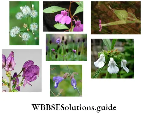
Zygomorphic or irregular) flower: The flower that is equally and symmetrically divisible only through a single vertical plane passing through the axis, is known as a zygomorphic or irregular flower. example Pisum sativum, Clitoria ternatea, etc.
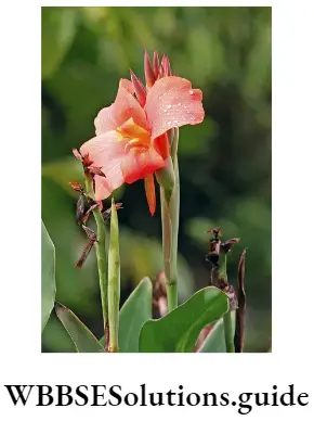
Asymmetrical flower: There are some flowers, that can not be divided into two equal halves through any vertical plane are known as asymmetrical flowers. example Canna indica.
Classification of flowers on the basis of arrangement of floral whorls on the thalamus:
Flowers on the basis of the arrangement of floral whorls on the thalamus are of the following types.
Cyclic flower: When the sepals, petals, stamens, and carpels are arranged on the thalamus in separate whorls, then the flower is termed a cyclic flower. Most angiosperms have cyclic flowers. example Hibiscus rosa-chinensis (china rose), Brassica nigra, etc.
Acyclic flower: When all the floral leaves of a flower are spirally arranged on the thalamus but not in distinct whorls, then it is termed an acyclic flower. example Nelumbo nucifera, Michelia champaca, etc.
Spirocyclic (hemicyclic flower: When some floral leaves are arranged in whorls and some are arranged spirally, then the flower is called a spirocyclic or hemicyclic flower. example, Nymphaea stellata, rose, etc.

Classification of flowers on the basis of number of the members in floral whorls: Flowers on the basis of number of the members in floral whorls are of the following types.
Isomerous flower: In this type of flower, the number of sepals, petals, stamens, and carpels are of the same number or present in multiples of the same number. It may be of the following types.
- Bimerous flower: The number of floral members in each whorl is two or multiple. example Circaea lutetiana.
- Trimerous flower: The number of floral members in each whorl is three or multiple. example Tulipa clusiana, Annona squamosa, etc. Monocotyledonous flowers are mostly trimerous.
- Tetramerous flower: The number of floral members in each whorl is four or multiple.example radish and mustard, Gynandropsis gynandra, etc.
- Pentamerous flower: The number of floral members in each whorl is five or multiple. It is commonly found in dicotyledons. example Hibiscus rosa-sinensis.
Heteromerous flower: The number of floral leaves of different whorls is not the same. example Beilis perennis.

Classification of flowers on the basis of presence or absence of bract: Flowers on the basis of a number of presence or absence of bract are of the following types.
Bracteate flower: The flower which arises from the axil of a bract is known as a bracteate flower. example Clitoria ternatea.
Ebracteate flower: The flower that does not arise from any bract is known as an ebracteate flower.example Mangifera indica.
Classification of flowers on the basis of insertion of floral leaves on the thalamus in respect to the ovary: Flowers on the basis of a number of insertion of floral leaves on the thalamus in respect to the ovary are of the following types.
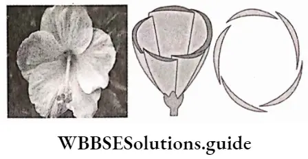
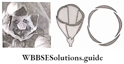
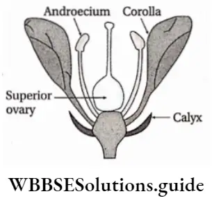
Hypogynous flower: The ovary (gynoecium) is seated at the uppermost position on the convex or conical thalamus. The other whorls arise below the ovary. Thus, the thalamus is present below the gynoecium. Hence, the ovary is superior and all other floral members are inferior, by position example Hibiscus rosa-sinensis.
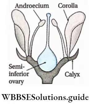
Perigynous flower: The thalamus is cup-shaped or concave, and the ovary remains in the center of the cup. The other floral whorls remain attached to the rim of the cup-shaped thalamus.
The ovary is superior by position in this flower. Sometimes the ovary is said to be half inferior instead of inferior. example pea, rose, Portulaca oleracea, etc.
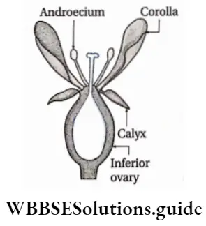
Epigynous flower: The deep concave cup-like thalamus completely encloses the ovary and fuses with the ovary wall. Here, the other floral whorls remain above the ovary. The ovary in such cases is inferior and the rest of the floral members are superior. Common examples are sunflower, pumpkin, etc.
Classification of plants, on the basis of the presence of male and female flowers
Plants on the basis of the presence of male and female flowers are of the following types.
- Monoecious plant: When both the male (staminate) and female (pistillate) flowers are borne on the same plant, then the plant is known as a monoecious plant. example Trichosanthes dioica and Cucurbita maxima.
- Dioecious plant: When male and female flowers are borne on different plants, then such plants are known as dioecious plants. example Borassus flabellifer (palm) and Carica papaya.
- Trioecious plant: If the male, female, and bisexual flowers are borne on different plants, then they are known as trioecious plants. example Silene sp.
- Polygamous plant: If unisexual, bisexual, and sterile flowers are borne on the same plant, then the plant is known as a polygamous plant. example Mangifera indica.
Placentation
The ridge of soft parenchymatous tissue on which the ovules grow by means of funicles (stalk) is known as the placenta.
Placentation Definition: The arrangement of the placenta bearing the ovules inside the ovary is known as placentation.
Types of placentation
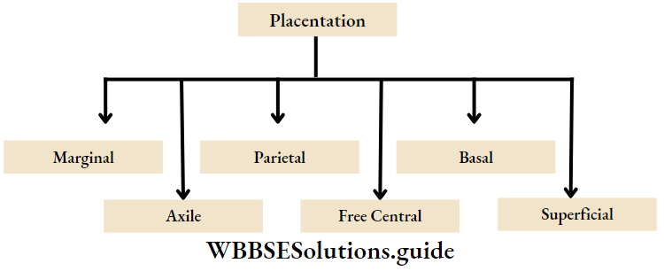
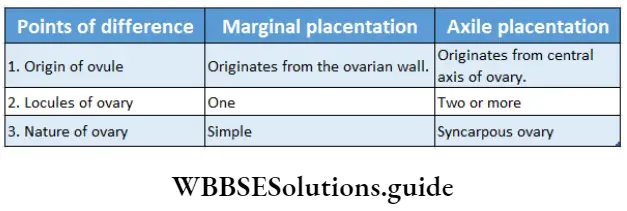
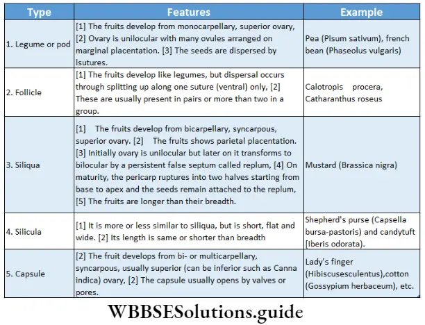
Marginal: This type of placentation is found in the monocarpellary, and unilocular ovary. The placenta develops on one side of the ovary. Ovules are present at the margins of the carpel in one or two rows. Example Dolichos lablab.
Axile: This type of placentation is found in polycarpellary, and multilocular ovary. The carpels remain joined together to form an axis. The placenta with ovules develops around this axis. Example Citrus limon.
Parietal: This type of placentation is found in the polycarpellary, and unilocular ovary. Several false partition walls are formed in the ovary due to the fusion of carpels, known as replum. The placenta occurs along the wall of the ovary. Example Brassica nigra.
Morphology of flowering plants notes with diagrams
Free central: This type of placentation is found in polycarpellary, and unilocular ovary. The placenta with ovules develops on the central axis. Example Scoparia dulcis.
Basal: This type of placentation is found in the monocarpellary, and unilocular ovary. The placenta grows at the base of the ovary. Example Helianthus annuus.
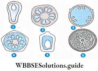
Superficial: This type of placentation is found in the polycarpellary, and multilocular ovary. The placenta with ovules is present all around the inner wall of the ovary. Example Nymphaea sp.
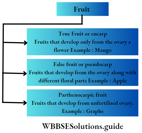
The Fruit
After fertilization, the floral parts, except the ovary, dry up and fall off. The ovary enlarges and the ovules get modified into seeds. This enlarged ovary with seeds is the fruit. Besides this, fruits may also develop from a whole inflorescence. Sometimes different parts of a flower may also develop into fruits.
The Fruit Types:
Fruits can be of various types—
Structure Of A True Fruit
A true fruit consists of mainly two parts
- Seeds and
- Pericarp.
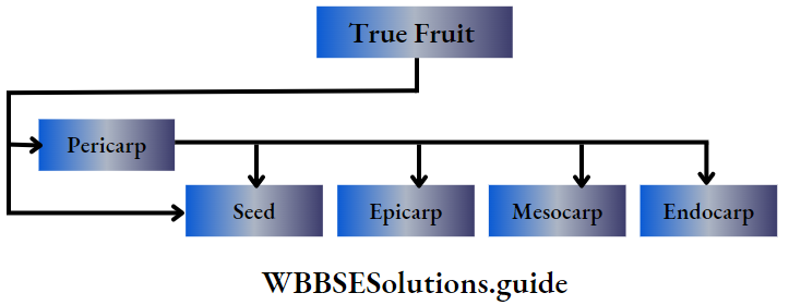
Pericarp: The pericarp can be thin or thick and fleshy or dry.
A well-developed pericarp is differentiated into three layers—
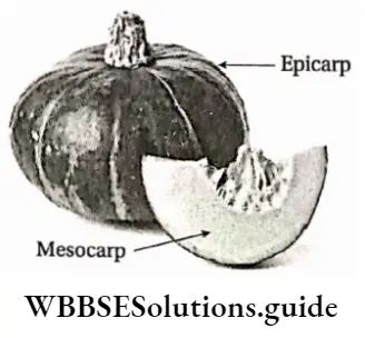
Epicarp or exocarp: It is the outermost thin layer of the fruit, i.e., the skin of the fruit.
Mesocarp: It is the thick, fibrous, or fleshy middle layer of the fruit. It is present just below the epicarp and forms the pulp. In most of the fruits, this part is edible.
Endocarp: It is the innermost layer of the fruit and encloses seeds or seeds. It may be membranous or hard.
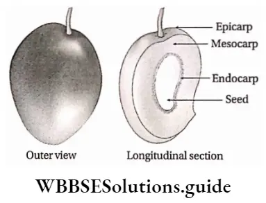
Seed: Fruits contain one or more seeds. After fertilization, the ovules are transformed into seeds. The seed coat may remain attached or separated from the inner wall of the fruit.
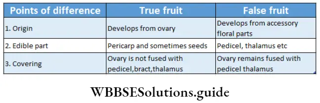
Structure of a true Function of fruit: it protects the seeds and helps in seed dispersal.
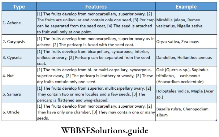
Classification Of Fruits
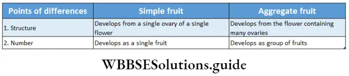
Based on origin, texture, and dehiscence, fruits are mainly of three types—
- Simple fruit,
- Aggregate fruit and
- Composite or compound or multiple fruit.
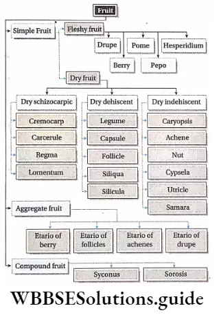
Simple fruits
The fruit, which develops from the ovary of a solitary pistil is known as a simple fruit. Example Pisum sativum (pea), and Oryza sativa (rice).
These types of fruits are further classified into two categories—
- Dry fruits and
- Succulent or fleshy fruits.
Dry fruits: The fruit in which the pericarp is simple, dry, and cannot be differentiated into three layers, i.e.,ectocarp, mesocarp, and endocarp, is known as dry fruit.
The dry fruits are further divided into the following three types
- Dry dehiscent fruit,
- Dry indehiscent three and
- Dry schizocarpic fruit.
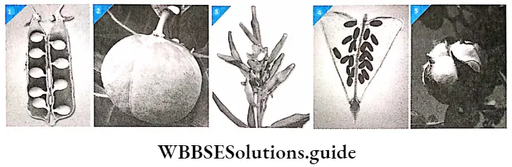
Dry dehiscent fruits: In this type of fruit, the pericarp ruptures at maturity, and then the seeds are dispersed. These fruits contain numerous seeds. These fruits are further divided into five types.
Dry indehiscent fruits: In this type of fruit, the pericarp does not rupture even after maturity or ripening. The seeds remain inside the fruits. These fruits mostly contain single seed. These fruits can be divided into six types as given in the.
Morphology of flowering plants chapter summary
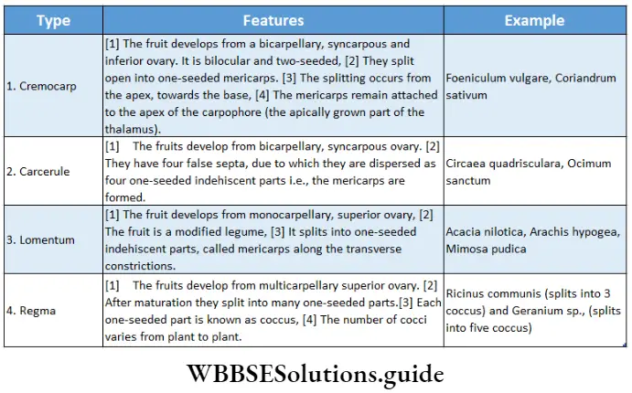
Dry schizocarpic fruits (splitting fruits): In this type, the ripe fruits are divided into two or more indehiscent segments. These segments are called mericarps. Each mericarp contains only one seed. These fruits are divided into four types as given in the.
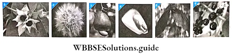
Succulent or fleshy fruits: These fruits become succulent and juicy after ripening. The pericarp of the fruit is differentiated into three layers—epicarp, mesocarp, and endocarp.
They have thick, fleshy, or fibrous mesocarp. The fruits are indehiscent. Hence, the seeds are released only after the decay of the fleshy tissue enclosing them.
These fruits are of the following types—
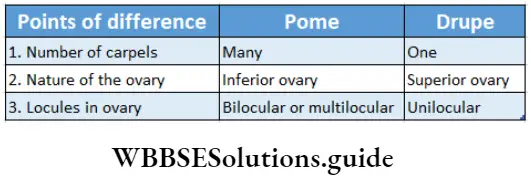
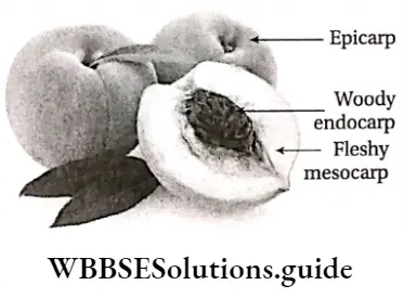
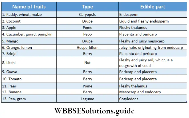
Drupe (Stone fruit): The fruits develop from the monocarpellary, superior ovary. They are generally one-seeded. The pericarp is differentiated into an outer exocarp or epicarp, a middle fleshy mesocarp, and an inner hard (stony) endocarp. Examples are mango, peach (Prunus persica), etc.
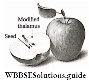
Pome: The fruits develop from syncarpous, bi- or multicarpellary, inferior ovary. They are mostly false fruits. The edible part of this fruit is the fleshy thalamus which surrounds the true fruit. Seeds are surrounded by a thin ovarian wall. For example pear(Pyrus communis) and apple (Malus sylvestris), etc.
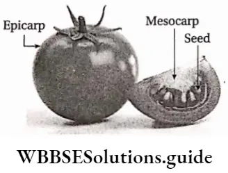
Berry or Bacca: The fruits develop from the multicarpellary, syncarpous, superior, or inferior ovary. The seeds are embedded freely in the massive pulp from by mesocarp and endocarp. The epicarp remains as the outer skin of the fruit. Example brinjal (Solanum melongena), tomato (Lycopersicon esculentum), etc.
Berry or Bacca: The fruits develop from the multicarpellary, syncarpous, superior, or inferior ovary. The seeds are embedded freely in the massive pulp from by mesocarp and endocarp. The epicarp remains as the outer skin of the fruit. Example brinjal (Solanum melongena), tomato (Lycopersicon esculentum), etc.
Date palm—berry or drupe
An example of a one-seeded berry is the date palm, (Phoenix sylvestris). In the case of date palms, the endocarp is thin and paper-like. But, it is also considered as a type of drupe.
Pepo: The fruits developed from tri carpellary, syncarpous, and inferior ovary. The seeds are firmly attached to the placenta. The exocarp is tough and leathery. Example cucumber (Cucumis sativa); pumpkin (Cucurbita maxima), etc.
Hesperidium: The fruits are multi-chambered and developed from the multicarpellary, syncarpous, superior ovary. In these fruits, the epicarp and mesocarp remain fused together and form the skin.
The endocarp projects inwards forming distinct chambers or lobe-like structures. The inner part of the endocarp contains unicellular juicy hairs. Example sweet orange, (Citrus sinensis) lemon (Citrus aurantium), etc.

Balausta: The fruits are many-chambered and develop from the polycarpellary, syncarpous, and inferior ovary. They contain many seeds. The seeds have seed coats known as outer fleshy testa and inner hard tegmen. The fleshy testa is the edible part.
Seeds are irregularly arranged inside the fruit. The pericarp is rough and leathery with persistent calyx. Example pomegranate (Punica granatum).
Amphisarca: The fruits are many-chambered and develop from the polycarpellary, syncarpous, superior ovary. They contain many seeds scattered within the fruit.
They have hard epicarp fleshy mesocarp and endocarp. The mesocarp, endocarp, and swollen placenta are the edible parts. For example wood apple (Aegle marmelos) and Feronia limonia.
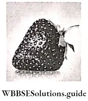
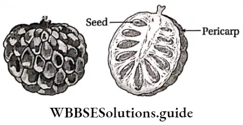
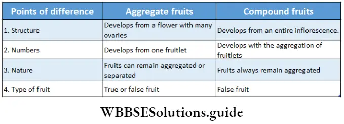
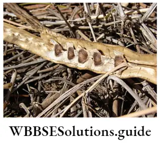
Aggregate Fruit
The fruit that develops from a single flower containing polycarpellary, and apocarpous ovary (many carpels and ovary) is known as aggregate fruit. This type of fruit is composed of many small fruits, known as etaerio of fruitlets.
These fruits are divided into the following four types—
Etaerio of follicles: In this type, each free carpel grows into a follicle and remains arranged together on the enlarged thalamus. This scenario may be composed of two follicles as in Calotropis procera or many as in Magnolia grandiflora.
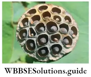
Etaerio of achenes: In this type, the fruits are achenes and remain aggregated on the thalamus. Many such achenes are arranged in different forms. In lotus (Nelumbo nucifera), the thalamus becomes spongy and achenes are embedded inside it. In Naravelia zeylanica, the hairy achenes are aggregated on the thalamus.
Etaerio of drupes: In this type, the fruits are drupes. Many small drupes are aggregated on the fleshy thalamus. Example strawberry (Fragaria vesca) raspberry {Rubus idaeus), etc.
Etaerio of berries: In this type, the fruits are berries. Many such small berries are arranged on the sides of the fleshy thalamus. The apical part is fused with each other forming a common rind. For example Artabotrys odoratissimus and custard apple (Anona squamosa), etc.
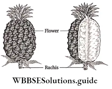
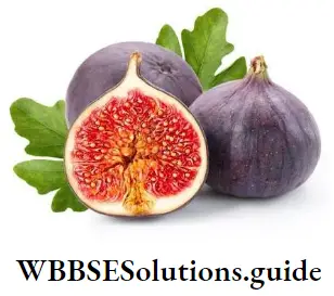
Composite or compound or multiple fruit
The fruits that develop from the complete inflorescence are known as composite compounds or multiple fruits. These are also called infructescences or syncarps.
These fruits are of the following two types—
Sorosis: These fruits develop from a spike, spadix, or catkin inflorescence, where the axis and the ovaries are fused together to form a single fruit. Example pineapple (Ananas comosus), and jackfruit (Artocarpus heterophyllus).
Syconus: These fruits develop from entire hypanthium or coenanthium inflorescence, where the receptacle contains many seeds. Example fig (Ficus hispida), banyan (F. benghajensis), etc.
The Seed
The Seed Definition: A seed is a fertilized matured ovule, consisting of an embryo enclosed by protective seed coats.
Generally, all flowering plants bear seeds. It contains a fully developed embryo in it. This embryo develops into a seedling during germination.
Formation of seeds after fertilization: After fertilization, different parts of the ovule modify to form different parts of the seed. Integuments are modified into testa and tegmen. Funiculus is modified into the stalk of the seed.
Micropyle and hilum of ovule become that of the seed. Egg (n) cells are modified into a zygote (2n) while the definitive nucleus (2n) is modified into the endosperm (3n) of the seed.
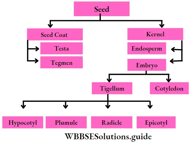
Different Parts Of A Typical Seed
A typical matured seed of angiosperms consists of the following parts
Types of seeds: Depending on the number of cotyledons and the presence of endosperm, seeds are of various types. Based on the number of cotyledons, seeds are of three types- monocotyledonous, dicotyledonous, and polycotyledonous and polycotyledonous based on the presence of endosperm seeds are of two types endospermic and non-endospermic.
On the basis of the number of cotyledons
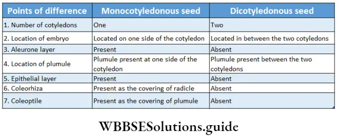
- Monocotyledonous: The seeds with one cotyledon, are known as monocotyledonous seeds. These types of seeds are found in rice (Oryza sativa) wheat (Triticum aestivum), maize (Zeo mays), etc.
- Dicotyledonous: The seeds with two cotyledons, are known as dicotyledonous seeds. These types of seeds are found in mango (Mangifera indica), gram (Cicer arietinum), pea (Pisum sativum), castor (Ricinus communis), gourd (Cucurbita maxima), etc.
- Polycotyledonous: The seeds that bear more than two cotyledons are known as polycotyledonous seeds. These types of seeds are found in pine and in most conifers.
On the basis of the presence of endosperm
Exalbuminous (non-endospermic): In these seeds, the food is stored in the cotyledons and the endosperm is inconspicuous. This type of seeds is found in both dicot (Pisum sativum) and monocot (Alisma sp.) plants.
Albuminous (endospermic): In these seeds, the food is stored in a separate tissue, called the endosperm. The cotyledons of these seeds are thin papery structures without reserve food. This type of seed is found in both dicotyledonous (Ricinus communis, Carica papaya) and monocotyledonous (Zea mays, Oryza sativa) plants.
Structure of some important seeds
The structures of some seeds are discussed below.
Structure of non-endospermic dicotyledonous seed:
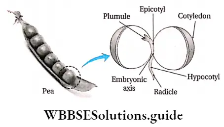
Gram (Cicer arietinum), mango (Mangifera indica), and pea (Pisum sativum) are examples of dicotyledonous, non-endospermic seeds The structure of a gram seed is discussed below.
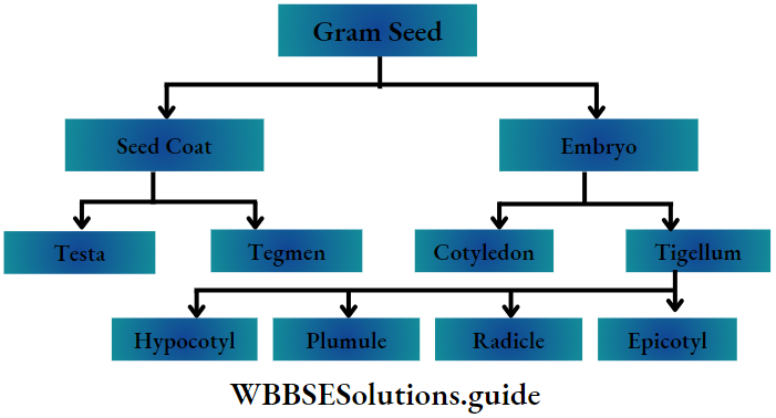
The two main parts of this seed are—
- Seed coat and
- kernel.
Seed coat: The outer covering of the seed is known as the seed coat. It has two parts—
Testa: It is the brown-colored, thick, leathery outer covering of the seed.
Tegmen: It is present below the testa. It is a thin white-colored membrane that remains attached to the testa. The pointed part of the seed has an oval scar on its surface, known as a hilum. The seed remains attached to the placenta inside the ovary, through this hilum.
A small opening, called micropyle is present near the hilum. The lens-shaped scar present on the middle of the testa is known as strophiole or chalaza.
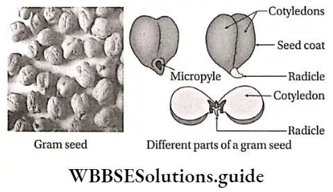
Kernel: After removal of the seed coat a thick, fleshy, spherical part can be seen within the seed. It is known as the kernel.
It is the embryo. It has two parts—
- Cotyledons: These are the thick, fleshy, pale yellow-colored hemispherical structures. They store food for the embryo.
- Tigellum or embryo axis:
- This is a hook-like structure, present in between the cotyledons.
- The tigellum remains attached to the cotyledons at a point. This point of attachment is known as the cotyledonary node or nodal zone.
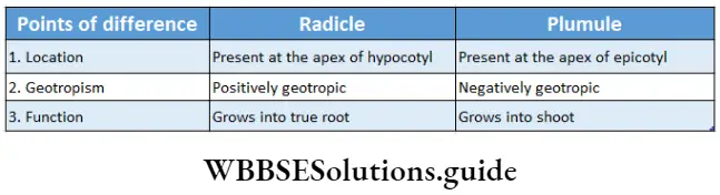
- The upper growing region of the embryo axis is known as the plumule and the lower growing region is known as the radicle,
- The region between the plumule and the cotyledonary node is known as epicotyl.
- The region between the radicle and cotyledonary node is known as hypocotyl.
Structure of the endospermic dicotyledonous seed: Castor (Ricinus communis), cotton (Gossypium herbaceum), and jute (Corchorus olitorius) are examples of endospermic dicotyledonous seeds. The structure of castor seed is described below.
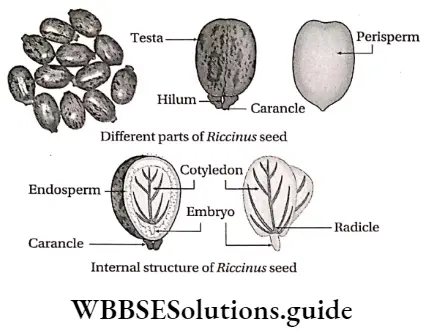
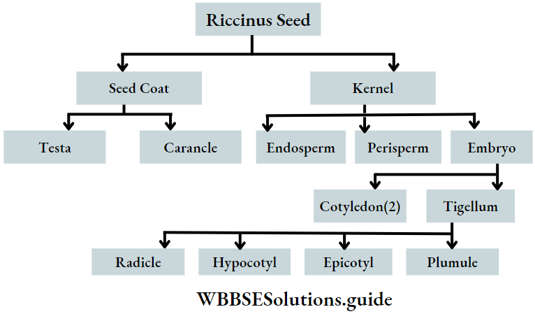
The seed is flat and oval in shape. Its two main parts are—
- Seed coat and
- Kernel.
Seed coat: These seeds are composed of thick, hard, and fragile testes. Tegmen is absent in these seeds. Testa has black, brown, and white colored ornamentations. The white-colored fleshy outgrowth on the narrow region of the seed is known as a caruncle.
This structure covers the micropyle and hilum. A ridge that runs from hilum to the strophiole of the seed, is known as raphe. It helps to absorb water into the seeds.
Kernel: It is the spherical fleshy structure present below the seed coat. It has three parts. They
- Perisperm: The thin transparent membrane, that covers the endosperm, is known as the perisperm.
- Endosperm: The thick, whitish-flat region below the perisperm is known as the endosperm. The endosperm provides nutrients to the embryo.
- Embryo: It is the part that consists of two cotyledons and a tigellum or embryo axis.
- Cotyledons: Cotyledons of this seed are thin leaf-like structures with veins and veinlets.
- Embryo axis or tigellum: The minute rod-shaped structure attached to the cotyledons is known as the embryo axis or tigellum. The upper growing region of the embryo axis is known as plumule and the lower growing region is known as radicle. Both the radicle and plumule of these seeds are very small. Thus, the epicotyl and hypocotyl regions are not clearly visible.
Structure of endospermic monocotyledonous seed: Rice {Oryza sativa), wheat (Triticum aestivum), maize (Zea mays) are examples of endospermic monocotyledonous seeds. The structure of maize seed is discussed as follows.
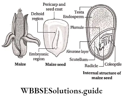
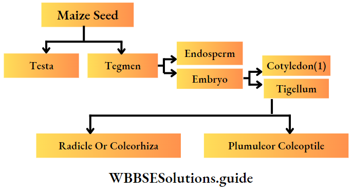
Covering layer: The outer covering of the maize seed is formed by the combination of pericarp and seed coat. This is translucent, thick, and golden yellow in color.
Kernel: The part below the outer coat is known as the kernel. It has two parts—endosperm and embryo.
- Endosperm: 75% of the kernel is the endosperm. This region stores a large amount of starch. Due to the presence of endosperm, this seed is known as endospermic seed. The endosperm is surrounded by a proteinaceous layer known as the aleurone layer.
- Embryo: The embryo is present at the triangular swollen region, below the endosperm. The embryo is very small and formed of two parts.
- Cotyledons: Maize contains only one cotyledon, known as scutellum. It is present between the embryo axis and endosperm. The scutellum supplies food from the endosperm to the embryo.
- Embryo axis: A small rod-shaped embryo axis is present beside the scutellum. The upper part of the embryo axis is known as plumule and the lower part is known as radicle. The plumule is covered with a protective covering known as coleoptile. The covering of the radicle is known as coleorhiza.
Functions Of Different Parts Of A Typical Seed
The function of seed coat:
- It protects the kernel from the external environment.
- It helps in the absorption of water and oxygen.
- It also provides the passage for the germ tube to come out during germination.
Function of cotyledons:
- They provide nutrition to the embryo and help it to grow.
- Cotyledon protects the embryo axis.
- It also holds the embryo tightly.
The function of the embryo axis:
- It joins the two cotyledons.
- It helps in the formation of root from the radicle and shoot from the plumule.
Function of endosperm:
- It provides nutrients to the embryo in the endospermic seed.
- It helps in the development of the embryo.
Dispersal Of Fruits And Seeds
A plant bears numerous fruits and seeds. If all these fruits or seeds fall and germinate in the same place then all of them will compete with each other for necessary requirements, such as water, minerals, sunlight, etc., for survival.
Thus, they must be dispersed away from the mother plant to avoid overcrowding and unwanted struggle for existence. Fruits and seeds can not move by their own.
Morphology of flowering plants detailed notes PDF
So, they depend on different agents such as water, animals, wind, etc., for dispersal. In this regard, they produce varied external outgrowths on bpsis of different dispersal agencies.
Dispersal Of Fruits And Seeds Definition: Seed or fruit dispersal is the process of carrying the seeds or fruits away from the parent plant for proper germination under suitable conditions.
Mechanisms of fruit and seed dispersal
Fruits and seeds are dispersed in various ways.
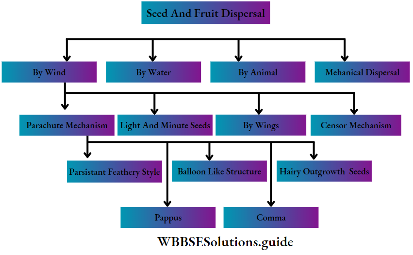
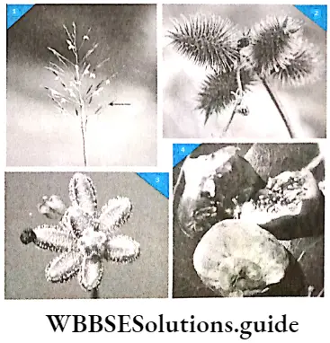
Dispersal by wind: The fruits or seeds that are dispersed by wind bear certain modified structures. Due to these structures, they can easily carried away by the wind. Some of these structures are discussed below.
Parachute mechanism: Some hairy appendages are found in certain fruits and seeds. These appendages help them to float in the air for a long time. These seeds are dispersed to greater distances.
These are of the following types—
Ucture is formed by the cluster of persistent hairy calyx. Pappi (plural) are present at the upper region of the fruits and help the fruit to float in the air. These are found in Mikania cordata, Helianthus annuus, etc.
Comma: The tuft of hairs is the outgrowth of the testa and is found on both sides of the seed. These tufts help them to float in the air just like a parachute. These are found in Calotropis procera, Alstonia scholaris, etc.
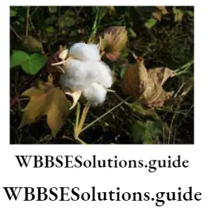
A hairy outgrowth of seeds: In some seeds, long hairy outgrowths develop from the testa and help them to float in the air. These hairy outgrowths are known as lint. These are found in Gossypium herbaceum, Bombax ceiba, etc.
Persistent feathery style: Some seeds contain persistent feathery styles, which help them to float with the wind. These are found in Clematis gouriana.
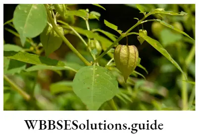
Balloon-like structure: In some plants, some floral parts become air-filled, swollen, balloon-like structures. These help the seeds to remain in the air for a longer period of time. These modifications are found in Cardiospermum halicacabum (inflated fruit), Physalis minima (inflated persistent calyx), etc.
Light and minute seeds: The seeds of some grasses and orchids are minute and lightweight. So, they can float in air very easily.
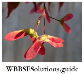
Seeds and fruits with wings: Seeds and fruits of many plants have wing-like appendages. They can be easily blown away In the air. Winged fruits are found In Shorea sp., Acer sp., and winged seeds are found In Moringa sp. (drumstick), Cinchona sp.
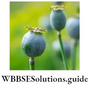
Censer mechanism: Fruits-like capsules of some plants have minute pores, from which a few seeds are dispersed when are being shaken by air. This mechanism is observed in Papaver somniferum (poppy), and Argemone maxicana.
Dispersal by water: The fruits and seeds of the plants, that grow near water bodies, are mainly dispersed through water. Such fruits and seeds must be provided with a waterproof, salt-resistant coat that will make the fruits and seeds buoyant.
Spongy thalamus: The thalamus of lotus (Nelumbo nucifera) is soft and spongy containing clusters of fruits. This helps the thalamus to remain afloat.
Water current carries it to a certain distance. The thalamus degenerates and the fruits are released. They settle down on the soil at the bottom of the water body and germinate.

Fibrous mesocarp: The fibrous mesocarp of coconut (Cocos nuclfera) fruit can entrap air. Its outer coat Is impermeable to water and keeps the fruit afloat for longer periods. Water current carries it to a certain distance.
Dispersal by animals: Some seeds and fruits are also dispersed by animals, birds, and human beings. There are different methods of fruit and seed dispersal by animals.
Some of these are described below—
Spiny outgrowth or appendages: Hard spiny outgrowths on the outer surface of the fruits are found in some plants such as Aristida sp., Chrysopogon aciculatus, etc. These fruits are attached to the animal’s body by the spiny outgrowths and are dispersed by them.
Hook-like appendages: Hooked appendages are found on the outer surface of the fruits in some plants such as Xanthium strumarium, Aerva aspera, etc. These appendages get anchored to the animal’s fur and are dispersed as the animals go to different places for grazing.
Secretion of sticky juice: Fruits of some plants such as Boerhavia repens, Gynandropsis gynandra, etc., secretes sticky juice from the glands present on their outer surface. This sticky substance helps the fruits to attach to the bodies of the grazing animals and get dispersed with them.
Remains of the food: The fruits of Azadirachta indica, and Ficus benghalensis, are usually eaten by animals and birds. But they cannot digest the seeds, as they have hard testa.
So, these seeds are dispersed through the excreta of animals or birds, many miles away from the place of their origin. In many cases, animals only eat the fleshy part of the fruit and throw away the seeds. Under favorable conditions, these seeds germinate.
Mechanical dispersal of fruits and seeds:
- The fruits of Oxalis corniculata, Impatiens balsamina burst open suddenly when touched or due to air current and the seeds dispersed far away from the parent plant
- The fruits of some plants such as Ruellia tuberosa Andrographis paniculate, and burst into two valves when get soaked in rain or dew thereby ejecting the seeds.
- In Luffa aegyptiaca, a pore develops on the ripe fruits through which the seeds get dispersed out in the outer environment.
- The fruits of Clitoria ternatea etc., twist after their pericarp bursts open, thus dispersing the seeds.
Semi-Technical Description Of Typical Flowering Plants
The following factors are important to describe a flowering plant—
- Hierarchical position of the plant,
- Habit and habitat,
- The morphological features of root, stem, leaves, flower, fruits, etc.
Some other factors that are also important for describing a flowering plant are discussed below—
Dissection of a flower: A vertical and transverse section of a flower as well as its bud provide the shape of the thalamus and the arrangement of floral whorls on it,
- Shape and size of the different floral whorls,
- Adhesion or cohesion between different floral whorls,
- Type of placentation,
- The number of ovules in the locules of the ovary.
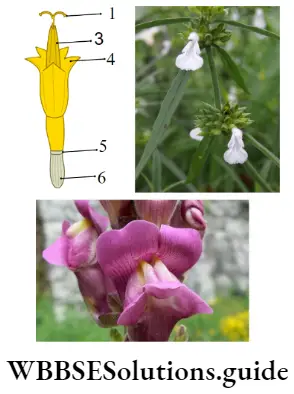
Floral formula: The representation of the number of floral whorls and their interrelationship by using digits, letters, and various symbols, is known as its floral formula.
By this formula, we get to know about the number of floral whorls, the number of bracts, the position of the floral whorls, the adhesion, and cohesion of different floral whorls, sexuality, and other characteristics of a flower.
NEET biology morphology of flowering plants notes
Symbols used in floral formula: The following symbols are used in the floral formula—
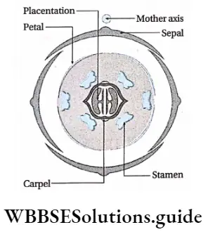
Floral diagram: The floral diagram is the diagrammatic representation of the number and their relative arrangement of different floral whorls as observed in the transverse section of a flower bud, with respect to the mother axis (stalk of the flower).
Examples of floral formula and floral diagram: A sample floral formula and floral diagram are given below
Floral formula:

Description of the flora! formula:
- The flower is ebracteate, complete, actinomorphic, and bisexual.
- Calyx is polysepalous with 4 sepals arranged in two whorls. Hence, 2 sepals are present in each round.
- The corolla is composed of 4 free petals.
- There are six stamens, arranged in two whorls, 2 in one whorl and 4 in another.
- The two groups of stamens may be of different lengths.
- Gynoecium is bi-carpellary and fused
- The ovary is superior.
Taxonomic Description Of Some Plants Of Selected Angiospermic Families
Descriptions of some selected angiosperm families are given below along with their economic importance. One member from each family is also described.
Description of Solanaceae Family
Distinguishing Characteristics
- Plants are mostly terrestrial. Some are aquatic (For example Solanum tampicense).
- Plants are mostly herbs, some are shrubs, and rarely soft woody trees (For example Solanum grandiflorum). They are either annual or perennial.
- Plants have a tap root system.
- Aerial stem is herbaceous or woody, erect or prostrate (For example Solanum surattense), climbing (For example Solanum dulcamara), branched, with solid internodes, hairy or with prickles. The underground stem is tuberous (For example Solanum tuberosum).
- Leaves are either simple or pinnately compound, exstipulate, entire or incised, petiolate or sessile, alternate or opposite, unicostate type of reticulate venation.
- The inflorescence is either solitary or axillary cyme helicoid cyme or cymose pannicle.
- Flowers are bracteate or ebracteate bisexual, actinomorphic or zygomorphic (Example Schizanthus sp., sp.), hypogynous, and pentamerous. Floral whorls are arranged in a tetracyclic pattern.
- Calyx is made of five, green colored sepals. It is gamosepalous, rotate, tube or bell-shaped, aestivation valvate. Sepals are hairy, persistent, or accresent (Example Physalis sp.).
- Corolla is usually made of five petals. It is gamapetalous, may have hair, rotate or campanulate or tubular or infundibuliform in shape. Aestivation valvate or imbricate.
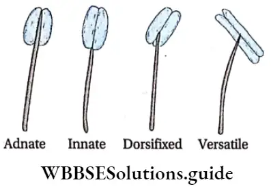
- Stamens are five and epipetalous. Anthers are pithecoid, basifixed, or dorsifixed and dehiscence by an apical pore or longitudinal slit.
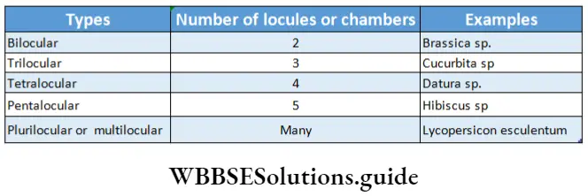
- Gynoecium bicarpellary and syncarpous. Nectarine glands are present at the base. Stigma is bifid or capitate and style is simple and terminally placed. Ovary oblique, superior, bi- or trilocular, placentation axile, ovule numerous.
- Fruit usually berry, sometimes capsule (Example Datura).
- Seeds are dicotyledonous and with endosperm.
Economic Importance:
The economic importance of the Solanaceae family is as follows—
- There are many food-yielding plants belonging to the family Solanaceae. Examples Solanum tuberosum (potato), Solanum melongena (brinjal), Capsicum annum (chilli), Lycopersicum esculentum (tomato), etc.
- Tobacco is obtained from leaves and branches of Nicotiana tabacum and Nicotiana rustica.
- Various alkaloids are obtained from Datura stramonium, anoxia, and Hyoscymous niger.
- Several medicinally important plants belong to this family. Examples are Datura, Withania somnifera, Solanum xanthocarpum, Atropa belladonna (yield atropine), etc.
- Cestrum nocturnum, Petunia sp., etc are some of the ornamental plants belonging to this family.
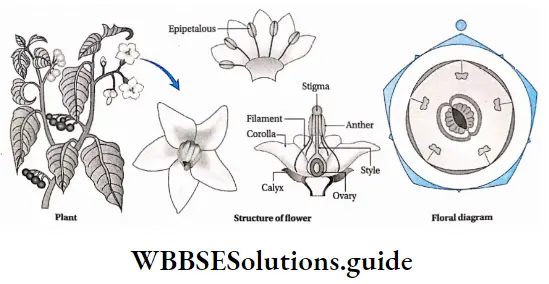
Description of a member of the Solanaceae family
Scientific name: Solanum nigrum
Common name: Indian nightshade
Systematic position:
Class—Dicotyledonae
Sub-class—Gamopetalae
Series—Bicarpellatae
Order—Polemoniales
Family—Solanaceae
Genus—Solanum
Species—nigrum
Description of the plant
- Habit and habitat: Annual, wild herb, that grows mainly in shaded regions.
- Root: Tap root.
- Stem: Herbaceous, aerial, erect, cylindrical, hard and branched.
- Leaves: Simple, exstipulate, unicostate reticulate venation, petiolate, oval-shaped, acute apex, entire leaf margin, smooth leaf surface, and arranged in alternate phyllotaxy.
- Inflorescence: Cymose, scorpioid, uniparous with 5-8 flowers (rhipidium).
- Flower: Bracteate, pedicellate, complete, bisexual, actinomorphic, hypogynous, white-colored, valvate aestivation.
- Calyx: 5 sepals, gamosepalous, persistent
- Corolla: 5 petals, gamopetalous, white-coloured.
- Androecium: 5 stamens, polyandrous, epipetalous stamens, basifixed, long filament, dehiscence of anther occurs through terminal pores.
- Gynoecium: Bicarpellary, syncarpous, obliquely placed superior ovary, thick placenta, containing multiple ovules, axile placentation, elongated pistil, flattened stigma.
- Fruit: Fleshy, indehiscent, berry type with persistent stalk.
- Floral formula:

Description of floral formula: Ebr—Ebracteate;
 —Actinomorphic flower;
 Bisexual, K(5)— 5 Sepals and gamosepalous;
 —Epipetalous stamen where the number of petals is 5, gamopetalous, 5 free stamens;
 —2 pistils, joined and ovary superior.
Description of Fabaceae family
The description of the Fabaceae family is given below along with its economic importance.
Distinguishing Characters
- Plants are mostly terrestrial, but some are aquatic (For example Neptunia natans).
- Plants are mostly herbs, some are shrubs or woody trees. They are either annual or perennial.
- Most plants have a tap root system. Roots bear nodules which carry symbiotic nitrogen fixing bacteria Rhizobium sp.
- Stem erect, twinning or climbing, herbaceous or woody, hair may present.
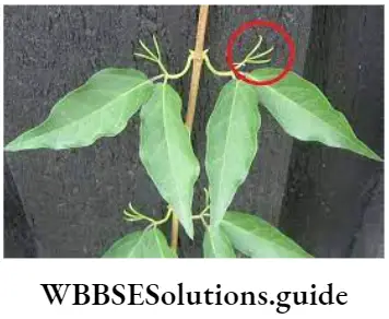
- Leaves are simple or pinnately compound, alternately arranged, stipulated, leaf base pulvinus, venation reticulate, whole leaf or upper leaflets of the compound leaf may modify into tendrils.
- Inflorescence was racemose or rarely solitary axillary.
- Flowers are bracteate or ebracteate, pedicellate or sessile bisexual, complete, zygomorphic, hypogynous or perigynous, cyclic, and pentamerous.
- Calyx is made of five, green colored sepals. It is either free or gamosepalous, aestivation imbricate, and often persistent.
- Corolla is usually made of five petals. It is polypetalous and may be papilionaceous (For example Pisum sativum), aestivation imbricate (For example Mimosa pudica), or vexillum (For example Clitoria ternate)
- The androecium is composed of 10 or more stamens present. Stamens are either free or united (monadelphous or diadelphous), vitreous, and basifixed.
- Gynoecium monocarpellary. Stigma is simple or capitate. The style is long and flattened. Ovary superior, unilocular, elongated, placentation marginal, with many ovules.
- Fruit legume or pod, rarely lomentum (Example Desmodium sp.)
- Seeds are dicotyledonous and non-endospermic.
Economic Importance:
The economic importance of the Fabaceae family is as follows—
- There are many members of Fabaceae that yield pulses. Examples are Pisum sativum (pea), Cicer arietinum (gram), Lens culinaris (lentil), Vigna radiata (green mung), etc.
- Green young pods of Dolicos lablab, Vicia faba, etc., are used as vegetables.
- Glycine max (soybean) and Archls hypogea (groundnut) are important sources of cooking oil. Soybean is used to extract soya milk which Is a substitute for milk.
- Fibers are obtained from the stem of Crotolaria juncea, and Sesbania bispinosa. These fibers are used to prepare ropes, canvas, mats, etc.
- Timber is obtained from Delbargia sisso, latifolia, Pterocarpus sp., etc.
- Roots of Glycyrrhiza glabra (licorice), leaves and seeds of Abrus sp., flowers of Sesbania grandiflora, etc., are known to have different medicinal properties.
- Different dyes are obtained from plants like Indigofera tinctoria, Pterocarpus santalinus, Sophora japonica, etc.
- Lathyrus odoratus, Clitoria ternatea, etc., are used as ornamental plants.
Description of a member of the Fabaceae family
Scientific name: Pisum sativum
Common name: Sweet pea
Systematic position:
Class—Dicotyledonae
Sub-class—Polypetalae
Series—Calyciflorae
Order—Rosales
Family—Fabaceae
Genus—Pisum
Species—sativum
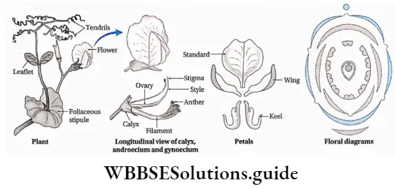
Description of the plant
- Habit and habitat: Annual, climbing herb
- Root: Branched tap root, nodules present.
- Stem: Herbaceous, weak, branched, hollow, green colored, climber, glabrous.
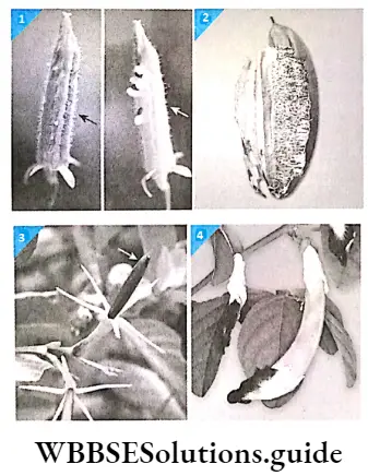
- Leaf: Arranged in alternate phyllotaxy, sessile, foliaceous stipules, compound imparipinnate, leaf apex is transformed into tendrils. Leaves are oval-shaped with unicostate reticulate venation. The entire margin with acute leaf apex.
- Inflorescence: Racemose or solitary inflorescence.
- Flower: Bracteate, ebracteolate, pedicellate, bisexual, zygomorphic, complete, cyclic, partially perigynous, variously colored.
- Calyx: 5 sepals, gamosepalous, valvate aestivation, green-colored, hairy, persistent.
- Corolla: 5 petals, papilionaceous, vexillary aestivaion.
- Androecium: Stamens 10, diadelphous, 9 stamens fused to form a sheath around the pistil and the tenth stamen remains free. Anther is dithecous, basifixed.
- Gynoecium: Monocarpellary, unilocular, superior ovary, marginal placentation with many ovules, style is long and curved, single hairy stigma.
- Fruit: Legume or pod.
- Floral formula:

- Description of the floral formula: Br—Bracteate; flower; K(5) -5 sepals, gamosepalous; C1+2+(2) Corolla consists of one single large petal (standard), two free petals (wings) and two fused petals (keel), A(9)+1—9 fused stamen and1 free stamen; G(1)—Monocarpellary, superior ovary.
Description ofLiliaceae family
The description of the Liliaceae family is given below along with its economic importance.
Distinguishing characters
- Plants are mostly perennial or annual herbs. Some are shrubs (Example Yucca, Dracaena, etc.) or climbers (Example Smilax sp.). Trees are rare (For example, Xanthorhoea sp.).
- Roots are adventitious, generally, fibrous roots and fasciculated tuberous roots may present (Example Asparagus sp.).
- Stem aerial or underground. Aerial stems may be erect or climbing. Underground stems may be rhizomes, bulbs or corms. Internodes were solid or fistular. Stem branches may be modified into cladode (Example Aspergus sp.)
- Leaves are either simple, stipulate or exstipulate, sessile or petiolate with sheathing bases, cauline or cylindrical, alternate, whorled or opposite, leaves may be fleshy and leathery (Example Yucca sp.), or reduced to scales (Example Ruscus sp.), leaf margin may be entire or spiny, venation mostly parallel.
- The inflorescence may be solitary terminal (For example tulip.), solitary axillary (For example glory lily.), racemose (For example Yucca sp.), panini (For example Asphodelus sp.), or umbel (For example Allium sp.)
- Flowers are bracteate or ebracteate, bisexual or unisexual, actinomorphic or zygomorphic, hypogynous, incomplete, and trimerous.
- Perianth is composed of 4-8 tepals, gamophyllous to form a tube or polyphyllous (For example tulip, glory lily, etc.). Tepals are petaloid or sapaloid, arranged in 2 whorls, aestivation valvate or imbricate.
- The androecium is composed of 3-12 stamens. Stamens polyandrous, monadelphous, arranged in two whorls, antitepalous. Anther basifixed or versatile, bithecous and dehiscence longitudinal or by apical pore.
- Gynoecium bi- ortricarpellary and syncarpous. Ovary superior, bi- or trilocular, placentation parietal, ovules two or many. Style simple, united, or free. Stigma is often trilobed.
- Fruits berry (For example Asparagus sp.) or capsule (For example Asphodelus sp.)
- Seeds are monocotyledonous and endospermic.
Economic Importance:
The economic importance of the Liliaceae family is as follows—
- Leaves and pedicels of Allium cepa, A. ascolium, tender shoots and roots or Asperagus sp., bulbs of Allium cepa, and A. sativum are used in cooking.
- Fibers are obtained from leaves of Yucca sp., Agave sp., Phormium sp., etc.
- Several medicinally important plants belong to this family. Examples are Aloe vera, Smilax sp., Asparagus sp., Gloriosa sp., Colchicum sp., etc.
- Corms of Colchicum autumnale are used to yield colchicine used to stain chromosomes and to induce polyploidy in organisms.
- There are many important ornamental plants that belong to this family. Examples Tulipa (tulip), Lilium (tiger lily), Gloriosa (glory lily), etc.
Description of a member of the LlUiaceac family
Scientific name: Allium cepa
Common name: Onion
Systematic position:
Class—Monocotyledonae
Series—Coronarieae
Order—Liliales
Family—Liliaceae
Genus—Allium
Species—cepa
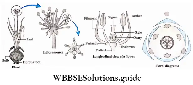
Description of the plant
- Habit and habitat: Annual, herbs, cultivated plants.
- Root: Adventitious, fibrous.
- Shoot: Underground tunicated bulb, disc-like stem is present at the lower portion of the bulb. The floral axis is leafless and known as a scape.
- Leaf: Leaves bear in whorls, fleshy and scaly leaves arise from small bulbs, aerial leaves green, cylindrical, leaf base, hollow, venation parallel.
- Inflorescence: Umbel.
- Flower: Ebracteate, pedicellate, ebracteolate, incomplete, bisexual, white in color, cyclic, trimerous.
- Perianth: The number of tepals is 6, present in alternately placed two whorls, 3 tepals in each whorl. Tepal is white in color, petaloid.
- Androecium: Stamens 6, arranged in two whorls. Each whorl contains 3 stamens. The base of the stamens may be free or attached to the perianth. Anther is dithecous.
- Gynoecium: Tricarpellary and fused, superior ovary, trilocular, axile placentation, each locule bears two ovules, short style, and minute stigma.
- Fruit: Capsule.
- Seed: Monocotyledonous endospermic seed.
- Floral formula:
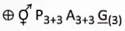
- Description of floral formula:
- —Actinomorphic (regular) flower;
- —bisexual;
- P3+3—Perianth consists of 6 free tepals, arranged in two whorls, each whorl has 3 tepals;
- A3+3—6 free stamens, arranged in two whorls;
- G(3)— Tricarpellary, fused, superior ovary.
Morphology Of Flowering Plants Notes
- Alkaloids: Any of a class of nitrogenous organic compounds of plant origin that have pronounced physiological actions on humans. These may include drugs or poisons.
- Canopy: The upper layer or habitat zone, formed by mature tree crowns and biological organisms (For example epiphytes, lianas, and arboreal animals).
- Definitive nucleus: The diploid nucleus found in the center of the embryo sac, produced after the fusion of the two haploid polar nuclei.
- Epiphyte: A plant that grows harmlessly upon another plant and drives its moisture and nutrients from the air.
- Funiculus: A slender rope-like stalk attaching an ovule to the ovary wall.
- Gynofaasic style: A style that appears to be inserted at the base of the ovary because of the infolding of the ovary wall.
- Hilum: A scar or mark left on a seed coat by the former attachment to the ovary wall or to the funiculus.
- Integuments: A tough outer protective layer of the ovule.
- Line of suture: A fairly rigid joint between two anther lobes, through which anther dehisces.
- Micropyle: A small opening in the surface of an ovule, through which the pollen tube penetrates the ovule.
- Nectaries: A nectar-secreting glandular organ found mainly in flower and also on leaf or stem.
- Staminode: A sterile or abortive stamen, frequently resembling a stamen.
Points To Remember
- Assimiiatory roots of Tinospora and epiphytic roots of Venda can synthesize food by photosynthesis.
- The flowering plant that lacks root is called epiposium. Example duckweed.
- Solanum tuberosum (potato) is a modified stem, but Ipomoea batatus (sweet potato) is a modified root.
- Asparagus sp. is a diode.
- A rhizome with weak, horizontal, and longer internodes, is called sobole. Example Cynodon dactylon (durba ghas).
- When two leaves originate opposite to each other from the same node with a difference in size between them, this condition is called anisophilly. ExampleGoldfussia.
- The small, flattened ear-like outgrowths that develop from two sides of the junction of leaf base (hypopodium) and leaf lamina (epipodium) in monocot leaves, is called auricle.
- The epipodium of Dischidia rafflesiana gets modified into a pitcher and stores rainwater. This stored water is utilized later as required. This type of modified leaf is called a water reservoir.
- Phyllotaxy in Acalypha hispida is of a special type due to its variable length of petiole. This type of phyllotaxy is known as leaf mosaic.
- The plants in which bisexual flowers, unisexual flowers, and neuter or neutral flowers grow in a single inflorescence, is called polygamous plant. Example mango.
- Bisexual, incomplete flower is found in Polianthes tuberosa.
- Coconut sap is the liquid endosperm.
- The smallest flowering plant is duckweed (Lemna sp.).
- The smallest flower is Wollfia microscopic.
- The largest flower is Rafflesia arnoldii.
- The world’s largest inflorescence is that of Puya Raimondi (about 32ft).
- The dicotyledonous plant that lacks cotyledon is Cuscuta reflexa.
- The banana plant is the largest perennial herb.
- Double fertilization occurs in angiosperms.
- Growth in width occurs in dicot plants but not in monocot plants.
- The modified underground stem of potatoes is used as seeds. Underground roots of the stripped gourd is used as plantlets. New plantlets also develop from the stem of rose, berry, etc., and from the leaves of Bryophyllum sp., Begonia sp.
- The phenomenon of opening of flower buds is called anthesis.
- The junction point of the flagellum and cotyledon is called the nodal zone.
- Fleshy scale leaves or cataphylls are present in Allium cepa (onion).
- When different types of flowers grow in the same plant, then the condition is known as heterophylly. Example Sagittaria sp., Limnophila sp.